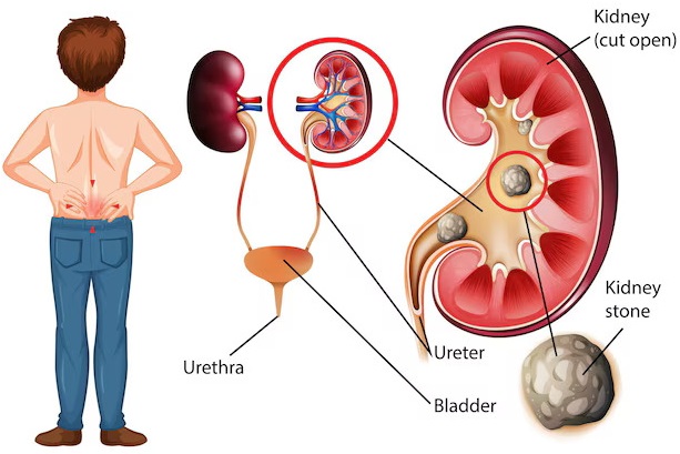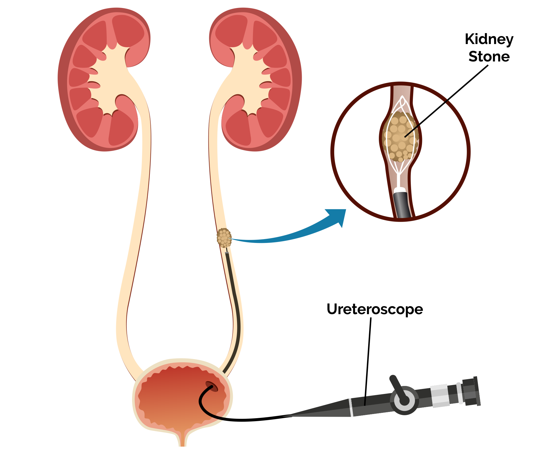
Ureteroscopy (URS) is a minimally invasive urological procedure used to diagnose and treat conditions of the ureter and kidney, such as stones, tumors, or strictures. It involves the insertion of a ureteroscope (a thin, flexible or rigid scope) through the urethra and bladder into the ureter.
Here is a list of instrument names used in Ureteroscopy (URS):
1. Ureteroscope
Types:
Rigid ureteroscope – for lower ureter.
Flexible ureteroscope – for upper ureter and kidney.
Semi-rigid ureteroscope – for mid-ureter and some upper ureter access.
Allows visualization and access to the ureter and renal pelvis.
2. Guidewires
Hydrophilic Guidewire – for easy ureteral navigation.
Zebra Wire / Amplatz Wire – for support and safety.
PTFE Guide Wire – To safely access and guide instruments into the ureter.
Fluoroscopy – confirms stent position.
3. Access Sheaths
Ureteral Access Sheath (UAS) – facilitates repeated passage of instruments, reduces intrarenal pressure, and protects the ureter.
URS Forceps – To grasp and retrieve stones or tissue samples.
Bugbee Electrode – For cauterization and tumor ablation during URS.
4. Irrigation System
Maintains clear vision by flushing the field.
Manual syringe or pressurized bag system.
5. Stone Retrieval Devices
Stone retrieval baskets (Nitinol / Stainless) – To capture and remove stone fragments from the ureter/kidney.
Grasping Forceps – to retrieve stones or tissue.
6. Lithotripsy Devices (for stone fragmentation)
Holmium:YAG Laser – most commonly used.
Ultrasonic lithotripter
Pneumatic lithotripter
7. Dilators and Catheters
Ureteral Dilators or metal dilators – to widen the ureteral orifice.
Balloon Dilators – for precise dilation.
8. Stents and Tubes
Double-J (DJ) Stents – placed post-procedure to prevent obstruction and promote healing.
Ureteral Catheters – for dye injection or drainage.
9. Contrast Media and Monitoring Tools
Ureteroscope withdrawal – under vision to avoid trauma.
C-arm Fluoroscopy Unit – real-time X-ray guidance.
LED Light Source – Provides bright illumination for endoscopic visualization.
Fiber Optic Cable – Transmits light from the LED source to the ureteroscope.