Percutaneous Nephrolithotomy (PCNL) is a minimally invasive kidney surgery to remove large or complex kidney stones directly from the kidney through a small cut in the back.
When is PCNL recommended?
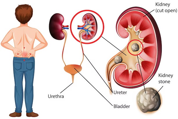
Kidney stones larger than 2 cm
Stones that don’t respond to other treatments
Staghorn calculi (complex, branching stones)
Multiple or hard stones
Step-by-Step Procedure:
1. Pre-Operative Preparation
Pre-Surgical Evaluation:
Imaging tests like CT scan, ultrasound, or X-ray are done to assess stone size, location, and type.
Blood tests and other health evaluations are done to ensure you’re fit for surgery.
Anesthesia:
The patient is given general anesthesia, which means you will be asleep and pain-free during the surgery.
Positioning:
The patient is placed in a prone position (lying on the stomach) to allow access to the back.
2. Accessing the Kidney
Locating the Stone:
Using ultrasound or fluoroscopy (X-ray), the surgeon locates the stone and marks the best entry point.
Making a Small Incision:
A small 1 cm incision is made in the flank area (the side of the body), usually in the lower back.
Inserting the Needle:
A thin needle is inserted through the incision into the kidney under imaging guidance.
The needle allows access to the renal pelvis, where the stones are located.
3. Creating a Pathway to the Kidney
Guidewire Insertion:
Once the needle is in the kidney, a guidewire is threaded through the needle into the kidney.
This guidewire provides a secure path for the rest of the instruments.
Dilation of the Track:
The opening created by the needle is gradually expanded using dilators.
Dilators are progressively larger tubes used to widen the track from the skin to the kidney, ensuring there’s enough space to pass instruments safely.
4. Inserting the Amplatz Sheath
A hollow tube (Amplatz sheath) is inserted into the expanded tract to maintain the pathway open during the procedure.
The sheath allows the surgeon to pass various tools into the kidney while keeping the tract safe and stable.
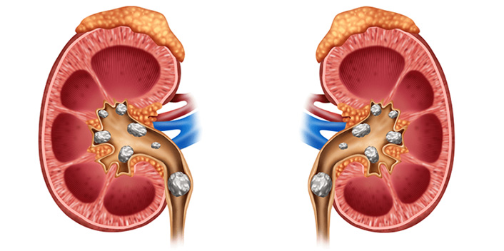
5. Removing the Stones
Nephroscope Insertion:
A nephroscope (a small, flexible camera) is inserted through the sheath to allow the surgeon to see inside the kidney and locate the stones.
Breaking the Stones:
The stones are broken into smaller fragments using ultrasonic, laser, or pneumatic energy.
Laser lithotripsy is commonly used for stone fragmentation.
Removing the Stones:
The stone fragments are removed using specialized tools that grab and pull out the pieces.
6. Drainage and Closing
Nephrostomy Tube:
A temporary nephrostomy tube (a small drainage tube) may be placed in the kidney to allow urine to drain and prevent swelling or infection.
Closing the Incision:
After all the stones are removed, the incision is closed with a few sutures.
A sterile dressing is applied to cover the incision site.
7. Post-Surgery Care
Recovery and Monitoring:
The patient is monitored in the recovery room for a few hours.
Most patients stay in the hospital for 1–2 days to ensure there are no immediate complications.
Nephrostomy Tube Care:
If a nephrostomy tube was placed, it is usually removed after a few days or once healing is confirmed.
Follow-Up Imaging:
Imaging tests may be performed to ensure the stones have been completely removed and to monitor the healing process.
Post-Operative Recovery
Pain Management:
Mild pain is common after the procedure, but medications will be provided to manage it.
Activity and Diet:
Most patients can resume normal activities in about 1–2 weeks.
A healthy diet and hydration are recommended to help with healing.
Advantages of PCNL
Minimally invasive with only a small incision.
High success rate in removing large or complex stones.
Faster recovery compared to open surgery.