C-Arm Machine – Working Principle, Features, Uses and Benefits
A C-Arm Machine is a medical imaging system that provides X-ray images during selected diagnostic and interventional procedures. Its C-shaped structure allows an X-ray source and detector to face each other around the patient.
During surgery, clinicians use fluoroscopic imaging to visualize anatomical structures, medical instruments and treatment progress. Consequently, the equipment supports image-guided procedures in several medical specialties.
Furthermore, mobile C-arm systems offer flexible positioning within operating rooms and procedural environments.
JDMeditech supplies medical equipment, surgical instruments and hospital accessories for healthcare facilities and medical distributors.
What Is a C-Arm Machine?
A C-arm is an X-ray imaging system that uses a curved support structure to position an X-ray tube opposite an image detector.
The system can acquire images from different angles around the patient. Furthermore, many models provide fluoroscopy, which allows clinicians to view moving or sequential X-ray images.
Therefore, the equipment supports procedures that require real-time imaging guidance.
However, imaging capabilities vary according to the selected model and detector technology.
What Is Fluoroscopy?
Fluoroscopy is an imaging technique that uses X-rays to display internal anatomical structures during a medical procedure.
Unlike a single conventional radiograph, fluoroscopy can provide a sequence of images over time.
Consequently, clinicians can monitor instrument positioning and selected procedural steps.
However, fluoroscopy involves ionizing radiation. Therefore, healthcare professionals must follow appropriate radiation protection protocols.
How Does a C-Arm Machine Work?
The X-ray generator supplies electrical energy to the X-ray tube. Next, the tube produces X-rays that pass through the selected anatomical region.
Different tissues attenuate the X-ray beam to varying degrees. Subsequently, the image detector captures the transmitted radiation.
The imaging system processes the detector signals and displays the resulting image on a monitor.
Furthermore, clinicians can reposition the C-arm to obtain different viewing angles.
However, exposure settings and imaging protocols must match the clinical task and patient requirements.
Working Principle of C-Arm Fluoroscopy
A mobile fluoroscopy system uses X-ray transmission and digital image processing to create anatomical images.
First, the generator controls X-ray production. Next, the beam travels through the patient’s body toward the detector.
The detector converts the received radiation into image information. Consequently, the system displays the anatomy for clinical interpretation.
Additionally, selected systems offer pulsed fluoroscopy, image storage and digital enhancement features.
Key Features of a C-Arm Machine
- C-Shaped Imaging Structure
- Intraoperative X-Ray Visualization
- Mobile Equipment Configuration
- Adjustable Imaging Position
- Compatible Image Detector
- X-Ray Generator and Tube Assembly
- Operator Control Interface
- Image Display Monitor
- Model-Specific Fluoroscopy Modes
- Model-Specific Digital Image Processing
- Radiation Exposure Controls
- Manufacturer-Defined Safety Functions
Furthermore, customers should verify the exact features and available imaging modes before purchasing.
Main Components of a C-Arm Machine
A typical mobile C-arm system may include the following components:
- C-Shaped Gantry
- X-Ray Generator
- X-Ray Tube
- Image Detector
- Image Processing Unit
- Control Panel
- Display Monitor
- Mobile Equipment Base
- Positioning Mechanism
- Exposure Controls
- Power Supply
- Manufacturer-Approved Accessories
However, exact components depend on the system configuration.
1. C-Shaped Gantry
The gantry supports the X-ray tube and detector in opposing positions.
Furthermore, its curved design allows the imaging assembly to move around the patient.
2. X-Ray Generator
The generator provides controlled electrical power for X-ray production.
Additionally, its specifications influence the available exposure settings and imaging performance.
3. X-Ray Tube
The X-ray tube generates ionizing radiation for image acquisition.
However, tube ratings and thermal capabilities vary between equipment models.
4. Image Detector
The detector receives X-rays after they pass through the patient’s body.
Depending on the system, the detector may use an image intensifier or a flat-panel detector.
5. Image Processing Unit
The processing unit converts detector information into images for display.
Furthermore, selected systems offer contrast adjustment, image enhancement and storage functions.
6. Display Monitor
The monitor presents acquired images to the clinical team.
Additionally, some systems provide multiple displays for image comparison and procedural guidance.
7. Mobile Equipment Base
The mobile base supports movement and positioning within suitable clinical environments.
However, the equipment must remain stable during imaging and follow the manufacturer’s positioning requirements.
Types of C-Arm Machines
Healthcare facilities can choose from several imaging system configurations.
Mobile C-Arm Machine
A mobile C-arm provides imaging capabilities in different procedural areas.
Furthermore, its movable structure supports flexible equipment positioning.
Mini C-Arm Machine
Mini C-arm systems generally support imaging of selected extremities.
However, their intended applications and imaging capabilities differ from full-size systems.
Flat Panel Detector C-Arm
A flat-panel detector system uses digital detector technology to acquire X-ray images.
Furthermore, detector size and image performance vary by model.
Image Intensifier C-Arm
An image intensifier system converts incoming X-ray information into a visible image signal.
However, equipment design and processing technology vary between manufacturers.
Clinical Applications of C-Arm Machine
Mobile fluoroscopy supports a range of image-guided procedures.
- Orthopedic Surgery
- Trauma Surgery
- Selected Spine Procedures
- Urological Procedures
- Selected Pain Management Procedures
- Selected Vascular Procedures
- Interventional Procedures
- Other Approved Image-Guided Applications
However, clinicians must verify the intended use and imaging capabilities of the specific equipment.
C-Arm Machine in Orthopedic Surgery
Orthopedic surgeons use fluoroscopy to assess bone alignment and instrument positioning during selected procedures.
For example, imaging may support fracture fixation and placement of orthopedic implants.
Furthermore, different imaging angles help clinicians evaluate anatomical relationships.
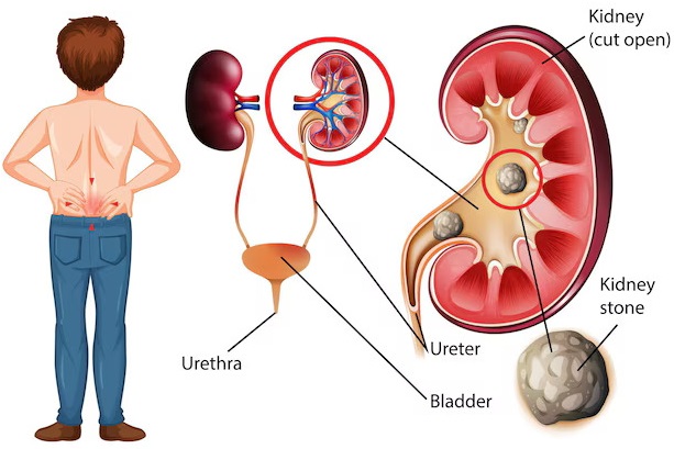
C-Arm Machine in Urology
Urologists use fluoroscopy during selected procedures that require visualization of radiopaque instruments or contrast-filled urinary structures.
For example, imaging may support ureteral stent placement, selected ureteroscopy procedures and percutaneous kidney interventions.
Furthermore, fluoroscopy can help clinicians assess the position of guidewires, catheters and other compatible devices.
However, not every urological procedure requires fluoroscopic guidance.
C-Arm Machine in Spine Surgery
Selected spine procedures require imaging to identify anatomical landmarks and assess instrument positioning.
Consequently, mobile fluoroscopy may support surgical guidance.
However, the surgical team must follow procedure-specific imaging and radiation protection protocols.
C-Arm Machine in Pain Management
Clinicians may use fluoroscopy to guide selected interventional pain procedures.
Furthermore, imaging helps identify anatomical landmarks and monitor instrument placement.
However, procedure selection and imaging requirements depend on the patient’s condition.
Potential Benefits of C-Arm Imaging
- Supports Real-Time Imaging Guidance
- Provides Multiple Viewing Angles
- Supports Selected Minimally Invasive Procedures
- Helps Visualize Instruments and Implants
- Offers Mobile Imaging Capability
- Supports Intraoperative Assessment
- Provides Digital Imaging Functions on Compatible Systems
However, imaging performance depends on equipment configuration, operator expertise and clinical conditions.
C-Arm Machine vs Conventional X-Ray
| Feature | C-Arm Fluoroscopy | Conventional X-Ray |
|---|---|---|
| Primary Imaging | Fluoroscopy and Radiography, Depending on Model | Radiographic Images |
| Real-Time Imaging | Available | Not Typical |
| Intraoperative Use | Common | Depends on Equipment |
| Equipment Mobility | Commonly Mobile | Fixed or Mobile |
| Radiation Exposure | Depends on Fluoroscopy Time and Settings | Depends on Examination and Settings |
Technical Specifications of C-Arm Machine
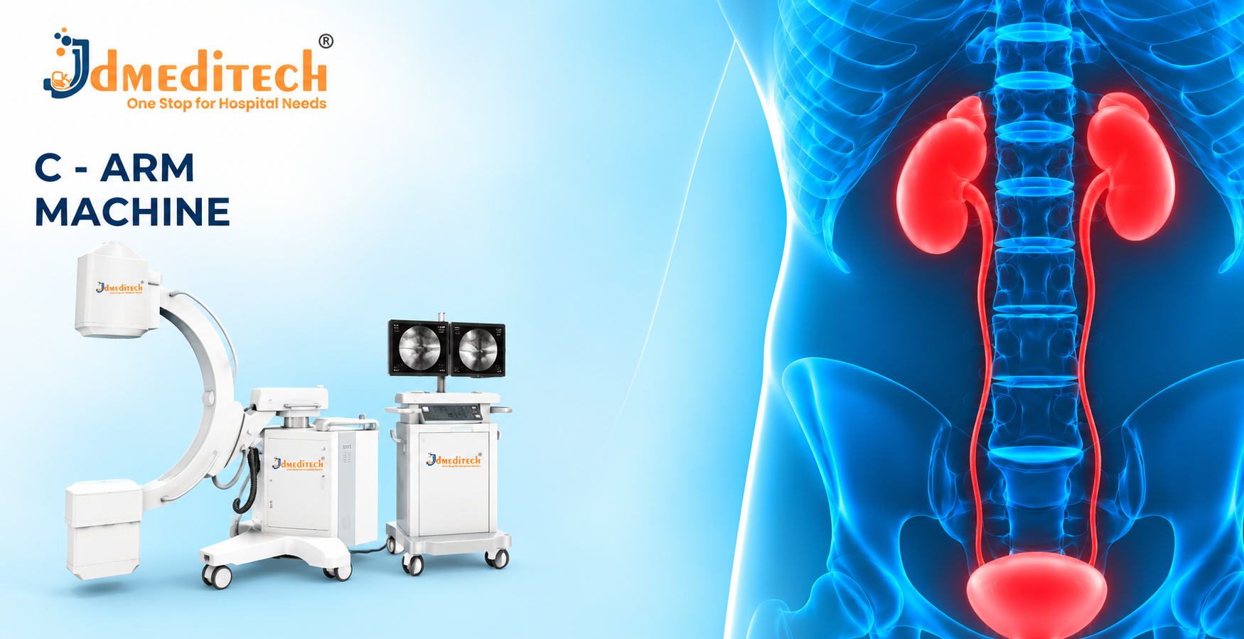
The supplied product image shows a mobile C-arm imaging system with a C-shaped gantry and display equipment.
However, the image does not confirm its generator rating, detector dimensions, imaging modes or radiation output.
Therefore, buyers should verify the manufacturer’s official technical datasheet.
| Parameter | Product Information |
|---|---|
| Product Name | C-Arm Machine |
| Equipment Category | Surgical Imaging Equipment |
| Imaging Technology | X-Ray Fluoroscopy |
| System Configuration | Mobile C-Arm |
| Generator Power | Confirm Manufacturer Specifications |
| Tube Voltage Range | Confirm Manufacturer Specifications |
| Tube Current Range | Confirm Manufacturer Specifications |
| Detector Type | Confirm Manufacturer Specifications |
| Detector Size | Confirm Manufacturer Specifications |
| Fluoroscopy Modes | Confirm Available Configuration |
| Pulse Rate | Confirm Manufacturer Specifications |
| Image Resolution | Confirm Manufacturer Specifications |
| Monitor Size | Confirm Manufacturer Specifications |
| Image Storage | Confirm Available Configuration |
| Gantry Movement | Confirm Manufacturer Specifications |
| Source-to-Image Distance | Confirm Manufacturer Specifications |
| Radiation Dose Display | Confirm Available Configuration |
| Input Voltage | Confirm Manufacturer Specifications |
| Dimensions | Confirm Manufacturer Specifications |
| Weight | Confirm Manufacturer Specifications |
| Regulatory Documentation | Verify for Destination Market |
Importance of Image Quality
Image quality affects the visibility of anatomical structures and medical instruments.
Furthermore, detector performance, X-ray settings and image processing influence the final image.
Therefore, clinical teams should use appropriate imaging protocols.
Importance of Radiation Dose Management
Fluoroscopy exposes patients and potentially nearby personnel to ionizing radiation.
Therefore, operators should follow the ALARA principle, which means keeping radiation exposure as low as reasonably achievable while obtaining adequate clinical images.
Furthermore, appropriate collimation, exposure settings, positioning and protective measures help manage radiation exposure.
Radiation Safety Precautions
- Operate Equipment Only With Trained Personnel
- Follow Local Radiation Safety Regulations
- Use Appropriate Radiation Protection
- Apply the ALARA Principle
- Use Appropriate Beam Collimation
- Minimize Unnecessary Fluoroscopy Time
- Use Suitable Imaging Protocols
- Monitor Radiation Dose Where Supported
- Verify Equipment Safety Functions
- Follow Manufacturer Instructions
- Perform Required Quality Assurance Checks
Potential Risks and Limitations
- Ionizing Radiation Exposure
- Occupational Radiation Exposure
- Potential Skin Injury at High Radiation Doses
- Image Artifacts
- Limited Soft Tissue Contrast Compared With Some Other Imaging Modalities
- Equipment Positioning Constraints
- Mechanical Collision Risks
- Electrical or Equipment Malfunction
Furthermore, risk levels depend on procedure complexity, imaging settings and safety practices.
Inspection Before Equipment Use
- Confirm Equipment Identification
- Inspect the C-Arm Gantry
- Check Mechanical Movement
- Inspect the X-Ray Tube Assembly
- Verify Detector Condition
- Check Display Monitors
- Inspect Electrical Connections
- Verify Exposure Controls
- Check Radiation Safety Functions
- Confirm Equipment Maintenance Status
- Follow Manufacturer Pre-Use Procedures
Cleaning and Maintenance Guidelines
Healthcare facilities should clean equipment surfaces according to the manufacturer’s instructions.
Furthermore, authorized technical personnel should inspect the imaging system regularly.
Additionally, facilities must perform required radiation quality assurance and equipment safety testing.
However, servicing and calibration must follow the specific equipment manual and applicable regulations.
Storage and Handling Recommendations
- Store in a Suitable Clinical Environment
- Protect Equipment From Excessive Moisture
- Avoid Mechanical Impact
- Secure Equipment During Movement
- Protect Imaging Components
- Inspect Power Connections
- Follow Manufacturer Environmental Requirements
- Remove Malfunctioning Equipment From Service
How to Choose the Right C-Arm Machine
Hospitals and procurement teams should evaluate several factors before purchasing a mobile fluoroscopy system.
- Clinical Applications
- Generator Power
- Detector Technology
- Detector Size
- Image Resolution
- Fluoroscopy Modes
- Radiation Dose Management Features
- Gantry Movement Range
- Monitor Configuration
- Image Storage and Export
- Operating Room Space Requirements
- Electrical Requirements
- Regulatory Documentation
- Installation Requirements
- Operator Training
- Maintenance and Technical Support
Why Choose JDMeditech?
JDMeditech supplies medical equipment, surgical instruments and hospital accessories for healthcare facilities and medical distributors.
Furthermore, our team assists customers with product inquiries, available configurations and quotation requests.
Customers can contact JDMeditech regarding:
- C-Arm Machine
- Urology Surgical Equipment
- Endoscopic Instruments
- Orthopedic Surgical Instruments
- Hospital Imaging Equipment
- Medical Equipment Procurement
Frequently Asked Questions
1. What Is a C-Arm Machine?
A C-arm is an X-ray imaging system that supports real-time visualization during selected medical procedures.
2. Why Is It Called a C-Arm?
The name refers to the C-shaped support structure that holds the X-ray source and detector.
3. What Is the Main Use of a C-Arm?
Its main purpose is to provide imaging guidance during selected diagnostic and interventional procedures.
4. Does a C-Arm Use X-Rays?
Yes. C-arm systems use ionizing radiation to create medical images.
5. Can a C-Arm Be Used in Urology?
Yes. Urologists use fluoroscopy during selected stone management and urinary tract procedures.
6. Is a C-Arm Used in Orthopedic Surgery?
Yes. It commonly supports fracture fixation, implant positioning and other selected orthopedic procedures.
7. What Is the Difference Between a C-Arm and an X-Ray Machine?
A C-arm commonly provides fluoroscopic imaging, while conventional radiographic systems primarily acquire individual X-ray images.
8. What Is a Flat Panel Detector?
A flat-panel detector is a digital imaging component that converts received X-ray information into image data.
9. Does Every C-Arm Have the Same Imaging Features?
No. Imaging capabilities vary by model, detector type and system configuration.
10. Is Fluoroscopy Safe?
Fluoroscopy can provide important clinical benefits, but it uses ionizing radiation and requires appropriate safety controls.
11. Does a C-Arm Require Regular Maintenance?
Yes. Facilities should follow manufacturer servicing schedules and applicable radiation quality assurance requirements.
12. Where Can Customers Request Product Information?
Hospitals and medical distributors can contact JDMeditech for product details and quotations.
Conclusion
In conclusion, the C-Arm Machine is an important imaging system for selected surgical and interventional procedures.
Furthermore, fluoroscopy supports real-time visualization of anatomical structures and medical instruments.
However, safe operation requires trained personnel, appropriate radiation protection and regular equipment maintenance.
Therefore, healthcare facilities should verify complete technical specifications before purchasing.
JDMeditech – One Stop for Hospital Needs
Website:
www.jdmeditech.com
Related Medical Equipment
- Urology Surgical Equipment
- Endoscopic Instruments
- Orthopedic Surgical Instruments
- Hospital Imaging Equipment
- Medical Equipment Accessories
Medical Imaging Reference
For general information about radiation protection in medical imaging, visit the
International Atomic Energy Agency – Radiation Protection of Patients
.
Medical Disclaimer
This article provides general educational and product information only. It does not replace professional medical advice, qualified radiological practice, clinical judgment, operator training or manufacturer instructions. Healthcare professionals must verify approved indications, X-ray specifications, radiation safety requirements and regulatory documentation before clinical use.