Biliary Stricture Dilation Set for Bile Duct Expansion

Biliary strictures can obstruct the normal flow of bile and lead to serious digestive and liver-related complications. These narrowings may develop due to inflammation, surgical trauma, chronic pancreatitis, bile duct injuries, or malignant conditions. Consequently, patients often experience symptoms such as jaundice, abdominal pain, recurrent infections, and impaired liver function.

To restore proper biliary drainage, physicians frequently perform minimally invasive dilation procedures. A Biliary Stricture Dilation Set provides a precise and reliable solution for expanding narrowed bile ducts safely and effectively. Moreover, these specialized systems help gastroenterologists and interventional specialists achieve optimal treatment outcomes while minimizing procedural risks.

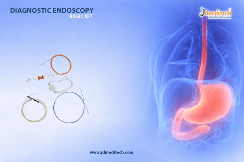
What Is a Biliary Stricture Dilation Set?

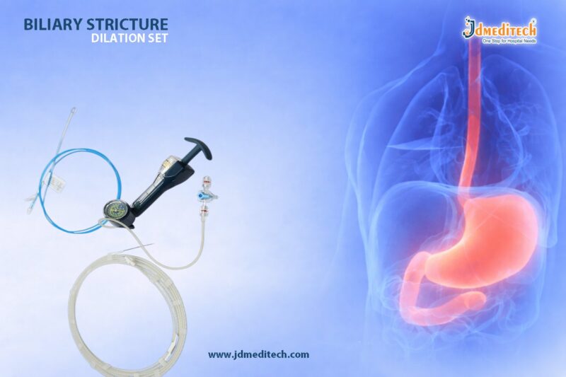
A Biliary Stricture Dilation Set is a collection of specialized devices designed to dilate narrowed segments of the bile duct during endoscopic or percutaneous procedures. Typically, the set includes guidewires, dilation catheters, balloon dilators, introducer components, and procedural accessories.

Furthermore, these systems enable physicians to restore bile flow by gradually expanding the constricted area. As a result, patients often experience symptom relief and improved biliary function.

Why Is Biliary Stricture Dilation Important?

Biliary strictures can restrict bile drainage from the liver to the intestine. Therefore, untreated strictures may increase the risk of infection, liver damage, and recurrent biliary obstruction.

Common indications for biliary dilation include:

  • Benign biliary strictures
  • Post-surgical bile duct narrowing
  • Chronic pancreatitis-related strictures
  • Anastomotic strictures
  • Primary sclerosing cholangitis
  • Bile duct injury repair
  • Obstructive jaundice
  • Preparation for biliary stent placement

Consequently, timely dilation helps restore bile flow and prevents long-term complications.

Components of a Biliary Stricture Dilation Set
Guidewire

The guidewire allows physicians to navigate the biliary anatomy accurately. In addition, it provides a stable pathway for the advancement of dilation devices.

Dilation Catheter

The dilation catheter helps enlarge narrowed bile ducts progressively. Therefore, physicians can perform controlled expansion with precision.

Balloon Dilator

The balloon dilator expands when inflated within the stricture. As a result, the narrowed segment widens gradually and safely.

Introducer System

The introducer system facilitates smooth access to the biliary tract. Moreover, it supports accurate placement of procedural devices.

Contrast Injection Accessories

These components allow clinicians to visualize the biliary anatomy during fluoroscopic imaging. Consequently, they can identify strictures and assess treatment effectiveness.

Procedural Accessories

Additional accessories such as connectors, syringes, and tubing help streamline workflow and maintain sterile conditions.

How Biliary Stricture Dilation Is Performed
Patient Evaluation

First, the physician reviews imaging studies, laboratory findings, and clinical symptoms. Proper evaluation helps determine the severity and location of the stricture.

Biliary Access

Next, the physician accesses the biliary system through an endoscopic or percutaneous approach. Subsequently, a guidewire advances through the narrowed segment.

Stricture Assessment

The physician evaluates the length and severity of the narrowing using contrast imaging. Therefore, an appropriate dilation strategy can be selected.

Device Placement

The physician advances the dilation catheter or balloon over the guidewire and positions it within the stricture.

Controlled Dilation

The physician gradually expands the stricture using the selected dilation device. Consequently, the bile duct diameter increases and bile flow improves.

Final Evaluation

Finally, imaging confirms successful dilation and adequate biliary drainage. Afterward, the physician may place a stent if additional support is necessary.

Benefits of Using a Biliary Stricture Dilation Set
Precise Bile Duct Expansion

The system enables controlled and targeted dilation of strictures. Therefore, physicians can achieve predictable treatment results.

Minimally Invasive Treatment

Unlike traditional surgical procedures, biliary dilation requires only endoscopic or percutaneous access. Consequently, patients often recover faster.

Improved Biliary Drainage

The procedure restores bile flow and reduces symptoms associated with obstruction. As a result, patients experience better digestive and liver function.

Enhanced Procedural Efficiency

A complete dilation set provides all essential components in one package. Moreover, it simplifies procedural preparation and workflow.

Increased Patient Safety

Modern dilation systems support controlled expansion while minimizing trauma to surrounding tissues. Therefore, procedural risks remain lower.

Clinical Applications of Biliary Stricture Dilation Sets
Benign Biliary Stricture Management

Physicians commonly use these systems to treat benign bile duct narrowings caused by inflammation or previous surgery.

Pre-Stent Placement Procedures

Dilation often prepares the bile duct for stent insertion. Consequently, stents can expand more effectively and maintain long-term drainage.

Chronic Pancreatitis-Related Strictures

Patients with chronic pancreatitis frequently develop biliary narrowing. Therefore, dilation helps restore normal bile flow and reduce symptoms.

Post-Operative Bile Duct Narrowing

Following hepatobiliary surgery, some patients develop strictures. In such cases, dilation provides a minimally invasive treatment option.

Features to Consider When Selecting a Biliary Stricture Dilation Set

Healthcare providers should evaluate several factors before selecting a dilation system:

  • High-quality balloon or catheter design
  • Smooth guidewire compatibility
  • Reliable fluoroscopic visibility
  • Controlled expansion capability
  • Sterile packaging
  • Easy device handling
  • Durable construction
  • Compliance with international quality standards

Consequently, choosing a high-quality system can improve procedural success and patient outcomes.

Importance in Modern Biliary Interventions

As minimally invasive biliary procedures continue to evolve, stricture dilation remains a cornerstone of bile duct management. Furthermore, advances in catheter and balloon technology have improved precision, safety, and treatment effectiveness.

Today, gastroenterologists and interventional specialists rely on biliary stricture dilation systems to manage complex biliary disorders efficiently. Ultimately, these specialized sets contribute to better patient care and improved long-term biliary health.

Conclusion

A Biliary Stricture Dilation Set offers a precision solution for safe and effective bile duct expansion. It equips healthcare professionals with the tools required to restore biliary drainage and manage strictures with confidence.

Moreover, these specialized systems improve procedural accuracy, enhance patient safety, and support successful treatment outcomes. As a result, biliary stricture dilation sets remain an essential part of modern gastroenterology and biliary intervention practice.


Explore More Product >>

Get Connected:
+91 79909 93062 | +91 63513 72032 | exports@jdmeditech.com 

Pressure Monitoring & Contrast Delivery Set

Accurate hemodynamic monitoring and reliable contrast media delivery are essential components of modern diagnostic and interventional procedures. Physicians rely on precise pressure measurements and controlled contrast injection to evaluate cardiovascular conditions, guide interventions, and improve patient outcomes. Consequently, healthcare facilities require dependable systems that support both monitoring and imaging procedures.

A Pressure Monitoring & Contrast Delivery Set provides a comprehensive solution for pressure measurement and contrast administration during cardiac catheterization, angiography, vascular interventions, and other diagnostic procedures. Moreover, these advanced systems help clinicians obtain accurate data while maintaining procedural efficiency and patient safety.

What Is a Pressure Monitoring & Contrast Delivery Set?

A Pressure Monitoring & Contrast Delivery Set is a specialized collection of tubing, connectors, manifolds, transducers, and accessories designed to facilitate pressure monitoring and contrast media delivery during diagnostic and interventional procedures.

Furthermore, these systems enable healthcare professionals to measure intravascular pressures accurately while administering contrast agents for imaging studies. As a result, physicians can make informed clinical decisions based on real-time hemodynamic data and high-quality imaging results.

Why Are Pressure Monitoring and Contrast Delivery Important?

Accurate pressure monitoring helps clinicians assess cardiovascular function and vascular status. Meanwhile, contrast media enhances visualization of blood vessels and anatomical structures during imaging procedures.

These systems play a critical role in:

  • Cardiac catheterization procedures
  • Coronary angiography
  • Peripheral angiography
  • Interventional cardiology procedures
  • Structural heart interventions
  • Vascular surgery procedures
  • Hemodynamic assessment
  • Diagnostic imaging studies

Consequently, reliable monitoring and contrast delivery improve diagnostic accuracy and procedural success.

Components of a Pressure Monitoring & Contrast Delivery Set
Pressure Monitoring Line

The pressure monitoring line transmits pressure signals from the patient to the monitoring equipment. Therefore, clinicians can obtain real-time hemodynamic measurements.

Pressure Transducer

The transducer converts pressure signals into electrical data that monitoring systems can interpret. As a result, accurate pressure readings become available throughout the procedure.

Manifold System

The manifold controls fluid flow between monitoring lines, contrast media, and saline solutions. Moreover, it simplifies pressure management and contrast administration.

High-Pressure Tubing

High-pressure tubing supports safe delivery of contrast media during angiographic procedures. Consequently, healthcare professionals can perform injections with confidence.

Stopcocks and Connectors

These components allow clinicians to manage fluid pathways efficiently. Furthermore, they improve procedural flexibility and workflow organization.

Contrast Delivery Components

Contrast delivery accessories facilitate controlled administration of imaging contrast agents. Therefore, physicians can obtain clear and detailed diagnostic images.

How a Pressure Monitoring & Contrast Delivery Set Works
System Preparation

First, the healthcare team assembles and primes the monitoring system. Proper preparation eliminates air bubbles and ensures accurate measurements.

Vascular Access

Next, the physician gains vascular access and positions the diagnostic or interventional catheter. Subsequently, the monitoring line connects to the catheter system.

Pressure Monitoring

The pressure transducer continuously records intravascular pressure values. As a result, clinicians can monitor hemodynamic changes in real time.

Contrast Injection

The physician administers contrast media through the delivery system. Consequently, imaging equipment can capture detailed views of vascular structures.

Data Evaluation

Throughout the procedure, physicians evaluate pressure readings and imaging findings simultaneously. Therefore, they can make precise treatment decisions.

Procedure Completion

Finally, the healthcare team disconnects the system and secures vascular access. Afterward, the patient enters the recovery phase.

Applications of Pressure Monitoring & Contrast Delivery Sets
Cardiac Catheterization

These systems support accurate pressure measurements during diagnostic and interventional cardiac procedures.

Coronary Angiography

Physicians use pressure monitoring and contrast delivery systems to evaluate coronary artery disease and guide treatment planning.

Peripheral Vascular Procedures

The systems help visualize peripheral arteries and assess blood flow abnormalities. Consequently, clinicians can identify vascular diseases effectively.

Structural Heart Interventions

Advanced structural heart procedures require precise hemodynamic monitoring and detailed imaging guidance. Therefore, these systems play a vital role in procedural success.

Benefits of Using a Pressure Monitoring & Contrast Delivery Set
Accurate Hemodynamic Assessment

The system provides reliable pressure measurements throughout the procedure. As a result, clinicians can monitor cardiovascular function effectively.

Enhanced Imaging Quality

Controlled contrast delivery improves visualization of blood vessels and cardiac structures. Consequently, diagnostic accuracy increases significantly.

Improved Workflow Efficiency

A complete set provides all necessary components in one package. Moreover, this reduces setup time and simplifies procedural management.

Increased Procedural Safety

High-quality components support secure fluid management and accurate monitoring. Therefore, healthcare professionals can minimize procedural risks.

Broad Clinical Compatibility

These systems support a wide range of cardiovascular and vascular procedures. Furthermore, they integrate easily into modern catheterization laboratories.

Key Features to Consider When Selecting a Pressure Monitoring & Contrast Delivery Set

When choosing a monitoring and contrast delivery system, healthcare providers should consider:

  • Accurate pressure transducer performance
  • High-pressure tubing reliability
  • Durable manifold construction
  • Easy-to-use stopcock configuration
  • Secure connectors
  • Sterile packaging
  • Fluid management efficiency
  • Compliance with international medical device standards

Consequently, selecting a high-quality system can improve both procedural performance and patient care.

Importance in Modern Interventional Medicine

As cardiovascular and vascular interventions continue to advance, accurate hemodynamic monitoring remains essential for successful outcomes. Furthermore, high-quality imaging depends on precise contrast delivery systems that provide consistent performance.

Today, physicians rely on integrated pressure monitoring and contrast delivery sets to perform complex procedures efficiently and safely. Ultimately, these systems contribute to improved diagnostic accuracy, enhanced treatment planning, and better patient outcomes.

Conclusion

A Pressure Monitoring & Contrast Delivery Set is an essential component of modern diagnostic and interventional procedures. It combines accurate hemodynamic monitoring with reliable contrast administration to support informed clinical decision-making.

Moreover, these advanced systems improve workflow efficiency, enhance imaging quality, and contribute to safer patient care. As a result, pressure monitoring and contrast delivery sets remain indispensable tools in cardiac catheterization laboratories, angiography suites, and vascular intervention centers.


Explore More Product >>

Get Connected:
+91 79909 93062 | +91 63513 72032 | exports@jdmeditech.com 

ERCP Stone Extraction Set for CBD Stone Removal

Common bile duct (CBD) stones can obstruct the normal flow of bile and trigger serious health complications. Patients often experience symptoms such as abdominal pain, jaundice, fever, and pancreatitis. Therefore, healthcare professionals must remove these stones quickly and effectively to prevent further complications.

Endoscopic Retrograde Cholangiopancreatography (ERCP) remains one of the most effective procedures for treating biliary stone disease. Moreover, an ERCP Stone Extraction Set provides physicians with all the essential tools required for safe and efficient stone removal. As a result, these specialized systems have become indispensable in modern therapeutic endoscopy.

Understanding Common Bile Duct Stones

CBD stones develop when gallstones migrate from the gallbladder into the bile duct or form directly within the biliary system. Consequently, these stones can block bile drainage and increase pressure within the biliary tract.

If physicians do not treat the condition promptly, patients may develop:

  • Obstructive jaundice
  • Acute cholangitis
  • Biliary pancreatitis
  • Recurrent biliary pain
  • Bile duct infections
  • Liver-related complications

Therefore, timely stone extraction plays a critical role in restoring normal biliary function.

What Is an ERCP Stone Extraction Set?

An ERCP Stone Extraction Set is a specialized collection of devices designed to remove stones from the common bile duct during ERCP procedures. Typically, the set contains guidewires, extraction balloons, stone retrieval baskets, catheters, and supporting accessories.

Furthermore, these devices help gastroenterologists navigate the biliary system with greater precision. Consequently, physicians can remove stones efficiently while reducing procedural challenges.

Key Components of an ERCP Stone Extraction Set
Guidewire

The guidewire helps physicians access and navigate the biliary tract. In addition, it creates a stable pathway for other devices.

Stone Extraction Balloon

The extraction balloon sweeps stones from the bile duct. Physicians position the balloon above the stone, inflate it, and gently pull it toward the intestine.

Stone Retrieval Basket

The basket captures and removes stones that balloons cannot easily extract. Therefore, it provides additional flexibility during complex procedures.

ERCP Catheter

The catheter delivers contrast media and supports guidewire placement. As a result, physicians can visualize the biliary anatomy clearly.

Contrast Injection Accessories

These accessories help clinicians identify stone location and evaluate ductal anatomy under fluoroscopic guidance.

Procedural Accessories

Additional accessories such as syringes, tubing, and connectors improve workflow efficiency and maintain sterile conditions throughout the procedure.

How ERCP Stone Extraction Works
Patient Preparation

First, the healthcare team evaluates the patient through imaging studies, blood tests, and clinical assessment. This preparation helps physicians plan the procedure effectively.

Endoscope Advancement

Next, the physician advances a duodenoscope through the mouth, esophagus, and stomach to reach the duodenum.

Biliary Cannulation

The physician inserts a guidewire and catheter into the common bile duct. Consequently, the treatment area becomes accessible.

Cholangiogram Assessment

The physician injects contrast media into the biliary system and obtains fluoroscopic images. Therefore, the exact size and location of the stones become visible.

Stone Removal

The physician uses a balloon catheter or retrieval basket to extract the stones. In many cases, the physician clears multiple stones during a single procedure.

Final Inspection

Finally, the physician performs a follow-up cholangiogram to confirm complete stone clearance. Afterward, the physician completes the procedure and monitors the patient during recovery.

Benefits of Using an ERCP Stone Extraction Set
Supports Efficient Stone Removal

The specialized design of extraction devices allows physicians to remove stones quickly and effectively. As a result, procedure times often decrease.

Promotes Minimally Invasive Treatment

ERCP eliminates the need for open surgery in many patients. Consequently, recovery times remain shorter and hospital stays become less frequent.

Improves Procedural Accuracy

The combination of guidewires, catheters, balloons, and baskets helps physicians perform stone extraction with greater precision.

Enhances Patient Safety

Modern extraction devices give physicians better control during stone retrieval. Therefore, they can minimize trauma to surrounding tissues.

Streamlines Clinical Workflow

A complete extraction set provides all required components in one package. Moreover, it reduces preparation time and improves procedural organization.

Clinical Applications of ERCP Stone Extraction Sets
Choledocholithiasis Treatment

Physicians frequently use these systems to remove stones from the common bile duct and restore bile flow.

Management of Obstructive Jaundice

When stones block the bile duct, ERCP extraction helps relieve the obstruction and reduce jaundice symptoms.

Treatment of Acute Cholangitis

Physicians often perform emergency ERCP procedures to remove infected biliary obstructions and restore drainage.

Post-Cholecystectomy Stone Management

Some patients develop retained bile duct stones after gallbladder surgery. In such cases, ERCP offers an effective treatment solution.

Features to Consider When Selecting an ERCP Stone Extraction Set

Healthcare providers should evaluate several factors before choosing an extraction system:

  • High-quality extraction balloon
  • Durable stone retrieval basket
  • Smooth guidewire compatibility
  • Reliable catheter performance
  • Sterile packaging
  • Easy device handling
  • Excellent fluoroscopic visibility
  • Compliance with international quality standards

Consequently, a high-quality extraction set can improve both procedural success and patient outcomes.

Importance in Modern Gastroenterology

As therapeutic endoscopy continues to evolve, physicians increasingly rely on ERCP for biliary stone management. Furthermore, advances in extraction technology have improved both efficiency and safety.

Today, gastroenterologists use ERCP stone extraction systems to treat complex biliary disorders with confidence. Ultimately, these specialized kits help physicians deliver faster treatment, achieve higher stone clearance rates, and improve patient care.

Conclusion

An ERCP Stone Extraction Set provides a complete solution for safe and efficient CBD stone removal. It equips healthcare professionals with the tools needed to perform precise and effective therapeutic ERCP procedures.

Moreover, these specialized systems support minimally invasive treatment, improve procedural efficiency, and enhance patient outcomes. As a result, ERCP stone extraction sets remain an essential part of modern gastroenterology and biliary intervention practice.


Explore More Product >>

Get Connected:
+91 79909 93062 | +91 63513 72032 | exports@jdmeditech.com 

Thrombus Management Set for Acute Clot Removal

Acute thrombotic occlusions can lead to serious and potentially life-threatening complications if left untreated. Blood clots that obstruct arteries or veins may restrict blood flow to critical organs and tissues, increasing the risk of conditions such as deep vein thrombosis, pulmonary embolism, myocardial infarction, and acute limb ischemia. Consequently, rapid diagnosis and effective thrombus removal are essential for preserving tissue function and improving patient outcomes.

A Thrombus Management Set provides healthcare professionals with the specialized tools required to perform acute thrombus removal procedures efficiently and safely. Moreover, these comprehensive systems support a variety of minimally invasive techniques designed to restore blood flow and reduce thrombotic burden.

What Is a Thrombus Management Set?

A Thrombus Management Set is a collection of interventional devices and procedural accessories used to remove or manage blood clots within the vascular system. Typically, the set includes catheters, guidewires, introducer components, aspiration systems, and other accessories required for thrombus management procedures.

Furthermore, these systems are widely used in interventional cardiology, vascular surgery, interventional radiology, and endovascular procedures. As a result, clinicians can perform targeted clot removal while minimizing procedural complexity.

Why Is Thrombus Removal Important?

A thrombus can significantly impair blood circulation and may cause severe complications if not treated promptly. Therefore, early intervention is often necessary to restore normal blood flow and prevent tissue damage.

Common conditions requiring thrombus management include:

  • Acute limb ischemia
  • Deep vein thrombosis (DVT)
  • Pulmonary embolism
  • Coronary artery thrombosis
  • Peripheral arterial occlusions
  • Dialysis access thrombosis
  • Venous thromboembolic disease
  • Acute vascular emergencies

Consequently, effective thrombus removal can reduce complications and improve long-term patient outcomes.

Components of a Thrombus Management Set
Aspiration Catheter

The aspiration catheter is designed to remove thrombotic material from the vessel lumen. As a result, blood flow can be restored more rapidly.

Guidewire

The guidewire facilitates safe navigation through the vascular anatomy. Furthermore, it supports accurate placement of thrombus management devices.

Introducer Sheath

The introducer sheath provides secure vascular access and enables smooth insertion of interventional devices.

Aspiration Syringe or Vacuum System

These components generate suction for thrombus extraction. Consequently, clot burden can be reduced efficiently during the procedure.

Dilators

Dilators assist in vessel access and help facilitate smooth device introduction. Moreover, they contribute to procedural efficiency.

Hemostasis Components

Hemostasis accessories help manage bleeding at the vascular access site. Therefore, patient safety is enhanced during and after the procedure.

How Acute Thrombus Removal Procedures Are Performed
Patient Assessment

First, the physician evaluates the patient’s symptoms, imaging findings, and clinical condition. Proper assessment helps determine the most appropriate treatment strategy.

Vascular Access

Next, vascular access is obtained through an appropriate artery or vein. Subsequently, an introducer sheath is placed to facilitate device advancement.

Guidewire Navigation

A guidewire is advanced through the affected vessel and positioned beyond the thrombus. As a result, treatment devices can be delivered accurately.

Device Placement

The aspiration catheter or thrombectomy device is carefully advanced to the clot location. Therefore, targeted thrombus removal can begin.

Thrombus Extraction

The physician removes thrombotic material using aspiration or other clot-management techniques. Consequently, vessel patency can be restored.

Final Evaluation

Finally, imaging is performed to assess blood flow and confirm successful clot removal. Afterward, the devices are withdrawn, and hemostasis is achieved.

Applications of Thrombus Management Sets
Interventional Cardiology

These systems are commonly used to manage thrombotic complications during coronary interventions. As a result, coronary blood flow can be restored effectively.

Peripheral Vascular Procedures

Thrombus management sets play an important role in treating peripheral arterial and venous occlusions.

Interventional Radiology

Interventional radiologists frequently utilize these systems for minimally invasive clot removal procedures. Consequently, many patients can avoid more invasive surgical treatments.

Dialysis Access Management

Thrombus removal techniques are often used to restore functionality in thrombosed dialysis access sites. Therefore, continuity of dialysis treatment can be maintained.

Benefits of Using a Thrombus Management Set
Rapid Restoration of Blood Flow

The primary goal of thrombus removal is to re-establish circulation quickly. As a result, tissue damage and ischemic complications may be reduced.

Minimally Invasive Treatment

Many thrombus management procedures are performed through catheter-based techniques. Consequently, patients often experience shorter hospital stays and faster recovery.

Enhanced Procedural Efficiency

A complete kit provides all necessary components in one package. Moreover, this streamlines workflow and reduces preparation time.

Improved Clinical Outcomes

Early clot removal can help preserve organ and tissue function. Therefore, patient outcomes may improve significantly.

Versatile Clinical Applications

These systems can be used across multiple specialties and treatment settings. Furthermore, they support a wide range of thrombus management techniques.

Key Features to Consider When Selecting a Thrombus Management Set

When choosing a thrombus management system, healthcare providers should consider:

  • High-performance aspiration capability
  • Smooth catheter tracking
  • Reliable guidewire compatibility
  • Efficient clot extraction design
  • Sterile packaging
  • Biocompatible materials
  • Ease of handling
  • Compliance with international medical device standards

Consequently, selecting a high-quality system can improve procedural performance and treatment success.

Importance in Modern Endovascular Therapy

As minimally invasive vascular interventions continue to advance, thrombus management systems have become essential tools in acute care settings. Furthermore, improvements in catheter technology and aspiration techniques have enhanced both procedural safety and effectiveness.

Today, physicians rely on specialized thrombus management sets to address complex vascular occlusions rapidly and efficiently. Ultimately, these systems contribute to improved patient care, reduced complication rates, and better clinical outcomes.

Conclusion

A Thrombus Management Set is a vital component of modern vascular and endovascular intervention. It provides healthcare professionals with the tools necessary to perform acute thrombus removal procedures safely, efficiently, and effectively.

Moreover, these specialized systems support rapid restoration of blood flow, enhance procedural precision, and improve patient outcomes. As a result, thrombus management sets continue to play a critical role in the treatment of acute thrombotic conditions across multiple medical specialties.


Explore More Product >>

Get Connected:
+91 79909 93062 | +91 63513 72032 | exports@jdmeditech.com 

Esophageal Bougie Dilation Set for Stricture Treatment

Esophageal strictures can significantly affect swallowing function and overall quality of life. These narrowed sections of the esophagus may develop due to chronic acid reflux, surgical scarring, radiation therapy, or various gastrointestinal disorders. Consequently, patients often experience difficulty swallowing, discomfort, and nutritional challenges.

Esophageal dilation is one of the most effective methods for restoring the normal diameter of the esophagus. Among the available treatment options, an Esophageal Bougie Dilation Set provides a structured and controlled approach to progressive esophageal expansion. Moreover, these specialized sets enable healthcare professionals to treat esophageal narrowing with precision and consistency.

What Is an Esophageal Bougie Dilation Set?

An Esophageal Bougie Dilation Set is a collection of graduated dilators designed to widen narrowed segments of the esophagus gradually. Typically, the set contains multiple bougie dilators of increasing diameters that allow progressive expansion of strictures during treatment.

Furthermore, these sets are widely used in gastroenterology and endoscopy procedures to manage benign esophageal narrowing. As a result, physicians can improve esophageal patency while minimizing procedural risks.

Why Is Esophageal Dilation Necessary?

Esophageal narrowing can restrict the passage of food and liquids from the mouth to the stomach. Therefore, patients may experience symptoms that interfere with daily eating and drinking activities.

Common conditions requiring esophageal dilation include:

  • Benign esophageal strictures
  • Peptic strictures caused by gastroesophageal reflux disease (GERD)
  • Post-surgical esophageal narrowing
  • Radiation-induced strictures
  • Esophageal rings and webs
  • Anastomotic strictures
  • Eosinophilic esophagitis
  • Chronic swallowing difficulties (dysphagia)

Consequently, progressive dilation can help restore normal swallowing and improve patient comfort.

Components of an Esophageal Bougie Dilation Set
Bougie Dilators

Bougie dilators are the primary components of the set. They are available in multiple sizes and are designed to gradually expand the narrowed esophageal segment. As a result, controlled dilation can be achieved without excessive force.

Guidewire Compatibility

Many modern bougie systems are designed for guidewire-assisted procedures. Therefore, physicians can achieve greater accuracy during dilator placement.

Graduated Sizing System

The structured sizing sequence allows clinicians to increase dilation progressively. Furthermore, this approach helps reduce the risk of complications.

Flexible Construction

High-quality bougie dilators are manufactured from flexible materials that support smooth passage through the esophagus. Consequently, patient comfort and procedural safety are improved.

Sterile Packaging

Most bougie dilation systems are supplied in sterile packaging. In addition, this helps maintain infection control standards throughout the procedure.

How Esophageal Bougie Dilation Is Performed
Patient Evaluation

First, the physician reviews the patient’s medical history, symptoms, and diagnostic findings. Proper assessment helps determine the severity and location of the stricture.

Endoscopic Examination

Next, an endoscopic evaluation may be performed to visualize the narrowing. Consequently, the physician can plan the appropriate dilation strategy.

Guidewire Placement

If a guidewire-assisted technique is used, the guidewire is advanced through the narrowed segment. Subsequently, it serves as a pathway for the bougie dilators.

Progressive Dilation

The physician introduces progressively larger bougie dilators through the esophagus. Therefore, the narrowed area expands gradually and safely.

Treatment Assessment

After achieving the desired diameter, the physician evaluates the treatment outcome. Moreover, additional dilation sessions may be scheduled if necessary.

Procedure Completion

Finally, the dilators are removed, and the patient is monitored for recovery. As a result, most patients can resume normal activities shortly after the procedure.

Benefits of Using an Esophageal Bougie Dilation Set
Controlled Progressive Expansion

The graduated sizing system allows gradual dilation of strictures. Therefore, physicians can achieve precise treatment outcomes.

Effective Stricture Management

Bougie dilation remains a proven method for treating various benign esophageal narrowings. Consequently, it is widely utilized in gastroenterology practice.

Improved Swallowing Function

By restoring the esophageal diameter, the procedure enhances the passage of food and liquids. As a result, patients experience better swallowing comfort.

Minimally Invasive Treatment

Unlike surgical interventions, bougie dilation can often be performed endoscopically. Moreover, this contributes to faster recovery times.

Reliable Clinical Performance

The structured design of the dilation set supports consistent treatment results. Furthermore, it enables healthcare professionals to perform procedures efficiently.

Applications of Esophageal Bougie Dilation Sets
Gastroenterology Procedures

These sets are commonly used for managing benign esophageal strictures and related swallowing disorders.

Endoscopy Centers

Endoscopy specialists frequently utilize bougie dilators as part of comprehensive esophageal treatment programs.

Post-Surgical Stricture Treatment

Patients who develop narrowing after esophageal surgery often benefit from progressive bougie dilation. Consequently, normal swallowing function can be restored.

Radiation-Induced Esophageal Narrowing

Radiation therapy may cause scar tissue formation within the esophagus. Therefore, bougie dilation can provide effective symptom relief.

Bougie Dilation vs Balloon Dilation
Progressive Mechanical Expansion

Bougie dilators widen the esophagus through gradual mechanical expansion. In contrast, balloon systems rely on radial pressure generated by balloon inflation.

Multiple Diameter Options

Bougie sets provide several dilator sizes within a single system. As a result, physicians can tailor treatment according to patient requirements.

Proven Clinical Technique

Bougie dilation has been used successfully for many years in gastroenterology practice. Furthermore, it remains a trusted treatment option for many benign strictures.

Key Features to Consider When Selecting an Esophageal Bougie Dilation Set

When choosing an esophageal bougie dilation system, healthcare providers should consider:

  • Comprehensive size range
  • Flexible and durable construction
  • Smooth surface finish
  • Guidewire compatibility
  • Sterile packaging
  • Ease of handling
  • Consistent diameter progression
  • Compliance with international medical device standards

Therefore, selecting a high-quality dilation set can improve both procedural performance and patient outcomes.

Importance in Modern Gastroenterology

As minimally invasive gastrointestinal treatments continue to advance, esophageal bougie dilation remains an essential therapeutic technique. Furthermore, modern bougie systems provide greater precision, improved safety, and enhanced procedural efficiency.

Today, gastroenterologists rely on these structured dilation sets to manage esophageal strictures effectively. Ultimately, they play a crucial role in restoring swallowing function and improving patient quality of life.

Conclusion

An Esophageal Bougie Dilation Set is a valuable solution for the progressive treatment of esophageal strictures and swallowing disorders. It provides healthcare professionals with a structured approach to controlled esophageal expansion while supporting procedural safety and effectiveness.

Moreover, these specialized sets enhance treatment precision, improve patient comfort, and contribute to successful clinical outcomes. As a result, esophageal bougie dilation continues to be an important component of modern gastroenterology and endoscopic care.


Explore More Product >>

Get Connected:
+91 79909 93062 | +91 63513 72032 | exports@jdmeditech.com 

Coronary Stenting Set for Angioplasty Procedures

Coronary artery disease (CAD) remains one of the leading causes of cardiovascular complications worldwide. When plaque buildup narrows or blocks coronary arteries, blood flow to the heart muscle can become restricted, leading to chest pain, shortness of breath, or even heart attacks. Consequently, timely intervention is essential to restore adequate blood circulation and prevent severe cardiac events.

Coronary angioplasty and stenting procedures have become highly effective minimally invasive treatments for coronary artery blockages. Moreover, a Coronary Stenting Set provides healthcare professionals with the essential instruments required to perform these procedures safely and efficiently. As a result, these specialized kits play a vital role in modern interventional cardiology.

What Is a Coronary Stenting Set?

A Coronary Stenting Set is a comprehensive collection of medical devices and accessories used during percutaneous coronary intervention (PCI), commonly known as coronary angioplasty and stenting. Typically, the set includes guidewires, guiding catheters, introducer components, inflation devices, and other procedural accessories.

Furthermore, these kits are designed to support smooth workflow and precise device delivery during coronary interventions. Therefore, physicians can effectively treat narrowed or blocked coronary arteries while minimizing procedural complexity.

Why Are Coronary Angioplasty and Stenting Important?

Coronary artery narrowing can significantly reduce blood supply to the heart. Therefore, patients may experience symptoms that affect both quality of life and cardiovascular health.

Coronary angioplasty and stenting are commonly performed for:

  • Coronary artery disease (CAD)
  • Acute myocardial infarction (heart attack)
  • Chronic coronary artery blockages
  • Angina (chest pain)
  • Ischemic heart disease
  • Coronary restenosis
  • Multivessel coronary disease
  • Emergency cardiac interventions

Consequently, these procedures help restore blood flow, reduce symptoms, and improve overall cardiac function.

Components of a Coronary Stenting Set
Guiding Catheter

The guiding catheter provides access to the coronary arteries and serves as a pathway for interventional devices. As a result, physicians can accurately navigate through complex vascular anatomy.

Guidewire

The guidewire is carefully advanced through the narrowed coronary artery. Furthermore, it acts as a track for balloons, stents, and other devices.

Balloon Catheter

Balloon catheters are used to dilate narrowed arterial segments before or during stent placement. Therefore, adequate vessel expansion can be achieved.

Stent Delivery System

The stent delivery system enables accurate positioning and deployment of the coronary stent. Consequently, the treated artery remains open and supports improved blood flow.

Inflation Device

The inflation device controls balloon pressure during angioplasty and stent deployment. Moreover, it allows precise expansion according to procedural requirements.

Introducer Sheath

The introducer sheath facilitates vascular access and supports smooth insertion of catheters and guidewires.

Hemostasis Accessories

These components help maintain blood control during and after the procedure. As a result, procedural safety and efficiency are enhanced.

How Coronary Angioplasty and Stenting Are Performed
Patient Preparation

First, the patient undergoes clinical evaluation, imaging studies, and laboratory assessments. Proper preparation helps ensure procedural success.

Vascular Access

Next, the physician gains access through the radial or femoral artery. Subsequently, an introducer sheath is inserted to facilitate device advancement.

Coronary Artery Navigation

A guiding catheter is positioned at the coronary artery opening. Meanwhile, a guidewire is advanced through the narrowed or blocked vessel segment.

Balloon Angioplasty

The balloon catheter is positioned within the blockage and inflated carefully. Therefore, the narrowed artery can be expanded.

Stent Deployment

Once adequate vessel preparation is achieved, the stent is delivered and deployed at the treatment site. Consequently, the artery remains open and blood flow improves.

Final Assessment

Finally, angiographic imaging confirms successful stent placement and vessel patency. Afterward, the devices are removed, and vascular access is secured.

Benefits of Using a Coronary Stenting Set
Improved Procedural Efficiency

A complete kit provides all essential components in a single package. Therefore, procedural preparation and workflow become more efficient.

Precise Device Placement

Specialized tools support accurate navigation and stent deployment. As a result, treatment outcomes are improved.

Enhanced Patient Safety

Modern coronary intervention systems are designed to minimize procedural risks. Moreover, they help physicians perform complex procedures with greater confidence.

Minimally Invasive Treatment

Unlike open-heart surgery, coronary angioplasty and stenting require only small vascular access points. Consequently, patients often experience faster recovery times.

Reliable Clinical Performance

High-quality components support consistent procedural success. Furthermore, they contribute to better long-term cardiovascular outcomes.

Applications of Coronary Stenting Sets
Elective Coronary Interventions

These kits are widely used for planned angioplasty and stenting procedures in patients with stable coronary artery disease.

Emergency Cardiac Care

Coronary stenting sets play a critical role in emergency treatment of acute myocardial infarction. Therefore, rapid restoration of blood flow can help preserve heart muscle function.

Complex Coronary Procedures

Interventional cardiologists frequently use these systems for multivessel disease and challenging coronary anatomy. Consequently, they remain indispensable in advanced cardiac care.

Hospital Cardiac Catheterization Laboratories

Cardiac catheterization labs rely on comprehensive stenting kits to support daily interventional procedures and optimize workflow.

Key Features to Consider When Selecting a Coronary Stenting Set

When choosing a coronary stenting system, healthcare providers should consider:

  • High-performance guidewire compatibility
  • Smooth catheter navigation
  • Reliable balloon inflation systems
  • Precision stent delivery capability
  • Sterile packaging
  • Biocompatible materials
  • Ease of handling
  • Compliance with international medical device standards

As a result, selecting a high-quality coronary stenting set can improve both procedural efficiency and patient outcomes.

Importance in Modern Cardiac Care

As cardiovascular disease continues to affect millions of people worldwide, coronary angioplasty and stenting remain among the most effective treatment options. Furthermore, technological advancements have significantly improved the safety and success rates of these procedures.

Today, interventional cardiologists depend on specialized coronary stenting sets to perform life-saving procedures with precision and confidence. Ultimately, these kits contribute to improved patient care, reduced complications, and enhanced cardiovascular health.

Conclusion

A Coronary Stenting Set is an essential cardiac care kit designed to support coronary angioplasty and stenting procedures. It provides healthcare professionals with the tools required to restore coronary blood flow safely and effectively.

Moreover, these specialized kits enhance procedural efficiency, improve treatment accuracy, and support successful cardiovascular interventions. As a result, coronary stenting sets continue to play a crucial role in modern interventional cardiology and patient-centered cardiac care.


Explore More Product >>

Get Connected:
+91 79909 93062 | +91 63513 72032 | exports@jdmeditech.com 

Esophageal Balloon Dilation Kit for Stricture Management

Esophageal strictures can significantly affect a patient’s ability to swallow and maintain proper nutrition. These narrowings of the esophagus often result from chronic acid reflux, post-surgical scarring, radiation therapy, or other gastrointestinal conditions. Consequently, timely diagnosis and treatment are essential to restore normal esophageal function and improve quality of life.

An Esophageal Balloon Dilation Kit provides healthcare professionals with a safe and effective solution for managing esophageal strictures. Moreover, these specialized kits enable controlled dilation of narrowed esophageal segments, helping patients experience improved swallowing and reduced discomfort.

What Is an Esophageal Balloon Dilation Kit?

An Esophageal Balloon Dilation Kit is a collection of medical devices specifically designed to dilate narrowed areas of the esophagus. Typically, the kit includes a balloon dilator, inflation device, guidewire, and other accessories required for controlled expansion of strictures.

Furthermore, these kits are commonly used during endoscopic procedures to restore the normal diameter of the esophagus. As a result, physicians can effectively treat swallowing difficulties while minimizing patient discomfort.

Why Is Esophageal Balloon Dilation Necessary?

Esophageal strictures can restrict the passage of food and liquids through the digestive tract. Therefore, patients may experience symptoms that affect their daily activities and overall health.

Common indications for esophageal balloon dilation include:

  • Benign esophageal strictures
  • Peptic strictures caused by acid reflux
  • Post-surgical esophageal narrowing
  • Radiation-induced strictures
  • Anastomotic strictures
  • Esophageal rings and webs
  • Eosinophilic esophagitis-related narrowing
  • Difficulty swallowing (dysphagia)

Consequently, balloon dilation often serves as an effective minimally invasive treatment option for restoring esophageal patency.

Components of an Esophageal Balloon Dilation Kit
Balloon Dilator

The balloon dilator is the primary component of the kit. It expands in a controlled manner within the narrowed section of the esophagus, thereby widening the passage and improving swallowing function.

Inflation Device

The inflation device allows precise control of balloon pressure. Furthermore, it helps physicians achieve gradual and accurate dilation.

Guidewire

The guidewire facilitates accurate placement of the balloon catheter through the stricture. As a result, procedural precision and safety are enhanced.

Balloon Catheter

The catheter delivers the balloon to the targeted treatment area. Moreover, it allows smooth navigation through the gastrointestinal tract.

Contrast Media Port

Some systems include ports for contrast media injection. Therefore, clinicians can verify balloon positioning under fluoroscopic guidance when necessary.

Sterile Accessories

Additional accessories may include syringes, connectors, and tubing. In addition, these components support procedural efficiency and infection control.

How Esophageal Balloon Dilation Is Performed
Patient Evaluation

First, the physician evaluates the patient’s symptoms and diagnostic findings. Proper assessment helps determine the severity and location of the stricture.

Endoscopic Access

Next, an endoscope is introduced into the esophagus to visualize the narrowed segment. Consequently, the treatment area can be accurately identified.

Guidewire Placement

A guidewire is carefully advanced through the stricture. Subsequently, it serves as a pathway for balloon catheter placement.

Balloon Positioning

The balloon catheter is advanced over the guidewire and positioned within the narrowed area. Therefore, targeted treatment can be achieved.

Controlled Balloon Inflation

The physician gradually inflates the balloon to a predetermined diameter and pressure. As a result, the stricture is dilated safely and effectively.

Procedure Completion

Finally, the balloon is deflated and removed. The physician then assesses the treatment outcome and confirms adequate esophageal expansion.

Benefits of Using an Esophageal Balloon Dilation Kit
Controlled Dilation

The balloon system provides gradual and controlled expansion of the stricture. Therefore, physicians can achieve precise treatment outcomes.

Minimally Invasive Procedure

Unlike surgical interventions, balloon dilation is performed endoscopically. Consequently, patients often experience shorter recovery times.

Improved Swallowing Function

By widening the narrowed segment, balloon dilation helps restore normal food and liquid passage. As a result, patients can eat and drink more comfortably.

Enhanced Patient Safety

Modern balloon dilation systems are designed for precise pressure control. Moreover, this reduces the risk of excessive tissue trauma.

Efficient Clinical Workflow

Complete kits provide all essential components in one package. Therefore, healthcare professionals can perform procedures more efficiently.

Applications of Esophageal Balloon Dilation Kits
Gastroenterology Procedures

These kits are widely used in gastroenterology departments for the treatment of benign esophageal strictures and related conditions.

Endoscopy Centers

Endoscopy specialists frequently utilize balloon dilation systems to manage swallowing disorders and gastrointestinal narrowing.

Post-Surgical Stricture Management

Following certain esophageal surgeries, scar tissue formation may cause narrowing. Consequently, balloon dilation can help restore esophageal function.

Radiation-Induced Stricture Treatment

Patients who undergo radiation therapy may develop esophageal narrowing. Therefore, balloon dilation often provides effective symptom relief.

Key Features to Consider When Selecting an Esophageal Balloon Dilation Kit

When choosing an esophageal balloon dilation system, healthcare providers should consider:

  • Controlled radial expansion technology
  • High-quality balloon material
  • Accurate pressure monitoring capability
  • Smooth catheter tracking performance
  • Guidewire compatibility
  • Sterile packaging
  • Ease of use
  • Compliance with international medical device standards

As a result, selecting a reliable dilation kit can improve both procedural outcomes and patient satisfaction.

Importance in Modern Gastroenterology

As minimally invasive gastrointestinal procedures continue to evolve, balloon dilation remains a preferred treatment option for many esophageal strictures. Furthermore, advancements in balloon technology have improved treatment precision and procedural safety.

Today, gastroenterologists rely on these specialized kits to manage complex esophageal narrowing with confidence. Ultimately, esophageal balloon dilation systems contribute significantly to effective stricture management and enhanced patient care.

Conclusion

An Esophageal Balloon Dilation Kit is an essential tool for the controlled treatment of esophageal strictures and swallowing disorders. It enables healthcare professionals to perform precise, minimally invasive dilation procedures while supporting patient safety and comfort.

Moreover, these specialized kits improve procedural efficiency, enhance treatment accuracy, and promote better clinical outcomes. As a result, they remain a valuable component of modern gastroenterology and endoscopic practice.


Explore More Product >>

Get Connected:
+91 79909 93062 | +91 63513 72032 | exports@jdmeditech.com 

Transseptal Puncture Set for Heart Procedures

Modern cardiac interventions increasingly rely on minimally invasive techniques to diagnose and treat complex heart conditions. Among these procedures, transseptal access has become a critical step for reaching the left atrium and other left-sided cardiac structures. Consequently, healthcare professionals require specialized instruments that ensure precision, safety, and procedural efficiency.

A Transseptal Puncture Set is specifically designed to facilitate safe access through the interatrial septum during electrophysiology and structural heart procedures. Moreover, these sets provide clinicians with the tools necessary to perform complex interventions with greater confidence and accuracy.

What Is a Transseptal Puncture Set?

A Transseptal Puncture Set is a specialized collection of medical devices used to gain access from the right atrium to the left atrium through the interatrial septum. Typically, the set includes a transseptal needle, introducer sheath, dilator, and guidewire components.

Furthermore, these kits are widely used in cardiac catheterization laboratories and electrophysiology suites. As a result, physicians can safely access the left side of the heart for various diagnostic and therapeutic procedures.

Why Is Transseptal Access Important?

Transseptal puncture provides a minimally invasive route to the left atrium. Therefore, physicians can perform advanced cardiac procedures without requiring open-heart surgery.

Additionally, transseptal access is essential for:

  • Atrial fibrillation ablation
  • Left atrial mapping procedures
  • Mitral valve interventions
  • Left atrial appendage closure
  • Structural heart defect repairs
  • Electrophysiology studies
  • Catheter-based cardiac therapies
  • Complex cardiac diagnostic procedures

Consequently, transseptal techniques have become fundamental to modern interventional cardiology and electrophysiology.

Components of a Transseptal Puncture Set
Transseptal Needle

The transseptal needle is the primary instrument used to puncture the interatrial septum. Furthermore, it is engineered to provide controlled penetration while minimizing tissue trauma.

Introducer Sheath

The introducer sheath provides a stable pathway for advancing catheters and other interventional devices into the left atrium. As a result, procedural efficiency is significantly improved.

Dilator

The dilator helps facilitate smooth insertion of the sheath into the vascular system. Moreover, it reduces resistance during device advancement.

Guidewire

The guidewire supports accurate navigation through the vascular anatomy. Therefore, physicians can position devices with greater precision.

Hemostatic Valve System

Many transseptal systems include hemostatic valves that help maintain blood control during catheter exchanges. Consequently, procedural safety is enhanced.

Accessory Components

Additional accessories may include connectors, flushing systems, and procedural aids. In addition, these components contribute to a streamlined workflow.

How a Transseptal Puncture Procedure Is Performed
Patient Preparation

First, the patient undergoes clinical evaluation and imaging assessment. Proper preparation helps ensure procedural success and patient safety.

Venous Access

Next, the physician gains venous access, typically through the femoral vein. Subsequently, guidewires and sheaths are advanced toward the right atrium.

Septal Localization

Using fluoroscopy, intracardiac echocardiography, or transesophageal echocardiography, the interatrial septum is identified. Therefore, the puncture site can be accurately targeted.

Transseptal Puncture

The transseptal needle is carefully advanced through the sheath and positioned against the septum. Once the optimal location is confirmed, the physician performs the puncture.

Left Atrial Access

After successful puncture, the sheath is advanced into the left atrium. Consequently, diagnostic or therapeutic catheters can be introduced safely.

Procedure Completion

Finally, the intended cardiac intervention is performed. Upon completion, all devices are removed carefully, and vascular access is secured.

Applications of Transseptal Puncture Sets
Electrophysiology Procedures

Transseptal puncture is widely used during atrial fibrillation ablation and other electrophysiology procedures. As a result, physicians can access the left atrium for mapping and treatment.

Structural Heart Interventions

Many structural heart procedures require left atrial access. Therefore, transseptal puncture sets play a vital role in minimally invasive structural heart therapies.

Left Atrial Appendage Closure

These procedures require accurate navigation within the left atrium. Furthermore, transseptal access enables safe device deployment.

Mitral Valve Procedures

Catheter-based mitral valve repair and replacement procedures frequently utilize transseptal techniques. Consequently, patients may benefit from less invasive treatment options.

Benefits of Using a Transseptal Puncture Set
Precise Cardiac Access

The specialized design of the set enables accurate entry into the left atrium. Therefore, procedural precision is improved.

Enhanced Procedural Safety

Modern transseptal systems are designed to support controlled septal puncture. As a result, the risk of complications can be minimized.

Improved Workflow Efficiency

A complete kit provides all essential components in a single package. Moreover, this reduces setup time and improves procedural organization.

Minimally Invasive Treatment Support

Transseptal access facilitates catheter-based interventions that avoid open-heart surgery. Consequently, patients often experience shorter recovery times.

Broad Clinical Applications

These sets support numerous electrophysiology and structural heart procedures. Therefore, they are valuable tools in modern cardiac care.

Key Features to Consider When Selecting a Transseptal Puncture Set

When choosing a transseptal puncture system, healthcare providers should consider:

  • High-quality transseptal needle design
  • Smooth sheath and dilator performance
  • Reliable guidewire compatibility
  • Hemostatic valve functionality
  • Sterile packaging
  • Biocompatible materials
  • Ease of handling
  • Compliance with international medical device standards

As a result, selecting a high-quality set can improve both procedural outcomes and clinician confidence.

Importance in Modern Cardiac Care

As minimally invasive cardiac procedures continue to evolve, transseptal access remains an essential technique. Furthermore, advancements in transseptal puncture technology have enhanced procedural accuracy and safety.

Today, electrophysiologists and interventional cardiologists rely on these specialized sets to perform complex procedures efficiently. Ultimately, transseptal puncture systems contribute significantly to improved patient care and successful cardiac interventions.

Conclusion

A Transseptal Puncture Set is an indispensable tool for electrophysiology and structural heart procedures. It provides safe and reliable access to the left atrium while supporting a wide range of diagnostic and therapeutic interventions.

Moreover, these specialized kits enhance procedural precision, improve workflow efficiency, and support minimally invasive treatment approaches. As a result, they play a crucial role in modern cardiac care and contribute to better clinical outcomes for patients undergoing advanced heart procedures.


Explore More Product >>

Get Connected:
+91 79909 93062 | +91 63513 72032 | exports@jdmeditech.com 

Diagnostic Endoscopy Basic Kit – Essential GI Tools

Diagnostic endoscopy has become one of the most effective methods for examining the gastrointestinal (GI) tract. It allows healthcare professionals to visualize internal organs, identify abnormalities, and diagnose digestive disorders with precision. Consequently, hospitals and diagnostic centers rely on specialized equipment to ensure safe and efficient procedures.

A Diagnostic Endoscopy Basic Kit contains the essential instruments required for routine gastrointestinal examinations. Moreover, these kits help clinicians perform diagnostic procedures accurately while maintaining patient safety and procedural efficiency.

What Is a Diagnostic Endoscopy Basic Kit?

A Diagnostic Endoscopy Basic Kit is a collection of medical instruments and accessories used during endoscopic examinations of the digestive system. Typically, the kit supports procedures involving the esophagus, stomach, duodenum, colon, and other gastrointestinal structures.

Furthermore, these kits are designed to assist physicians in obtaining clear visualization and collecting diagnostic information. As a result, healthcare providers can identify gastrointestinal disorders at an early stage and develop effective treatment plans.

Why Is Diagnostic Endoscopy Important?

Diagnostic endoscopy plays a critical role in modern gastroenterology. Unlike many imaging techniques, endoscopy provides direct visualization of the digestive tract. Therefore, physicians can assess tissue conditions more accurately.

Additionally, endoscopy helps diagnose various gastrointestinal conditions, including:

  • Gastroesophageal reflux disease (GERD)
  • Gastric ulcers
  • Esophageal disorders
  • Gastritis
  • Colon polyps
  • Colorectal cancer
  • Inflammatory bowel disease (IBD)
  • Gastrointestinal bleeding
  • Barrett’s esophagus
  • Celiac disease

Consequently, early diagnosis often leads to more effective treatment and improved patient outcomes.

Components of a Diagnostic Endoscopy Basic Kit
Endoscope

The endoscope is the primary instrument used during the procedure. It consists of a flexible tube equipped with a camera and light source. As a result, physicians can obtain detailed images of the gastrointestinal tract.

Biopsy Forceps

Biopsy forceps are used to collect tissue samples for laboratory examination. Furthermore, these instruments help confirm diagnoses when abnormal tissue is detected.

Cytology Brush

A cytology brush collects cellular samples from suspicious areas. Therefore, clinicians can perform further pathological evaluation when necessary.

Injection Needle

Injection needles allow physicians to administer medications directly into targeted tissues. In addition, they support certain diagnostic and therapeutic interventions.

Retrieval Devices

These devices help retrieve foreign bodies, tissue samples, or small objects during endoscopic examinations. Consequently, procedures can be completed more efficiently.

Suction and Irrigation Accessories

Suction and irrigation tools maintain a clear visual field throughout the procedure. Moreover, they improve visibility by removing fluids and debris.

Sterile Procedure Accessories

The kit may also include sterile accessories such as syringes, tubing, and connectors. As a result, procedural efficiency and infection control are enhanced.

Common Diagnostic Endoscopy Procedures
Upper Gastrointestinal Endoscopy

This procedure examines the esophagus, stomach, and upper small intestine. Therefore, it is commonly used to investigate symptoms such as acid reflux, abdominal pain, and swallowing difficulties.

Colonoscopy

Colonoscopy allows physicians to evaluate the large intestine and rectum. Furthermore, it plays a crucial role in colorectal cancer screening and polyp detection.

Sigmoidoscopy

Sigmoidoscopy focuses on the lower portion of the colon. Consequently, it helps identify abnormalities affecting the rectum and sigmoid colon.

Endoscopic Biopsy Procedures

Biopsies are often performed during endoscopy when suspicious lesions are identified. As a result, tissue samples can be analyzed for cancer, inflammation, or infection.

How a Diagnostic Endoscopy Procedure Is Performed
Patient Preparation

First, the patient receives instructions regarding fasting and medication adjustments. Proper preparation ensures accurate examination results.

Sedation and Monitoring

Next, healthcare professionals administer sedation if required. Meanwhile, patient vital signs are continuously monitored.

Endoscope Insertion

The physician carefully inserts the endoscope through the appropriate anatomical pathway. Subsequently, the digestive tract is examined in detail.

Visualization and Assessment

During the examination, high-definition images are displayed on a monitor. Therefore, physicians can identify abnormalities, lesions, inflammation, or bleeding.

Sample Collection

If necessary, biopsy forceps or cytology brushes are used to collect tissue samples. Furthermore, these samples assist in confirming diagnoses.

Procedure Completion

Finally, the endoscope is withdrawn carefully, and the patient is observed during recovery. As a result, most patients can return home the same day.

Benefits of Using a Diagnostic Endoscopy Basic Kit
Accurate Diagnosis

The kit supports precise visualization and sample collection. Consequently, healthcare providers can make accurate clinical decisions.

Early Disease Detection

Diagnostic endoscopy helps identify gastrointestinal disorders in their early stages. Therefore, treatment can begin before complications develop.

Minimally Invasive Approach

Unlike traditional surgical exploration, endoscopy requires minimal tissue disruption. Moreover, patients typically experience faster recovery times.

Improved Procedural Efficiency

A complete kit ensures that all essential instruments are readily available. As a result, procedures can be performed smoothly and efficiently.

Enhanced Patient Safety

Sterile and high-quality instruments reduce procedural risks. Furthermore, they support compliance with infection control standards.

Applications of Diagnostic Endoscopy Basic Kits

These kits are widely used in:

  • Gastroenterology departments
  • Endoscopy centers
  • Hospitals
  • Ambulatory surgical centers
  • Diagnostic clinics
  • Teaching hospitals
  • Research institutions

Consequently, they have become indispensable tools in modern gastrointestinal healthcare.

Key Features to Consider When Selecting a Diagnostic Endoscopy Basic Kit

When choosing a diagnostic endoscopy kit, healthcare providers should consider:

  • High-quality instrument construction
  • Sterile packaging
  • Compatibility with endoscopic systems
  • Reliable biopsy and sampling devices
  • Ease of use
  • Infection control compliance
  • Durable components
  • International quality standards certification

Therefore, selecting the right kit can significantly improve procedural outcomes and patient care.

Conclusion

A Diagnostic Endoscopy Basic Kit is an essential component of modern gastrointestinal diagnostics. It provides healthcare professionals with the instruments needed to visualize the digestive tract, collect tissue samples, and diagnose a wide range of gastrointestinal conditions.

Moreover, these kits enhance procedural efficiency, support accurate diagnosis, and contribute to better patient outcomes. Ultimately, investing in high-quality diagnostic endoscopy equipment helps healthcare facilities deliver safe, reliable, and effective gastrointestinal care.


Explore More Product >>

Get Connected:
+91 79909 93062 | +91 63513 72032 | exports@jdmeditech.com 

Temporary Pacing Surgical Set – Complete Kit for Emergency Cardiac Pacing Procedures

What Is a Temporary Pacing Surgical Set?

A Temporary Pacing Surgical Set is a sterile, ready-to-use collection of surgical instruments and accessories used for temporary cardiac pacing procedures. Essentially, the set enables physicians to insert pacing electrodes into the heart through a venous access route and connect them to an external pacemaker.

Furthermore, these kits are commonly utilized in emergency departments, intensive care units (ICUs), cardiac catheterization laboratories, operating rooms, and cardiac care centers. As a result, healthcare providers can respond quickly during life-threatening cardiac emergencies.

Temporary pacing serves as a bridge therapy until the underlying cardiac condition is resolved or a permanent pacemaker can be implanted.

When Is Temporary Cardiac Pacing Required?

Temporary pacing may be necessary in various clinical situations. For example, patients with symptomatic bradycardia often require immediate pacing support. Similarly, individuals experiencing complete heart block may benefit from temporary pacing while awaiting definitive treatment.

Additionally, temporary pacing is commonly used in:

  • Symptomatic bradycardia
  • Complete heart block
  • Acute myocardial infarction with conduction disturbances
  • Drug-induced cardiac rhythm abnormalities
  • Post-cardiac surgery arrhythmias
  • Temporary support before permanent pacemaker implantation
  • Electrophysiology studies and cardiac interventions
  • Emergency management of unstable cardiac rhythms

Therefore, prompt pacing can restore adequate cardiac output and help prevent serious complications.

Components of a Temporary Pacing Surgical Set
Temporary Pacing Electrode Catheter

The pacing electrode is advanced through a central vein into the right ventricle to deliver electrical impulses that regulate heartbeats.

Introducer Needle

The introducer needle helps establish initial venous access during the procedure.

Guidewire

The guidewire facilitates safe navigation and accurate placement of the pacing catheter.

Dilators

Dilators enlarge the access tract and allow smooth insertion of the sheath and pacing catheter.

Introducer Sheath

The introducer sheath provides a stable pathway for advancing the pacing electrode.

Connecting Accessories

These accessories ensure a secure connection between the pacing lead and the external pacemaker generator.

How Temporary Cardiac Pacing Is Performed
Step 1: Patient Preparation

First, the patient is monitored continuously, and the insertion site is prepared under sterile conditions.

Step 2: Venous Access

Next, a suitable vein, typically the internal jugular, subclavian, or femoral vein, is accessed using the introducer needle.

Step 3: Guidewire Placement

After that, the guidewire is advanced through the needle into the venous system.

Step 4: Sheath Insertion

Subsequently, dilators and introducer sheaths are used to establish a secure access channel.

Step 5: Electrode Advancement

Meanwhile, the pacing electrode is carefully guided into the right ventricle under fluoroscopic or ECG guidance.

Step 6: Connection to External Pacemaker

Once the electrode is correctly positioned, it is connected to an external pulse generator.

Step 7: Pacing Verification

Finally, electrical capture and pacing effectiveness are confirmed before securing the catheter.

Advantages of Using a Complete Temporary Pacing Surgical Set
Rapid Emergency Response

Pre-packaged kits reduce preparation time during critical cardiac emergencies. Consequently, medical teams can initiate treatment faster.

Enhanced Procedural Efficiency

All necessary instruments are available in one sterile package. In addition, this minimizes delays caused by missing equipment.

Improved Safety

The set is designed to support accurate catheter placement and minimize procedural complications. As a result, patient safety is significantly improved.

Reduced Infection Risk

Sterile, single-use components help maintain infection control standards. Moreover, they reduce the likelihood of cross-contamination.

Consistent Clinical Performance

Standardized kits support procedural consistency across healthcare facilities. Therefore, healthcare providers can achieve more predictable outcomes.

Importance of Temporary Pacing in Critical Care

Temporary cardiac pacing remains a vital intervention for managing life-threatening rhythm disturbances. Importantly, it helps maintain adequate heart rate and cardiac output during acute cardiac events. Furthermore, it provides valuable time for clinicians to evaluate long-term treatment options.

Ultimately, temporary pacing stabilizes patients and supports recovery until definitive cardiac management can be performed.

Conclusion

A Temporary Pacing Surgical Set is an indispensable medical device kit designed to support emergency and critical cardiac pacing procedures. Moreover, its comprehensive collection of sterile instruments helps healthcare professionals perform procedures efficiently and safely.

As a result, clinicians can deliver rapid treatment for patients experiencing serious cardiac rhythm abnormalities. Overall, high-quality temporary pacing surgical sets play a crucial role in improving procedural efficiency, enhancing patient safety, and supporting successful clinical outcomes.


Explore More Product >>

Get Connected:
+91 79909 93062 | +91 63513 72032 | exports@jdmeditech.com