Ovarian Drilling Surgery | Effective Ovulation Induction Treatment

Ovulation disorders are among the most common causes of female infertility. In particular, women with Polycystic Ovary Syndrome (PCOS) often experience irregular or absent ovulation, making natural conception difficult. Although fertility medications help many patients, some women do not respond adequately to medical treatment.

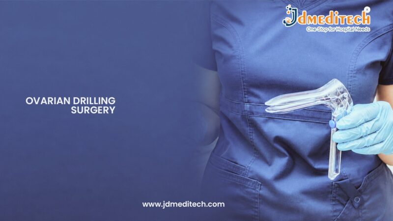
Fortunately, Ovarian Drilling Surgery offers an effective alternative for inducing ovulation in selected patients. This minimally invasive laparoscopic procedure helps restore hormonal balance and improve ovulation rates. As a result, many women experience improved fertility and increased chances of natural pregnancy. Moreover, ovarian drilling may reduce the need for long-term fertility medication use.

What is Ovarian Drilling Surgery?

Ovarian Drilling Surgery is a minimally invasive laparoscopic procedure used primarily to treat ovulation disorders associated with PCOS.

Purpose of Ovarian Drilling

The main goal of the procedure is to stimulate regular ovulation by reducing the amount of hormone-producing ovarian tissue. Consequently, hormonal balance may improve, allowing the ovaries to release eggs more consistently.

How the Procedure Works

During surgery, the surgeon creates several small punctures on the surface of the ovaries using laser energy or electrocautery. Subsequently, hormone production is altered, which may help restore normal ovulation patterns.

Understanding Polycystic Ovary Syndrome (PCOS)

PCOS is one of the leading causes of female infertility worldwide. Furthermore, it affects hormone production, menstrual cycles, and ovarian function.

Common Symptoms of PCOS

Women with PCOS may experience:

  • Irregular menstrual cycles
  • Absent ovulation
  • Difficulty conceiving
  • Weight gain
  • Excess facial or body hair
  • Acne
  • Multiple ovarian cysts

As a result, many women seek fertility treatment to improve their chances of pregnancy.

Why is Ovarian Drilling Performed?

Ovarian drilling is usually considered when conventional fertility treatments have not been successful.

Medical Indications

The procedure may be recommended for women who have:

  • PCOS-related infertility
  • Failure to respond to ovulation induction medications
  • Resistance to clomiphene citrate
  • Irregular ovulation
  • Hormonal imbalance affecting fertility

Therefore, ovarian drilling may serve as an effective second-line fertility treatment.

Who is a Candidate for Ovarian Drilling Surgery?

Not all women with PCOS require surgery. However, certain patients may benefit significantly from this approach.

Ideal Candidates

The procedure is often suitable for women who:

  • Have PCOS with anovulation
  • Have not responded to fertility medications
  • Desire natural conception
  • Have normal fallopian tube function
  • Have a partner with normal sperm parameters
  • Are suitable candidates for laparoscopic surgery

A fertility specialist can determine eligibility through a comprehensive evaluation.

Preoperative Evaluation

Before surgery, several assessments are performed to ensure the procedure is appropriate.

Diagnostic Tests

The evaluation may include:

  • Hormonal blood tests
  • Pelvic ultrasound
  • Ovulation assessment
  • Fertility workup
  • Medical history review
  • General health evaluation

Additionally, these investigations help identify other factors that may affect fertility outcomes.

Ovarian Drilling Surgery Procedure

The procedure is usually performed under general anesthesia using a laparoscopic approach.

Step 1: Administration of Anesthesia

General anesthesia is provided to ensure patient comfort during surgery.

Step 2: Laparoscopic Access

Small incisions are made in the abdomen. Subsequently, a laparoscope is inserted to visualize the pelvic organs.

Step 3: Examination of the Ovaries

The surgeon carefully evaluates both ovaries and surrounding reproductive structures.

Step 4: Ovarian Drilling

Several small punctures are created in each ovary using controlled energy sources. As a result, excess androgen-producing tissue is reduced.

Step 5: Completion of the Procedure

Finally, the instruments are removed, and the incisions are closed with minimal scarring.

Benefits of Ovarian Drilling Surgery

Compared with prolonged medication use, ovarian drilling offers several advantages.

Key Benefits
  • Improved ovulation rates
  • Enhanced fertility potential
  • Reduced androgen levels
  • More regular menstrual cycles
  • Decreased dependence on fertility medications
  • Minimally invasive treatment
  • Short recovery period
  • Potential for natural conception

Furthermore, many women experience ovulation within a few months after surgery.

Success Rates of Ovarian Drilling

Success rates vary depending on age, overall fertility health, and the severity of PCOS.

Factors Influencing Success

Several factors can affect outcomes, including:

  • Age of the patient
  • Body mass index (BMI)
  • Duration of infertility
  • Severity of hormonal imbalance
  • Presence of other fertility issues

Nevertheless, ovarian drilling has demonstrated favorable results in appropriately selected patients.

Recovery After Ovarian Drilling Surgery

Recovery is generally rapid because the procedure is performed laparoscopically.

Immediate Recovery

Patients may experience:

  • Mild abdominal discomfort
  • Temporary bloating
  • Shoulder pain from laparoscopic gas
  • Mild fatigue

However, these symptoms usually improve within a few days.

Recovery Timeline

First 24 Hours

Most patients return home on the same day or after a short observation period.

Days 2–7

Physical discomfort gradually decreases. In addition, normal daily activities can often be resumed.

Within Two Weeks

Most women return to their regular routines and continue fertility monitoring.

Risks and Complications

Although Ovarian Drilling Surgery is generally safe, every surgical procedure carries certain risks.

Possible Risks
  • Infection
  • Bleeding
  • Adhesion formation
  • Ovarian damage
  • Injury to nearby organs
  • Anesthesia-related complications

Fortunately, serious complications are uncommon when the procedure is performed by experienced laparoscopic surgeons.

Postoperative Care

Following postoperative instructions can support healing and improve outcomes.

Recovery Recommendations
  • Stay hydrated
  • Avoid strenuous exercise initially
  • Follow medication instructions
  • Attend follow-up appointments
  • Monitor menstrual cycles
  • Maintain a healthy lifestyle

Consequently, proper postoperative care can contribute to better fertility results.

Frequently Asked Questions
Is Ovarian Drilling Surgery painful?

The procedure is performed under general anesthesia. Therefore, patients do not feel pain during surgery and usually experience only mild discomfort afterward.

How soon can ovulation occur after surgery?

Many women begin ovulating within a few weeks to several months after treatment.

Can pregnancy occur naturally after ovarian drilling?

Yes. In many cases, natural conception becomes possible once regular ovulation is restored.

Is ovarian drilling a permanent cure for PCOS?

No. While the procedure can improve ovulation and fertility, it does not cure PCOS completely.

Additional Resources

For reliable information regarding PCOS and fertility treatments, readers can visit:

  • The American Society for Reproductive Medicine (ASRM)
  • The European Society of Human Reproduction and Embryology (ESHRE)
  • The American College of Obstetricians and Gynecologists (ACOG)

These organizations provide evidence-based guidance on infertility treatment and reproductive health.

Conclusion

Ovarian Drilling Surgery is an effective minimally invasive procedure for women with PCOS-related infertility who have not responded to fertility medications. By improving hormonal balance and restoring ovulation, the surgery can significantly increase the chances of natural conception. Moreover, its minimally invasive approach, short recovery time, and favorable success rates make it a valuable treatment option for selected patients. Therefore, women struggling with ovulation disorders should discuss ovarian drilling with their fertility specialist to determine whether it is the right solution for their reproductive goals.


Explore More Product >>

Get Connected:
+91 79909 93062 | +91 63513 72032 | exports@jdmeditech.com 

Egg Retrieval in IVF Surgery | IVF Egg Collection Procedure

In Vitro Fertilization (IVF) has transformed the field of reproductive medicine by helping millions of couples overcome infertility. Among the various stages of IVF treatment, egg retrieval is one of the most important procedures. During this process, mature eggs are collected from the ovaries and prepared for fertilization in a specialized laboratory.

Fortunately, modern fertility treatments have made egg retrieval a safe and minimally invasive procedure. Moreover, advances in controlled ovarian stimulation allow fertility specialists to maximize the number of mature eggs available for fertilization. As a result, patients have a greater chance of achieving successful embryo development and pregnancy.

What is Egg Retrieval in IVF Surgery?

Egg retrieval, also known as oocyte retrieval, is a minimally invasive procedure used to collect mature eggs from the ovaries during an IVF cycle.

Purpose of Egg Retrieval

The primary goal of egg retrieval is to obtain healthy eggs that can be fertilized with sperm outside the body. Subsequently, the resulting embryos may be transferred into the uterus or preserved for future use.

Importance in IVF Treatment

Egg retrieval is a critical step in the IVF process. Without egg collection, fertilization and embryo development cannot take place. Therefore, the procedure plays a central role in assisted reproductive technology.

Understanding Controlled Ovarian Stimulation

Before egg retrieval can occur, the ovaries must be stimulated to produce multiple mature follicles.

Why Ovarian Stimulation is Necessary

Under normal circumstances, a woman typically releases one egg per menstrual cycle. However, IVF treatment aims to collect multiple eggs to increase the chances of successful fertilization. Consequently, fertility medications are used to encourage the development of several mature follicles.

Medications Used During Stimulation

Controlled ovarian stimulation may include:

  • Follicle-Stimulating Hormone (FSH)
  • Luteinizing Hormone (LH)
  • Gonadotropin injections
  • Ovulation suppression medications
  • Trigger injections

As a result, the ovaries produce multiple mature eggs instead of a single egg.

Who Needs Egg Retrieval in IVF?

Egg retrieval is recommended for women undergoing IVF or fertility preservation procedures.

Common Candidates

The procedure may be suitable for women with:

  • Blocked fallopian tubes
  • Unexplained infertility
  • Male factor infertility
  • Endometriosis
  • Ovulation disorders
  • Advanced maternal age
  • Recurrent IVF failures
  • Fertility preservation needs

Additionally, women planning egg freezing before medical treatments may also require egg retrieval.

Preoperative Evaluation

Before beginning ovarian stimulation, fertility specialists perform a comprehensive assessment.

Diagnostic Tests

The evaluation may include:

  • Hormonal blood tests
  • Ovarian reserve testing
  • Pelvic ultrasound
  • Infectious disease screening
  • Medical history review

Furthermore, these investigations help create a personalized treatment plan that maximizes IVF success.

Monitoring During Ovarian Stimulation

Careful monitoring is essential throughout the stimulation phase.

Ultrasound Monitoring

Regular transvaginal ultrasounds are performed to assess follicle growth and ovarian response.

Hormonal Assessment

Blood tests help measure hormone levels and determine the ideal timing for egg retrieval.

Consequently, fertility specialists can schedule the procedure when the eggs have reached optimal maturity.

Egg Retrieval in IVF Surgery Procedure

The procedure is typically performed under sedation or light anesthesia and usually takes less than 30 minutes.

Step 1: Administration of Sedation

The patient receives sedation or anesthesia to ensure comfort throughout the procedure.

Step 2: Ultrasound-Guided Needle Insertion

A transvaginal ultrasound probe guides a thin aspiration needle toward the ovarian follicles.

Step 3: Follicular Fluid Aspiration

The fluid within each follicle is carefully aspirated. As a result, the eggs contained inside the follicles are collected.

Step 4: Laboratory Identification of Eggs

Embryologists immediately examine the collected fluid and identify the retrieved eggs.

Step 5: Completion of the Procedure

Finally, the instruments are removed, and the patient is transferred to a recovery area for observation.

What Happens After Egg Retrieval?

Once the eggs are collected, the IVF laboratory begins the fertilization process.

Fertilization of Eggs

The retrieved eggs are combined with sperm in a controlled laboratory environment. Depending on the treatment plan, conventional IVF or Intracytoplasmic Sperm Injection (ICSI) may be used.

Embryo Development

Following fertilization, embryos are carefully monitored for several days to assess their growth and quality.

Embryo Transfer or Cryopreservation

Healthy embryos may be transferred into the uterus or frozen for future fertility treatment cycles.

Benefits of Egg Retrieval in IVF Surgery

Egg retrieval offers several advantages for individuals undergoing fertility treatment.

Key Benefits
  • Minimally invasive procedure
  • High egg collection success rates
  • Short treatment duration
  • Quick recovery
  • Improved IVF outcomes
  • Fertility preservation opportunities
  • Safe and effective technique
  • Enhanced reproductive options

Furthermore, the procedure provides a critical opportunity for successful embryo creation and pregnancy.

Recovery After Egg Retrieval

Recovery is generally quick, allowing most patients to return home on the same day.

Immediate Recovery

Patients may experience:

  • Mild cramping
  • Temporary bloating
  • Light spotting
  • Mild fatigue

However, these symptoms usually improve within a few days.

Recovery Timeline

First 24 Hours

Most patients rest at home and avoid strenuous activities.

Days 2–3

Discomfort gradually subsides. Additionally, many women can return to normal daily activities.

Within One Week

Most patients feel completely recovered and can proceed with the next stages of IVF treatment.

Risks and Complications

Although egg retrieval is considered safe, every medical procedure carries some risks.

Possible Risks
  • Bleeding
  • Infection
  • Ovarian injury
  • Pelvic discomfort
  • Adverse reaction to anesthesia
  • Ovarian Hyperstimulation Syndrome (OHSS)

Fortunately, serious complications are uncommon when the procedure is performed by experienced fertility specialists.

Tips for a Smooth Recovery

Following medical advice can support a comfortable recovery.

Recovery Recommendations
  • Stay hydrated
  • Avoid heavy exercise
  • Follow medication instructions
  • Attend follow-up appointments
  • Monitor symptoms carefully
  • Contact your doctor if unusual symptoms occur

Consequently, proper postoperative care helps ensure a successful IVF journey.

Frequently Asked Questions
Is Egg Retrieval in IVF Surgery painful?

Because sedation is usually provided, most patients experience little to no discomfort during the procedure.

How long does egg retrieval take?

The procedure generally takes between 20 and 30 minutes.

How many eggs are usually collected?

The number varies depending on age, ovarian reserve, and response to fertility medications.

Can I go home the same day?

Yes. Most patients are discharged within a few hours after the procedure.

Additional Resources

For reliable information regarding IVF and fertility treatments, readers can visit:

  • The American Society for Reproductive Medicine (ASRM)
  • The European Society of Human Reproduction and Embryology (ESHRE)
  • The American College of Obstetricians and Gynecologists (ACOG)

These organizations provide evidence-based guidance on fertility care and assisted reproductive technologies.

Conclusion

Egg Retrieval in IVF Surgery is a crucial component of modern fertility treatment. Through controlled ovarian stimulation and advanced egg collection techniques, fertility specialists can obtain mature eggs for successful fertilization and embryo development. Moreover, the procedure is minimally invasive, highly effective, and associated with a relatively quick recovery. Therefore, understanding the egg retrieval process can help patients feel more confident and prepared as they progress through their IVF journey.


Explore More Product >>

Get Connected:
+91 79909 93062 | +91 63513 72032 | exports@jdmeditech.com

Tubal Recanalization Surgery | Blocked Fallopian Tube Treatment

Blocked fallopian tubes are one of the leading causes of female infertility. Since the fallopian tubes play a crucial role in fertilization, any blockage can prevent the sperm and egg from meeting naturally. As a result, women with tubal obstruction may experience difficulty conceiving despite having normal ovulation and healthy reproductive organs.

Fortunately, Tubal Recanalization Surgery offers a minimally invasive solution for selected patients with blocked fallopian tubes. This advanced procedure helps restore tubal patency by opening the blocked segment of the tube. Consequently, many women may regain the possibility of natural conception without requiring more invasive fertility treatments. Moreover, the procedure is safe, effective, and associated with a relatively quick recovery.

Why are Fallopian Tubes Important for Fertility?

The fallopian tubes serve as the pathway through which the egg travels from the ovary to the uterus. In addition, fertilization typically occurs within the tube itself.

Functions of the Fallopian Tubes

Healthy fallopian tubes are responsible for:

  • Capturing the released egg
  • Transporting sperm toward the egg
  • Supporting fertilization
  • Transporting the fertilized embryo to the uterus

Therefore, any blockage can significantly reduce the chances of natural pregnancy.

Benefits of Tubal Recanalization Surgery

Compared with more invasive fertility procedures, tubal recanalization offers several advantages.

Key Benefits
  • Minimally invasive technique
  • No major surgical incisions
  • Short recovery period
  • Outpatient procedure
  • Restoration of natural fertility potential
  • Reduced treatment costs
  • High success rates for selected patients
  • Minimal postoperative discomfort

Furthermore, many women can attempt conception soon after successful treatment. As a result, the procedure has become an attractive fertility-preserving option for appropriately selected patients.

Success Rates of Tubal Recanalization

Success depends on the type and location of the blockage. Nevertheless, outcomes are generally favorable in properly selected patients.

Factors Affecting Success

Success rates may be influenced by:

  • Patient age
  • Fertility status
  • Type of tubal blockage
  • Duration of infertility
  • Presence of additional reproductive disorders

Therefore, individualized evaluation remains essential. Additionally, early diagnosis may improve treatment outcomes and future pregnancy chances.

Recovery After Tubal Recanalization Surgery

Recovery is usually quick because the procedure is minimally invasive. In most cases, patients return to normal activities within a few days.

Immediate Recovery

Patients may experience:

  • Mild cramping
  • Light spotting
  • Temporary pelvic discomfort
  • Mild fatigue

However, these symptoms generally resolve within a short period.

Recovery Timeline

First 24 Hours

Most patients return home the same day. Meanwhile, rest and hydration are encouraged.

Days 2–3

Discomfort gradually improves. Additionally, normal activities can often be resumed.

Within One Week

Most women return to their usual routines. Consequently, the procedure has minimal disruption to daily life.

Risks and Complications

Although Tubal Recanalization Surgery is considered safe, certain risks may occur. However, serious complications are uncommon.

Possible Complications
  • Infection
  • Bleeding
  • Tubal perforation
  • Contrast dye reactions
  • Recurrent blockage
  • Pelvic discomfort

Fortunately, experienced specialists use advanced imaging techniques to minimize these risks. Furthermore, careful patient selection improves overall safety and success rates.

Conclusion

Tubal Recanalization Surgery is an advanced minimally invasive technique designed to restore fertility in women with blocked fallopian tubes. By reopening obstructed tubes and improving reproductive function, the procedure offers many patients the opportunity to achieve natural pregnancy.

Moreover, its outpatient nature, rapid recovery, and favorable success rates make it an attractive option for appropriately selected candidates. Therefore, women experiencing infertility due to tubal blockage should discuss tubal recanalization with a fertility specialist. Ultimately, early intervention and proper evaluation can significantly improve reproductive outcomes and fertility success.


Explore More Product >>

Get Connected:
+91 79909 93062 | +91 63513 72032 | exports@jdmeditech.com 

Hysteroscopy for Infertility Evaluation | Advanced Fertility Diagnosis

Female infertility affects millions of women worldwide and can result from a variety of reproductive health conditions. While several diagnostic tests are available, identifying the exact cause of infertility often requires direct visualization of the uterine cavity. Therefore, Hysteroscopy for Infertility Evaluation Surgery has become an essential tool in modern fertility assessment.

This minimally invasive procedure allows fertility specialists to examine the inside of the uterus using a thin, lighted instrument called a hysteroscope. As a result, abnormalities that may interfere with conception or implantation can be detected and, in some cases, treated during the same procedure. Moreover, hysteroscopy provides highly accurate diagnostic information that can improve fertility treatment outcomes.

What is Hysteroscopy for Infertility Evaluation?

Hysteroscopy is a minimally invasive procedure that enables doctors to directly inspect the uterine cavity. Unlike imaging tests that provide indirect views, hysteroscopy offers real-time visualization of the uterus.

Purpose of Hysteroscopy in Infertility

The primary goal of hysteroscopy is to identify uterine abnormalities that may contribute to infertility or recurrent pregnancy loss. Furthermore, the procedure helps fertility specialists develop more effective treatment plans.

How the Procedure Works

A thin hysteroscope is inserted through the vagina and cervix into the uterus. Subsequently, a small camera transmits high-definition images to a monitor, allowing the doctor to carefully examine the uterine cavity.

Why is Hysteroscopy Important for Infertility Evaluation?

Successful pregnancy depends on a healthy uterine environment. However, structural abnormalities inside the uterus may prevent embryo implantation or increase the risk of miscarriage.

Benefits for Fertility Assessment

Hysteroscopy helps:

  • Detect uterine abnormalities
  • Identify implantation issues
  • Evaluate recurrent pregnancy loss
  • Improve IVF success rates
  • Guide fertility treatment decisions
  • Confirm findings from ultrasound or HSG

Consequently, many fertility specialists consider hysteroscopy an important component of comprehensive infertility evaluation.

Conditions Diagnosed Through Hysteroscopy

The procedure can identify several uterine conditions that may affect fertility.

Endometrial Polyps

Polyps are growths within the uterine lining that can interfere with embryo implantation. Therefore, identifying and removing them may improve fertility outcomes.

Uterine Fibroids

Submucosal fibroids that distort the uterine cavity can reduce fertility and increase miscarriage risk.

Uterine Septum

A uterine septum is a congenital abnormality that divides the uterine cavity. As a result, it may contribute to infertility or recurrent pregnancy loss.

Intrauterine Adhesions

Scar tissue inside the uterus can impair implantation and menstrual function. Fortunately, hysteroscopy can both diagnose and treat these adhesions.

Endometrial Abnormalities

The procedure also helps evaluate abnormalities of the uterine lining that may affect reproductive success.

Who Needs Hysteroscopy for Infertility Evaluation?

Not every infertility patient requires hysteroscopy. Nevertheless, the procedure is highly beneficial in specific situations.

Ideal Candidates

Hysteroscopy may be recommended for women who have:

  • Unexplained infertility
  • Recurrent miscarriages
  • Abnormal uterine bleeding
  • Failed IVF cycles
  • Suspected uterine abnormalities
  • Abnormal ultrasound findings
  • Repeated implantation failure

A fertility specialist can determine whether the procedure is appropriate based on individual circumstances.

Preoperative Preparation

Proper preparation helps ensure a safe and successful procedure.

Before the Procedure

Patients may be advised to:

  • Schedule the procedure after menstruation
  • Undergo pregnancy testing
  • Review medications with their physician
  • Complete routine laboratory investigations
  • Follow fasting instructions if anesthesia is planned

Additionally, patients receive detailed instructions regarding the procedure and recovery process.

Hysteroscopy for Infertility Evaluation Procedure

The procedure is typically performed as an outpatient treatment and often requires only a short recovery period.

Step 1: Anesthesia Administration

Depending on the patient’s condition, local, regional, or general anesthesia may be used.

Step 2: Insertion of the Hysteroscope

The hysteroscope is gently inserted through the cervix and into the uterine cavity.

Step 3: Expansion of the Uterus

A sterile fluid is introduced to expand the uterus and improve visibility.

Step 4: Examination of the Uterine Cavity

The specialist carefully examines the uterine lining, cervical canal, and tubal openings.

Step 5: Treatment of Identified Abnormalities

If abnormalities are detected, certain conditions such as polyps or adhesions may be treated during the same procedure.

Step 6: Completion of the Procedure

Finally, the instruments are removed, and the patient is transferred to the recovery area.

Benefits of Hysteroscopy for Infertility Evaluation

Compared with many traditional diagnostic methods, hysteroscopy offers several advantages.

Key Benefits
  • Direct visualization of the uterus
  • High diagnostic accuracy
  • Minimally invasive approach
  • Short recovery time
  • Simultaneous diagnosis and treatment
  • Improved fertility treatment planning
  • Better IVF preparation
  • Reduced need for exploratory surgery

Furthermore, the procedure provides valuable information that may not be visible on imaging studies alone.

Recovery After Hysteroscopy

Recovery is generally quick and uncomplicated.

Immediate Recovery

Patients may experience:

  • Mild cramping
  • Light vaginal spotting
  • Temporary fatigue
  • Mild pelvic discomfort

However, these symptoms usually resolve within a few days.

Recovery Timeline

First 24 Hours

Most patients return home the same day. Meanwhile, rest and hydration are recommended.

Days 2–3

Cramping and spotting gradually decrease. In addition, normal activities can often be resumed.

Within One Week

Most women experience complete recovery and can return to their usual routines.

Risks and Complications

Although hysteroscopy is considered very safe, potential complications should be discussed.

Possible Risks
  • Infection
  • Bleeding
  • Uterine perforation
  • Fluid overload
  • Cervical injury
  • Adverse reaction to anesthesia

Fortunately, serious complications are rare when the procedure is performed by experienced specialists.

Postoperative Care

Following medical instructions can help ensure a smooth recovery.

Recovery Recommendations
  • Rest for the remainder of the day
  • Avoid sexual intercourse temporarily
  • Use prescribed medications as directed
  • Monitor for unusual symptoms
  • Attend follow-up appointments
  • Maintain proper hygiene

As a result, patients can recover comfortably while minimizing complications.

Frequently Asked Questions
Is hysteroscopy painful?

Most women experience only mild discomfort. Additionally, anesthesia options help improve comfort during the procedure.

How long does the procedure take?

The procedure typically takes between 15 and 45 minutes, depending on the complexity of the evaluation.

Can hysteroscopy improve fertility?

Yes. In many cases, identifying and treating uterine abnormalities can improve fertility outcomes and increase the chances of successful pregnancy.

Is hospitalization required?

No. Most hysteroscopy procedures are performed on an outpatient basis, allowing patients to return home the same day.

Additional Resources

For evidence-based information about infertility evaluation and hysteroscopy, readers can visit:

  • The American Society for Reproductive Medicine (ASRM)
  • The American College of Obstetricians and Gynecologists (ACOG)
  • The European Society of Human Reproduction and Embryology (ESHRE)

These organizations provide reliable resources on fertility treatments, reproductive health, and hysteroscopic procedures.

Conclusion

Hysteroscopy for Infertility Evaluation Surgery is an advanced and highly effective diagnostic procedure that helps identify uterine conditions affecting fertility. By providing direct visualization of the uterine cavity, the procedure enables accurate diagnosis and, when necessary, immediate treatment of abnormalities. Furthermore, its minimally invasive nature, quick recovery, and high diagnostic accuracy make it an invaluable tool in modern reproductive medicine. Therefore, women experiencing infertility or recurrent pregnancy loss should discuss hysteroscopy with their fertility specialist as part of a comprehensive evaluation.


Explore More Product >>

Get Connected:
+91 79909 93062 | +91 63513 72032 | exports@jdmeditech.com 

Diagnostic Laparoscopy Surgery | Modern Abdominal Diagnosis Technique

Diagnosing abdominal and pelvic conditions can sometimes be challenging, especially when imaging tests do not provide clear answers. In such situations, Diagnostic Laparoscopy Surgery offers a highly effective solution. This minimally invasive procedure allows surgeons to directly examine organs inside the abdomen and pelvis using a specialized camera called a laparoscope.

Moreover, Diagnostic Laparoscopy Surgery helps identify conditions that may not be visible through ultrasound, CT scans, or MRI. As a result, healthcare providers can make accurate diagnoses and develop appropriate treatment plans. Today, this advanced endoscopic technique plays an important role in modern surgical practice.

What is Diagnostic Laparoscopy Surgery?

Diagnostic Laparoscopy Surgery is a minimally invasive procedure used to examine the abdominal and pelvic organs. During the procedure, a thin tube equipped with a high-definition camera is inserted through a small incision in the abdomen.

Purpose of Diagnostic Laparoscopy

The primary goal of the procedure is to identify abnormalities that may be causing symptoms. Additionally, surgeons can collect tissue samples, evaluate organ function, and determine the extent of certain diseases.

Organs Examined During Laparoscopy

The procedure may be used to assess:

  • Liver
  • Gallbladder
  • Appendix
  • Stomach
  • Small intestine
  • Large intestine
  • Pancreas
  • Spleen
  • Female reproductive organs
  • Peritoneum

Consequently, Diagnostic Laparoscopy Surgery provides valuable information for a wide range of medical conditions.

Why is Diagnostic Laparoscopy Performed?

Doctors may recommend this procedure when non-invasive tests fail to identify the cause of symptoms.

Common Medical Indications

Diagnostic laparoscopy may be performed for:

  • Chronic abdominal pain
  • Pelvic pain
  • Unexplained infertility
  • Suspected endometriosis
  • Pelvic inflammatory disease
  • Abdominal masses
  • Internal bleeding
  • Liver disorders
  • Tumor evaluation
  • Cancer staging

Furthermore, the procedure can help confirm diagnoses before major surgical interventions.

Conditions Diagnosed with Diagnostic Laparoscopy

This advanced technique can identify numerous abdominal and pelvic disorders.

Gynecological Conditions

In women, Diagnostic Laparoscopy Surgery is frequently used to diagnose:

  • Endometriosis
  • Ovarian cysts
  • Pelvic adhesions
  • Ectopic pregnancy
  • Uterine abnormalities
  • Infertility-related conditions
Gastrointestinal Conditions

The procedure can also evaluate:

  • Appendicitis
  • Abdominal infections
  • Intestinal obstruction
  • Crohn’s disease
  • Abdominal tumors
  • Peritoneal diseases
Oncological Assessment

In addition, laparoscopy may help determine the extent of cancers involving the abdomen or pelvis.

Who is a Candidate for Diagnostic Laparoscopy Surgery?

Not every patient requires laparoscopic evaluation. However, the procedure is often recommended when symptoms remain unexplained after routine testing.

Suitable Candidates

Patients may benefit from the procedure if they have:

  • Persistent abdominal pain
  • Chronic pelvic pain
  • Unexplained infertility
  • Suspicious imaging findings
  • Suspected abdominal tumors
  • Unclear diagnoses despite medical investigations

A thorough medical evaluation helps determine whether diagnostic laparoscopy is appropriate.

Preoperative Preparation

Proper preparation helps ensure a safe and successful procedure.

Before the Surgery

Patients may be advised to:

  • Fast for several hours
  • Undergo blood tests
  • Complete imaging studies
  • Review medications with their doctor
  • Arrange transportation after surgery

Additionally, the surgical team explains the procedure and answers any questions before treatment.

Diagnostic Laparoscopy Surgery Procedure

The surgery is typically performed under general anesthesia and usually takes between 30 minutes and 2 hours.

Step 1: Administration of Anesthesia

General anesthesia ensures that the patient remains comfortable throughout the procedure.

Step 2: Creation of Small Incisions

A small incision is made near the navel. Subsequently, carbon dioxide gas is introduced into the abdomen to create working space.

Step 3: Insertion of the Laparoscope

The laparoscope is carefully inserted through the incision. As a result, high-definition images of the abdominal cavity are displayed on a monitor.

Step 4: Examination of Internal Organs

The surgeon systematically evaluates the abdominal and pelvic organs for abnormalities.

Step 5: Tissue Sampling if Necessary

If abnormal tissue is identified, a biopsy may be obtained for laboratory analysis.

Step 6: Completion of the Procedure

Finally, the instruments are removed, the gas is released, and the incisions are closed.

Benefits of Diagnostic Laparoscopy Surgery

Compared with traditional exploratory surgery, laparoscopy offers numerous advantages.

Key Benefits
  • Minimally invasive approach
  • Smaller incisions
  • Reduced postoperative pain
  • Faster recovery
  • Shorter hospital stay
  • Improved diagnostic accuracy
  • Lower risk of complications
  • Better cosmetic results

Furthermore, patients often return to normal activities much sooner than with open surgical procedures.

Recovery After Diagnostic Laparoscopy

Recovery is generally quick because the procedure involves only small incisions.

Immediate Recovery

Patients may experience:

  • Mild abdominal discomfort
  • Temporary bloating
  • Shoulder pain caused by residual gas
  • Mild fatigue

However, these symptoms typically improve within a few days.

Recovery Timeline

First 24 Hours

Most patients are discharged on the same day. Nevertheless, rest is recommended.

Days 2–7

Energy levels gradually improve. In addition, light activities can usually be resumed.

Week 2 and Beyond

Most patients return to their normal routines, although recovery times may vary.

Risks and Complications

Although Diagnostic Laparoscopy Surgery is considered safe, every surgical procedure carries some risks.

Possible Complications
  • Infection
  • Bleeding
  • Injury to internal organs
  • Adverse reactions to anesthesia
  • Blood clots
  • Hernia at incision sites

Fortunately, serious complications are uncommon when the procedure is performed by experienced surgeons.

Postoperative Care

Following postoperative instructions helps promote healing and reduce complications.

Recovery Recommendations
  • Keep incision sites clean and dry
  • Follow medication instructions
  • Avoid strenuous activities initially
  • Attend follow-up appointments
  • Stay hydrated
  • Report unusual symptoms promptly

Consequently, proper care contributes to a smoother recovery process.

Frequently Asked Questions
Is Diagnostic Laparoscopy Surgery painful?

Most patients experience only mild discomfort, which is usually controlled with prescribed medications.

How long does the procedure take?

The surgery generally takes between 30 minutes and 2 hours, depending on the complexity of the evaluation.

When can normal activities be resumed?

Many patients return to light activities within a few days and resume normal routines within one to two weeks.

Can Diagnostic Laparoscopy detect endometriosis?

Yes. In fact, diagnostic laparoscopy is considered one of the most effective methods for diagnosing endometriosis.

Additional Resources

For reliable information about minimally invasive surgery and abdominal health, consider visiting:

  • The American College of Obstetricians and Gynecologists (ACOG)
  • The Society of American Gastrointestinal and Endoscopic Surgeons (SAGES)
  • The American College of Surgeons (ACS)

These organizations provide evidence-based information regarding laparoscopic procedures and surgical care.

Conclusion

Diagnostic Laparoscopy Surgery is a modern and highly effective endoscopic technique used to diagnose a wide range of abdominal and pelvic conditions. By providing direct visualization of internal organs, the procedure allows for accurate diagnosis and timely treatment planning. Moreover, its minimally invasive nature offers numerous benefits, including faster recovery, reduced discomfort, and improved outcomes. Therefore, Diagnostic Laparoscopy Surgery remains an essential tool in contemporary medical practice.

Explore More Product >>

Get Connected:
+91 79909 93062 | +91 63513 72032 | exports@jdmeditech.com 

Vaginal Vault Suspension Surgery | Post-Hysterectomy Prolapse Repair

Vaginal vault prolapse is a condition that can occur after a hysterectomy when the upper portion of the vagina loses its normal support and descends into the vaginal canal. As a result, women may experience pelvic pressure, vaginal bulging, urinary problems, and discomfort during everyday activities. Furthermore, severe prolapse can affect both physical well-being and emotional confidence.

Fortunately, Vaginal Vault Suspension Surgery provides an effective and long-lasting solution for restoring pelvic support. By repositioning the vaginal vault and securing it to strong pelvic structures, this procedure helps relieve symptoms and improve quality of life. Moreover, modern surgical techniques have enhanced both safety and recovery outcomes.

What is Vaginal Vault Prolapse?

Vaginal vault prolapse occurs when the top of the vagina drops from its normal position due to weakened pelvic floor tissues and ligaments. Although the condition most commonly develops after a hysterectomy, other factors can also contribute to pelvic support failure.

Understanding Post-Hysterectomy Prolapse

After the uterus is removed, the upper end of the vagina is closed and supported by surrounding tissues. However, if these support structures weaken over time, the vaginal vault may gradually descend. Consequently, patients may develop symptoms that interfere with daily activities and overall comfort.

Types of Vaginal Vault Prolapse
  • Mild prolapse
  • Moderate prolapse
  • Severe prolapse

The severity of the prolapse often determines the most appropriate treatment approach.

Symptoms of Vaginal Vault Prolapse

Symptoms can vary from mild discomfort to significant functional impairment. In many cases, symptoms become more noticeable during physical activity or prolonged standing.

Common Symptoms

Women with vaginal vault prolapse may experience:

  • A feeling of vaginal bulging
  • Pelvic heaviness or pressure
  • Lower back discomfort
  • Urinary urgency
  • Difficulty emptying the bladder
  • Urinary leakage
  • Bowel dysfunction
  • Pain during sexual intercourse

Additionally, some women report a sensation of tissue protruding through the vaginal opening.

Causes of Vaginal Vault Prolapse

Several factors may weaken the structures that support the vaginal apex. Therefore, understanding these causes can help identify individuals at higher risk.

Previous Hysterectomy

Removal of the uterus changes the natural support system of the pelvis. Consequently, the vaginal vault may become vulnerable to prolapse if adequate support is not maintained.

Childbirth-Related Pelvic Floor Damage

Pregnancy and vaginal delivery place significant stress on pelvic muscles and connective tissues. Over time, this damage may contribute to prolapse development.

Aging and Menopause

As women age, tissues naturally lose strength and elasticity. Furthermore, reduced estrogen levels after menopause can accelerate pelvic floor weakening.

Other Risk Factors

Additional factors include:

  • Obesity
  • Chronic constipation
  • Heavy lifting
  • Chronic coughing
  • Connective tissue disorders
  • Previous pelvic surgeries
What is Vaginal Vault Suspension Surgery?

Vaginal Vault Suspension Surgery is a pelvic reconstructive procedure that restores support to the vaginal apex. During the procedure, the vaginal vault is elevated and attached to strong pelvic ligaments or supportive structures.

Goals of the Procedure

The surgery aims to:

  • Restore normal vaginal anatomy
  • Reduce prolapse symptoms
  • Improve bladder and bowel function
  • Enhance quality of life
  • Prevent further prolapse progression

As a result, patients often experience significant long-term improvement.

Who Needs Vaginal Vault Suspension Surgery?

While mild prolapse may respond to conservative treatment, surgery is often recommended for women with persistent or severe symptoms.

Ideal Candidates

This procedure may be suitable for women who have:

  • Moderate to severe vaginal vault prolapse
  • Persistent pelvic pressure
  • Vaginal bulging
  • Urinary difficulties
  • Recurrent prolapse
  • Failure of non-surgical treatments

A comprehensive pelvic examination helps determine whether surgery is the best option.

Preoperative Evaluation

Before proceeding with surgery, healthcare providers conduct a detailed assessment to ensure optimal treatment planning.

Diagnostic Tests

The evaluation may include:

  • Pelvic examination
  • Medical history review
  • Urinalysis
  • Pelvic ultrasound
  • MRI in selected cases
  • Blood investigations

In addition, the surgeon discusses expectations, potential risks, and recovery requirements.

Types of Vaginal Vault Suspension Surgery

Several surgical techniques are available. Therefore, the choice of procedure depends on the patient’s anatomy, health status, and treatment goals.

Uterosacral Ligament Suspension

This procedure uses the uterosacral ligaments to provide support for the vaginal apex.

Sacrospinous Ligament Fixation

During this technique, the vaginal vault is attached to the sacrospinous ligament to restore pelvic support.

Sacrocolpopexy

Sacrocolpopexy uses surgical mesh to connect the vaginal vault to the sacrum. Consequently, it provides durable and long-lasting support.

Laparoscopic and Robotic-Assisted Suspension

Minimally invasive approaches offer enhanced precision, reduced pain, and faster recovery.

Vaginal Vault Suspension Surgery Procedure

The operation is typically performed under general anesthesia and may be completed through a vaginal, laparoscopic, robotic, or abdominal approach.

Step 1: Surgical Access

The surgeon gains access to the pelvic support structures using the selected surgical technique.

Step 2: Identification of Support Structures

Strong ligaments and connective tissues are carefully identified to provide durable support.

Step 3: Repositioning of the Vaginal Vault

The prolapsed vaginal apex is elevated and returned to its normal anatomical position.

Step 4: Suspension and Fixation

The vaginal vault is securely attached to supportive structures to maintain long-term stability.

Step 5: Closure

Finally, the surgical site is closed, and the patient is transferred to the recovery area.

Benefits of Vaginal Vault Suspension Surgery

This procedure offers numerous advantages for women with post-hysterectomy prolapse.

Key Benefits
  • Restores pelvic support
  • Reduces vaginal bulging
  • Improves bladder function
  • Enhances bowel function
  • Relieves pelvic pressure
  • Improves sexual function
  • Provides long-term symptom relief

Furthermore, many patients report greater confidence and improved daily comfort after treatment.

Recovery After Vaginal Vault Suspension Surgery

Recovery varies depending on the surgical approach. Nevertheless, most women experience steady improvement during the healing process.

Immediate Recovery

Patients may notice:

  • Mild discomfort
  • Temporary fatigue
  • Light vaginal spotting
  • Short hospital stay
Recovery Timeline

Week 1–2

Rest and light activity are recommended. Meanwhile, gentle walking can promote circulation and support healing.

Week 3–6

Energy levels gradually improve. Additionally, many patients resume routine daily activities during this period.

Week 6–8

Most women return to normal activities. However, heavy lifting should still be avoided until medical clearance is provided.

Postoperative Care Guidelines

Proper postoperative care is essential for achieving the best possible outcomes.

Recovery Recommendations
  • Avoid heavy lifting
  • Prevent constipation
  • Stay well hydrated
  • Follow prescribed medications
  • Attend follow-up appointments
  • Avoid strenuous exercise
  • Resume activities gradually

Consequently, these measures support healing and reduce the risk of complications.

Risks and Complications

Although Vaginal Vault Suspension Surgery is considered safe, all surgical procedures carry some degree of risk.

Potential Risks
  • Infection
  • Bleeding
  • Urinary retention
  • Injury to surrounding organs
  • Pelvic pain
  • Recurrence of prolapse
  • Anesthesia-related complications

Fortunately, serious complications are uncommon when surgery is performed by experienced pelvic reconstructive specialists.

Long-Term Outcomes

Vaginal Vault Suspension Surgery has a high success rate and provides durable symptom relief. Therefore, it remains one of the most effective treatments for post-hysterectomy prolapse.

Maintaining Surgical Results

Women can improve long-term outcomes by:

  • Maintaining a healthy weight
  • Performing pelvic floor exercises
  • Avoiding chronic straining
  • Managing constipation
  • Following medical recommendations

As a result, the likelihood of prolapse recurrence can be significantly reduced.

Conclusion

Vaginal Vault Suspension Surgery is a safe, reliable, and highly effective treatment for post-hysterectomy prolapse. By restoring support to the vaginal apex and correcting pelvic anatomy, the procedure helps improve comfort, function, and quality of life. Moreover, advances in minimally invasive surgery have enhanced recovery and long-term outcomes. Therefore, women experiencing symptoms of vaginal vault prolapse should seek evaluation from a qualified pelvic floor specialist to explore the most appropriate treatment options.


Explore More Product >>

Get Connected:
+91 79909 93062 | +91 63513 72032 | exports@jdmeditech.com 

Sacrocolpopexy Surgery | Safe & Reliable Prolapse Management

Pelvic organ prolapse is a common condition that affects many women, particularly after childbirth, menopause, or hysterectomy. As pelvic support structures weaken, organs such as the vagina, bladder, or uterus may descend from their normal position. Consequently, women may experience pelvic pressure, vaginal bulging, urinary problems, and discomfort during daily activities.

Fortunately, Sacrocolpopexy Surgery is considered one of the most effective and durable treatments for pelvic organ prolapse. By restoring the normal position of the vaginal vault, this procedure provides long-term support and significantly improves quality of life. Moreover, modern surgical techniques have made sacrocolpopexy safer and less invasive than ever before.

What is Sacrocolpopexy Surgery?

Sacrocolpopexy is a pelvic reconstructive procedure that corrects vaginal vault prolapse by attaching the vagina to the sacrum using a surgical mesh. As a result, the vagina is restored to its natural position and receives strong long-term support.

Purpose of Sacrocolpopexy

The primary goal of the surgery is to restore pelvic anatomy while preserving vaginal function. In addition, the procedure helps relieve symptoms associated with pelvic organ prolapse and improves overall pelvic floor support.

Understanding Vaginal Vault Prolapse

Vaginal vault prolapse occurs when the top portion of the vagina loses support and descends into the vaginal canal. Although this condition often develops after a hysterectomy, it can also occur due to severe weakening of the pelvic floor.

Common Symptoms

Women with vaginal vault prolapse may experience:

  • A feeling of vaginal bulging
  • Pelvic heaviness or pressure
  • Lower back discomfort
  • Difficulty emptying the bladder
  • Urinary incontinence
  • Bowel dysfunction
  • Discomfort during sexual intercourse

Furthermore, symptoms often worsen after prolonged standing or physical activity.

Causes of Pelvic Organ Prolapse

Several factors can contribute to pelvic floor weakness.

Childbirth and Pregnancy

Repeated pregnancies and vaginal deliveries place significant stress on pelvic support tissues. Consequently, the risk of prolapse increases over time.

Menopause and Aging

As estrogen levels decline, connective tissues become weaker and less elastic. Therefore, pelvic organs are more likely to lose their normal support.

Additional Risk Factors

Other contributing factors include:

  • Obesity
  • Chronic constipation
  • Heavy lifting
  • Chronic coughing
  • Previous pelvic surgery
  • Genetic predisposition
Who Needs Sacrocolpopexy Surgery?

Not every prolapse requires surgical treatment. However, surgery may be recommended when symptoms become severe or significantly affect daily life.

Ideal Candidates

Sacrocolpopexy may be suitable for women who have:

  • Vaginal vault prolapse
  • Recurrent prolapse after previous surgery
  • Significant pelvic pressure
  • Urinary or bowel symptoms
  • Failure of conservative treatments
  • Desire for durable long-term repair

A comprehensive pelvic examination helps determine whether this procedure is the best treatment option.

Preoperative Evaluation

Before surgery, the healthcare team performs a detailed assessment to ensure safe and effective treatment.

Diagnostic Assessment

The evaluation may include:

  • Pelvic examination
  • Medical history review
  • Urinalysis
  • Pelvic ultrasound
  • MRI in selected cases
  • Blood investigations

Additionally, the surgeon will discuss treatment goals and expected outcomes.

Sacrocolpopexy Surgery Procedure

The procedure is typically performed under general anesthesia. Depending on the patient’s condition, the surgery may be completed using open, laparoscopic, or robotic-assisted techniques.

Step 1: Access to the Pelvic Cavity

Small abdominal incisions are created during minimally invasive surgery. Alternatively, an open approach may be used in certain situations.

Step 2: Identification of the Vaginal Apex

The surgeon carefully identifies the top portion of the vagina that requires support.

Step 3: Mesh Attachment

A surgical mesh is attached to the vaginal apex. Subsequently, the other end of the mesh is secured to the sacrum.

Step 4: Restoration of Pelvic Support

The mesh creates a strong support structure that restores the vagina to its normal anatomical position.

Step 5: Surgical Closure

Finally, all incisions are closed, and the patient is moved to the recovery area.

Advanced Surgical Approaches

Modern technology has significantly improved prolapse surgery outcomes.

Laparoscopic Sacrocolpopexy

This minimally invasive technique uses small incisions, resulting in reduced pain and faster recovery.

Robotic-Assisted Sacrocolpopexy

Robotic systems provide enhanced visualization and precision. Consequently, surgeons can perform complex repairs with greater accuracy.

Open Sacrocolpopexy

Although less common today, open surgery may still be appropriate for selected patients.

Benefits of Sacrocolpopexy Surgery

Sacrocolpopexy is widely regarded as the gold standard treatment for vaginal vault prolapse.

Key Benefits
  • Excellent long-term success rates
  • Restoration of normal vaginal anatomy
  • Reduced pelvic pressure
  • Improved bladder function
  • Better bowel function
  • Enhanced sexual function
  • Lower recurrence rates compared to some other prolapse procedures

Furthermore, many women experience substantial improvements in daily comfort and confidence.

Recovery After Sacrocolpopexy Surgery

Recovery varies depending on the surgical approach used. Nevertheless, most patients recover successfully with appropriate postoperative care.

Immediate Recovery

Patients may experience:

  • Mild discomfort
  • Temporary fatigue
  • Light vaginal spotting
  • Short hospital stay
Recovery Timeline

Week 1–2

Rest is essential during the initial healing period. However, light walking is usually encouraged.

Week 3–6

Energy levels gradually improve. In addition, many women can resume light daily activities.

Week 6–8

Most patients return to normal routines. Nevertheless, heavy lifting should still be avoided unless approved by the surgeon.

Postoperative Care Guidelines

Proper postoperative care supports healing and reduces complications.

Important Recommendations
  • Avoid heavy lifting
  • Prevent constipation
  • Stay hydrated
  • Follow medication instructions
  • Attend follow-up visits
  • Avoid strenuous exercise initially
  • Resume activities gradually

As a result, recovery is often smoother and more successful.

Risks and Complications

Although Sacrocolpopexy Surgery is generally safe, every surgical procedure carries some risk.

Potential Complications
  • Infection
  • Bleeding
  • Mesh-related complications
  • Bladder injury
  • Bowel injury
  • Urinary retention
  • Pain
  • Recurrence of prolapse

Fortunately, serious complications are uncommon when the procedure is performed by experienced pelvic reconstructive surgeons.

Long-Term Outcomes

Sacrocolpopexy has one of the highest success rates among prolapse surgeries. Therefore, it remains a preferred option for women seeking durable symptom relief.

Maintaining Surgical Results

Women can help maintain long-term success by:

  • Maintaining a healthy weight
  • Performing pelvic floor exercises
  • Managing constipation
  • Avoiding excessive straining
  • Following medical recommendations

Consequently, the risk of prolapse recurrence can be significantly reduced.

Frequently Asked Questions
Is Sacrocolpopexy Surgery painful?

Most patients experience mild to moderate discomfort, which is typically controlled with prescribed medications.

How long does recovery take?

Recovery generally takes six to eight weeks, although complete healing may continue for several months.

Is mesh always used during Sacrocolpopexy?

Yes, surgical mesh is commonly used because it provides durable support and long-term anatomical correction.

Can prolapse return after surgery?

Although recurrence is possible, sacrocolpopexy has one of the lowest recurrence rates among prolapse repair procedures.

Conclusion

Sacrocolpopexy Surgery is a safe, reliable, and highly effective treatment for pelvic organ prolapse. By restoring normal pelvic anatomy and providing durable support, the procedure helps relieve symptoms and improve quality of life. Furthermore, advances in laparoscopic and robotic-assisted techniques have made recovery faster and outcomes even more successful. Therefore, women experiencing significant prolapse symptoms should consult a pelvic floor specialist to determine whether sacrocolpopexy is the right treatment option.


Explore More Product >>

Get Connected:
+91 79909 93062 | +91 63513 72032 | exports@jdmeditech.com 

Rectocele Repair Surgery | Posterior Vaginal Wall Prolapse

Rectocele is a common type of pelvic organ prolapse that occurs when the rectum bulges into the posterior vaginal wall due to weakened pelvic floor muscles and connective tissues. As a result, many women experience discomfort, bowel dysfunction, and a reduced quality of life. Although mild cases may respond to conservative treatment, more severe prolapse often requires surgical correction.

Fortunately, Rectocele Repair Surgery offers an effective and long-lasting solution. By restoring the normal anatomy of the pelvic floor, this procedure helps improve bowel function and relieve uncomfortable symptoms. Furthermore, successful treatment can significantly enhance overall quality of life.

Causes of Rectocele

Several factors can contribute to the development of a rectocele. Most commonly, vaginal childbirth places considerable stress on the pelvic floor. In addition, aging and menopause can weaken the connective tissues that support pelvic organs.

Childbirth Trauma

During vaginal delivery, pelvic muscles and supportive tissues may stretch or become damaged. Consequently, the risk of developing posterior vaginal wall prolapse increases over time.

Chronic Constipation

Repeated straining during bowel movements places continuous pressure on the pelvic floor. Therefore, women with long-term constipation are more likely to develop a rectocele.

Aging and Menopause

As estrogen levels decline, pelvic tissues gradually lose strength and elasticity. Moreover, age-related weakening of muscles further contributes to prolapse development.

Benefits of Rectocele Repair Surgery

Rectocele Repair Surgery provides several important benefits. First, it restores the normal position of the rectum and strengthens weakened support structures. Additionally, many patients experience improved bowel function and reduced pelvic discomfort.

Key Benefits
  • Improved bowel emptying
  • Reduced vaginal bulging
  • Better pelvic support
  • Enhanced comfort during daily activities
  • Improved sexual function
  • Long-term symptom relief

Furthermore, many women report increased confidence and a better quality of life following surgery.

Recovery After Rectocele Repair Surgery

Recovery is generally straightforward when postoperative instructions are carefully followed. Initially, patients may experience mild discomfort and swelling. However, these symptoms usually improve within the first few weeks.

Recovery Timeline

Week 1–2

During the early recovery period, rest is important. Meanwhile, light walking can help improve circulation and promote healing.

Week 3–6

At this stage, discomfort gradually decreases. Additionally, most patients can return to many normal daily activities.

Week 6–8

By this time, healing is well advanced. Consequently, many women can resume their regular routines after receiving medical clearance.

Conclusion

Rectocele Repair Surgery is a highly effective treatment for posterior vaginal wall prolapse. By repairing weakened tissues and restoring normal pelvic anatomy, the procedure helps improve bowel function and reduce discomfort. Furthermore, long-term outcomes are generally excellent when patients follow recommended lifestyle modifications. Therefore, women experiencing significant symptoms should consult a qualified specialist to discuss the most appropriate treatment options.


Explore More Product >>

Get Connected:
+91 79909 93062 | +91 63513 72032 | exports@jdmeditech.com 

Cystocele Repair Surgery | Advanced Bladder Prolapse Treatment

Cystocele, commonly known as bladder prolapse, occurs when the bladder drops into the vaginal wall due to weakened pelvic floor muscles and connective tissues. The condition can cause discomfort, urinary problems, and a significant reduction in quality of life. When non-surgical treatments fail to provide relief, Cystocele Repair Surgery offers a reliable and long-lasting solution.

What is a Cystocele?

A cystocele develops when the supportive tissues between the bladder and vaginal wall weaken, allowing the bladder to descend into the vagina.

Types of Cystocele
  • Grade 1 (Mild): Slight bladder descent.
  • Grade 2 (Moderate): Bladder reaches the vaginal opening.
  • Grade 3 (Severe): Bladder protrudes outside the vaginal opening.
Causes of Bladder Prolapse

Several factors contribute to the development of a cystocele.

Childbirth and Pregnancy

Repeated vaginal deliveries can stretch and weaken pelvic support structures.

Menopause

Reduced estrogen levels after menopause can weaken pelvic tissues.

Chronic Pressure on the Pelvic Floor

Factors such as obesity, chronic coughing, constipation, and heavy lifting increase pelvic pressure and contribute to prolapse.

Symptoms of Cystocele

Women with bladder prolapse may experience a variety of symptoms.

Common Symptoms
  • Pelvic pressure or heaviness
  • Vaginal bulging
  • Frequent urination
  • Difficulty emptying the bladder
  • Urinary leakage
  • Recurrent urinary tract infections
  • Discomfort during sexual intercourse
What is Cystocele Repair Surgery?

Cystocele Repair Surgery, also known as Anterior Colporrhaphy, is a surgical procedure designed to restore the bladder to its normal position and strengthen the weakened vaginal wall.

Purpose of the Procedure

The surgery repairs damaged support tissues and improves bladder function while relieving prolapse symptoms.

Who is a Candidate for Cystocele Repair Surgery?

Surgical treatment may be recommended for women experiencing significant symptoms.

Ideal Candidates
  • Moderate to severe bladder prolapse
  • Persistent urinary symptoms
  • Failure of conservative treatments
  • Recurrent urinary infections
  • Significant impact on daily activities
Preoperative Evaluation

A detailed assessment is performed before surgery.

Diagnostic Tests
  • Pelvic examination
  • Urinalysis
  • Urodynamic studies
  • Ultrasound
  • MRI in selected cases
  • Blood investigations
Cystocele Repair Surgery Procedure

The surgery is usually performed under general or spinal anesthesia.

Step 1: Vaginal Incision

A small incision is made in the anterior vaginal wall.

Step 2: Tissue Separation

The bladder is carefully separated from the vaginal tissue.

Step 3: Tissue Reinforcement

Weakened fascia and connective tissues are repaired and tightened.

Step 4: Bladder Repositioning

The bladder is restored to its normal anatomical position.

Step 5: Closure

The incision is closed using absorbable sutures.

Advanced Surgical Techniques

Modern pelvic reconstructive surgery offers several advanced approaches.

Native Tissue Repair

Uses the patient’s own tissues to reinforce pelvic support.

Laparoscopic Cystocele Repair

A minimally invasive technique that reduces recovery time.

Robotic-Assisted Surgery

Provides enhanced precision and visualization during complex repairs.

Benefits of Cystocele Repair Surgery

The procedure offers numerous long-term benefits.

Key Advantages
  • Restores normal bladder position
  • Improves urinary control
  • Reduces pelvic pressure
  • Enhances quality of life
  • Improves sexual comfort
  • Decreases recurrent infections
Recovery After Surgery

Most patients recover successfully with proper postoperative care.

Recovery Timeline

First Week

  • Mild discomfort and swelling
  • Limited physical activity

Weeks 2–4

  • Gradual improvement in symptoms
  • Increased daily activity

Weeks 6–8

  • Return to normal routine
  • Complete healing in most cases
Postoperative Care

Following recovery guidelines helps ensure successful outcomes.

Important Instructions
  • Avoid heavy lifting
  • Prevent constipation
  • Stay hydrated
  • Follow medication instructions
  • Attend follow-up appointments
  • Avoid sexual intercourse until cleared by the surgeon
Risks and Complications

Although generally safe, every surgical procedure carries some risks.

Possible Complications
  • Bleeding
  • Infection
  • Urinary retention
  • Bladder injury
  • Pain
  • Recurrence of prolapse
  • Anesthesia-related complications
Long-Term Outcomes

Cystocele repair surgery has a high success rate and provides long-lasting symptom relief.

Maintaining Surgical Results
  • Perform pelvic floor exercises
  • Maintain a healthy weight
  • Avoid chronic straining
  • Manage chronic cough and constipation
Frequently Asked Questions
Is Cystocele Repair Surgery painful?

Most patients experience mild to moderate discomfort that is manageable with medication.

How long does recovery take?

Most women recover within six to eight weeks.

Can bladder prolapse recur after surgery?

Recurrence is possible but can be minimized with proper lifestyle modifications and pelvic floor strengthening.

Will urinary symptoms improve after surgery?

Most patients experience significant improvement in urinary function and bladder control.

Conclusion

Cystocele Repair Surgery is an advanced and effective treatment for bladder prolapse. By restoring pelvic support structures and correcting bladder position, the procedure significantly improves urinary function, comfort, and overall quality of life. Early diagnosis and timely surgical intervention can help women regain confidence and maintain long-term pelvic health.


Explore More Product >>

Get Connected:
+91 79909 93062 | +91 63513 72032 | exports@jdmeditech.com 

Sling Surgery (TVT / TOT) for Stress Urinary Incontinence

What is Sling Surgery?

Sling surgery is a minimally invasive procedure designed to support the urethra and bladder neck, thereby preventing urine leakage during activities that increase abdominal pressure.

During the procedure, surgeons place a synthetic mesh sling beneath the urethra to provide additional support. As a result, the urethra remains properly positioned during movement, which helps reduce or eliminate episodes of stress urinary incontinence.

Understanding Stress Urinary Incontinence

Stress urinary incontinence develops when pelvic floor muscles and supportive tissues become weakened over time. Under these circumstances, the urethra may not remain adequately closed during increases in abdominal pressure.

Common Causes of Stress Urinary Incontinence
  • Pregnancy and childbirth
  • Aging
  • Menopause
  • Obesity
  • Chronic coughing
  • Pelvic floor muscle weakness
  • Previous pelvic surgery
  • Heavy physical activity
  • Hormonal changes

For this reason, women with one or more risk factors may be more likely to experience urinary leakage symptoms.

Why is Sling Surgery Performed?

Sling surgery is recommended for women with stress urinary incontinence that does not respond adequately to conservative treatment measures. In many cases, patients seek surgery after lifestyle modifications and pelvic floor therapies fail to provide sufficient improvement.

Common Indications
  • Moderate stress urinary incontinence
  • Severe stress urinary incontinence
  • Failed pelvic floor exercises
  • Persistent urinary leakage
  • Reduced quality of life due to incontinence
  • Recurrent stress urinary incontinence

Additionally, surgery may provide long-term symptom relief when non-surgical treatments are no longer effective.

Procedure of Sling Surgery

The procedure is typically performed under local, regional, or general anesthesia. Depending on the surgical technique used, treatment can usually be completed within a relatively short time.

Step-by-Step Procedure
  1. Administration of anesthesia.
  2. Creation of a small vaginal incision.
  3. Placement of the sling beneath the urethra.
  4. Adjustment of sling positioning.
  5. Verification of urethral support.
  6. Closure of the incision.
  7. Postoperative monitoring.

Throughout the procedure, surgeons carefully evaluate sling placement to ensure optimal support. Meanwhile, modern surgical techniques help improve accuracy and long-term treatment success.

Benefits of Sling Surgery

Sling surgery offers numerous advantages for women suffering from stress urinary incontinence. Most importantly, it addresses the underlying cause of urine leakage rather than simply managing symptoms.

Key Benefits
  • Improved bladder control
  • Reduced urine leakage
  • Enhanced quality of life
  • Increased confidence
  • Better participation in physical activities
  • Long-term symptom relief
  • Minimally invasive treatment
  • Short hospital stay
  • Faster recovery

Consequently, sling surgery remains one of the most effective treatment options available for stress urinary incontinence.

Recovery After Sling Surgery

Recovery is generally quick, and most patients return to normal daily activities within a few weeks. However, following recovery guidelines is essential for achieving the best possible outcome.

Recovery Recommendations
  • Follow postoperative instructions carefully.
  • Avoid heavy lifting during recovery.
  • Attend follow-up appointments.
  • Maintain proper hydration.
  • Take medications as prescribed.
  • Gradually resume physical activities.

At the same time, adherence to postoperative recommendations helps maximize surgical success and reduce recovery-related complications.

Conclusion

Sling surgery, including TVT and TOT procedures, is a highly effective treatment option for women suffering from stress urinary incontinence. By providing additional urethral support, these minimally invasive procedures help restore bladder control, reduce urine leakage, and improve overall quality of life.

Whether symptoms are moderate or severe, sling surgery offers reliable long-term results for appropriately selected patients. Ultimately, experienced surgeons, advanced surgical technology, and personalized patient care work together to deliver successful treatment outcomes and enhanced well-being.

Explore More Product >>

Get Connected:
+91 79909 93062 | +91 63513 72032 | exports@jdmeditech.com