Bladder Scanner – Working Principle, Features, Uses and Benefits
A Bladder Scanner is a noninvasive ultrasound device that estimates the amount of urine present inside the urinary bladder. Healthcare professionals use this equipment to evaluate bladder volume and assess selected urinary problems.
The device uses ultrasound technology to examine the bladder without routinely requiring catheter insertion. Consequently, clinicians can obtain useful bladder volume information at the point of care.
Furthermore, portable scanning systems support examinations in hospitals, clinics, nursing facilities and other suitable healthcare environments.
JDMeditech supplies urology diagnostic equipment, surgical instruments and medical accessories for hospitals, clinics and medical distributors.
What Is a Bladder Scanner?
A bladder scanner is a specialized ultrasound instrument that estimates urinary bladder volume.
Typically, the system includes a handheld ultrasound probe and an electronic display or processing unit.
During an examination, the operator positions the probe over the lower abdomen and acquires ultrasound measurements.
The equipment then calculates an estimated bladder volume using its measurement algorithm.
However, measurement performance depends on device design, patient anatomy and operator technique.
What Is Bladder Volume Measurement?
Bladder volume measurement estimates the quantity of urine inside the bladder at the time of examination.
Clinicians may assess bladder volume before or after urination, depending on the clinical question.
Furthermore, post-void measurements can help evaluate how much urine remains after the patient attempts to empty the bladder.
How Does a Bladder Scanner Work?
The scanner transmits ultrasound pulses through a compatible probe placed against the patient’s lower abdomen.
Next, internal tissues reflect portions of the ultrasound energy back toward the transducer.
The equipment receives these echoes and processes the resulting signals.
Subsequently, the system estimates bladder dimensions or volume using its approved measurement technology.
Therefore, clinicians can review the estimated urine volume on the display.
Working Principle of Ultrasound Bladder Scanning
Ultrasound imaging uses high-frequency sound waves to evaluate internal anatomical structures.
Unlike X-ray imaging, diagnostic ultrasound does not use ionizing radiation.
The transducer emits sound pulses and receives returning echoes.
Next, the scanner processes the signals to estimate bladder volume.
However, different models use different scanning methods and calculation algorithms.
Key Features of a Bladder Scanner
- Noninvasive Bladder Volume Assessment
- Ultrasound Measurement Technology
- Portable Equipment Configuration
- Compatible Handheld Probe
- Digital Measurement Display
- Estimated Urine Volume Calculation
- Post-Void Residual Assessment
- Model-Specific Imaging Functions
- Model-Specific Data Storage
- Compatible Patient Examination Modes
- Operator Control Interface
- Manufacturer-Defined Safety Functions
Furthermore, exact features depend on the selected product model and configuration.
Main Components of a Bladder Scanner
A typical portable bladder scanning system may include the following components:
- Ultrasound Probe
- Ultrasound Transducer
- Electronic Control Unit
- Display Screen
- Processing System
- Operator Control Buttons
- Probe Connection Cable
- Power Supply or Battery
- Compatible Ultrasound Coupling Gel
- Manufacturer-Approved Accessories
However, exact components and included accessories vary by manufacturer.
1. Ultrasound Probe
The ultrasound probe transmits and receives sound waves during examination.
Furthermore, its design supports external scanning of the lower abdominal region.
2. Ultrasound Transducer
The transducer converts electrical energy into ultrasound pulses and receives returning echoes.
Consequently, the system can collect information for bladder volume estimation.
3. Electronic Control Unit
The control unit manages the equipment’s approved operating functions.
Additionally, it supports communication between the probe and processing system.
4. Digital Display
The display presents estimated bladder volume and other available examination information.
Furthermore, selected systems may provide ultrasound images or scanning guidance.
5. Processing System
The processing system analyzes ultrasound signals and calculates an estimated bladder volume.
However, calculation methods vary between models.
6. Probe Connection Cable
The cable connects the ultrasound probe to the main equipment unit when the system uses a wired configuration.
Additionally, appropriate cable handling helps protect the equipment.
7. Power Supply
Portable systems may use rechargeable batteries or compatible external power supplies.
However, battery performance and operating time depend on the model.
Clinical Applications of Bladder Scanner
Healthcare professionals use bladder scanning to assess selected urinary conditions and monitor bladder volume.
- Post-Void Residual Urine Assessment
- Suspected Urinary Retention
- Assessment of Incomplete Bladder Emptying
- Selected Postoperative Urinary Assessments
- Bladder Volume Monitoring
- Selected Neurogenic Bladder Evaluations
- Support for Catheterization Decisions
- Selected Urology Follow-Up Examinations
- Other Clinically Indicated Assessments
However, bladder scanning results require appropriate clinical interpretation.
Bladder Scanner for Post-Void Residual Measurement
Post-void residual urine, commonly called PVR, refers to the amount of urine remaining in the bladder after urination.
Clinicians may use a bladder scanner shortly after the patient voids to estimate the remaining urine volume.
Furthermore, PVR measurement can support assessment of incomplete bladder emptying.
However, an abnormal result does not independently establish the underlying diagnosis.
Bladder Scanner for Urinary Retention
Urinary retention occurs when the bladder cannot empty adequately.
A bladder scanner may help clinicians assess the amount of urine present when retention is suspected.
Consequently, the findings may support decisions about additional assessment or treatment.
However, acute urinary retention requires prompt clinical evaluation.
Bladder Scanner in Postoperative Care
Some patients experience difficulty urinating after surgery or anesthesia.
Therefore, healthcare teams may use bladder volume measurements to assess selected postoperative patients.
Furthermore, scanning may support monitoring according to an approved clinical protocol.
Bladder Scanner in Neurogenic Bladder Assessment
Neurological conditions can affect bladder storage and emptying.
Consequently, selected patients may benefit from bladder volume monitoring.
However, bladder scanning alone does not provide a complete assessment of neurogenic lower urinary tract dysfunction.
Potential Benefits of Bladder Scanning
- Noninvasive Examination
- No Routine Catheter Insertion
- Point-of-Care Bladder Volume Estimation
- No Ionizing Radiation
- Supports Post-Void Residual Assessment
- May Reduce Unnecessary Diagnostic Catheterization
- Supports Selected Urinary Retention Assessments
- Portable Equipment Options
- Useful for Selected Clinical Monitoring Applications
However, clinical benefits depend on appropriate use and accurate interpretation.
Bladder Scanner vs Urinary Catheterization
| Feature | Bladder Scanner | Urinary Catheterization |
|---|---|---|
| Method | External Ultrasound Measurement | Catheter Insertion Into Bladder |
| Invasiveness | Noninvasive | Invasive |
| Urine Volume Assessment | Estimated Volume | Measures Drained Volume |
| Bladder Drainage | No | Yes |
| Catheter-Related Infection Risk | Not Associated With Catheter Insertion | Present |
| Primary Purpose | Assessment and Monitoring | Drainage and Selected Diagnostic Uses |
Technical Specifications of Bladder Scanner
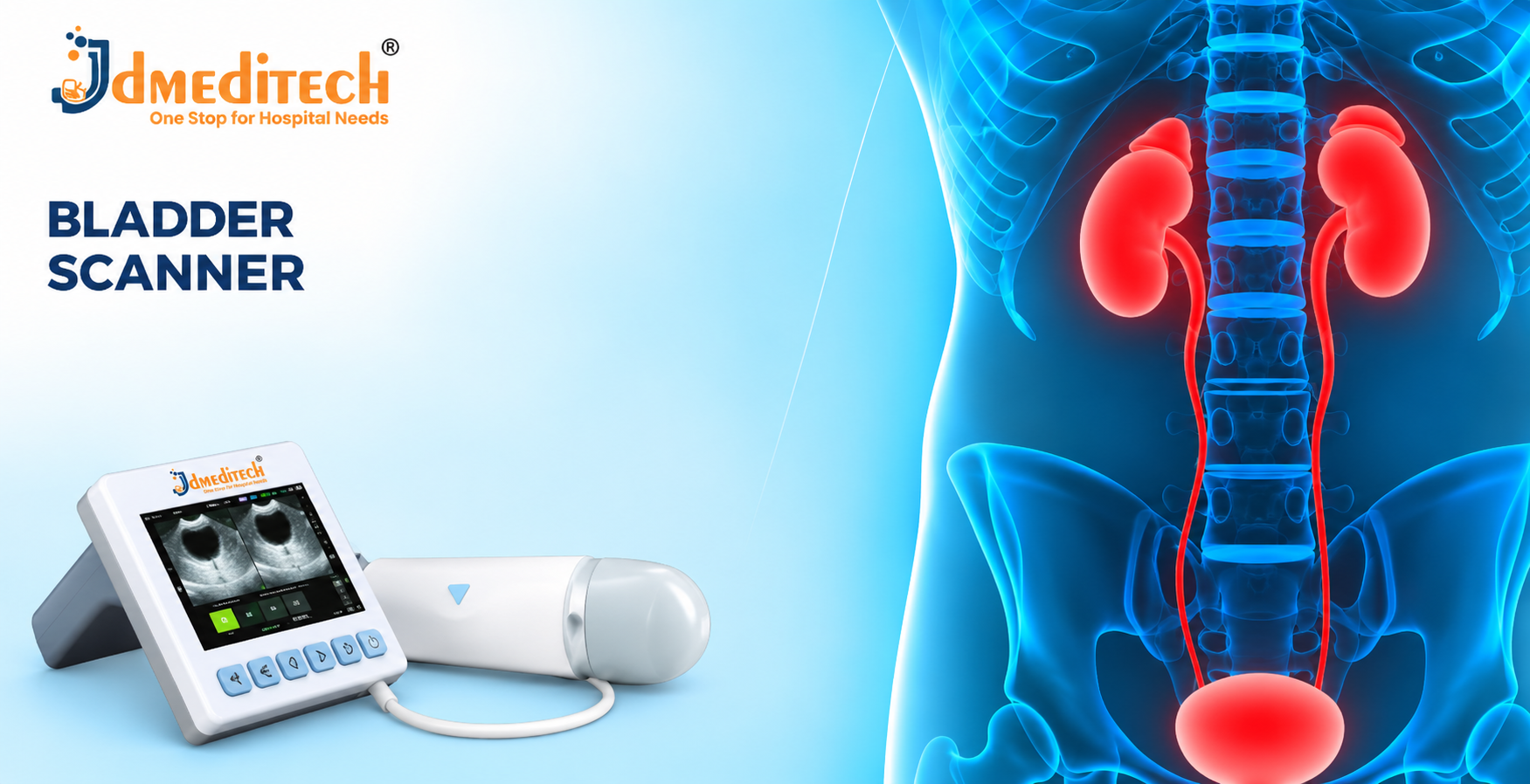
The supplied product image shows a portable ultrasound bladder scanning device with a display and handheld probe.
However, the image does not confirm measurement range, ultrasound frequency, volume accuracy or battery performance.
Therefore, buyers should verify the manufacturer’s official technical datasheet.
| Parameter | Product Information |
|---|---|
| Product Name | Bladder Scanner |
| Equipment Category | Urology Diagnostic Equipment |
| Measurement Technology | Ultrasound |
| Primary Function | Bladder Volume Estimation |
| Equipment Configuration | Portable Scanner With Handheld Probe |
| Measurement Range | Confirm Manufacturer Specifications |
| Volume Measurement Accuracy | Confirm Manufacturer Specifications |
| Ultrasound Frequency | Confirm Manufacturer Specifications |
| Probe Type | Confirm Manufacturer Specifications |
| Display Type | Confirm Manufacturer Specifications |
| Imaging Modes | Confirm Available Configuration |
| Patient Examination Modes | Confirm Available Configuration |
| Data Storage | Confirm Available Configuration |
| Report Generation | Confirm Available Configuration |
| Battery Type | Confirm Manufacturer Specifications |
| Battery Operating Time | Confirm Manufacturer Specifications |
| Power Requirements | Confirm Manufacturer Specifications |
| Dimensions | Confirm Manufacturer Specifications |
| Weight | Confirm Manufacturer Specifications |
| Regulatory Documentation | Verify for Destination Market |
How Is a Bladder Scan Performed?
A trained healthcare professional first explains the examination and prepares the equipment.
Next, the operator applies compatible ultrasound coupling gel to the appropriate lower abdominal area or probe surface.
The operator then positions the probe according to the manufacturer’s instructions.
Subsequently, the system acquires ultrasound information and calculates an estimated bladder volume.
Finally, the clinician reviews the result alongside the patient’s symptoms and clinical condition.
Patient Preparation for Bladder Scanning
Preparation depends on the purpose of the examination.
For example, clinicians may perform scanning before urination or shortly afterward to assess residual urine.
Furthermore, the healthcare team may review relevant symptoms and medical history.
However, patients should follow instructions from their healthcare professional.
Importance of Accurate Bladder Volume Measurement
Reliable volume estimation supports clinical decision-making.
However, measurement errors can occur because of patient anatomy, incorrect probe positioning or other fluid-filled structures.
Therefore, clinicians should repeat or confirm unexpected results when clinically necessary.
Limitations of Bladder Scanning
- Provides Estimated Rather Than Directly Drained Volume
- Accuracy Varies by Device and Clinical Conditions
- Operator Technique Can Affect Results
- Ascites May Interfere With Some Measurements
- Pelvic Masses or Other Fluid Collections May Affect Readings
- Does Not Independently Diagnose the Cause of Urinary Retention
- Additional Investigations May Be Required
Safety Precautions for Bladder Scanner
- Use Only With Appropriately Trained Personnel
- Follow Manufacturer Instructions
- Use Approved Ultrasound Coupling Gel
- Inspect the Probe Before Use
- Follow Appropriate Cleaning Procedures
- Check Electrical Connections
- Verify Equipment Readiness
- Protect Patient Privacy
- Interpret Results in Clinical Context
- Perform Required Equipment Maintenance
Inspection Before Equipment Use
- Confirm Equipment Identification
- Inspect the Main Control Unit
- Check the Ultrasound Probe
- Inspect Probe Cable Connections
- Verify Display Function
- Check Battery Status
- Confirm Measurement Readiness
- Verify Maintenance Status
- Follow Manufacturer Pre-Use Procedures
Cleaning and Maintenance Guidelines
Healthcare facilities should clean and disinfect the ultrasound probe according to the manufacturer’s instructions and applicable infection prevention policies.
Furthermore, operators should protect the probe and electronic components from damage.
Additionally, authorized technical personnel should perform required inspections and performance checks.
However, maintenance procedures depend on the specific equipment model.
Storage and Handling Recommendations
- Store According to Manufacturer Instructions
- Protect Equipment From Excessive Moisture
- Avoid Mechanical Impact
- Protect the Ultrasound Probe
- Handle Connection Cables Carefully
- Follow Battery Storage Instructions
- Inspect Equipment Before Examination
- Remove Malfunctioning Equipment From Service
How to Choose the Right Bladder Scanner
Hospitals and procurement teams should evaluate several factors before purchasing diagnostic equipment.
- Measurement Range
- Volume Measurement Accuracy
- Ultrasound Technology
- Probe Configuration
- Display Quality
- Portability
- Battery Performance
- Patient Examination Modes
- Data Storage Functions
- Reporting Capabilities
- Cleaning Requirements
- Regulatory Documentation
- Technical Support Availability
Why Choose JDMeditech?
JDMeditech supplies urology diagnostic equipment, surgical instruments and medical accessories for hospitals, clinics and medical distributors.
Furthermore, our team assists customers with product inquiries, available configurations and quotation requests.
Customers can contact JDMeditech regarding:
- Portable Bladder Scanner
- Uroflowmetry Equipment
- Urodynamic Testing Equipment
- Urodynamic Catheters
- Rectal Balloon Catheters
- Urology Diagnostic Accessories
- Hospital Equipment Procurement
Frequently Asked Questions
1. What Is a Bladder Scanner?
A bladder scanner is an ultrasound device that estimates urinary bladder volume without routine catheter insertion.
2. What Is a Bladder Scanner Used For?
Clinicians use it to assess bladder volume, suspected urinary retention and post-void residual urine.
3. Is Bladder Scanning Noninvasive?
Yes. Standard bladder scanning uses an external ultrasound probe.
4. Does a Bladder Scanner Use Radiation?
No. Diagnostic ultrasound does not use ionizing radiation.
5. What Is Post-Void Residual Urine?
Post-void residual urine is the amount of urine remaining in the bladder after urination.
6. Can a Bladder Scanner Diagnose Urinary Retention?
It can support assessment of suspected retention, but clinicians must interpret the findings alongside other clinical information.
7. Does Bladder Scanning Replace Catheterization?
No. A scanner estimates bladder volume but does not drain urine when catheterization is clinically necessary.
8. Is Bladder Scanning Painful?
Standard external bladder scanning is generally painless, although probe pressure may cause mild discomfort.
9. Can a Bladder Scanner Be Used After Surgery?
Yes. Healthcare teams may use it to assess bladder volume in selected postoperative patients.
10. Are Bladder Scanner Results Always Accurate?
No. Accuracy depends on equipment performance, patient anatomy and operator technique.
11. Does the Equipment Require Maintenance?
Yes. Healthcare facilities should follow manufacturer requirements for inspection, cleaning and performance verification.
12. Where Can Customers Request Product Information?
Hospitals, clinics and medical distributors can contact JDMeditech for product details and quotations.
Conclusion
In conclusion, the Bladder Scanner provides a noninvasive method for estimating urinary bladder volume.
Furthermore, ultrasound measurement supports post-void residual assessment and evaluation of selected urinary conditions.
However, clinicians must interpret measurements alongside symptoms, medical history and other investigations.
Therefore, healthcare facilities should verify technical specifications and equipment compatibility before purchasing.
JDMeditech – One Stop for Hospital Needs
Website:
www.jdmeditech.com
Related Urology Equipment
- Uroflowmetry Equipment
- Urodynamic Testing Equipment
- Urodynamic Catheters
- Rectal Balloon Catheters
- Urology Diagnostic Accessories
Medical Equipment Reference
For professional information about lower urinary tract function and urodynamic terminology, visit the
International Continence Society
.
Medical Disclaimer
This article provides general educational and product information only. It does not replace professional medical advice, clinical judgment, specialist training or manufacturer instructions. Healthcare professionals must verify approved indications, measurement specifications, probe compatibility, safety requirements and regulatory documentation before clinical use.