Introduction to Laparoscopic Myomectomy Surgery
Laparoscopic Myomectomy Surgery is a minimally invasive gynecological procedure used to remove uterine fibroids while preserving the uterus. It may be recommended for selected patients experiencing heavy menstrual bleeding, pelvic pain, pressure symptoms, or fertility-related concerns associated with fibroids.
Uterine fibroids, also called leiomyomas or myomas, are usually benign growths that develop in or around the muscular wall of the uterus.
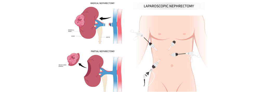
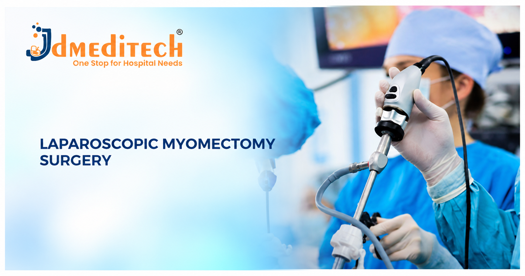
During laparoscopic myomectomy, surgeons use a laparoscope, camera system, and specialized instruments introduced through small abdominal incisions.
Compared with conventional open abdominal myomectomy, the laparoscopic approach may offer smaller incisions, reduced postoperative discomfort, and faster early recovery in appropriately selected patients.
However, treatment selection depends on fibroid size, number, location, symptoms, fertility goals, and overall health.
In this guide, JDMeditech explains laparoscopic myomectomy surgery, its indications, types, instruments, benefits, complications, recovery, and frequently asked questions.
What Is Laparoscopic Myomectomy Surgery?
Laparoscopic myomectomy is a uterus-preserving operation performed to remove selected uterine fibroids.
Instead of a large abdominal incision, the surgeon introduces a laparoscope and compatible instruments through several small openings.
The laparoscope provides magnified images of the uterus and surrounding pelvic anatomy.
The surgeon removes the selected fibroids and repairs the uterine tissue as required.
Unlike hysterectomy, the uterus remains in place after myomectomy.
However, new fibroids can develop in the future, and some patients may require additional treatment.
What Are Uterine Fibroids?
Uterine fibroids are usually noncancerous growths of smooth muscle and fibrous tissue that develop in the uterus.
Fibroids vary considerably in size, number, and location.
Some patients have a single small fibroid, while others develop multiple fibroids of different sizes.
Many fibroids cause no symptoms and do not require treatment.
Types of Uterine Fibroids
1. Intramural Fibroids
Intramural fibroids develop within the muscular wall of the uterus.
2. Subserosal Fibroids
Subserosal fibroids grow toward the outer surface of the uterus.
3. Submucosal Fibroids
Submucosal fibroids grow toward or into the uterine cavity and may contribute to abnormal bleeding or fertility problems.
4. Pedunculated Fibroids
Pedunculated fibroids are attached to the uterus by a stalk and may project into the uterine cavity or outward from the uterine surface.
5. Cervical Fibroids
Cervical fibroids develop in the cervix and may require specialized surgical planning.
Common Symptoms of Uterine Fibroids
- Heavy menstrual bleeding
- Prolonged menstrual periods
- Pelvic pain or pressure
- Abdominal fullness
- Frequent urination
- Constipation
- Menstrual cramps
- Pain during intercourse
- Iron-deficiency anemia caused by heavy bleeding
- Fertility or pregnancy-related concerns in selected cases
However, these symptoms may also occur with other gynecological conditions. A clinical assessment is required to identify the cause.
Why Is Laparoscopic Myomectomy Performed?
Laparoscopic myomectomy may be considered when uterine fibroids cause significant symptoms or complications and preserving the uterus is important.
For example, selected patients may experience heavy bleeding that does not respond adequately to medical treatment.
Additionally, fibroids may cause pelvic pressure or affect the uterine cavity.
Some patients may choose myomectomy because they wish to preserve the possibility of future pregnancy.
However, myomectomy does not guarantee improved fertility or successful pregnancy.
Who May Be Suitable for Laparoscopic Myomectomy?
Potential candidates may include selected patients with:
- Symptomatic uterine fibroids
- Heavy menstrual bleeding associated with fibroids
- Fibroid-related pelvic pressure
- Fibroids affecting the uterine cavity
- A desire to preserve the uterus
- Future pregnancy plans
- Fibroids suitable for minimally invasive removal
- Appropriate overall surgical fitness
However, fibroid number, size, location, surgical complexity, and the surgeon’s expertise determine whether laparoscopy is appropriate.
How Are Uterine Fibroids Diagnosed?
1. Medical History and Pelvic Examination
The gynecologist reviews symptoms, menstrual history, fertility goals, and relevant medical conditions.
2. Pelvic Ultrasound
Ultrasound is commonly used to identify fibroids and assess their size and location.
3. Transvaginal Ultrasound
Transvaginal ultrasound provides detailed images of the uterus and pelvic structures.
4. MRI
MRI may help map complex or multiple fibroids and support surgical planning.
5. Hysteroscopy
Hysteroscopy may be used to assess fibroids involving the uterine cavity.
6. Blood Tests
Blood tests may be used to evaluate anemia or other relevant medical concerns.
Types of Myomectomy Surgery
1. Laparoscopic Myomectomy
Fibroids are removed using a laparoscope and specialized instruments through small abdominal incisions.
2. Open Abdominal Myomectomy
Fibroids are removed through a larger abdominal incision. This approach may be appropriate for selected large, numerous, or complex fibroids.
3. Hysteroscopic Myomectomy
Selected fibroids projecting into the uterine cavity may be removed through the cervix using a hysteroscope, without abdominal incisions.
4. Robot-Assisted Myomectomy
Robot-assisted myomectomy uses a surgeon-controlled robotic system. It is one of several minimally invasive approaches.
5. Mini-Laparotomy Myomectomy
Selected fibroids may be removed through a smaller abdominal incision using an appropriate surgical technique.
Laparoscopic vs Open vs Hysteroscopic Myomectomy
| Feature | Laparoscopic | Open | Hysteroscopic |
|---|---|---|---|
| Access | Small abdominal incisions | Larger abdominal incision | Through the cervix |
| Typical Fibroid Suitability | Selected intramural and subserosal fibroids | Selected large or complex fibroids | Selected submucosal fibroids |
| Uterus Preserved | Yes | Yes | Yes |
| Early Recovery | Often faster than open surgery | May take longer | Often relatively short |
| Major Considerations | Fibroid mapping and uterine repair | Larger incision | Limited to suitable cavity-related fibroids |
Alternatives to Myomectomy
Not every patient with fibroids requires surgical removal.
Depending on symptoms, fibroid characteristics, and fertility goals, alternatives may include:
- Observation and follow-up
- Medication for heavy menstrual bleeding
- Hormonal treatments
- Selected gonadotropin-releasing hormone therapies
- Uterine artery embolization
- Radiofrequency ablation
- Other suitable fibroid treatments
- Hysterectomy when appropriate and desired
The effects of some treatments on future fertility are uncertain or may make them unsuitable for patients planning pregnancy.
Preparation Before Laparoscopic Myomectomy
Preparation begins with a detailed gynecological evaluation and assessment of the fibroids.
First, the surgical team reviews ultrasound or MRI findings.
Next, the patient undergoes appropriate anesthesia and medical assessment.
Additional preparation may include:
- Blood tests
- Assessment and treatment of anemia
- Review of current medications
- Discussion of fertility plans
- Evaluation of fibroid size and location
- Discussion of possible conversion to open surgery
- Discussion of blood loss and transfusion risks
- Discussion of tissue extraction options
- Assessment of malignancy risk
- Informed consent
How Is Laparoscopic Myomectomy Surgery Performed?
Laparoscopic myomectomy is generally performed under general anesthesia by a qualified gynecological surgical team.
First, the surgeon establishes laparoscopic access through small abdominal incisions.
Next, a laparoscope and camera system provide visualization of the uterus and surrounding pelvic structures.
The surgical team identifies the fibroids selected for removal.
Appropriate surgical techniques are used to separate the fibroids from the uterine tissue while controlling bleeding.
After fibroid removal, the uterine muscle is repaired using suitable suturing techniques.
The removed tissue is extracted using an approach appropriate to the patient’s condition and applicable safety guidance.
Finally, the surgeon assesses the operative field and closes the incisions.
However, the exact surgical technique depends on the number, size, depth, and location of the fibroids.
Why Is Uterine Reconstruction Important?
Myomectomy may involve an incision in the uterine muscle.
After fibroid removal, the surgeon repairs the affected uterine tissue.
Appropriate uterine reconstruction is particularly important when the patient may wish to become pregnant in the future.
However, the risk of pregnancy-related complications depends on the extent of surgery and individual circumstances.
Patients should discuss future pregnancy timing and delivery planning with their gynecologist.
Essential Instruments Used in Laparoscopic Myomectomy Surgery
Laparoscopic myomectomy instruments support pelvic visualization, tissue handling, fibroid removal, bleeding control, uterine reconstruction, and specimen extraction.
The following instrument categories are examples and do not represent a verified JDMeditech myomectomy surgical kit.
1. Laparoscope Telescope
Provides magnified visualization of the pelvic cavity.
2. Laparoscopic Camera System
Transfers images from the laparoscope to a surgical monitor.
3. Medical Light Source
Provides illumination during laparoscopic surgery.
4. Fiber-Optic Light Cable
Transfers illumination to the laparoscope.
5. Laparoscopic Trocar and Cannula
Provide access for compatible laparoscopic instruments.
6. Veress Needle
May be used for selected laparoscopic access techniques.
7. Uterine Manipulator
May support positioning and mobilization of the uterus during suitable procedures.
8. Laparoscopic Myoma Screw
Supports controlled traction on selected fibroids during appropriate surgical techniques.
9. Laparoscopic Atraumatic Grasper
Supports delicate handling of pelvic tissues.
10. Laparoscopic Grasping Forceps
Support controlled manipulation of selected tissues.
11. Laparoscopic Maryland Dissector
Supports fine dissection and anatomical exposure.
12. Laparoscopic Scissors
Support selected tissue cutting tasks.
13. Laparoscopic Metzenbaum Scissors
Support delicate tissue dissection.
14. Laparoscopic Electrosurgical Hook
May support selected dissection and coagulation tasks.
15. Laparoscopic Bipolar Forceps
Support selected hemostasis tasks.
16. Advanced Vessel Sealing Instrument
May support selected tissue and vessel management within the device’s approved intended use.
17. Laparoscopic Needle Holder
Supports intracorporeal suturing and uterine tissue repair.
18. Laparoscopic Knot Pusher
May assist with selected laparoscopic knot-tying techniques.
19. Laparoscopic Suction and Irrigation Instrument
Helps manage fluids and maintain visibility.
20. CO2 Insufflator
Creates the laparoscopic working space.
21. Insufflation Tubing
Connects the insufflator to compatible laparoscopic access equipment.
22. Laparoscopic Retractor
Supports exposure of selected pelvic structures.
23. Surgical Sutures
Support uterine muscle reconstruction and selected tissue closure tasks.
24. Specimen Retrieval Bag
May support contained retrieval of removed fibroid tissue.
25. Laparoscopic Morcellation System
In selected cases, an appropriately indicated morcellation system may be considered for tissue extraction.
Power morcellation requires careful patient selection and a compatible containment system in accordance with applicable safety guidance.
26. Electrosurgical Generator
Supplies energy to compatible surgical instruments.
27. Surgical Monitor
Displays laparoscopic images.
28. Sterile Surgical Drapes
Support preparation of the operative field.
29. Sterile Surgical Accessories
Compatible gloves, tubing, dressings, and other accessories support the surgical setup.
Laparoscopic Myomectomy Instruments and Their Functions
| Instrument | Primary Function |
|---|---|
| Laparoscope Telescope | Provides pelvic visualization |
| Camera System | Displays operative images |
| Light Source | Illuminates the surgical field |
| Trocar and Cannula | Provide laparoscopic access |
| Uterine Manipulator | Supports uterine positioning |
| Myoma Screw | Supports selected fibroid traction tasks |
| Atraumatic Grasper | Supports delicate tissue handling |
| Maryland Dissector | Supports fine dissection |
| Laparoscopic Scissors | Support tissue cutting |
| Bipolar Forceps | Support hemostasis |
| Vessel Sealing Instrument | Supports suitable vessel management |
| Needle Holder | Supports uterine suturing |
| Knot Pusher | Supports selected knot-tying techniques |
| Suction and Irrigation Instrument | Helps maintain visualization |
| CO2 Insufflator | Creates the laparoscopic working space |
| Specimen Retrieval Bag | Supports selected tissue retrieval |
| Morcellation System | Supports selected tissue extraction procedures |
| Electrosurgical Generator | Supplies compatible surgical energy |
Important Safety Considerations for Tissue Morcellation
Morcellation refers to dividing tissue into smaller pieces for extraction.
However, tissue believed to be a benign fibroid may occasionally contain an unsuspected uterine malignancy.
Morcellation can spread malignant or benign tissue within the abdominal cavity.
Therefore, power morcellation should only be considered in appropriately selected patients after suitable evaluation and informed consent.
The US FDA recommends that laparoscopic power morcellation be performed only with a compatible tissue containment system when the procedure is appropriate.
The FDA also advises against power morcellation for suspected or confirmed malignancy and in certain higher-risk patient groups.
Importantly, containment systems cannot eliminate every risk of tissue spread.
Potential Benefits of Laparoscopic Myomectomy Surgery
1. Uterus Preservation
The uterus remains in place after removal of the selected fibroids.
2. Relief of Fibroid-Related Symptoms
Many patients experience improvement in heavy bleeding, pelvic pressure, or other symptoms caused by fibroids.
3. Smaller Surgical Incisions
Laparoscopic techniques generally use smaller incisions than conventional open abdominal myomectomy.
4. Reduced Postoperative Discomfort
Selected patients may experience less postoperative pain.
5. Faster Early Recovery
Recovery may be faster after uncomplicated laparoscopic surgery compared with open surgery.
6. Magnified Surgical Visualization
The laparoscope provides magnified images of pelvic anatomy.
7. Preservation of Future Pregnancy Possibility
Because the uterus is preserved, future pregnancy may remain possible, although fertility outcomes are not guaranteed.
Risks and Complications of Laparoscopic Myomectomy
Although laparoscopic myomectomy is an established procedure, complications can occur.
- Bleeding
- Need for blood transfusion
- Wound infection
- Pelvic infection
- Injury to nearby organs
- Bladder injury
- Bowel injury
- Adhesion formation
- Conversion to open surgery
- Incomplete fibroid removal
- Development of new fibroids
- Need for additional surgery
- Possible effects on future pregnancy
- Rare uterine rupture during a future pregnancy
- Risks associated with tissue morcellation
- Anesthesia-related complications
Importantly, complication risks vary according to fibroid characteristics, patient health, and surgical complexity.
Recovery After Laparoscopic Myomectomy Surgery
Recovery depends on the number and size of fibroids removed, the extent of uterine repair, and the patient’s general health.
Initially, patients may experience abdominal discomfort, fatigue, bloating, and light vaginal bleeding.
Many patients gradually resume light activities within one to two weeks after uncomplicated surgery.
However, full recovery and internal uterine healing take longer.
Therefore, patients should follow their surgeon’s instructions regarding exercise, lifting, intercourse, and pregnancy planning.
Postoperative Care After Myomectomy
- Take prescribed medicines as directed
- Follow wound-care instructions
- Gradually resume walking
- Follow lifting restrictions
- Maintain hydration and appropriate nutrition
- Attend scheduled follow-up appointments
- Discuss future pregnancy timing
- Monitor menstrual symptoms
- Review pathology results when available
- Seek medical assessment for concerning symptoms
Can Fibroids Return After Myomectomy?
Yes. Myomectomy removes existing fibroids, but new fibroids may develop later.
The likelihood of future fibroids depends on several factors, including age, fibroid number, and individual biology.
Some patients may require additional medical or surgical treatment in the future.
Pregnancy After Laparoscopic Myomectomy
Pregnancy may be possible after myomectomy because the uterus is preserved.
However, the surgical repair of the uterine wall may affect pregnancy and delivery planning.
Some patients may be advised to have a cesarean delivery depending on the extent and location of uterine incisions.
There is no universally appropriate waiting period before attempting pregnancy.
Therefore, patients should discuss individualized pregnancy timing and delivery recommendations with their gynecologist.
Warning Signs After Laparoscopic Myomectomy
Patients should seek urgent medical assessment if concerning symptoms develop.
- Heavy vaginal bleeding
- Severe or worsening abdominal pain
- Persistent fever
- Foul-smelling vaginal discharge
- Persistent vomiting
- Increasing wound redness or discharge
- Difficulty passing urine
- New leg swelling or pain
- Chest pain or breathing difficulty
- Sudden severe weakness or fainting
Severe breathing difficulty, collapse, or major bleeding requires emergency medical care.
How to Choose Laparoscopic Myomectomy Instruments
Hospitals and gynecological surgery departments should evaluate their procedural requirements before purchasing laparoscopic myomectomy instruments.
First, purchasing teams should identify the types of myomectomy procedures performed by their department.
Next, they should review laparoscope dimensions, trocar compatibility, and instrument working lengths.
Furthermore, hospitals should assess uterine manipulators, myoma screws, atraumatic graspers, dissectors, scissors, and needle holders.
Additionally, surgical teams should verify camera, light source, electrosurgical generator, and CO2 insufflator compatibility.
Finally, purchasing departments should confirm approved intended uses, cleaning and sterilization instructions, and applicable regulatory documentation.
Important Equipment Selection Factors
- Approved intended use
- Planned myomectomy technique
- Laparoscope diameter and viewing angle
- Trocar and cannula dimensions
- Instrument working length
- Uterine manipulator compatibility
- Myoma screw specifications
- Atraumatic grasper design
- Dissector specifications
- Scissor design
- Vessel sealing device limitations
- Needle holder compatibility
- Suture specifications
- Specimen extraction requirements
- Morcellation safety requirements
- Camera system specifications
- Light source compatibility
- CO2 insufflator capabilities
- Reusable or single-use classification
- Cleaning and sterilization instructions
- Manufacturer technical documentation
- Applicable regulatory documentation
Laparoscopic Myomectomy Instruments – JDMeditech
JDMeditech welcomes inquiries from hospitals, gynecology departments, laparoscopic surgery centers, healthcare institutions, and international medical distributors regarding laparoscopic surgical instruments and related accessories.
For example, customers can inquire about available laparoscopic grasping forceps, scissors, trocars, needle holders, dissectors, and compatible surgical equipment.
Furthermore, the JDMeditech team can discuss available product configurations, technical specifications, intended uses, and applicable documentation.
Therefore, hospitals and medical distributors can contact JDMeditech to discuss their laparoscopic gynecological surgery equipment requirements.
For product information and international business inquiries, visit www.jdmeditech.com.
Frequently Asked Questions About Laparoscopic Myomectomy Surgery
1. What is laparoscopic myomectomy surgery?
It is a minimally invasive operation used to remove uterine fibroids while preserving the uterus.
2. What are uterine fibroids?
Fibroids are usually benign growths that develop in or around the muscular wall of the uterus.
3. Why is myomectomy performed?
It may be performed for selected patients with heavy bleeding, pelvic pressure, or other fibroid-related problems.
4. Is myomectomy the same as hysterectomy?
No. Myomectomy removes fibroids while preserving the uterus. Hysterectomy removes the uterus.
5. What is laparoscopic myomectomy?
It is a myomectomy performed using small abdominal incisions and specialized laparoscopic instruments.
6. What is hysteroscopic myomectomy?
It is removal of selected fibroids projecting into the uterine cavity using instruments introduced through the cervix.
7. Is laparoscopic myomectomy minimally invasive?
Yes. It uses small incisions and camera-based visualization.
8. What instruments are used in laparoscopic myomectomy?
Common instruments include laparoscopes, trocars, myoma screws, graspers, dissectors, scissors, needle holders, and compatible tissue extraction equipment.
9. What is a laparoscopic myomectomy set?
It is a collection of compatible instruments and accessories used for selected laparoscopic fibroid removal procedures.
10. Does myomectomy require general anesthesia?
Laparoscopic myomectomy is generally performed under general anesthesia.
11. How long does laparoscopic myomectomy take?
Operation time varies according to the number, size, and location of fibroids and surgical complexity.
12. Is laparoscopic myomectomy painful?
Anesthesia prevents pain during surgery. Temporary postoperative abdominal discomfort may occur.
13. Can myomectomy improve heavy menstrual bleeding?
Yes. Many patients experience improvement when fibroids are responsible for heavy bleeding.
14. Can pregnancy occur after myomectomy?
Yes. Pregnancy may remain possible because the uterus is preserved, but outcomes are not guaranteed.
15. What are the main risks of myomectomy?
Risks include bleeding, infection, injury to nearby organs, adhesion formation, and other complications.
16. Can fibroids return after myomectomy?
Yes. New fibroids may develop after surgery.
17. How long is recovery after laparoscopic myomectomy?
Many patients gradually resume light activities within one to two weeks, although full recovery varies.
18. Can patients eat normally after myomectomy?
Many patients return to their usual diet as tolerated according to postoperative instructions.
19. Is laparoscopic myomectomy better than open surgery?
It may provide faster early recovery in suitable patients, but open surgery may be preferable for certain complex fibroids.
20. Where can hospitals inquire about JDMeditech laparoscopic instruments?
Hospitals and medical distributors can visit JDMeditech to inquire about available laparoscopic instruments and accessories.
Conclusion
Laparoscopic Myomectomy Surgery is an established minimally invasive treatment option for selected patients with symptomatic uterine fibroids.
The procedure removes fibroids while preserving the uterus and may offer smaller incisions, reduced postoperative discomfort, and faster early recovery.
However, successful outcomes depend on accurate diagnosis, appropriate patient selection, surgical expertise, and careful postoperative follow-up.
For laparoscopic myomectomy instrument inquiries and international business requirements, contact JDMeditech – One Stop for Hospital Needs.
Website: www.jdmeditech.com
Medical Disclaimer: This article provides general educational information only. It does not replace professional medical advice, diagnosis, or surgical training. Qualified healthcare professionals must make treatment and equipment decisions according to current clinical guidance, applicable regulations, and approved manufacturer instructions.