Introduction to PUJ Obstruction Surgery
PUJ Obstruction Surgery is a urological procedure that treats a blockage or narrowing where the kidney joins the ureter. Doctors call this area the pelvi-ureteric junction (PUJ) or ureteropelvic junction (UPJ).
Normally, urine collects in the renal pelvis before flowing through the ureter into the bladder. However, a narrow junction may slow or block this flow.
As a result, urine can collect inside the kidney and cause swelling, known as hydronephrosis. Furthermore, severe or long-lasting obstruction may damage kidney function.
Therefore, a urologist may recommend surgery when symptoms, poor drainage, or declining kidney function make treatment necessary.
In this guide, JDMeditech explains PUJ obstruction, its causes, symptoms, diagnosis, pyeloplasty techniques, surgical instruments, benefits, risks, recovery, and frequently asked questions.
What Is PUJ Obstruction?
PUJ obstruction occurs when urine cannot pass normally from the renal pelvis into the upper ureter.
Sometimes, the narrowing develops before birth. In other cases, scarring, inflammation, or pressure from a nearby blood vessel may contribute.
Consequently, urine may collect above the narrowed area. Over time, this pressure can affect kidney drainage and function.
However, not every enlarged renal pelvis indicates a harmful obstruction. Therefore, doctors evaluate both kidney appearance and function before recommending treatment.
Understanding the Kidney and Ureter
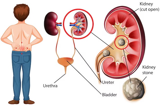
The kidneys filter blood and produce urine. Next, urine collects in a funnel-shaped area called the renal pelvis.
From there, the ureter carries urine toward the bladder. Normally, the junction between these structures allows urine to pass without significant restriction.
However, a narrow PUJ may interfere with drainage. As a result, the renal pelvis can become enlarged.
What Causes PUJ Obstruction?
PUJ obstruction may develop because of several anatomical or acquired conditions.
1. Congenital Narrowing
Some children are born with a narrow or poorly functioning PUJ. Consequently, doctors may detect kidney swelling during prenatal ultrasound.
2. Crossing Blood Vessel
A nearby blood vessel may press against the junction. Therefore, the surgeon may need to account for the vessel during reconstruction.
3. Scar Tissue
Previous inflammation, injury, or surgery may cause scarring. As a result, the junction may become narrow.
4. Abnormal Ureter Position
Sometimes, the ureter joins the renal pelvis at an unusual angle. Consequently, urine drainage may become less effective.
5. Previous Stones or Procedures
Selected patients develop narrowing after stone-related inflammation or medical procedures. However, the exact cause requires clinical evaluation.
Common Symptoms of PUJ Obstruction
Symptoms depend on age, obstruction severity, and kidney function.
For example, some children have no symptoms, while adults may experience repeated flank pain.
Common symptoms include:
- Pain in the side or back
- Abdominal discomfort
- Recurrent urinary tract infections
- Nausea or vomiting
- Kidney swelling
- Blood in the urine
- Pain after increased fluid intake
- Kidney stones in selected cases
- Reduced kidney function
- Poor feeding or unexplained symptoms in some infants
However, these symptoms can occur with other urinary conditions. Therefore, patients need appropriate medical assessment.
PUJ Obstruction in Children
Doctors often identify PUJ obstruction during pregnancy because ultrasound shows an enlarged renal pelvis.
After birth, the healthcare team may monitor the kidneys with repeat ultrasound examinations.
Furthermore, selected children need a diuretic renogram to assess urine drainage and kidney function.
However, many children with urinary tract dilation do not require immediate surgery.
Therefore, pediatric urologists consider the degree of swelling, changes over time, infections, symptoms, and kidney function.
PUJ Obstruction in Adults
Adults may develop symptoms from a narrowing that has existed for years or from an acquired problem.
For example, intermittent flank pain may become more noticeable after increased fluid intake.
Additionally, some adults experience urinary infections or kidney stones.
Therefore, doctors use clinical findings and imaging to determine whether the junction requires treatment.
How Is PUJ Obstruction Diagnosed?
Diagnosis begins with a medical history, physical examination, and suitable imaging.
1. Renal Ultrasound
Ultrasound helps doctors identify kidney swelling and assess the collecting system. Furthermore, repeat scans help monitor changes over time.
2. Diuretic Renogram
A diuretic renogram evaluates urine drainage and the relative function of each kidney. For example, doctors may use a MAG3 renal scan.
3. CT Urography
Selected adults may undergo CT imaging to assess urinary anatomy and possible causes of obstruction. However, radiation exposure must be considered.
4. MR Urography
Magnetic resonance imaging may provide additional anatomical information in selected patients.
5. Blood and Urine Tests
Blood tests help assess kidney health. Additionally, urine tests may identify infection or blood in the urine.
Consequently, the combination of imaging, symptoms, and kidney function guides treatment planning.
When Is PUJ Obstruction Surgery Recommended?
Not every patient with PUJ obstruction requires surgery.
However, doctors may recommend surgical correction when the obstruction causes significant symptoms or threatens kidney function.
Possible reasons include:
- Repeated flank pain caused by obstruction
- Declining kidney function
- Poor drainage on functional imaging
- Worsening hydronephrosis
- Recurrent infections related to obstruction
- Progressive kidney damage
- Selected cases involving associated kidney stones
- Significant obstruction confirmed by specialist assessment
Furthermore, pediatric decisions depend on serial imaging and the affected kidney’s contribution to overall kidney function.
Therefore, the surgeon should assess each patient individually.
Treatment Options for PUJ Obstruction
Treatment depends on symptoms, kidney function, and the cause of obstruction.
1. Observation and Monitoring
Doctors may monitor selected patients with ultrasound and functional imaging. However, follow-up is important when kidney swelling persists.
2. Temporary Urinary Drainage
A ureteral stent or nephrostomy tube may help drain urine in selected circumstances. However, temporary drainage does not always correct the underlying narrowing.
3. Pyeloplasty Surgery
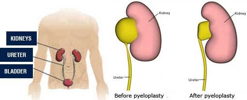
Pyeloplasty reconstructs the narrowed junction to improve urine drainage. Therefore, it is a standard surgical treatment for suitable cases.
4. Endopyelotomy
Endopyelotomy treats selected narrow junctions through an internal incision. However, its success can be lower than pyeloplasty, so doctors reserve it for carefully selected cases.
5. Other Surgical Options
Rarely, severe kidney damage or complex anatomy requires a different treatment plan. Therefore, the specialist considers the patient’s remaining kidney function and overall health.
What Is Pyeloplasty Surgery?
Pyeloplasty is a reconstructive operation that improves urine flow between the renal pelvis and ureter.
In a common technique, the surgeon removes the narrowed junction and reconnects healthy tissue.
As a result, urine can pass more freely from the kidney toward the bladder.
Furthermore, pyeloplasty may help preserve remaining kidney function and reduce obstruction-related symptoms.
However, it cannot guarantee recovery of kidney function that has already been permanently lost.
Types of PUJ Obstruction Surgery
1. Open Pyeloplasty
Open pyeloplasty uses a conventional surgical incision to reach the kidney and ureter.
Furthermore, the surgeon directly reconstructs the narrowed junction.
However, recovery may take longer than with suitable minimally invasive approaches.
2. Laparoscopic Pyeloplasty
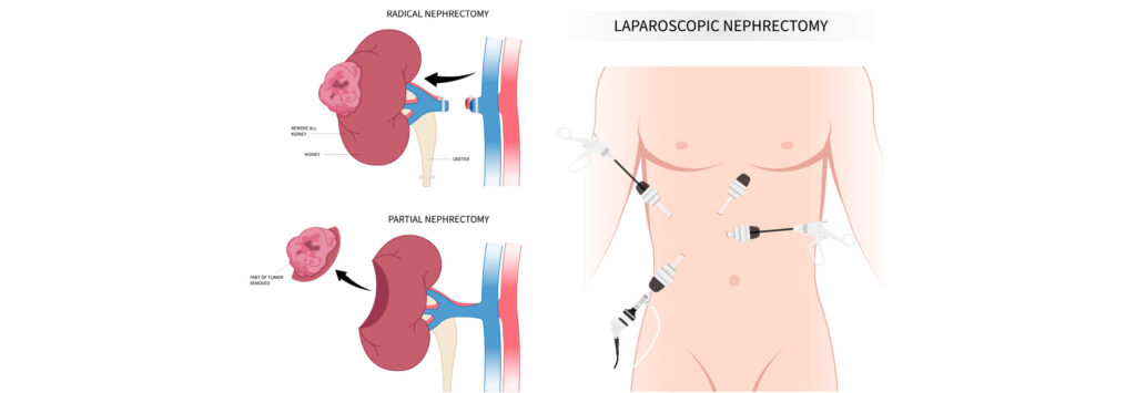
Laparoscopic pyeloplasty uses a camera and long surgical instruments through small abdominal incisions.
As a result, selected patients may experience smaller scars and a shorter hospital stay.
However, the operation requires specialized surgical training and equipment.
3. Robotic-Assisted Pyeloplasty
Robotic-assisted pyeloplasty uses surgeon-controlled robotic instruments to perform reconstruction through small incisions.
Furthermore, the system supports precise instrument movement and suturing.
However, robotic surgery is not necessary or available for every patient.
4. Anderson-Hynes Dismembered Pyeloplasty
Anderson-Hynes pyeloplasty is a widely used reconstructive technique.
The surgeon removes the narrowed junction and reconnects the ureter to the renal pelvis.
Additionally, the technique can address selected cases involving crossing blood vessels.
Therefore, specialists commonly use this approach for suitable PUJ obstructions.
5. Endopyelotomy
Endopyelotomy uses an endoscopic approach to release a selected narrowing.
However, it is not the preferred treatment for many patients because outcomes can be less reliable than pyeloplasty.
Comparison of PUJ Obstruction Surgical Techniques
| Technique | Approach | Important Consideration |
|---|---|---|
| Open Pyeloplasty | Conventional incision | Established reconstructive option |
| Laparoscopic Pyeloplasty | Small incisions and camera | Requires laparoscopic expertise |
| Robotic Pyeloplasty | Robot-assisted small-incision surgery | Requires specialized equipment |
| Anderson-Hynes Repair | Reconstruction of narrowed junction | Can use open, laparoscopic, or robotic access |
| Endopyelotomy | Internal incision | Suitable only for selected cases |
Preparation Before PUJ Obstruction Surgery
Preparation begins with a detailed urological assessment.
First, the surgeon reviews imaging results and kidney function. Next, the healthcare team evaluates medical history and current medicines.
Furthermore, the doctor checks for urinary infection and discusses the planned surgical approach.
Preparation may include:
- Medical history review
- Physical examination
- Renal ultrasound assessment
- Review of diuretic renogram results
- Selected CT or MRI review
- Kidney function testing
- Urine testing
- Medication and allergy review
- Anesthesia assessment
- Discussion of stent placement
- Review of possible complications
- Informed consent
- Recovery planning
Finally, the healthcare team provides instructions about fasting, medicines, and hospital admission.
How Is PUJ Obstruction Surgery Performed?
Pyeloplasty is a reconstructive procedure that aims to improve urine drainage.
First, the surgical team confirms the planned operation and provides appropriate anesthesia.
Next, the surgeon reaches the kidney and ureter through an open, laparoscopic, or robotic approach.
Afterward, the surgeon identifies the narrowed junction and reconstructs the connection between the renal pelvis and ureter.
Furthermore, the surgeon may place a temporary ureteral stent to support drainage during healing.
Finally, the surgical team checks the repair and closes the surgical wounds.
However, the exact technique depends on the patient’s anatomy and clinical requirements.
What Is a Double J Stent in Pyeloplasty?
A Double J Stent is a flexible tube that helps maintain urine drainage from the kidney to the bladder.
Its curved ends help keep the stent in position.
During selected pyeloplasty procedures, surgeons place a temporary Double J stent across the reconstructed junction.
Consequently, urine can drain while the repaired tissue heals.
However, not every pyeloplasty requires a stent. Furthermore, the timing of removal varies according to the surgeon’s plan.
How Long Does PUJ Obstruction Surgery Take?
Operating time varies according to the technique and complexity of the obstruction.
For example, laparoscopic pyeloplasty may take several hours.
Furthermore, anesthesia preparation and postoperative observation increase the total hospital time.
Therefore, patients should ask their surgical team for an individualized estimate.
Essential Instruments Used in PUJ Obstruction Surgery
PUJ obstruction surgical instruments support surgical access, tissue handling, reconstruction, suturing, and urinary drainage.
However, open, laparoscopic, robotic, and endoscopic procedures require different equipment.
The following list describes general instrument categories. It does not represent a verified JDMeditech pyeloplasty surgical set.
1. Surgical Scalpel
A surgical scalpel supports controlled tissue incision during open surgical access.
2. Metzenbaum Scissors
Metzenbaum scissors support careful soft-tissue dissection.
3. Mayo Scissors
Mayo scissors support selected tissue and suture cutting tasks.
4. Adson Tissue Forceps
Adson forceps support precise tissue handling.
5. Atraumatic Tissue Forceps
Atraumatic forceps help surgeons handle delicate tissue gently.
6. Dissecting Forceps
Dissecting forceps support suitable tissue-handling tasks.
7. Mosquito Artery Forceps
Mosquito forceps support control of small bleeding points.
8. Kelly Forceps
Kelly forceps support selected clamping and tissue-handling tasks.
9. Fine Needle Holder
A fine needle holder supports delicate reconstructive suturing.
10. Mayo-Hegar Needle Holder
A suitable Mayo-Hegar needle holder supports selected wound-closure tasks.
11. Surgical Retractor
A compatible retractor helps maintain surgical visibility.
12. Self-Retaining Retractor
A suitable self-retaining retractor supports exposure during selected open procedures.
13. Laparoscopic Telescope
A laparoscopic telescope provides visualization during minimally invasive pyeloplasty.
14. Laparoscopic Camera System
The camera system displays the surgical field on a monitor.
15. Laparoscopic Trocar
A compatible trocar provides access for selected laparoscopic instruments.
16. Laparoscopic Grasper
A laparoscopic grasper supports tissue manipulation during minimally invasive surgery.
17. Laparoscopic Dissector
A suitable dissector supports careful tissue separation.
18. Laparoscopic Scissors
Laparoscopic scissors support controlled cutting through minimally invasive access.
19. Laparoscopic Needle Holder
A laparoscopic needle holder supports reconstructive suturing.
20. Laparoscopic Clip Applicator
A compatible clip applicator may support selected bleeding-control tasks.
21. Electrosurgical Instrument
A compatible electrosurgical instrument may support tissue dissection or bleeding control.
22. Electrosurgical Generator
An electrosurgical generator supplies energy to compatible instruments.
23. Laparoscopic Suction and Irrigation Instrument
A suitable instrument helps remove fluid and maintain surgical visibility.
24. Surgical Sutures
Appropriate sutures support reconstruction and wound closure.
25. Double J Ureteral Stent
A Double J stent may provide temporary urinary drainage after reconstruction.
26. Ureteral Guide Wire
A compatible guide wire may support selected stent-placement procedures.
27. Ureteral Catheter
A suitable ureteral catheter may support selected drainage or imaging requirements.
28. Urinary Foley Catheter
A Foley catheter may support temporary bladder drainage during perioperative care.
29. Surgical Drain
A suitable surgical drain may help manage postoperative fluid in selected cases.
30. Sterile Surgical Drapes
Sterile drapes help establish the operative field.
31. Sterile Gauze
Gauze supports appropriate wound care and bleeding management.
32. Surgical Dressing
A suitable dressing helps protect surgical wounds.
33. Surgical Instrument Tray
A compatible tray supports instrument organization and reprocessing.
PUJ Obstruction Surgery Instruments and Their Functions
| Instrument | Primary Function | Typical Use |
|---|---|---|
| Surgical Scalpel | Controlled incision | Open surgery |
| Metzenbaum Scissors | Soft-tissue dissection | Open surgery |
| Fine Needle Holder | Reconstructive suturing | Open pyeloplasty |
| Laparoscopic Telescope | Internal visualization | Laparoscopic pyeloplasty |
| Laparoscopic Trocar | Instrument access | Laparoscopic pyeloplasty |
| Laparoscopic Grasper | Tissue handling | Minimally invasive surgery |
| Laparoscopic Needle Holder | Precise suturing | Laparoscopic reconstruction |
| Suction and Irrigation | Operative field management | Selected procedures |
| Double J Stent | Temporary urinary drainage | Selected pyeloplasty procedures |
| Ureteral Guide Wire | Selected device placement support | Stent placement |
| Foley Catheter | Bladder drainage | Perioperative care |
| Surgical Drain | Postoperative fluid drainage | Selected cases |
Why Is Instrument Selection Important?
PUJ obstruction surgery involves delicate kidney and ureteral tissues.
Therefore, surgeons need instruments that match the selected procedure and patient anatomy.
Furthermore, laparoscopic surgery requires compatible cameras, access devices, and long instruments.
Consequently, hospitals should verify instrument dimensions, intended uses, and equipment compatibility before surgery.
Potential Benefits of PUJ Obstruction Surgery
1. Improved Urine Drainage
Pyeloplasty aims to restore urine flow through the narrowed junction.
2. Relief of Obstruction-Related Pain
Successful treatment may reduce flank pain caused by poor drainage.
3. Protection of Kidney Function
Correcting significant obstruction may help preserve remaining kidney function.
4. Reduced Urine Backflow Pressure
Improved drainage may reduce pressure within the renal collecting system.
5. Treatment of Recurrent Symptoms
Selected patients may experience fewer obstruction-related symptoms after successful reconstruction.
6. Minimally Invasive Recovery Options
Laparoscopic or robotic surgery may allow smaller incisions and earlier recovery in suitable patients.
Risks and Complications of PUJ Obstruction Surgery
Although pyeloplasty is an established procedure, complications can occur.
For example, patients may experience bleeding, infection, or temporary discomfort from a ureteral stent.
Possible complications include:
- Postoperative pain
- Bleeding
- Wound infection
- Urinary tract infection
- Urine leakage
- Persistent hydronephrosis
- Recurrent junction narrowing
- Stent-related discomfort
- Stent migration or blockage
- Injury to nearby structures
- Delayed wound healing
- Need for additional procedures
- Anesthesia-related complications
Furthermore, some patients may have limited kidney function improvement despite successful drainage.
Therefore, the surgeon should discuss realistic outcomes before treatment.
Recovery After PUJ Obstruction Surgery
Recovery depends on the surgical technique, age, and individual health.
Initially, patients may experience wound discomfort, fatigue, or mild urinary symptoms.
However, these symptoms often improve gradually.
Furthermore, patients with a temporary ureteral stent may experience urinary frequency, urgency, or discomfort until removal.
Therefore, the healthcare team should provide an individualized recovery plan.
Postoperative Care After Pyeloplasty
Appropriate postoperative care supports healing and helps identify complications.
- Take prescribed medicines as directed
- Follow wound-care instructions
- Attend scheduled follow-up visits
- Follow advice about fluid intake
- Avoid strenuous activity until cleared
- Monitor for fever or worsening pain
- Follow catheter or drain-care instructions
- Keep the scheduled stent-removal appointment
- Complete recommended follow-up imaging
- Report persistent urinary symptoms
Additionally, patients should ask their surgeon when they can return to work, school, or physical activity.
How Long Does a Double J Stent Stay After Pyeloplasty?
A temporary Double J stent may remain in place for several weeks after surgery.
For example, some surgeons remove it after approximately four to six weeks.
However, timing varies according to the patient, technique, and healing progress.
Therefore, patients should follow the specific removal plan provided by their healthcare team.
Can PUJ Obstruction Return After Surgery?
Pyeloplasty has a high success rate in appropriately selected patients.
However, recurrent narrowing can occur.
Furthermore, some patients continue to show kidney swelling on imaging even after drainage improves.
Therefore, follow-up scans help assess the repair and kidney function.
Does Pyeloplasty Restore Kidney Function?
Pyeloplasty aims to relieve obstruction and preserve kidney health.
In selected patients, kidney function may improve after successful treatment.
However, long-standing kidney damage may not fully reverse.
Therefore, the surgeon should explain expected results using the patient’s preoperative kidney function and imaging.
PUJ Obstruction Surgery in Children vs Adults
Children and adults may require different assessment and follow-up plans.
For example, pediatric specialists often monitor prenatal hydronephrosis with repeat ultrasound examinations.
In contrast, adults may present with pain, stones, or recurrent infections.
However, both groups require evaluation of kidney drainage and function before treatment.
Warning Signs After PUJ Obstruction Surgery
Patients should contact their healthcare team if concerning symptoms develop.
- Fever or chills
- Severe or worsening flank pain
- Persistent vomiting
- Heavy bleeding in the urine
- Difficulty passing urine
- Inability to pass urine
- Increasing wound redness
- Foul-smelling wound discharge
- Significant abdominal swelling
- Problems with a urinary stent or drain
Importantly, fever with urinary obstruction, severe illness, or inability to pass urine requires urgent medical assessment.
How to Choose PUJ Obstruction Surgical Instruments
Hospitals and urology departments should review their procedure requirements before purchasing surgical instruments.
First, purchasing teams should identify whether the facility performs open, laparoscopic, robotic, or endoscopic procedures.
Next, they should review suitable scissors, graspers, needle holders, cameras, and access devices.
Furthermore, hospitals should verify compatibility between guide wires, ureteral catheters, and Double J stents.
Additionally, teams should confirm material specifications, instrument dimensions, and cleaning requirements.
Finally, purchasing departments should review approved intended uses and applicable regulatory documentation.
Important Equipment Selection Factors
- Approved intended use
- Surgical technique
- Instrument dimensions
- Laparoscopic equipment compatibility
- Camera and telescope compatibility
- Trocar specifications
- Needle holder requirements
- Suture compatibility
- Guide wire compatibility
- Double J stent dimensions
- Ureteral catheter specifications
- Sterile packaging
- Single-use or reusable classification
- Cleaning and sterilization instructions
- Manufacturer technical documentation
- Applicable regulatory documentation
PUJ Obstruction Surgical Instruments – JDMeditech
JDMeditech welcomes inquiries from hospitals, urology departments, surgical centers, healthcare institutions, and international medical distributors regarding urological surgical instruments and related medical equipment.
For example, customers can inquire about available Double J stents, ureteral guide wires, ureteral catheters, surgical forceps, needle holders, and compatible accessories.
Furthermore, healthcare facilities may discuss instrument configuration requirements with the JDMeditech team.
However, customers should confirm product availability, approved intended uses, technical specifications, and applicable regulatory documentation before ordering.
For product information and international business inquiries, visit www.jdmeditech.com.
Frequently Asked Questions About PUJ Obstruction Surgery
1. What is PUJ obstruction?
PUJ obstruction is a narrowing that reduces urine flow between the renal pelvis and ureter.
2. What does PUJ stand for?
PUJ stands for pelvi-ureteric junction. Doctors also use the term ureteropelvic junction, or UPJ.
3. What causes PUJ obstruction?
Possible causes include congenital narrowing, crossing blood vessels, scarring, and unusual ureter anatomy.
4. What are the symptoms of PUJ obstruction?
Symptoms may include flank pain, kidney swelling, urinary infections, nausea, and reduced kidney function.
5. Does every patient need surgery?
No. Doctors monitor selected patients when symptoms and kidney function do not require immediate intervention.
6. What is pyeloplasty surgery?
Pyeloplasty reconstructs the narrowed junction to improve urine drainage from the kidney.
7. What is Anderson-Hynes pyeloplasty?
It is a widely used technique that removes the narrowed junction and reconnects the ureter to the renal pelvis.
8. What is laparoscopic pyeloplasty?
Laparoscopic pyeloplasty repairs the obstruction using a camera and long instruments through small incisions.
9. What is robotic pyeloplasty?
Robotic pyeloplasty uses surgeon-controlled robotic instruments for minimally invasive reconstruction.
10. What is endopyelotomy?
Endopyelotomy treats selected junction narrowings through an internal incision. However, it is not suitable for every patient.
11. What instruments are used in PUJ obstruction surgery?
Common instruments include scissors, forceps, needle holders, laparoscopic graspers, telescopes, and suitable drainage accessories.
12. What is a Double J stent?
A Double J stent is a flexible tube that helps drain urine from the kidney to the bladder.
13. Is a Double J stent always necessary?
No. Stent use depends on the surgical technique and patient needs.
14. How long does the stent stay after pyeloplasty?
Some stents remain for approximately four to six weeks. However, the surgeon determines the removal schedule.
15. Does pyeloplasty require anesthesia?
Yes. The surgical team uses suitable anesthesia according to the procedure and patient’s needs.
16. How long does PUJ obstruction surgery take?
Duration varies according to the surgical technique and complexity of the obstruction.
17. How long does recovery take?
Recovery often takes several weeks. However, individual healing and surgical approaches vary.
18. Can PUJ obstruction return after surgery?
Yes. Recurrent narrowing can occur, although pyeloplasty has a high success rate.
19. Does surgery restore kidney function?
Successful treatment may preserve or improve kidney function. However, permanent damage may not fully reverse.
20. Where can hospitals inquire about JDMeditech PUJ obstruction instruments?
Hospitals and medical distributors can visit JDMeditech to inquire about available urological surgical instruments and accessories.
Conclusion
PUJ Obstruction Surgery is an established treatment for selected patients with impaired urine drainage between the kidney and ureter.
Furthermore, pyeloplasty can help relieve obstruction, improve drainage, and protect remaining kidney function.
However, treatment depends on symptoms, imaging results, and the patient’s overall kidney health.
Therefore, a qualified urologist should evaluate the condition and recommend suitable treatment.
For urological surgical instrument inquiries and international business requirements, contact JDMeditech – One Stop for Hospital Needs.
Website: www.jdmeditech.com
Medical Disclaimer: This article provides general educational information only. It does not replace professional medical advice, diagnosis, informed consent, or surgical training. Qualified healthcare professionals must make treatment and equipment decisions according to current clinical guidance, applicable regulations, and approved manufacturer instructions.