Rigid Ureteroscopy Surgery: Procedure, Instruments, Benefits and Recovery
Rigid Ureteroscopy Surgery is a minimally invasive endoscopic procedure that helps urologists examine and treat selected conditions of the ureter. Doctors commonly use rigid or semi-rigid ureteroscopy to manage ureteral stones, especially stones in the lower and middle ureter.
During the procedure, the surgeon introduces a rigid or semi-rigid ureteroscope through the natural urinary passage. Therefore, the procedure usually does not require an external surgical incision.
Furthermore, ureteroscopy provides direct visualization of the ureter. When necessary, the surgeon may use compatible lithotripsy instruments or retrieval devices to treat a stone.
However, rigid ureteroscopy is not suitable for every patient. Therefore, doctors assess stone size, location, urinary anatomy, infection status, and overall health before recommending treatment.
JDMeditech supplies urology and endourology instruments for hospitals, healthcare facilities, and medical distributors.
What Is Rigid Ureteroscopy Surgery?
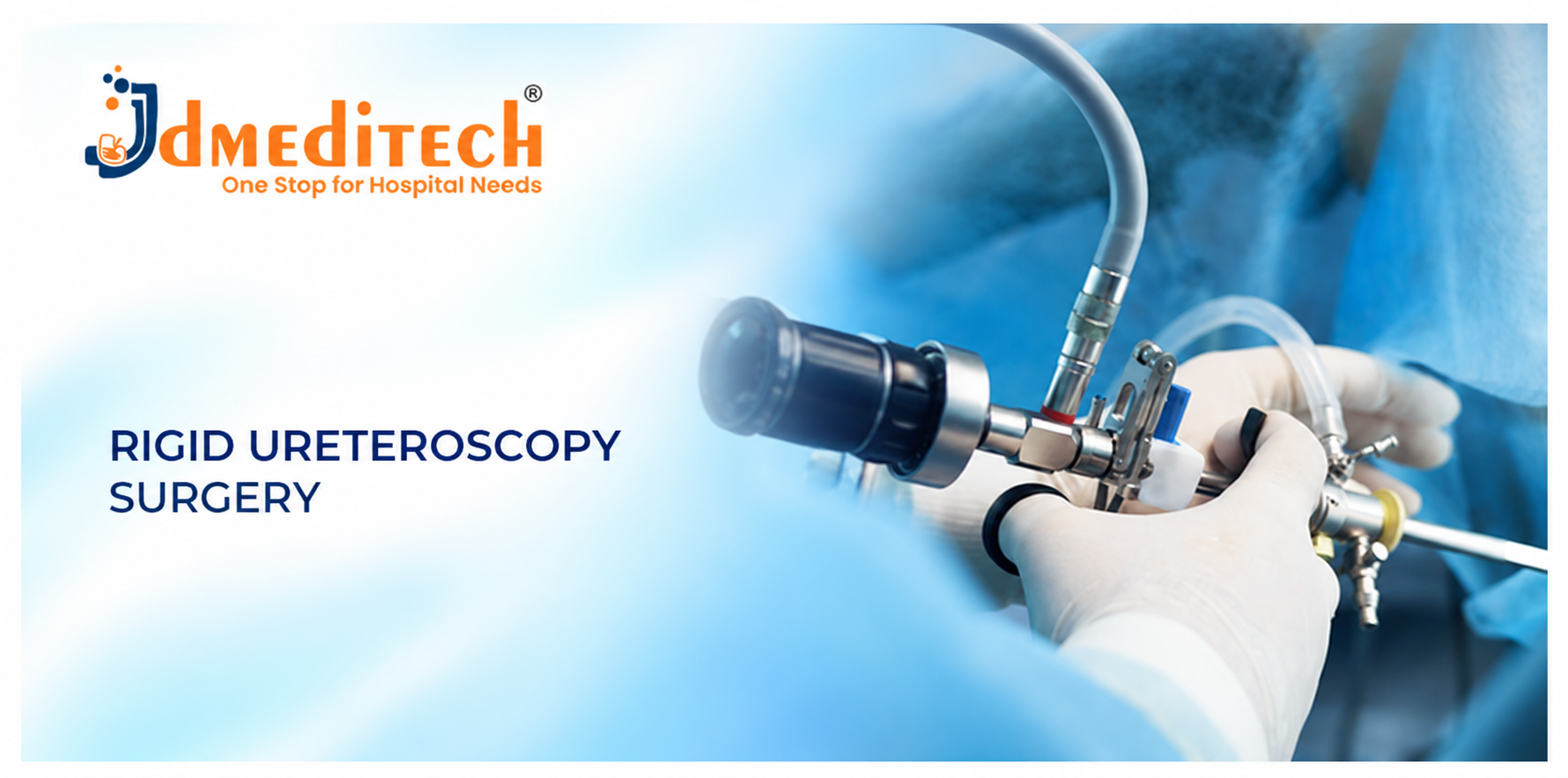
Rigid ureteroscopy is an endoscopic procedure that allows a urologist to inspect the ureter using a specialized rigid ureteroscope.
The instrument has a relatively firm shaft and provides visualization of selected urinary structures.
Additionally, compatible working channels allow the surgeon to use selected treatment instruments.
However, modern ureteroscopic procedures often use semi-rigid scopes because they provide some flexibility while maintaining suitable instrument control.
What Is the Full Form of URS?
URS commonly stands for Ureteroscopy or Ureteroscopic Surgery.
Furthermore, URSL often refers to Ureteroscopic Lithotripsy, a procedure that fragments ureteral stones.
Why Is Rigid Ureteroscopy Performed?
Doctors use rigid or semi-rigid ureteroscopy for selected conditions affecting the ureter.
- Lower ureteral stones requiring active treatment
- Selected middle ureteral stones
- Selected upper ureteral stones when access is suitable
- Assessment of certain ureteral abnormalities
- Selected diagnostic ureteral biopsy procedures
- Removal of selected urinary tract foreign bodies
- Selected ureteral narrowing assessments
However, the indication depends on the patient’s condition and available treatment alternatives.
What Are Ureteral Stones?
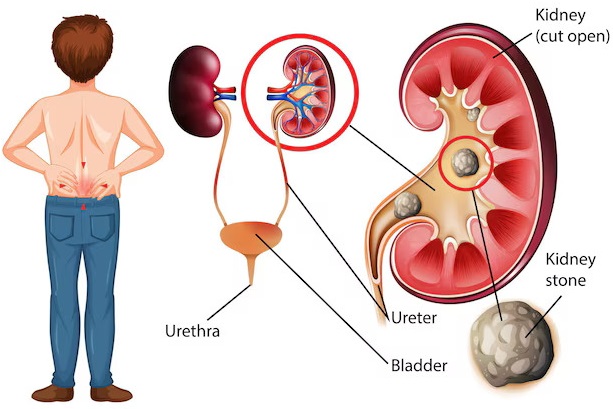
Ureteral stones are urinary calculi located within the ureter, the tube that carries urine from the kidney to the bladder.
Most ureteral stones originate in the kidney and then move into the ureter.
Consequently, stones may obstruct urine flow and cause significant pain.
Common Causes of Ureteral Stones
Several factors can increase the likelihood of urinary stone formation.
- Low fluid intake
- High urinary concentrations of stone-forming substances
- Family history of urinary stones
- Selected metabolic disorders
- Recurrent urinary infections
- Certain medications
- Selected anatomical abnormalities
- Dietary factors in susceptible individuals
However, stone formation often involves multiple factors. Therefore, doctors may recommend metabolic evaluation for recurrent stone disease.
Common Symptoms of Ureteral Stones
Ureteral stones may cause severe pain or urinary symptoms. However, some stones produce few symptoms.
- Severe pain in the side or back
- Pain extending toward the groin
- Blood in the urine
- Nausea or vomiting
- Frequent urination
- Urgency to urinate
- Painful urination
- Fever or chills when infection occurs
Important: Fever or chills with suspected urinary obstruction may indicate a medical emergency. Therefore, patients should seek urgent medical care.
When Is Rigid Ureteroscopy Recommended?
Urologists may recommend rigid or semi-rigid ureteroscopy when a ureteral stone requires intervention and endoscopic treatment is appropriate.
For example, persistent obstruction, uncontrolled pain, or failure of spontaneous stone passage may influence the decision.
Furthermore, stone location and size affect treatment selection.
However, some small ureteral stones pass naturally. Therefore, not every patient requires surgery.
Rigid Ureteroscopy for Lower Ureteral Stones
Rigid or semi-rigid ureteroscopy is commonly used to manage selected distal ureteral stones.
Furthermore, direct visualization supports assessment of the stone and surrounding ureter.
Rigid Ureteroscopy for Middle Ureteral Stones
Rigid or semi-rigid ureteroscopy can treat selected middle ureteral stones.
Additionally, compatible lithotripsy instruments support stone fragmentation when necessary.
Rigid Ureteroscopy for Upper Ureteral Stones
Rigid or semi-rigid ureteroscopy may treat selected upper ureteral stones when access is suitable.
However, flexible ureteroscopy may offer advantages in difficult anatomy.
Therefore, the surgeon selects the endoscope according to clinical requirements.
Rigid Ureteroscopy vs. Semi-Rigid Ureteroscopy
Rigid and semi-rigid ureteroscopes share several clinical applications. However, their shaft flexibility differs.
| Feature | Rigid Ureteroscope | Semi-Rigid Ureteroscope |
|---|---|---|
| Shaft Design | Relatively fixed shaft | Limited shaft flexibility |
| Common Application | Selected ureteral procedures | Commonly used for ureteral stone treatment |
| Flexibility | Minimal | Greater than a fully rigid model |
| Working Channel | According to model | According to model |
| Imaging | According to model | According to model |
Rigid Ureteroscopy vs. Flexible Ureteroscopy
Rigid ureteroscopy uses a relatively firm endoscope for selected ureteral procedures.
In contrast, flexible ureteroscopy uses a flexible instrument that can navigate selected curves within the upper urinary tract.
Therefore, flexible ureteroscopy is commonly used for selected kidney stone procedures.
Rigid Ureteroscopy vs. RIRS Surgery
Rigid ureteroscopy commonly focuses on selected ureteral conditions.
In contrast, RIRS uses flexible endoscopic techniques to treat selected conditions inside the kidney collecting system.
However, both procedures belong to the broader field of endourology.
Rigid Ureteroscopy vs. ESWL
Extracorporeal Shock Wave Lithotripsy, or ESWL, uses externally generated shock waves to fragment selected urinary stones.
In contrast, rigid ureteroscopy provides direct endoscopic visualization.
Furthermore, compatible lithotripsy instruments can fragment stones during the procedure.
However, each technique has different indications and limitations.
Rigid Ureteroscopy vs. PCNL
Percutaneous Nephrolithotomy, or PCNL, accesses the kidney through a small percutaneous tract.
In contrast, rigid ureteroscopy reaches the ureter through the natural urinary passage.
Therefore, the procedures serve different treatment needs.
Diagnostic Tests Before Rigid Ureteroscopy
Before surgery, the healthcare team evaluates stone characteristics, urinary anatomy, and patient health.
Ultrasound Examination
Ultrasound may identify hydronephrosis and selected urinary stones.
CT Scan
CT imaging provides detailed information about stone size, location, and urinary anatomy.
Furthermore, it supports treatment planning.
Urine Examination and Culture
Urine testing helps identify infection. Therefore, clinicians review infection status before elective surgery.
Blood Tests
Blood tests may assess kidney function, blood counts, and other relevant factors.
Anesthesia Assessment
The anesthesia team evaluates medical fitness and plans appropriate monitoring.
How Does Rigid Ureteroscopy Surgery Work?
Rigid ureteroscopy uses a specialized endoscope to examine the ureter.
During treatment, the surgeon introduces a compatible rigid or semi-rigid ureteroscope through the natural urinary passage.
Next, the endoscope provides visualization of the targeted area.
When necessary, compatible lithotripsy instruments fragment the stone. Additionally, retrieval devices may remove selected fragments.
Finally, the surgeon evaluates the operative findings and determines whether temporary urinary drainage is necessary.
General Steps of Rigid Ureteroscopy
The following overview explains the broad stages of care. However, it does not provide instructions for performing surgery.
Step 1: Preoperative Assessment
First, the urologist reviews imaging, urine tests, kidney function, and medical history.
Step 2: Patient Preparation
Next, the healthcare team prepares the patient according to established surgical and anesthesia protocols.
Step 3: Endoscopic Access
The surgeon establishes suitable access through the natural urinary passage.
Step 4: Ureteroscope Examination
The ureteroscope provides visualization of the targeted urinary structures.
Step 5: Stone Treatment
When necessary, compatible lithotripsy instruments fragment the stone.
Step 6: Stone Fragment Management
The surgeon manages selected fragments using appropriate endoscopic techniques.
Step 7: Final Assessment
Next, the surgical team evaluates the operative findings and checks for additional concerns.
Step 8: Drainage and Recovery
Finally, the surgeon determines whether a temporary DJ stent or another drainage device is necessary.
Important: Only appropriately trained healthcare professionals should perform ureteroscopy.
Surgical Instruments Used in Rigid Ureteroscopy
Rigid ureteroscopy requires specialized endourology instruments. However, the exact equipment list depends on the planned procedure.
- Rigid Ureteroscope: Provides visualization of the ureter.
- Semi-Rigid Ureteroscope: Supports selected ureteral procedures.
- Cystoscope: Supports selected lower urinary tract access tasks.
- Guide Wire: Supports selected instrument access and placement tasks.
- Ureteral Catheter: Supports selected urinary access or drainage tasks.
- Laser Lithotripsy System: Supports fragmentation of selected urinary stones.
- Laser Fiber: Delivers energy through a compatible endoscope.
- Pneumatic Lithotripter: May support fragmentation of selected ureteral stones.
- Stone Retrieval Basket: Supports removal of selected stone fragments.
- Stone Grasping Forceps: May support retrieval in selected procedures.
- DJ Ureteral Stent: May provide temporary internal urinary drainage.
- Endoscopic Camera System: Displays images from compatible equipment.
- Medical Light Source: Provides illumination when required.
- Irrigation System: Helps maintain endoscopic visibility.
- Imaging Equipment: Supports selected guidance tasks.
Furthermore, all instruments must be compatible with the selected endoscopic system.
Rigid Ureteroscope
A rigid ureteroscope is the primary instrument used during rigid ureteroscopy.
Additionally, different models provide different shaft dimensions, imaging systems, and working channels.
Therefore, hospitals should confirm the selected model’s specifications before use.
Semi-Rigid Ureteroscope
A semi-rigid ureteroscope provides limited shaft flexibility while supporting selected ureteral procedures.
Furthermore, it is commonly used for ureteral stone treatment.
Guide Wire for Rigid Ureteroscopy
A guide wire supports selected access and instrument placement tasks.
Furthermore, guide wires vary in diameter, length, tip design, and coating.
Therefore, clinicians must confirm compatibility before use.
Ureteral Catheter
A ureteral catheter supports selected urinary access, assessment, or drainage tasks.
However, catheter selection depends on the planned procedure.
Laser Lithotripsy System
Laser lithotripsy systems deliver energy through compatible fibers to fragment urinary stones.
For example, holmium laser systems may support selected ureteral stone procedures.
However, compatibility depends on the selected equipment.
Laser Fiber for Rigid Ureteroscopy
A laser fiber delivers energy from a compatible laser system to the treatment area.
Additionally, fiber diameter and handling requirements vary between models.
Therefore, users must follow manufacturer instructions for use.
Pneumatic Lithotripter
A pneumatic lithotripter uses mechanical energy to fragment selected urinary stones.
Furthermore, the surgeon must consider stone location and the risk of stone migration.
Stone Retrieval Basket
A stone retrieval basket may help remove selected stone fragments.
Furthermore, basket dimensions must match the ureteroscope’s working channel.
Stone Grasping Forceps
Stone grasping forceps may support retrieval of selected fragments.
However, the surgeon must choose a compatible instrument according to the clinical situation.
DJ Ureteral Stent
A Double J ureteral stent may provide temporary internal urinary drainage after ureteroscopy.
However, not every patient requires a postoperative stent.
Therefore, the surgeon evaluates operative findings before deciding.
Irrigation System
An irrigation system helps maintain visibility during rigid ureteroscopy.
However, excessive irrigation pressure can increase complications.
Therefore, the surgical team must manage irrigation according to appropriate clinical protocols.
Technical Specifications of Rigid Ureteroscopy Instruments
Instrument specifications vary between manufacturers and models. Therefore, customers should request verified product datasheets before purchasing.
| Parameter | Specification |
|---|---|
| Procedure Name | Rigid Ureteroscopy Surgery |
| Short Name | URS |
| Medical Specialty | Urology / Endourology |
| Primary Application | Diagnosis and treatment of selected ureteral conditions |
| Main Endoscope | Rigid or semi-rigid ureteroscope |
| Ureteroscope Diameter | According to selected model |
| Working Channel | Confirm manufacturer specifications |
| Guide Wire Diameter | According to selected device |
| Guide Wire Length | Confirm manufacturer specifications |
| Ureteral Catheter Size | According to clinical requirements |
| Lithotripsy Technology | Compatible laser or pneumatic system |
| Laser Fiber Diameter | According to system compatibility |
| Stone Basket Configuration | According to working channel compatibility |
| DJ Stent Size | According to patient requirements |
| Instrument Material | Confirm manufacturer specifications |
| Sterility | According to product labeling |
| Reusable or Single Use | According to manufacturer instructions |
| Cleaning and Reprocessing | According to validated manufacturer instructions |
| Supplier | JDMeditech, subject to product availability |
Benefits of Rigid Ureteroscopy Surgery
Rigid ureteroscopy offers several potential benefits for appropriately selected patients.
- Usually avoids an external surgical incision
- Provides direct endoscopic visualization
- Supports treatment of selected ureteral stones
- Allows compatible lithotripsy techniques
- May support retrieval of selected stone fragments
- Often involves a relatively short hospital stay
- May support faster recovery than open surgery
- Offers an alternative to shock wave treatment in selected cases
However, outcomes depend on stone characteristics, anatomy, and surgical expertise.
Advantages of Endoscopic Ureteral Stone Treatment
Endoscopic treatment allows the surgeon to visualize the targeted ureteral structures.
Furthermore, compatible instruments support stone fragmentation and retrieval.
However, difficult anatomy or large stone burdens may limit treatment effectiveness.
Possible Risks and Complications
Rigid ureteroscopy is a surgical procedure with potential risks. Therefore, patients should discuss complications with their urologist.
- Urinary tract infection
- Fever
- Sepsis
- Bleeding or blood in the urine
- Ureteral injury
- Ureteral perforation
- Ureteral stricture
- Postoperative discomfort
- Residual stone fragments
- Need for additional treatment
- Anesthesia-related complications
Furthermore, complication risks depend on infection status, stone complexity, and patient health.
Who Is a Suitable Candidate?
Rigid ureteroscopy may suit patients with selected ureteral stones or other ureteral conditions.
However, the urologist must evaluate stone location, anatomy, infection status, and treatment alternatives.
Therefore, suitability requires an individualized clinical assessment.
Recovery After Rigid Ureteroscopy
Recovery varies according to the procedure and patient condition.
After surgery, the healthcare team monitors pain, urinary symptoms, bleeding, and signs of infection.
Additionally, follow-up imaging may help assess residual stones when appropriate.
Postoperative Care After Rigid Ureteroscopy
Appropriate postoperative care supports recovery and helps identify complications.
- Take prescribed medications as directed.
- Maintain fluid intake according to medical advice.
- Follow activity restrictions provided by the surgeon.
- Attend scheduled follow-up appointments.
- Follow DJ stent instructions when applicable.
- Report fever or worsening pain.
- Seek urgent care for severe symptoms or inability to urinate.
How Long Does Rigid Ureteroscopy Take?
Operating time depends on stone size, location, number, and complexity.
Furthermore, the selected instruments and treatment technique may influence duration.
Therefore, the surgical team provides an individualized estimate.
How Long Does Recovery Take?
Many patients resume light activities within several days. However, recovery varies.
Therefore, patients should follow their surgeon’s instructions before returning to strenuous activity.
Is a DJ Stent Required After Rigid Ureteroscopy?
Not every patient requires a DJ stent after uncomplicated ureteroscopy.
However, surgeons may place a temporary stent when clinically indicated.
Additionally, patients with a stent may experience urinary frequency, discomfort, or blood in the urine.
Can Ureteral Stones Return After Surgery?
Yes. Rigid ureteroscopy treats existing stones but does not eliminate every cause of urinary stone formation.
Therefore, doctors may recommend stone analysis, metabolic evaluation, and preventive care.
How to Prevent Ureteral Stone Recurrence
Prevention depends on stone composition and individual risk factors.
- Maintain appropriate hydration according to medical advice.
- Follow a balanced diet.
- Limit excessive sodium intake when recommended.
- Follow individualized dietary guidance.
- Complete recommended metabolic testing.
- Take prescribed preventive medications when indicated.
- Attend follow-up appointments.
Furthermore, patients should consult their healthcare team before making major dietary changes.
How to Select Rigid Ureteroscopy Instruments
Hospitals should evaluate several factors before purchasing rigid ureteroscopy equipment.
- Planned surgical technique
- Ureteroscope type and diameter
- Working channel dimensions
- Image quality and system compatibility
- Guide wire compatibility
- Ureteral catheter specifications
- Laser or pneumatic lithotripsy compatibility
- Stone basket compatibility
- DJ stent specifications
- Sterility and reuse requirements
- Cleaning and reprocessing instructions
- Technical documentation
Additionally, hospitals should confirm the complete equipment list with the operating surgeon.
Why Choose JDMeditech for Rigid Ureteroscopy Instruments?
JDMeditech supplies urology and endourology instruments for hospitals, healthcare facilities, and medical distributors.
Furthermore, customers can contact JDMeditech to inquire about available rigid ureteroscopy instruments and accessories.
- Rigid ureteroscope inquiries
- Semi-rigid ureteroscope inquiries
- URS surgery set inquiries
- Guide wire inquiries
- Ureteral catheter inquiries
- Laser lithotripsy accessory inquiries
- Pneumatic lithotripter inquiries
- Stone retrieval basket inquiries
- DJ stent inquiries
- International export inquiries
- Technical specifications upon request
However, product availability and compatibility vary. Therefore, customers should confirm specifications before ordering.
Frequently Asked Questions About Rigid Ureteroscopy
1. What Is Rigid Ureteroscopy Surgery?
Rigid ureteroscopy is a minimally invasive procedure that uses a rigid ureteroscope to examine and treat selected ureteral conditions.
2. What Is the Full Form of URS?
URS commonly stands for Ureteroscopy or Ureteroscopic Surgery.
3. Why Is Rigid Ureteroscopy Performed?
Doctors commonly perform it to treat selected ureteral stones.
4. Is Rigid Ureteroscopy Major Surgery?
Rigid ureteroscopy is a surgical procedure that requires appropriate anesthesia and monitoring. However, it is less invasive than open surgery.
5. Is Rigid Ureteroscopy Painful?
Patients receive anesthesia during surgery. However, postoperative discomfort may occur.
6. How Long Does Rigid Ureteroscopy Take?
Operating time depends on stone complexity and the selected treatment technique.
7. How Long Is the Hospital Stay?
Many patients have a short hospital stay. However, duration depends on recovery and clinical findings.
8. How Long Does Recovery Take?
Many patients resume light activities within several days, although recovery varies.
9. What Is the Difference Between Rigid and Semi-Rigid Ureteroscopy?
Semi-rigid ureteroscopes provide limited shaft flexibility, while fully rigid models have relatively fixed shafts.
10. What Is the Difference Between Rigid and Flexible Ureteroscopy?
Flexible ureteroscopes can navigate selected bends within the upper urinary tract, while rigid models have limited flexibility.
11. What Instruments Are Used?
Surgeons may use ureteroscopes, guide wires, lithotripsy instruments, retrieval baskets, and other compatible accessories.
12. What Is a Semi-Rigid Ureteroscope?
A semi-rigid ureteroscope is an endoscope commonly used for selected ureteral procedures.
13. Is Laser Lithotripsy Used?
Yes. Compatible laser systems commonly support ureteral stone fragmentation.
14. Is a DJ Stent Required?
Not always. The surgeon determines whether a temporary stent is necessary.
15. Can Rigid Ureteroscopy Treat Kidney Stones?
Rigid ureteroscopy primarily targets selected ureteral conditions. Flexible ureteroscopy is generally more suitable for accessing the kidney collecting system.
16. Can Ureteral Stones Return?
Yes. Stone recurrence remains possible, so preventive care and follow-up are important.
17. Is Rigid Ureteroscopy Suitable for Children?
Specialists may use appropriately sized ureteroscopes for selected pediatric patients.
18. Who Performs Rigid Ureteroscopy?
Appropriately trained urologists perform the procedure.
19. Does Rigid Ureteroscopy Require an External Incision?
Usually not. The surgeon commonly accesses the ureter through the natural urinary passage.
20. Where Can I Inquire About Rigid Ureteroscopy Instruments?
Hospitals and medical distributors can contact JDMeditech for information about available endourology equipment.
Conclusion
Rigid Ureteroscopy Surgery is a minimally invasive endoscopic procedure used to diagnose and treat selected ureteral conditions.
Furthermore, rigid and semi-rigid ureteroscopes with compatible lithotripsy instruments support treatment of selected ureteral stones.
However, successful treatment requires careful patient selection, appropriate equipment, and experienced healthcare professionals.
Therefore, hospitals should verify instrument specifications and compatibility before selecting a rigid ureteroscopy surgical instrument set.
JDMeditech – One Stop for Hospital Needs
Explore our range of urology instruments, endourology equipment, and related medical devices.
Website:
www.jdmeditech.com
Medical References
- European Association of Urology – Urolithiasis Guidelines
- NIDDK – Kidney Stones
- MedlinePlus – Kidney Stones
Medical Disclaimer
This article provides general educational and product information only. It does not replace professional medical advice, surgical training, or manufacturer instructions for use. Qualified healthcare professionals must select treatment and medical equipment according to individual clinical requirements.