Ureterolithotomy Surgery: Procedure, Instruments, Benefits and Recovery
Ureterolithotomy Surgery is a surgical procedure that removes a stone directly from the ureter. The ureter carries urine from the kidney to the bladder. When a large or impacted stone blocks this passage, the patient may experience pain, urinary obstruction, or kidney-related complications.
Today, urologists commonly treat ureteral stones using ureteroscopy or other less invasive methods. However, selected patients may require ureterolithotomy when these treatments fail or are unlikely to work.
Furthermore, surgeons may perform ureterolithotomy through an open incision, laparoscopic approach, or robot-assisted technique. Therefore, the treatment plan depends on stone characteristics, urinary anatomy, available expertise, and patient health.
JDMeditech supplies urology and surgical instruments for hospitals, medical facilities, and international distributors.
What Is Ureterolithotomy Surgery?
Ureterolithotomy is an operation in which a surgeon accesses the ureter and removes a stone through a controlled opening in its wall.
Unlike ureteroscopy, ureterolithotomy requires surgical access to the ureter from outside its lumen.
However, surgeons generally reserve this procedure for selected complex cases because less invasive treatments are available for most ureteral stones.
What Is the Meaning of Ureterolithotomy?
The term combines three medical elements: uretero, lith, and otomy.
- Uretero: Refers to the ureter.
- Lith: Refers to a stone.
- Otomy: Refers to a surgical incision.
Therefore, ureterolithotomy describes surgical access to the ureter for stone removal.
What Are Ureteral Stones?
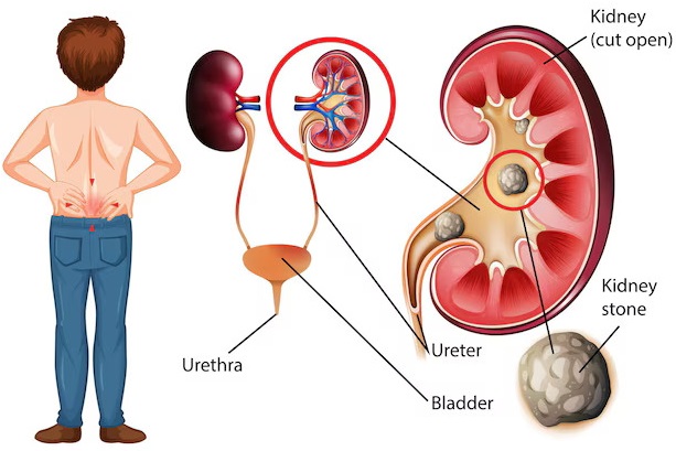
Ureteral stones are mineral deposits that become lodged in the ureter. Most originate inside the kidney and subsequently move into the ureter.
As a result, some stones interrupt normal urine flow. Additionally, obstruction may cause swelling of the kidney, known as hydronephrosis.
However, stone size and location influence the severity of symptoms and the need for treatment.
Common Causes of Ureteral Stones
Several factors can increase the risk of urinary stone formation.
- Insufficient fluid intake
- High urinary concentrations of stone-forming substances
- Family history of kidney stones
- Selected metabolic disorders
- Recurrent urinary tract infections
- Some medications
- Certain urinary tract abnormalities
- Dietary factors in susceptible individuals
Furthermore, recurrent stones may require a metabolic evaluation to identify preventable risk factors.
Symptoms of Ureteral Stones
Symptoms vary according to stone location and the degree of urinary obstruction.
- Severe pain in the side or lower back
- Pain extending toward the abdomen or groin
- Blood in the urine
- Nausea and vomiting
- Painful urination
- Frequent urination
- Urinary urgency
- Reduced urine output in selected cases
- Fever or chills when infection occurs
Important: Fever or chills with suspected urinary obstruction may indicate an infected obstructed kidney. Therefore, patients require urgent medical assessment and often immediate drainage rather than definitive stone surgery.
When Is Ureterolithotomy Recommended?
Doctors may recommend ureterolithotomy when a ureteral stone requires removal and standard endoscopic treatments are unsuitable or unsuccessful.
For example, a large impacted stone with difficult access may require an alternative surgical approach.
However, the surgeon must assess the expected benefits and risks before selecting ureterolithotomy.
Indications for Ureterolithotomy Surgery
- Selected large or impacted ureteral stones
- Failure of appropriate endoscopic stone treatment
- Complex ureteral anatomy that limits endoscopic access
- Selected cases requiring another reconstructive procedure
- Persistent obstruction when alternative treatment is unsuitable
- Selected complex stone cases requiring direct surgical access
Nevertheless, stone size alone does not automatically justify ureterolithotomy. Therefore, urologists compare available treatment options before surgery.
Types of Ureterolithotomy Surgery
Surgeons may use different surgical approaches depending on the clinical situation.
1. Open Ureterolithotomy
Open ureterolithotomy involves an incision that allows the surgeon to access the affected ureter directly.
Although this approach remains useful in selected complex cases, surgeons perform it less frequently than endoscopic stone treatment.
2. Laparoscopic Ureterolithotomy
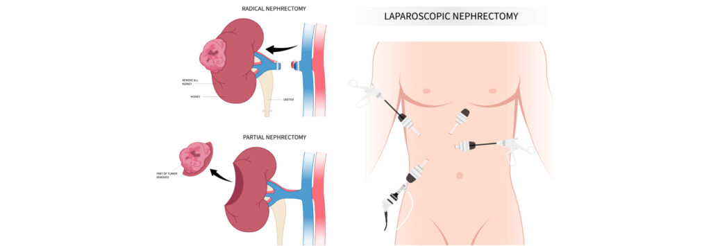
Laparoscopic ureterolithotomy uses small surgical incisions and specialized laparoscopic instruments.
Furthermore, a camera provides visualization of the operative field.
Compared with open surgery, laparoscopy may reduce postoperative discomfort and shorten recovery in appropriately selected patients.
3. Retroperitoneoscopic Ureterolithotomy
Retroperitoneoscopic ureterolithotomy accesses the ureter through the space behind the abdominal lining.
Consequently, the surgeon may reach selected ureteral locations without entering the main peritoneal cavity.
4. Robot-Assisted Ureterolithotomy
Robot-assisted ureterolithotomy uses a robotic surgical platform under the surgeon’s control.
However, availability, cost, and surgeon experience influence its use.
Open vs. Laparoscopic Ureterolithotomy
| Feature | Open Ureterolithotomy | Laparoscopic Ureterolithotomy |
|---|---|---|
| Surgical Access | Open incision | Small laparoscopic incisions |
| Visualization | Direct surgical view | Camera-assisted view |
| Instruments | Open surgical instruments | Laparoscopic instruments |
| Recovery | May require a longer recovery | May allow faster recovery |
| Selection | Selected complex cases | Selected patients suitable for laparoscopy |
Ureterolithotomy vs. Ureteroscopy
Ureteroscopy accesses the ureter through the natural urinary passage. In contrast, ureterolithotomy requires surgical access to the ureter from outside.
Therefore, ureteroscopy generally offers a less invasive option for most ureteral stones.
However, ureterolithotomy may remain appropriate when endoscopic treatment is unsuccessful or unsuitable.
Ureterolithotomy vs. ESWL
Extracorporeal Shock Wave Lithotripsy, or ESWL, uses externally generated shock waves to fragment selected urinary stones.
In contrast, ureterolithotomy removes a stone through a surgical opening in the ureter.
Therefore, doctors evaluate stone characteristics before recommending either treatment.
Ureterolithotomy vs. PCNL
Percutaneous Nephrolithotomy, or PCNL, primarily treats selected kidney stones through a percutaneous access tract.
In contrast, ureterolithotomy directly targets a stone within the ureter.
However, treatment planning may become more complex when patients have stones in multiple locations.
Diagnostic Tests Before Ureterolithotomy
Before surgery, the healthcare team evaluates the stone, urinary anatomy, and overall patient health.
Ultrasound Examination
Ultrasound may identify hydronephrosis and selected urinary stones.
CT Scan
CT imaging helps determine stone size, location, and surrounding anatomy.
Furthermore, imaging supports surgical planning.
Urine Examination and Culture
Urine testing helps identify infection. Therefore, clinicians assess infection status before elective surgery.
Blood Tests
Blood tests may evaluate kidney function, blood counts, and other relevant health factors.
Anesthesia Assessment
The anesthesia team evaluates the patient’s medical condition and plans appropriate monitoring.
How Does Ureterolithotomy Surgery Work?
Ureterolithotomy allows a surgeon to remove a ureteral stone through direct surgical access.
First, the healthcare team completes the necessary assessments and prepares the patient.
Next, the surgeon accesses the affected ureter using the selected surgical approach.
After identifying the stone, the surgeon removes it through an appropriate ureteral opening.
Finally, the surgeon manages the ureteral opening and determines whether temporary urinary drainage is necessary.
However, the exact technique varies according to anatomy, stone characteristics, and operative findings.
General Stages of Ureterolithotomy Surgery
The following overview explains the broad stages of clinical care. It does not provide instructions for performing surgery.
Step 1: Preoperative Evaluation
First, the urologist reviews imaging, laboratory results, medical history, and treatment alternatives.
Step 2: Anesthesia and Preparation
Next, the anesthesia and surgical teams prepare the patient according to established clinical protocols.
Step 3: Surgical Access
The surgeon accesses the ureter using an open, laparoscopic, or other suitable approach.
Step 4: Stone Localization
The surgical team identifies the affected ureteral segment and confirms the stone location.
Step 5: Stone Removal
The surgeon removes the stone using appropriate surgical instruments.
Step 6: Ureteral Management
Next, the surgeon evaluates the ureter and manages the surgical opening.
Step 7: Drainage Assessment
The surgeon determines whether a temporary ureteral stent or other drainage device is necessary.
Step 8: Postoperative Recovery
Finally, the healthcare team monitors recovery, urinary drainage, pain, and potential complications.
Surgical Instruments Used in Ureterolithotomy
Ureterolithotomy requires specialized urological and general surgical instruments. However, the exact equipment list depends on the surgical approach.
- Laparoscope: Provides visualization during laparoscopic surgery.
- Laparoscopic Camera System: Displays the operative field.
- Trocar and Cannula: Support laparoscopic instrument access.
- Laparoscopic Grasping Forceps: Assist with selected tissue-handling tasks.
- Laparoscopic Dissector: Supports controlled tissue dissection.
- Laparoscopic Scissors: Support selected cutting tasks.
- Needle Holder: Supports suturing during surgery.
- Surgical Suction and Irrigation System: Helps maintain operative visibility.
- Stone Retrieval Instrument: Supports removal of the ureteral stone.
- Ureteral Catheter: Supports selected urinary access or drainage tasks.
- DJ Ureteral Stent: May provide temporary internal urinary drainage.
- Surgical Sutures: Support tissue closure.
- Open Surgical Retractors: Support exposure during open surgery.
- Hemostatic Instruments: Support bleeding control.
Furthermore, surgeons select equipment according to the planned procedure and clinical requirements.
Laparoscope for Ureterolithotomy
A laparoscope provides visualization during laparoscopic ureterolithotomy.
Additionally, compatible camera and illumination systems help the surgical team observe the operative field.
Laparoscopic Grasping Forceps
Laparoscopic grasping forceps support selected tissue-handling tasks.
However, jaw design and working length vary between models.
Laparoscopic Dissector
A laparoscopic dissector supports controlled tissue separation during selected procedures.
Furthermore, instrument compatibility depends on the selected surgical system.
Laparoscopic Scissors
Laparoscopic scissors support selected tissue-cutting tasks.
Additionally, different models provide different jaw shapes and shaft dimensions.
Laparoscopic Needle Holder
A laparoscopic needle holder supports suturing during minimally invasive surgery.
Therefore, the surgeon selects a suitable model according to the procedure.
Trocar and Cannula
Trocar and cannula systems support access for compatible laparoscopic instruments.
However, the surgical team must select suitable dimensions and follow the manufacturer’s instructions.
Stone Retrieval Instruments
Stone retrieval instruments support removal of selected urinary calculi.
Furthermore, the surgeon chooses the instrument according to stone characteristics and surgical access.
Ureteral Catheter
A ureteral catheter may support selected urinary access or drainage tasks.
However, catheter selection depends on the patient’s anatomy and operative requirements.
DJ Ureteral Stent
A Double J ureteral stent may provide temporary internal drainage after ureterolithotomy.
However, the surgeon determines whether a stent is necessary.
Additionally, patients must follow the recommended stent removal or exchange schedule.
Surgical Suction and Irrigation System
A surgical suction and irrigation system helps maintain visibility during selected procedures.
Furthermore, the surgical team manages fluid use according to clinical protocols.
Technical Specifications of Ureterolithotomy Instruments
Instrument specifications vary by manufacturer and model. Therefore, customers should request verified datasheets before purchasing.
| Parameter | Specification |
|---|---|
| Procedure Name | Ureterolithotomy Surgery |
| Medical Specialty | Urology |
| Primary Application | Surgical removal of selected ureteral stones |
| Surgical Approach | Open, laparoscopic, retroperitoneoscopic, or robot-assisted |
| Laparoscope Diameter | According to selected model |
| Laparoscope Length | Confirm manufacturer specifications |
| Trocar Size | According to selected instrument system |
| Grasping Forceps | Confirm jaw design and working length |
| Dissector | According to selected model |
| Needle Holder | According to selected model |
| Suction and Irrigation | Compatible surgical system |
| Ureteral Catheter | According to clinical requirements |
| DJ Stent | According to patient requirements |
| Instrument Material | Confirm manufacturer specifications |
| Sterility | According to product labeling |
| Reusable or Single Use | According to manufacturer instructions |
| Reprocessing | According to validated manufacturer instructions |
| Supplier | JDMeditech, subject to product availability |
Benefits of Ureterolithotomy Surgery
Ureterolithotomy may offer important benefits in appropriately selected complex cases.
- Allows direct removal of a selected ureteral stone
- Provides an option after unsuccessful endoscopic treatment
- May help manage selected large impacted stones
- Supports treatment when anatomy limits standard access
- May allow treatment alongside another necessary surgical procedure
- Offers laparoscopic access in suitable patients
However, these potential benefits must be balanced against the greater invasiveness of surgery.
Advantages of Laparoscopic Ureterolithotomy
Laparoscopic ureterolithotomy uses smaller incisions than traditional open surgery.
Consequently, selected patients may experience less postoperative pain and shorter recovery.
However, outcomes depend on the patient’s condition, surgical complexity, and surgeon experience.
Limitations of Ureterolithotomy
Ureterolithotomy is more invasive than ureteroscopy or shock wave treatment.
Additionally, surgery requires anesthesia and carries risks associated with surgical access.
Therefore, urologists generally reserve it for selected cases rather than routine ureteral stone treatment.
Possible Risks and Complications
Ureterolithotomy carries potential surgical risks. Therefore, patients should discuss these risks with their urologist.
- Bleeding
- Urinary tract infection
- Wound infection
- Urine leakage
- Ureteral injury
- Ureteral narrowing or stricture
- Injury to nearby structures
- Postoperative pain
- Residual or recurrent stones
- Need for additional treatment
- Anesthesia-related complications
Furthermore, the complication profile differs between open, laparoscopic, and robot-assisted approaches.
Who Is a Suitable Candidate?
Ureterolithotomy may suit selected patients with complex ureteral stones that require direct surgical removal.
However, the urologist must review imaging, urinary anatomy, infection status, and previous treatment outcomes.
Therefore, suitability requires an individualized clinical assessment.
Recovery After Ureterolithotomy Surgery
Recovery depends on the surgical approach and the patient’s overall condition.
For example, laparoscopic surgery may allow a shorter recovery than open surgery in suitable cases.
However, postoperative monitoring remains important for all patients.
Postoperative Care After Ureterolithotomy
Appropriate postoperative care supports healing and helps identify complications.
- Take prescribed medications as directed.
- Follow wound-care instructions.
- Maintain fluid intake according to medical advice.
- Follow activity restrictions provided by the surgeon.
- Attend scheduled follow-up appointments.
- Follow DJ stent instructions when applicable.
- Report fever or worsening pain.
- Seek urgent care for severe symptoms or difficulty passing urine.
How Long Does Ureterolithotomy Take?
Operating time depends on stone location, anatomy, surgical approach, and procedural complexity.
Furthermore, previous surgery or associated urinary abnormalities may influence the duration.
Therefore, the surgical team provides an individualized estimate.
How Long Does Recovery Take?
Recovery varies between patients. Additionally, open surgery may require a longer recovery than minimally invasive approaches.
Therefore, patients should follow their surgeon’s guidance before returning to strenuous activity.
Is a DJ Stent Required After Ureterolithotomy?
Surgeons may place a temporary DJ stent to support urinary drainage and ureteral healing.
However, the decision depends on operative findings and clinical requirements.
Can Ureteral Stones Return After Surgery?
Yes. Ureterolithotomy removes the existing stone but does not eliminate every cause of urinary stone formation.
Therefore, doctors may recommend stone analysis, metabolic evaluation, and preventive care.
How to Prevent Ureteral Stone Recurrence
Prevention depends on stone composition and individual risk factors.
- Maintain appropriate hydration according to medical advice.
- Follow a balanced diet.
- Limit excessive sodium intake when recommended.
- Follow individualized dietary guidance.
- Complete recommended metabolic testing.
- Take prescribed preventive medications when indicated.
- Attend follow-up appointments.
Furthermore, patients should consult their healthcare team before making major dietary changes.
How to Select Ureterolithotomy Instruments
Hospitals should evaluate several factors before purchasing surgical instruments.
- Planned surgical approach
- Instrument dimensions
- Laparoscope and camera compatibility
- Trocar and cannula compatibility
- Grasping forceps design
- Dissector configuration
- Needle holder compatibility
- Suction and irrigation requirements
- Ureteral catheter specifications
- DJ stent specifications
- Sterility and reuse requirements
- Cleaning and reprocessing instructions
- Technical documentation
Additionally, hospitals should confirm the complete equipment list with the operating surgeon.
Why Choose JDMeditech for Ureterolithotomy Instruments?
JDMeditech supplies urology and surgical instruments for hospitals, healthcare facilities, and medical distributors.
Furthermore, customers can contact JDMeditech to inquire about available ureterolithotomy instruments and accessories.
- Laparoscopic instrument inquiries
- Open surgical instrument inquiries
- Laparoscope inquiries
- Trocar and cannula inquiries
- Grasping forceps inquiries
- Dissector inquiries
- Needle holder inquiries
- Suction and irrigation system inquiries
- Ureteral catheter inquiries
- DJ stent inquiries
- International export inquiries
- Technical specifications upon request
However, product availability and compatibility vary. Therefore, customers should confirm specifications before ordering.
Frequently Asked Questions About Ureterolithotomy
1. What Is Ureterolithotomy Surgery?
Ureterolithotomy is a surgical procedure that removes a stone directly from the ureter.
2. Why Is Ureterolithotomy Performed?
Doctors may perform it when selected ureteral stones require direct surgical removal and less invasive treatments are unsuitable or unsuccessful.
3. Is Ureterolithotomy Major Surgery?
Yes. Ureterolithotomy requires surgical access to the ureter and appropriate anesthesia.
4. What Is Open Ureterolithotomy?
Open ureterolithotomy removes a ureteral stone through a traditional surgical incision.
5. What Is Laparoscopic Ureterolithotomy?
Laparoscopic ureterolithotomy uses small incisions, a camera, and specialized instruments to remove a selected ureteral stone.
6. What Is Retroperitoneoscopic Ureterolithotomy?
It is a minimally invasive approach that accesses the ureter through the retroperitoneal space.
7. What Is Robot-Assisted Ureterolithotomy?
Robot-assisted ureterolithotomy uses a robotic surgical platform under the surgeon’s control.
8. How Long Does Ureterolithotomy Take?
Operating time depends on stone location, anatomy, and surgical complexity.
9. How Long Is the Hospital Stay?
Hospital stay varies according to the surgical approach and postoperative recovery.
10. How Long Does Recovery Take?
Recovery varies. However, laparoscopic approaches may allow faster recovery than open surgery in suitable cases.
11. What Is the Difference Between Ureterolithotomy and Ureteroscopy?
Ureteroscopy uses instruments introduced through the natural urinary passage, while ureterolithotomy requires direct surgical access to the ureter.
12. What Is the Difference Between Ureterolithotomy and ESWL?
ESWL uses externally generated shock waves, while ureterolithotomy removes a stone through a surgical opening in the ureter.
13. What Instruments Are Used in Ureterolithotomy?
Surgeons may use laparoscopes, grasping forceps, dissectors, needle holders, suction systems, and other suitable instruments.
14. Is a DJ Stent Required?
Not always. The surgeon determines whether temporary internal drainage is necessary.
15. Can Ureterolithotomy Treat Large Stones?
Yes. Surgeons may consider it for selected large or impacted ureteral stones when other treatments are unsuitable.
16. Can Ureteral Stones Return?
Yes. Stone recurrence remains possible, so preventive care and follow-up are important.
17. Is Ureterolithotomy Suitable for Children?
Specialists may consider surgery in selected pediatric cases after evaluating less invasive options.
18. Who Performs Ureterolithotomy?
Appropriately trained urologists perform the procedure.
19. Is Ureterolithotomy Better Than Ureteroscopy?
Not generally. Ureteroscopy is less invasive and is preferred for many ureteral stones. However, ureterolithotomy may suit selected complex cases.
20. Where Can I Inquire About Ureterolithotomy Instruments?
Hospitals and medical distributors can contact JDMeditech for information about available urology and laparoscopic instruments.
Conclusion
Ureterolithotomy Surgery remains an important treatment option for selected complex ureteral stones.
Furthermore, open and minimally invasive surgical approaches allow direct stone removal when standard treatments are unsuitable or unsuccessful.
However, successful treatment requires careful patient selection, appropriate surgical equipment, and experienced healthcare professionals.
Therefore, hospitals should verify instrument specifications and compatibility before selecting a ureterolithotomy surgical instrument set.
JDMeditech – One Stop for Hospital Needs
Explore our range of urology instruments, laparoscopic equipment, and related medical devices.
Website:
www.jdmeditech.com
Medical References
- European Association of Urology – Urolithiasis Guidelines
- NIDDK – Kidney Stones
- MedlinePlus – Kidney Stones
Medical Disclaimer
This article provides general educational and product information only. It does not replace professional medical advice, surgical training, or manufacturer instructions for use. Qualified healthcare professionals must select treatment and medical equipment according to individual clinical requirements.