Safe and Effective PTCA Balloon Catheter for Cardiology Experts

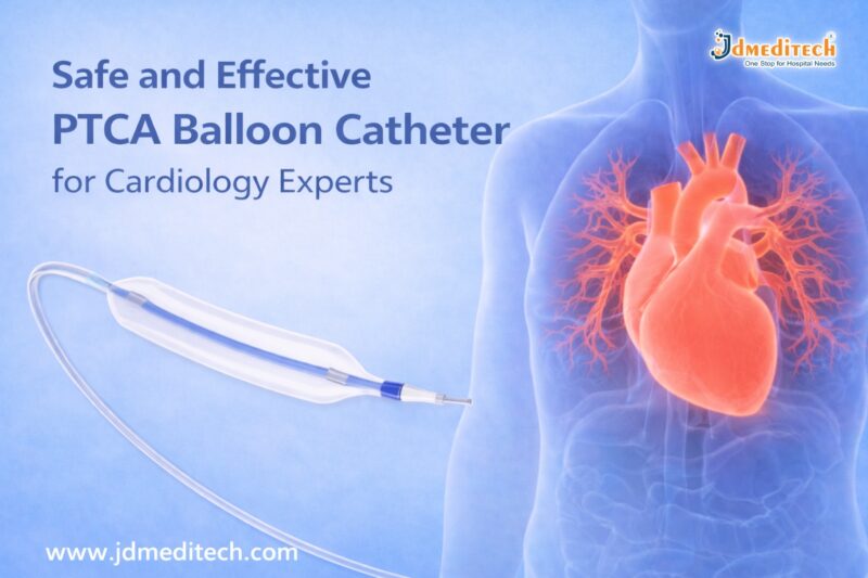
What is a PTCA Balloon Catheter?

A PTCA Balloon Catheter is a minimally invasive medical device used to open narrowed coronary arteries caused by plaque buildup. It consists of a small balloon attached to the tip of a catheter.

During the procedure:

  • The catheter is inserted into a blood vessel
  • Guided to the blocked coronary artery
  • The balloon is inflated to compress plaque
  • Blood flow is restored efficiently

This procedure is commonly known as angioplasty.

Why PTCA Balloon Catheters Are Essential in Cardiology

Cardiologists rely on PTCA balloon catheters because they offer:

  • Minimally invasive treatment
  • Quick restoration of blood flow
  • Reduced need for open-heart surgery
  • Faster patient recovery time

As a result, these catheters have become a cornerstone in treating coronary artery disease (CAD).

Key Features of Safe and Effective PTCA Balloon Catheters
1. High-Pressure Balloon Capability

Modern PTCA balloon catheters can withstand high pressure, allowing precise dilation of hardened plaques without damaging the artery.

2. Smooth Navigation

These catheters are designed for easy navigation through complex vascular pathways, ensuring accurate placement.

3. Excellent Crossability

They can cross tight lesions efficiently, making them ideal for challenging cases.

4. Controlled Balloon Inflation

Precise inflation and deflation provide better control, reducing procedural risks.

5. Biocompatible Materials

Manufactured using advanced materials, they minimize the risk of adverse reactions.

Types of PTCA Balloon Catheters
1. Semi-Compliant Balloons
  • Flexible and adaptable
  • Suitable for general angioplasty procedures
2. Non-Compliant Balloons
  • Maintain shape under high pressure
  • Ideal for precise dilation and post-stent expansion
3. Cutting Balloons
  • Equipped with micro blades
  • Used for resistant or calcified lesions
4. Drug-Coated Balloons
  • Release medication to prevent restenosis
  • Improve long-term outcomes
Benefits of Using PTCA Balloon Catheters

Using a safe and effective PTCA balloon catheter offers multiple benefits:

  • ✔ Minimally invasive procedure
  • ✔ Reduced hospital stay
  • ✔ Lower complication rates
  • ✔ Faster recovery
  • ✔ Improved patient comfort
  • ✔ High procedural success rate
Safety Considerations in PTCA Procedures

While PTCA is generally safe, proper technique and equipment selection are essential.

Important Safety Measures:
  • Use appropriate balloon size
  • Monitor inflation pressure carefully
  • Ensure proper imaging guidance
  • Follow sterilization protocols
  • Choose high-quality, certified devices

By adhering to these practices, cardiology experts can significantly reduce risks.

Applications of PTCA Balloon Catheters

PTCA balloon catheters are widely used in:

  • Coronary artery disease treatment
  • Pre-stent dilation
  • Post-stent expansion
  • Treatment of restenosis
  • Peripheral angioplasty procedures
How to Choose the Right PTCA Balloon Catheter

Selecting the right catheter is crucial for optimal results.

Consider the following factors:
  • Lesion type and severity
  • Vessel size
  • Balloon compliance type
  • Trackability and flexibility
  • Manufacturer quality and certification

Choosing the right device ensures both safety and effectiveness.

Future Innovations in PTCA Balloon Technology

Advancements in medical technology continue to improve PTCA outcomes.

Emerging trends include:

  • Drug-eluting balloon technologies
  • Enhanced catheter flexibility
  • Improved imaging compatibility
  • AI-assisted procedural planning

These innovations are shaping the future of interventional cardiology.

Conclusion

The PTCA Balloon Catheter remains an indispensable tool for cardiology experts. Its ability to safely and effectively restore blood flow makes it a preferred choice in modern cardiac care.

With continuous advancements in design and technology, PTCA balloon catheters are becoming even more efficient, ensuring better patient outcomes and procedural success.


Explore More Product >>

Get Connected:

+91 79909 93062 | +91 63513 72032 | exports@jdmeditech.com


Top-Quality Dialysis Chairs – Comfort & Support for Every Dialysis Session

Introduction

Dialysis is a life-saving procedure for patients with kidney failure. However, long treatment sessions can be physically and mentally exhausting. This is where top-quality dialysis chairs play a crucial role. These specialized chairs are designed to provide maximum comfort, safety, and support throughout every dialysis session.

In this blog, we will explore the importance, features, benefits, and selection criteria for dialysis chairs, helping healthcare providers make informed decisions.

What Are Top-Quality Dialysis Chairs?

Top-quality dialysis chairs are ergonomically designed medical chairs used during dialysis treatments. Patients often remain seated for several hours, so these chairs are built to ensure comfort, proper posture, and easy accessibility for healthcare professionals.

They are commonly used in:

  • Hospitals
  • Dialysis centers
  • Specialty clinics
Why Comfort Matters in Dialysis Sessions

Dialysis sessions typically last 3–5 hours. During this time, patients must remain still. Poor seating can lead to:

  • Back pain and discomfort
  • Poor blood circulation
  • Increased anxiety or restlessness

By using top-quality dialysis chairs, healthcare facilities can significantly enhance patient comfort and overall treatment experience.

Key Features of Top-Quality Dialysis Chairs
1. Ergonomic Design for Patient Comfort

Modern dialysis chairs are designed to support the natural body posture. Adjustable backrests, armrests, and footrests ensure optimal comfort.

2. Electric Adjustment Mechanism

High-end chairs come with motorized controls that allow smooth adjustments. This helps both patients and caregivers.

3. Durable and Hygienic Materials

These chairs are made with easy-to-clean, antimicrobial upholstery, ensuring hygiene and infection control.

4. Safety Features

Top-quality dialysis chairs include:

  • Lockable wheels
  • Stable base structure
  • Emergency positioning
5. Accessibility for Medical Procedures

The chair design allows easy access for healthcare professionals during dialysis procedures.

Benefits of Using Top-Quality Dialysis Chairs
Enhanced Patient Comfort

Patients experience less fatigue during long sessions, improving their overall well-being.

Improved Clinical Efficiency

Adjustable features help medical staff perform procedures more efficiently.

Better Patient Compliance

Comfortable patients are more likely to complete their sessions without interruption.

Increased Safety

Advanced safety mechanisms reduce the risk of accidents.

How to Choose the Best Dialysis Chair

Selecting the right dialysis chair is essential for both patient satisfaction and clinical performance.

Consider Patient Comfort

Look for chairs with cushioning, adjustability, and ergonomic support.

Evaluate Build Quality

Choose durable materials that withstand frequent use.

Check for Advanced Features

Electric controls, memory positions, and safety locks are important.

Ensure Easy Maintenance

Hygienic surfaces and easy cleaning are critical in healthcare settings.

Applications of Dialysis Chairs in Healthcare

Top-quality dialysis chairs are widely used in:

  • Hemodialysis units
  • Oncology treatment centers
  • Blood transfusion units
  • Daycare surgical centers

Their versatility makes them a valuable asset in modern healthcare facilities.

Maintenance Tips for Long-Term Use

To ensure longevity and performance:

  • Clean the chair regularly with approved disinfectants
  • Inspect mechanical and electrical components
  • Perform routine servicing
  • Train staff on proper usage
Why Choose JDMeditech Dialysis Chairs?

At JDMeditech, we provide top-quality dialysis chairs designed to meet international healthcare standards. Our chairs offer:

  • Superior comfort and ergonomic design
  • Advanced safety features
  • Durable and hygienic materials
  • Reliable performance for long-term use
Conclusion

Investing in top-quality dialysis chairs is essential for improving patient comfort, safety, and treatment efficiency. With the right chair, healthcare providers can deliver better care while enhancing operational performance.

If you are looking for reliable and high-performance dialysis chairs, JDMeditech offers solutions tailored to your needs.


Explore More Product >>

Get Connected:

+91 79909 93062 | +91 63513 72032 | exports@jdmeditech.com


Precision Cutting with Bugbee Electrodes – Ultimate Electrosurgical Tool

Precision Cutting with Bugbee Electrodes – The Ultimate Electrosurgical Tool

Introduction

Precision cutting with Bugbee electrodes has transformed modern electrosurgical procedures. Surgeons rely on this advanced tool for accurate tissue cutting and effective coagulation, especially in urology and endoscopy.

Bugbee electrodes provide excellent control, minimal tissue damage, and improved surgical outcomes. As a result, they have become an essential instrument in operating rooms worldwide.

What Are Bugbee Electrodes?

Bugbee electrodes are flexible, monopolar electrosurgical instruments designed for precise cutting and coagulation of soft tissues. They are commonly used in minimally invasive procedures, particularly in urology.

These electrodes consist of a thin wire tip that delivers controlled electrical energy. This energy allows surgeons to cut tissue while simultaneously controlling bleeding.

Why Precision Cutting with Bugbee Electrodes Matters

Precision cutting with Bugbee electrodes ensures clean incisions and reduces unnecessary tissue damage. This improves patient recovery and minimizes complications.

Key Benefits:
  • High precision during surgery
  • Minimal thermal spread
  • Reduced bleeding
  • Enhanced visibility in the surgical field
  • Faster healing time

Because of these advantages, surgeons prefer Bugbee electrodes for delicate procedures.

Applications of Bugbee Electrodes in Medical Procedures

Bugbee electrodes are widely used across various medical specialties. However, they are most popular in urology and endoscopic surgeries.

1. Urological Procedures

Surgeons use Bugbee electrodes during cystoscopy to remove small tumors, coagulate bleeding areas, and treat bladder conditions.

2. Endoscopic Surgeries

These electrodes allow precise cutting in minimally invasive procedures, reducing trauma to surrounding tissues.

3. Tumor Resection

Bugbee electrodes help in removing small growths with accuracy while maintaining hemostasis.

4. Coagulation of Bleeding Vessels

They effectively control bleeding by coagulating blood vessels during surgery.

How Bugbee Electrodes Work

Bugbee electrodes operate using monopolar electrosurgical energy. The electrical current passes through the electrode tip to the targeted tissue.

This process creates heat, which enables:

  • Cutting of tissue
  • Coagulation of blood vessels
  • Controlled surgical precision

The surgeon can adjust the energy level depending on the procedure, ensuring safe and effective outcomes.

Advantages of Using Bugbee Electrodes

Precision cutting with Bugbee electrodes offers several advantages over traditional surgical tools.

Improved Surgical Accuracy

The fine tip allows surgeons to perform delicate procedures with maximum control.

Reduced Tissue Damage

Minimal thermal spread protects surrounding tissues.

Enhanced Efficiency

Surgeries become quicker due to simultaneous cutting and coagulation.

Cost-Effective Solution

Reusable options make Bugbee electrodes a budget-friendly choice for hospitals.

Safety Considerations

Although Bugbee electrodes are highly effective, proper usage is essential for safety.

  • Always use appropriate power settings
  • Ensure proper grounding during monopolar procedures
  • Regularly inspect the electrode for damage
  • Follow sterilization protocols

By following these guidelines, healthcare professionals can ensure safe and successful procedures.

Choosing the Right Bugbee Electrode

Selecting the correct Bugbee electrode depends on the surgical requirement.

Factors to Consider:
  • Electrode length and flexibility
  • Tip size and shape
  • Compatibility with electrosurgical units
  • Reusable or disposable type

Choosing the right instrument enhances both precision and safety.

Future of Electrosurgical Tools

The demand for precision cutting with Bugbee electrodes continues to grow. Innovations in electrosurgical technology are improving efficiency, safety, and ease of use.

Modern designs now focus on better insulation, enhanced flexibility, and improved energy control. These advancements will further improve surgical outcomes in the future.

Conclusion

Precision cutting with Bugbee electrodes has become a cornerstone of modern electrosurgery. Their ability to deliver accurate cuts with minimal tissue damage makes them the ultimate electrosurgical tool.

From urology to endoscopic procedures, Bugbee electrodes provide unmatched performance and reliability. As technology advances, their role in surgery will only continue to expand.


Explore More Product >>

Get Connected:

+91 79909 93062 | +91 63513 72032 | exports@jdmeditech.com


ERCP Pancreatic Baskets: Uses, Types, Procedure & Benefits

What Are ERCP Pancreatic Baskets?

ERCP pancreatic baskets are small wire devices. Doctors use them to catch and remove stones from the pancreatic duct.

They pass through an endoscope during the ERCP procedure. These baskets are strong, flexible, and easy to control.

Key Features:
  • Flexible design for narrow ducts
  • Strong wires for firm grip
  • Smooth opening and closing system
  • Available in different sizes
Why Are ERCP Pancreatic Baskets Used?

Pancreatic stones can block the duct. Because of this, patients may feel severe pain. In some cases, it can lead to pancreatitis.

ERCP pancreatic baskets help to:
  • Remove stones safely
  • Improve enzyme flow
  • Reduce pain quickly
  • Prevent further complications
Types of ERCP Pancreatic Baskets

Different baskets are used for different cases. Therefore, doctors choose them based on the situation.

1. Four-Wire Basket

This is the most common type. It works well for medium-sized stones.

2. Eight-Wire Basket

It offers a stronger grip. So, it is useful for multiple or uneven stones.

3. Helical Basket

This basket moves easily in curved ducts. Hence, it is ideal for complex cases.

4. Nitinol (Memory) Basket

This basket keeps its shape. As a result, it works well even after repeated use.

ERCP Procedure Using Pancreatic Baskets

The procedure is simple and minimally invasive.

Steps:
  1. The doctor inserts an endoscope through the mouth
  2. The pancreatic duct is located
  3. A dye is used to view the duct clearly
  4. The basket is passed into the duct
  5. The stone is captured
  6. The basket is pulled out with the stone

This process is quick and effective.

Benefits of ERCP Pancreatic Baskets

These devices offer many benefits.

1. Minimally Invasive

No major surgery is needed. Therefore, recovery is faster.

2. High Success Rate

Most stones can be removed easily.

3. Better Control

Doctors can handle stones with precision.

4. Fewer Complications

There is less risk compared to open surgery.

5. Cost-Effective

Short hospital stays reduce overall cost.

Applications in Medical Practice

Doctors use ERCP pancreatic baskets in many situations.

  • Pancreatic stone removal
  • Chronic pancreatitis treatment
  • ERCP procedures
  • Endoscopic stone extraction
Risks and Considerations

The procedure is safe. However, some risks may occur.

Possible Risks:
  • Pancreatitis
  • Infection
  • Minor duct injury
  • Rare basket trapping

Even so, trained doctors can manage these risks well.

How to Choose the Right Basket

Choosing the right basket is important.

Doctors consider:

  • Stone size
  • Stone shape
  • Duct structure
  • Material type

Therefore, high-quality baskets give better results.

Future Developments

Medical tools are improving every year. As a result, ERCP baskets are becoming more advanced.

New Improvements:
  • Better flexibility
  • Stronger materials
  • Single-use options
  • Easy handling design

These changes help improve safety and performance.

Conclusion

ERCP Pancreatic Baskets are key tools for removing pancreatic stones. They are safe, effective, and easy to use.

Because they are minimally invasive, patients recover faster. In addition, new designs continue to improve results. Therefore, these baskets remain a top choice in modern ERCP procedures.


Explore More Product >>

Get Connected:

+91 79909 93062 | +91 63513 72032 | exports@jdmeditech.com


Embolectomy Catheter – Uses, Procedure & Benefits Explained

An embolectomy catheter is a medical device used to remove blood clots from blood vessels. These clots can block blood flow and cause serious health problems. Therefore, doctors must remove them quickly.

In many cases, this device helps restore circulation within minutes. As a result, it reduces the risk of stroke, tissue damage, and organ failure.

Today, the embolectomy catheter is widely used in cardiovascular procedures. It offers a fast, safe, and minimally invasive solution.

What is an Embolectomy Catheter?

An embolectomy catheter is a thin and flexible tube. Doctors insert it into a blocked artery or vein.

At the tip, there is a small balloon. First, the catheter reaches the clot. Then, the balloon inflates. After that, the doctor pulls the catheter back. As a result, the clot is removed from the vessel.

Because of this simple method, blood flow returns quickly.

Why is an Embolectomy Catheter Used?

Blood clots can be dangerous. In some cases, they can be life-threatening. Therefore, quick treatment is important.

Doctors use an embolectomy catheter in the following conditions:

  • Acute arterial blockage
  • Deep vein thrombosis (DVT)
  • Pulmonary embolism
  • Stroke-related clots
  • Peripheral artery disease

In addition, this device is useful in emergency situations. It helps save time and improves survival.

Types of Embolectomy Catheters

There are different types of embolectomy catheter devices. Each type serves a specific purpose.

Balloon Embolectomy Catheter

This is the most common type. It uses a balloon to capture and remove the clot.

Fogarty Catheter

Doctors often use this catheter in vascular surgery. It is reliable and easy to use.

Aspiration Catheter

This type removes clots using suction instead of a balloon.

Mechanical Devices

These devices break the clot into small pieces. Then, they remove it from the vessel.

Embolectomy Procedure

The procedure is simple and quick. Moreover, it is usually safe.

Step 1: Preparation

First, the patient receives anesthesia.

Step 2: Insertion

Next, the doctor inserts the embolectomy catheter into the blood vessel.

Step 3: Positioning

Then, imaging helps guide the catheter to the clot.

Step 4: Removal

After that, the balloon inflates and captures the clot. The doctor removes it carefully.

Step 5: Completion

Finally, blood flow returns to normal.

Benefits of Embolectomy Catheter

The embolectomy catheter offers many benefits.

  • Fast clot removal
  • Minimally invasive procedure
  • Quick recovery time
  • Lower risk of complications
  • High success rate

Most importantly, it helps prevent serious damage to tissues and organs.

Risks and Complications

Like any procedure, there are some risks. However, they are usually rare.

  • Bleeding
  • Infection
  • Vessel injury
  • Partial clot removal

Even so, experienced doctors manage these risks effectively.

Role in Cardiovascular Procedures

The embolectomy catheter plays a key role in modern medicine.

Doctors use it in:

  • Emergency vascular treatment
  • Stroke management
  • Cardiac procedures
  • Peripheral artery care

Therefore, it is an important tool in saving lives.

How to Choose the Right Embolectomy Catheter

Choosing the correct device is important. Doctors consider several factors.

  • Size of the vessel
  • Location of the clot
  • Type of blockage
  • Patient condition

As a result, the right choice improves treatment success.

Future of Embolectomy Catheters

Medical technology is improving every year. Therefore, newer devices are more advanced.

For example:

  • Better flexibility
  • Improved precision
  • Smart balloon systems
  • Combined clot removal techniques

In the future, these devices will become even safer and more effective.

Conclusion

The embolectomy catheter is a powerful tool for clot removal. It helps restore blood flow quickly and safely.

Because it is minimally invasive, patients recover faster. In addition, it reduces the risk of serious complications.

Overall, this device plays a vital role in cardiovascular procedures.


Explore More Product >>

Get Connected:

+91 79909 93062 | +91 63513 72032 | exports@jdmeditech.com

Functional Endoscopic Sinus Surgery (FESS)

Functional Endoscopic Sinus Surgery (FESS) is a minimally invasive surgical procedure used to treat chronic sinus problems. It helps restore normal drainage of the sinuses and improves breathing.

Today, FESS is one of the most advanced and commonly performed procedures for patients suffering from chronic sinusitis, nasal blockages, and sinus infections.

In this blog, we will explain everything about Functional Endoscopic Sinus Surgery, including its procedure, benefits, risks, and recovery.

What is Functional Endoscopic Sinus Surgery (FESS)?

Functional Endoscopic Sinus Surgery (FESS) is a modern surgical technique that uses a thin, flexible endoscope inserted through the nose to treat sinus issues.

Unlike traditional surgery, FESS:

  • Does not require external cuts
  • Is less painful
  • Offers faster recovery

This procedure focuses on restoring the natural function of the sinuses rather than removing large amounts of tissue.

Why is FESS Needed?

Doctors recommend Functional Endoscopic Sinus Surgery when medications fail to treat sinus conditions.

Common Conditions Treated:
  • Chronic sinusitis
  • Nasal polyps
  • Deviated nasal septum (in some cases)
  • Sinus infections
  • Blocked sinus drainage pathways
  • Fungal sinusitis

If you experience persistent nasal congestion, facial pain, or breathing difficulty, FESS may be the right solution.

Functional Endoscopic Sinus Surgery Procedure

The FESS procedure is safe and usually performed under general anesthesia.

Step-by-Step Procedure:
1. Preparation

The patient is given anesthesia to ensure comfort.

2. Endoscope Insertion

A small camera (endoscope) is inserted through the nostrils.

3. Identifying Blockages

The surgeon carefully examines sinus passages.

4. Removing Obstructions

Blocked tissues, polyps, or bone are removed to open sinus pathways.

5. Restoring Drainage

Normal airflow and sinus drainage are restored.

The entire procedure typically takes 1 to 3 hours, depending on the condition.

Benefits of Functional Endoscopic Sinus Surgery

FESS offers several advantages compared to traditional sinus surgery.

Key Benefits:
  • Minimally invasive procedure
  • No external scars
  • Faster recovery time
  • Reduced pain and bleeding
  • Improved breathing
  • Long-term relief from sinus symptoms

As a result, patients often experience a significant improvement in quality of life.

Risks and Complications of FESS

Although Functional Endoscopic Sinus Surgery is generally safe, some risks may occur.

Possible Risks:
  • Bleeding
  • Infection
  • Recurrence of sinus problems
  • Damage to surrounding structures (rare)
  • Vision-related complications (very rare)

However, choosing an experienced surgeon greatly reduces these risks.

Recovery After Functional Endoscopic Sinus Surgery

Recovery from FESS is usually quick and smooth.

What to Expect:
  • Mild discomfort for a few days
  • Nasal congestion or slight bleeding
  • Return to normal activities within 1–2 weeks
Post-Surgery Care Tips:
  • Avoid blowing your nose forcefully
  • Use prescribed nasal sprays
  • Attend follow-up appointments
  • Keep your nasal passages clean

Following proper care ensures faster healing and better results.

Who is a Good Candidate for FESS?

You may be a suitable candidate for Functional Endoscopic Sinus Surgery if:

  • You have chronic sinusitis lasting more than 12 weeks
  • Medications are not effective
  • You have nasal polyps or structural blockage
  • You frequently experience sinus infections

Consult an ENT specialist for proper evaluation.

Conclusion

Functional Endoscopic Sinus Surgery (FESS) is a safe, effective, and minimally invasive solution for chronic sinus problems. It helps restore normal sinus function, improves breathing, and enhances overall quality of life.

If you are struggling with long-term sinus issues, consult your doctor to see if FESS is the right option for you.

Explore More Product >>


Get Connected:

+91 79909 93062 | +91 63513 72032 | exports@jdmeditech.com

Pathology: Types, Importance & Role in Disease Diagnosis

Pathology is a vital branch of medical science that focuses on the study of diseases. It helps doctors understand the causes, development, and effects of various illnesses. Without pathology, accurate diagnosis and effective treatment would not be possible.

In modern healthcare, pathology plays a crucial role in detecting diseases early. As a result, it improves patient outcomes and supports life-saving treatments.

What is Pathology?

Pathology is the scientific study of disease through the examination of organs, tissues, blood, and bodily fluids. Pathologists analyze samples to identify abnormalities and determine the exact nature of a disease.

In simple terms, pathology answers key questions like:

  • What disease is present?
  • How severe is it?
  • What is causing it?

Therefore, it acts as the foundation of medical diagnosis.

Types of Pathology

Pathology is divided into several specialized branches. Each type focuses on a different aspect of disease analysis.

1. Clinical Pathology

Clinical pathology involves the analysis of blood, urine, and other body fluids. It includes:

  • Hematology (study of blood)
  • Microbiology (study of infections)
  • Biochemistry (study of chemical processes in the body)
2. Anatomical Pathology

Anatomical pathology focuses on examining tissues and organs. It includes:

  • Histopathology (study of tissues under a microscope)
  • Cytopathology (study of individual cells)
3. Molecular Pathology

Molecular pathology studies diseases at a genetic and molecular level. It is widely used in cancer diagnosis and personalized medicine.

4. Forensic Pathology

Forensic pathology determines the cause of death, especially in cases of accidents or criminal investigations.

Importance of Pathology in Healthcare

Pathology is essential in every stage of healthcare. It not only helps in diagnosis but also guides treatment decisions.

1. Accurate Disease Diagnosis

Pathology provides precise information about diseases. As a result, doctors can make correct diagnoses and avoid unnecessary treatments.

2. Early Detection of Diseases

Many diseases, including cancer, can be detected early through pathology tests. Early detection significantly improves survival rates.

3. Treatment Planning

Pathology reports help doctors choose the best treatment options. For example, cancer treatments often depend on biopsy results.

4. Monitoring Disease Progress

Pathology tests are used to track how a disease is progressing or responding to treatment.

Common Pathology Tests

Pathology laboratories perform a wide range of tests. Some of the most common include:

  • Blood tests (CBC, glucose levels)
  • Urine analysis
  • Biopsy tests
  • Pap smear tests
  • Culture tests for infections

These tests provide essential insights into a patient’s health.

Role of Pathologists

Pathologists are highly trained medical professionals who specialize in diagnosing diseases. Although they usually work behind the scenes, their role is critical.

They:

  • Analyze lab results
  • Interpret biopsy samples
  • Provide diagnostic reports
  • Support clinicians in treatment decisions

Therefore, pathologists are key contributors to patient care.

How Pathology Supports Modern Medicine

Pathology is the backbone of modern medicine. It supports advanced medical practices such as:

  • Cancer diagnosis and staging
  • Genetic testing and personalized medicine
  • Infectious disease detection
  • Organ transplant compatibility testing

As technology advances, pathology continues to evolve with digital pathology and AI-based diagnostics.

Future of Pathology

The future of pathology is rapidly advancing. New technologies are improving accuracy and efficiency.

Some key trends include:

  • Digital pathology systems
  • Artificial intelligence in diagnostics
  • Automated laboratory testing
  • Precision medicine

These innovations will make diagnosis faster and more reliable.

Conclusion

Pathology is a fundamental part of healthcare that ensures accurate diagnosis and effective treatment. From routine blood tests to advanced molecular analysis, pathology provides critical insights into diseases.

As healthcare continues to evolve, pathology will remain at the center of medical innovation and patient care.

Explore More Product >>


Get Connected:

+91 79909 93062 | +91 63513 72032 | exports@jdmeditech.com


Anesthesia: Types, Procedure, Risks, Benefits & Recovery Guide

Anaesthesia is a medical treatment that prevents pain during surgery or medical procedures. It works by blocking nerve signals in the body or inducing a temporary loss of sensation or consciousness.

A trained specialist called an anaesthesiologist administers anaesthesia and monitors the patient throughout the procedure.

Types of Anaesthesia

Different types of anaesthesia are used depending on the procedure, patient condition, and duration of surgery.

1. General Anaesthesia

General anaesthesia makes the patient completely unconscious.

Used for:

  • Major surgeries (heart surgery, brain surgery)
  • Long-duration procedures

Effects:

  • No awareness
  • No pain sensation
  • Temporary loss of reflexes
2. Regional Anaesthesia

Regional anaesthesia blocks pain in a specific part of the body.

Examples:

  • Spinal anaesthesia
  • Epidural anaesthesia

Used for:

  • Childbirth (C-section)
  • Orthopedic surgeries
3. Local Anaesthesia

Local anaesthesia numbs a small area of the body.

Used for:

  • Dental procedures
  • Minor surgeries

Benefits:

  • Patient remains awake
  • Quick recovery
4. Sedation (Conscious Sedation)

Sedation helps patients relax while remaining partially awake.

Used for:

  • Endoscopy
  • Minor procedures
How Does Anaesthesia Work?

Anaesthesia works by interrupting nerve signals in the brain and body. As a result, the patient does not feel pain during the procedure.

There are three main mechanisms:

  • Blocking pain signals
  • Inducing unconsciousness
  • Relaxing muscles
Anaesthesia Procedure: Step-by-Step
1. Pre-Anaesthesia Evaluation

Before surgery, the anaesthesiologist:

  • Reviews medical history
  • Checks allergies
  • Evaluates vital signs
2. Administration of Anaesthesia

Anaesthesia is given through:

  • Injection
  • Inhalation (gas)
  • Intravenous (IV)
3. Monitoring During Surgery

Doctors continuously monitor:

  • Heart rate
  • Blood pressure
  • Oxygen levels
4. Recovery Phase

After surgery, the patient is moved to a recovery room where medical staff monitor vital signs until the effects wear off.

Benefits of Anaesthesia

Anaesthesia offers several advantages:

  • Pain-free procedures
  • Improved surgical precision
  • Reduced anxiety
  • Controlled muscle relaxation
  • Safer complex surgeries
Risks and Side Effects of Anaesthesia

Although anaesthesia is generally safe, some risks may occur.

Common Side Effects
  • Nausea and vomiting
  • Drowsiness
  • Sore throat
  • Dizziness
Rare Risks
  • Allergic reactions
  • Breathing problems
  • Nerve damage (very rare)

However, modern techniques and monitoring systems significantly reduce these risks.

Recovery After Anaesthesia

Recovery depends on the type of anaesthesia used.

General Recovery Tips:
  • Follow doctor’s instructions
  • Avoid driving for 24 hours
  • Stay hydrated
  • Rest properly

Most patients recover quickly, especially after local or regional anesthesia.

Who Administers Anaesthesia?

Anaesthesia is administered by trained professionals such as:

  • Anaesthesiologists
  • Nurse anaesthetists
  • Anaesthesia assistants

They ensure patient safety before, during, and after the procedure.

When is Anaesthesia Required?

Anaesthesia is used in various medical procedures, including:

  • Surgical operations
  • Dental treatments
  • Diagnostic procedures
  • Cosmetic surgeries
Conclusion

Anaesthesia is a cornerstone of modern healthcare, making surgeries safe, painless, and effective. With advancements in medical technology, anaesthesia has become more precise and safer than ever before.

If you are scheduled for a procedure, understanding anaesthesia can help reduce anxiety and prepare you for a smooth experience.

Explore More Product >>


Get Connected:

+91 79909 93062 | +91 63513 72032 | exports@jdmeditech.com


Maintaining Your Surgical Instruments

Surgical instruments are the backbone of any healthcare facility. Therefore, maintaining your surgical instruments is essential for ensuring patient safety, improving surgical outcomes, and extending the lifespan of expensive tools.

Proper care not only reduces the risk of infection but also prevents instrument damage. In this guide, you will learn the best practices, step-by-step cleaning methods, and expert tips to maintain surgical instruments effectively.

Why Maintaining Your Surgical Instruments is Important

First and foremost, well-maintained instruments ensure precision during surgery. On the other hand, poorly maintained tools can lead to complications.

Key Benefits:
  • Enhances patient safety
  • Prevents surgical site infections (SSIs)
  • Increases instrument lifespan
  • Reduces replacement costs
  • Ensures compliance with healthcare standards
Types of Surgical Instruments That Require Maintenance

Different instruments need different care methods. Therefore, understanding categories is crucial.

1. Cutting Instruments
  • Scalpels
  • Scissors
  • Bone cutters
2. Grasping Instruments
  • Forceps
  • Clamps
3. Retracting Instruments
  • Surgical retractors
4. Specialized Instruments
  • Endoscopic tools
  • Microsurgical instruments
Step-by-Step Guide to Maintaining Surgical Instruments
1. Immediate Cleaning After Use

After surgery, instruments should be cleaned immediately. This step prevents blood and tissue from drying.

Tips:

  • Use lukewarm water
  • Avoid saline (it causes corrosion)
  • Wipe visible debris
2. Proper Cleaning Techniques

Cleaning is the most critical step. Therefore, it must be done carefully.

Manual Cleaning
  • Use a soft brush
  • Clean joints and hinges thoroughly
  • Use neutral pH detergents
Ultrasonic Cleaning
  • Removes microscopic debris
  • Ideal for complex instruments
  • Saves time and effort
3. Rinsing and Drying

After cleaning, rinse instruments with distilled water. Then, dry them completely.

Why this matters:
  • Prevents rust formation
  • Avoids water stains
4. Inspection and Function Testing

Next, inspect each instrument carefully.

Check for:
  • Cracks or corrosion
  • Loose joints
  • Sharpness of cutting tools

Replace or repair damaged instruments immediately.

5. Lubrication

Lubrication ensures smooth movement.

Best practices:
  • Use water-based lubricants
  • Apply to hinges and joints
  • Avoid oil-based products
6. Sterilization

Sterilization is essential to eliminate microorganisms.

Common Methods:
  • Autoclaving (steam sterilization)
  • Ethylene oxide (ETO)
  • Plasma sterilization

Always follow manufacturer guidelines for best results.

7. Proper Storage

Finally, store instruments correctly.

Storage tips:
  • Use dry, clean environments
  • Avoid overcrowding
  • Use protective trays
Common Mistakes to Avoid

Even small errors can damage instruments. Therefore, avoid these common mistakes:

  • Delaying cleaning after surgery
  • Using harsh chemicals
  • Skipping inspection
  • Improper sterilization techniques
  • Storing instruments while wet
Best Practices for Long-Term Maintenance

To maintain surgical instruments effectively, consistency is key.

Follow These Practices:
  • Train staff regularly
  • Use instrument tracking systems
  • Follow manufacturer instructions
  • Schedule routine maintenance checks
How Often Should You Maintain Surgical Instruments?

Maintenance should be a continuous process.

  • After every use: Cleaning and sterilization
  • Daily: Inspection and drying
  • Weekly: Deep cleaning and lubrication
  • Monthly: Detailed inspection and servicing
Latest Innovations in Instrument Maintenance

Modern technology has improved maintenance processes significantly.

New Trends:
  • Automated washer-disinfectors
  • Smart tracking systems
  • Anti-corrosion coatings
  • AI-based instrument monitoring

These innovations help reduce human error and improve efficiency.

Conclusion

Maintaining your surgical instruments is not just a routine task—it is a critical responsibility. Proper cleaning, sterilization, and storage ensure safety, efficiency, and cost savings.

By following the steps and best practices in this guide, healthcare facilities can significantly improve surgical outcomes while extending the life of their instruments.

Explore More Product >>


Get Connected:

+91 79909 93062 | +91 63513 72032 | exports@jdmeditech.com

Laryngoscopy: Procedure, Types, Uses, Risks & Recovery

Laryngoscopy is a medical procedure used to examine the larynx (voice box) and surrounding structures. Doctors commonly use it to diagnose throat problems, voice disorders, and breathing issues.

In addition, this procedure helps detect conditions such as infections, tumors, and vocal cord abnormalities. Because of its importance, laryngoscopy plays a key role in ENT (Ear, Nose, and Throat) care.

What is Laryngoscopy?

Laryngoscopy is a diagnostic and sometimes therapeutic procedure. It allows doctors to view the throat using a special instrument called a laryngoscope.

This procedure can be done in different ways. Therefore, the method used depends on the patient’s condition and the purpose of the examination.

Types of Laryngoscopy

There are three main types of laryngoscopy. Each type serves a specific purpose.

1. Indirect Laryngoscopy

This is the simplest method. Doctors use a small mirror or fiber-optic scope to examine the throat.

  • Quick and non-invasive
  • Performed in a clinic
  • No anaesthesia required
2. Direct Laryngoscopy

In this method, a rigid or flexible scope is inserted into the throat.

  • Provides a clear and detailed view
  • Often done under anesthesia
  • Used for diagnosis and minor procedures
3. Fiberoptic (Flexible) Laryngoscopy

This is the most commonly used modern technique.

  • Uses a thin, flexible tube with a camera
  • Inserted through the nose
  • Comfortable and widely used
Why is Laryngoscopy Done?

Doctors recommend laryngoscopy for several reasons. It helps diagnose and treat many conditions.

Common Uses Include:
  • Persistent sore throat
  • Hoarseness or voice changes
  • Difficulty swallowing (dysphagia)
  • Chronic cough
  • Breathing problems
  • Suspected throat cancer
  • Removal of foreign objects

Moreover, it helps evaluate vocal cord movement and detect abnormalities early.

Laryngoscopy Procedure

The procedure varies depending on the type. However, most steps remain similar.

Before the Procedure
  • The doctor reviews your medical history
  • You may be asked to avoid eating for a few hours
  • A local anaesthetic spray may be used
During the Procedure
  • A laryngoscope is gently inserted
  • The doctor examines the throat and vocal cords
  • The process usually takes 5 to 30 minutes
After the Procedure
  • Mild throat discomfort may occur
  • Avoid eating until numbness wears off
  • Most patients resume normal activities quickly
Benefits of Laryngoscopy

Laryngoscopy offers several advantages. As a result, it is widely used in medical practice.

  • Quick and effective diagnosis
  • Minimally invasive procedure
  • Helps detect serious conditions early
  • Can assist in minor treatments
Risks and Complications

.

Possible Risks:
  • Mild throat irritation
  • Gagging sensation
  • Bleeding (rare)
  • Infection (very rare)
  • Reaction to anaesthesia

However, serious complications are uncommon when performed by experienced professionals.

Recovery After Laryngoscopy

Recovery is usually quick and simple.

  • Most patients recover within a few hours
  • Drink warm fluids to soothe the throat
  • Avoid smoking or irritants
  • Follow doctor’s advice for medications

If symptoms such as severe pain or bleeding occur, seek medical attention immediately.

When to See a Doctor

You should consult a doctor if you experience:

  • Persistent hoarseness
  • Difficulty breathing
  • Pain while swallowing
  • Unexplained throat symptoms

Early diagnosis improves treatment outcomes significantly.

Conclusion

Laryngoscopy is an essential procedure for diagnosing and treating throat-related conditions. It is safe, effective, and widely used in modern healthcare.

By understanding the procedure, its types, and benefits, patients can feel more confident and prepared. If you have ongoing throat symptoms, consult an ENT specialist for proper evaluation.

Explore More Product >>


+91 79909 93062 | +91 63513 72032 | exports@jdmeditech.com

Importance of CSSD in Hospitals | Sterilization & Infection Control Guide

The importance of CSSD in hospitals cannot be overstated. CSSD, or Central Sterile Supply Department, plays a critical role in maintaining hygiene, preventing infections, and ensuring safe surgical procedures.

Every hospital relies on sterile instruments. Without proper sterilization, patients face serious risks such as hospital-acquired infections (HAIs). Therefore, CSSD acts as the backbone of infection control and patient safety.

In this blog, we will explore why CSSD is essential, how it functions, and its impact on healthcare quality.

What is CSSD in Hospitals?

CSSD stands for Central Sterile Supply Department. It is a specialized unit responsible for:

  • Cleaning medical instruments
  • Disinfecting equipment
  • Sterilizing surgical tools
  • Storing sterile supplies
  • Distributing sterile items across departments

This department ensures that all reusable medical devices are safe for patient use.

Importance of CSSD in Hospitals
1. Ensures Patient Safety

The primary importance of CSSD in hospitals is patient safety. Proper sterilization removes harmful microorganisms, reducing the risk of infections during medical procedures.

As a result, patients receive safe and reliable treatment.

2. Prevents Hospital-Acquired Infections (HAIs)

Hospital-acquired infections can be life-threatening. However, CSSD helps prevent these infections by:

  • Using advanced sterilization techniques
  • Maintaining strict hygiene protocols
  • Monitoring sterilization processes

Therefore, CSSD significantly reduces infection rates.

3. Maintains Quality Healthcare Standards

Hospitals must follow strict healthcare standards. CSSD ensures compliance with:

  • Infection control guidelines
  • Sterilization protocols
  • Regulatory standards

This improves overall healthcare quality and hospital reputation.

4. Supports Surgical Efficiency

Surgical procedures require sterile instruments at all times. CSSD ensures:

  • Timely availability of instruments
  • Proper packaging and labeling
  • Organized workflow

Because of this, surgeries can proceed without delays.

5. Extends Equipment Lifespan

Proper cleaning and sterilization help maintain medical instruments. CSSD:

  • Prevents damage caused by improper handling
  • Uses appropriate cleaning techniques
  • Ensures correct storage

As a result, hospitals save costs on equipment replacement.

6. Reduces Cross-Contamination

Cross-contamination can occur if instruments are not properly sterilized. CSSD minimizes this risk by:

  • Separating clean and contaminated areas
  • Following strict workflows
  • Using standardized procedures

Thus, it protects both patients and healthcare workers.

Key Functions of CSSD
Cleaning and Decontamination

All used instruments are thoroughly cleaned to remove biological material.

Disinfection

Chemical or thermal methods are used to reduce microorganisms.

Sterilization

High-pressure steam (autoclaving) or other methods ensure complete sterilization.

Storage

Sterile items are stored in controlled environments to maintain sterility.

Distribution

Sterile instruments are supplied to various hospital departments.

CSSD Workflow in Hospitals

The CSSD workflow follows a structured process:

  1. Collection of used instruments
  2. Cleaning and decontamination
  3. Inspection and packaging
  4. Sterilization
  5. Storage
  6. Distribution

This systematic approach ensures efficiency and safety.

Modern Technologies Used in CSSD

Modern CSSD units use advanced technologies such as:

  • Autoclaves
  • Ethylene Oxide (ETO) sterilizers
  • Plasma sterilization
  • Ultrasonic cleaners
  • Washer-disinfectors

These technologies improve accuracy and effectiveness.

Challenges Faced by CSSD

Despite its importance, CSSD faces several challenges:

  • High workload and time pressure
  • Need for skilled staff
  • Strict compliance requirements
  • Equipment maintenance

However, proper management can overcome these challenges.

Best Practices for Effective CSSD Management

To maximize the importance of CSSD in hospitals, follow these best practices:

  • Train staff regularly
  • Maintain strict hygiene protocols
  • Use advanced sterilization equipment
  • Monitor quality through audits
  • Ensure proper documentation

These steps enhance efficiency and safety.

Conclusion

The importance of CSSD in hospitals lies in its ability to ensure patient safety, prevent infections, and maintain high healthcare standards. It is a vital department that supports every medical procedure.

Without CSSD, hospitals cannot deliver safe and effective treatment. Therefore, investing in a well-equipped and properly managed CSSD is essential for any healthcare facility.


Explore More Product >>


Get Connected:

+91 79909 93062 | +91 63513 72032 | exports@jdmeditech.com

Artificial Intelligence in Healthcare: Benefits, Uses & Future Trends

Artificial Intelligence (AI) in healthcare is changing the way medical professionals diagnose, treat, and manage diseases. Today, hospitals and clinics use AI-powered tools to improve patient care and reduce errors. As a result, healthcare systems are becoming faster, smarter, and more efficient.

Moreover, AI helps doctors make better decisions by analyzing large amounts of medical data in seconds. Because of this, patients receive accurate diagnoses and personalized treatments. In this blog, you will learn how AI works in healthcare, its benefits, real-world applications, and future trends.

What is Artificial Intelligence in Healthcare?

Artificial Intelligence in healthcare refers to the use of advanced algorithms and machine learning to perform tasks that usually require human intelligence. For example, AI can analyze medical images, predict diseases, and assist in surgeries.

In addition, AI systems learn from past data and continuously improve their performance. Therefore, they become more accurate over time. This makes AI a powerful tool in modern medicine.

Benefits of AI in Healthcare

AI offers several advantages that improve both patient care and hospital efficiency.

1. Improved Diagnosis Accuracy

AI can analyze medical images such as X-rays, MRIs, and CT scans quickly. As a result, doctors can detect diseases like cancer at an early stage.

2. Faster Treatment Decisions

AI processes large datasets within seconds. Therefore, doctors can make quick and informed decisions, especially in emergencies.

3. Personalized Treatment Plans

AI helps create customized treatment plans based on patient history. In addition, it considers genetic information and lifestyle factors.

4. Reduced Human Errors

Medical errors can be dangerous. However, AI reduces these risks by providing accurate data analysis and recommendations.

5. Cost Efficiency

AI reduces unnecessary tests and hospital stays. Consequently, it lowers overall healthcare costs.

Applications of AI in Healthcare

AI is widely used across different areas of healthcare. Let’s explore some key applications.

1. Medical Imaging

AI tools analyze medical images with high precision. For instance, they detect tumors, fractures, and internal bleeding quickly.

2. Virtual Health Assistants

AI-powered chatbots and virtual assistants provide basic medical advice. Moreover, they help patients schedule appointments and manage medications.

3. Drug Discovery

Developing new drugs takes years. However, AI speeds up this process by analyzing chemical data and predicting drug effectiveness.

4. Robotic Surgery

AI-assisted robots help surgeons perform complex procedures. As a result, surgeries become more precise and less invasive.

5. Predictive Analytics

AI predicts disease outbreaks and patient risks. Therefore, healthcare providers can take preventive measures early.

Challenges of AI in Healthcare

Although AI offers many benefits, it also comes with challenges.

1. Data Privacy Concerns

Healthcare data is sensitive. Therefore, protecting patient information is crucial.

2. High Implementation Costs

AI technology requires significant investment. As a result, smaller hospitals may face difficulties adopting it.

3. Lack of Skilled Professionals

AI systems need trained experts to operate and maintain them. However, there is a shortage of such professionals.

4. Ethical Issues

AI decisions must be transparent. Otherwise, it may lead to trust issues among patients.

Future of AI in Healthcare

The future of AI in healthcare looks promising. In the coming years, AI will become more advanced and accessible.

For example, AI may help in early disease prediction even before symptoms appear. In addition, wearable devices will continuously monitor health and alert doctors in real time.

Furthermore, AI will improve telemedicine services, making healthcare accessible to remote areas. As technology evolves, AI will play a key role in transforming global healthcare systems.

Conclusion

Artificial Intelligence in healthcare is revolutionizing the medical industry. It improves diagnosis, enhances treatment, and reduces costs. Moreover, it supports doctors in delivering better patient care.

However, challenges such as data privacy and high costs must be addressed. Despite these issues, AI continues to grow rapidly. Therefore, adopting AI in healthcare is essential for a smarter and healthier future.


Explore More Product >>


Get Connected:

+91 79909 93062 | +91 63513 72032 | exports@jdmeditech.com


Intensive Care Unit (ICU): Meaning, Equipment, Treatment & Care Guide

The Intensive Care Unit (ICU) plays a vital role in modern healthcare. It provides advanced medical care to patients with life-threatening conditions. In most hospitals, the ICU is equipped with specialized technology and trained staff to monitor and treat critically ill patients.

Moreover, ICU care ensures constant supervision and quick medical response. Because of this, patients with severe injuries or illnesses receive the best possible support. In this blog, you will learn about ICU meaning, types, equipment, procedures, and patient care.

What is an Intensive Care Unit (ICU)?

An Intensive Care Unit (ICU) is a specialized hospital department designed to treat patients with serious and life-threatening conditions. Doctors and nurses monitor patients 24/7 using advanced machines.

In addition, ICU staff includes intensivists, critical care nurses, and respiratory therapists. Therefore, patients receive continuous and expert medical attention.

Types of ICU

Different hospitals provide various types of ICUs based on patient needs.

1. Medical ICU (MICU)

This ICU treats patients with severe medical conditions such as infections, organ failure, and respiratory diseases.

2. Surgical ICU (SICU)

Doctors use this ICU for patients recovering from major surgeries. As a result, they receive close monitoring after operations.

3. Cardiac ICU (CCU)

This unit focuses on heart-related conditions such as heart attacks and cardiac arrest.

4. Neonatal ICU (NICU)

NICU provides care for premature or critically ill newborn babies.

5. Pediatric ICU (PICU)

This ICU treats infants and children with serious health conditions.

Common Equipment Used in ICU

ICUs use advanced equipment to monitor and support patients.

1. Ventilators

Ventilators help patients breathe when they cannot do so on their own.

2. Cardiac Monitors

These machines track heart rate, blood pressure, and oxygen levels continuously.

3. Infusion Pumps

Doctors use these devices to deliver medications and fluids accurately.

4. Defibrillators

They help restore normal heart rhythm during emergencies.

5. Dialysis Machines

These machines support kidney function in patients with kidney failure.

Conditions Treated in ICU

Patients are admitted to the ICU when they require critical care. Some common conditions include:

  • Severe infections (sepsis)
  • Respiratory failure
  • Heart attack or cardiac arrest
  • Stroke
  • Major trauma or accidents
  • Post-surgical complications

Therefore, ICU care becomes essential for survival in such situations.

ICU Procedures and Treatment

ICU treatment involves continuous monitoring and advanced medical procedures.

1. Mechanical Ventilation

Doctors use ventilators to support breathing in critically ill patients.

2. Continuous Monitoring

Healthcare staff track vital signs such as heart rate, oxygen level, and blood pressure.

3. Medication Management

Patients receive critical medications through IV lines.

4. Life Support Systems

These systems support organs such as lungs, heart, and kidneys.

ICU Patient Care and Safety

Proper care in the ICU is essential for recovery.

  • Nurses provide round-the-clock care
  • Strict hygiene prevents infections
  • Doctors adjust treatment based on patient condition
  • Family visits are controlled for safety

In addition, emotional support plays a key role in patient recovery.

Challenges in ICU

Although ICUs provide life-saving care, they also face challenges.

1. High Cost of Treatment

ICU care can be expensive due to advanced equipment and specialized staff.

2. Risk of Infections

Patients may develop hospital-acquired infections.

3. Emotional Stress

Both patients and families experience stress during ICU stays.

Future of ICU Care

The future of ICU care looks advanced and technology-driven. AI and smart monitoring systems are improving patient outcomes.

For example, remote ICU monitoring allows doctors to track patients from different locations. In addition, robotic assistance and data analytics will enhance treatment accuracy.

Conclusion

The Intensive Care Unit (ICU) is an essential part of modern healthcare. It provides life-saving treatment and constant monitoring for critically ill patients.

Although ICU care can be costly and complex, it significantly improves survival rates. Therefore, understanding ICU functions helps patients and families make informed decisions during emergencies.

Explore More Product >>

Get Connected:

+91 79909 93062 | +91 63513 72032 | exports@jdmeditech.com


Hemodialysis

Hemodialysis is a life-saving treatment for people with severe kidney failure. When kidneys stop working properly, harmful waste and excess fluid build up in the body. Therefore, hemodialysis helps remove these toxins and keeps the body balanced.

Today, millions of patients rely on hemodialysis treatment to improve their quality of life. In this guide, you will learn how it works, who needs it, and what to expect during the procedure.

What is Hemodialysis?

Hemodialysis is a medical procedure that filters blood using a special machine. In other words, it acts as an artificial kidney.

During the process:

  • Blood is taken from the body
  • It passes through a dialyzer (artificial kidney)
  • Waste and extra fluids are removed
  • Clean blood returns to the body

As a result, the body maintains proper chemical balance.

Why is Hemodialysis Needed?

Doctors recommend hemodialysis treatment when kidneys lose about 85–90% of their function.

Common Causes Include:
  • Chronic Kidney Disease (CKD)
  • Diabetes
  • High blood pressure
  • Glomerulonephritis
  • Polycystic kidney disease

If untreated, kidney failure can become life-threatening. Therefore, timely dialysis is essential.

Types of Hemodialysis Access

Before starting treatment, doctors create a vascular access point. This allows easy blood flow during dialysis.

1. Arteriovenous (AV) Fistula
  • Best and most preferred option
  • Created by connecting an artery to a vein
  • Long-lasting and low risk of infection
2. AV Graft
  • Uses a synthetic tube
  • Suitable if veins are small
3. Central Venous Catheter
  • Temporary access
  • Used in emergency situations
Hemodialysis Procedure Step-by-Step

Understanding the procedure helps reduce anxiety. Here is how it works:

  1. First, medical staff connect you to the dialysis machine
  2. Blood flows through tubes into the dialyzer
  3. The dialyzer removes toxins and excess fluid
  4. Clean blood returns to your body
  5. The session usually lasts 3–5 hours

Typically, patients need dialysis 3 times per week.

Benefits of Hemodialysis

Hemodialysis offers several important benefits. For example:

  • Removes waste and toxins effectively
  • Balances electrolytes in the body
  • Controls blood pressure
  • Reduces swelling caused by fluid buildup
  • Improves overall quality of life

As a result, patients can live longer and healthier lives.

Risks and Side Effects

Although hemodialysis is safe, some side effects may occur.

Common Risks:
  • Low blood pressure
  • Muscle cramps
  • Fatigue
  • Itching
  • Sleep problems
Serious Complications (Rare):
  • Infection
  • Blood clots
  • Heart problems

However, proper care and regular monitoring can reduce these risks significantly.

Diet and Lifestyle During Hemodialysis

Diet plays a key role in treatment success. Therefore, patients must follow strict guidelines.

Recommended Diet Tips:
  • Limit salt intake
  • Control fluid consumption
  • Reduce potassium-rich foods (like bananas)
  • Eat high-quality protein
  • Avoid processed foods

Additionally, light exercise and regular check-ups improve outcomes.

Recovery and Daily Life

Many patients continue normal activities during treatment. However, some adjustments are necessary.

Helpful Tips:
  • Stick to your dialysis schedule
  • Take prescribed medications regularly
  • Monitor weight and fluid intake
  • Maintain hygiene around access site

Over time, patients adapt well to the routine.


Hemodialysis vs Peritoneal Dialysis

Both treatments help kidney failure patients, but they differ in approach.

Feature Hemodialysis Peritoneal Dialysis
Location Hospital/Center Home
Frequency 3 times/week Daily
Equipment Machine required No large machine
Flexibility Less More

Therefore, doctors choose the best option based on patient condition.

When to See a Doctor?

Seek medical help immediately if you notice:

  • Swelling in legs or face
  • Shortness of breath
  • Reduced urine output
  • Persistent fatigue

Early diagnosis can prevent complications.

Conclusion

Hemodialysis is a critical treatment for patients with kidney failure. Although it requires regular sessions, it significantly improves survival and quality of life.

By following medical advice, maintaining a proper diet, and attending scheduled sessions, patients can manage their condition effectively. Therefore, awareness and early treatment are key to better outcomes.


Explore More Product >>


Get Connected:

+91 79909 93062 | +91 63513 72032 | exports@jdmeditech.com

Diagnostic Radiology: Types, Procedures, Benefits & Imaging Tests

Diagnostic radiology plays a vital role in modern healthcare. It helps doctors detect, diagnose, and monitor various medical conditions using advanced imaging techniques. Because early diagnosis improves treatment outcomes, radiology has become essential in medical practice.

In this blog, you will learn what diagnostic radiology is, its types, procedures, benefits, and why it is important.

What is Diagnostic Radiology?

Diagnostic radiology is a medical specialty that uses imaging techniques to examine the inside of the body. These images help doctors identify diseases, injuries, and abnormalities without surgery.

For example, doctors use X-rays to detect fractures, while MRI scans help identify brain or spinal cord issues. As a result, radiology provides quick and accurate diagnoses.

Types of Diagnostic Radiology

There are several types of diagnostic radiology. Each method serves a different purpose.

1. X-rays

X-rays are the most common imaging test. They are mainly used to detect:

  • Bone fractures
  • Lung infections
  • Dental issues

Because X-rays are fast and cost-effective, they are widely used.

2. Computed Tomography (CT Scan)

CT scans provide detailed cross-sectional images of the body. Doctors use them to diagnose:

  • Internal injuries
  • Tumors
  • Infections

Moreover, CT scans offer more detail than standard X-rays.

3. Magnetic Resonance Imaging (MRI)

MRI uses magnetic fields and radio waves instead of radiation. It is ideal for:

  • Brain disorders
  • Joint injuries
  • Soft tissue evaluation

Therefore, MRI is safer for repeated imaging.

4. Ultrasound

Ultrasound uses sound waves to create images. It is commonly used for:

  • Pregnancy monitoring
  • Abdominal conditions
  • Heart evaluation

Additionally, it is painless and radiation-free.

5. Mammography

Mammography is a specialized X-ray used to detect breast cancer early. Early detection significantly improves survival rates.

Common Diagnostic Radiology Procedures

Radiology procedures are usually simple and non-invasive. However, some may require preparation.

Before the Procedure
  • You may need to fast for a few hours
  • Remove metal objects
  • Inform your doctor about allergies or pregnancy
During the Procedure
  • You must remain still for clear images
  • The process is usually painless
  • Some tests may use contrast dye
After the Procedure
  • Most patients can resume normal activities
  • Drink water to flush contrast dye
  • Your doctor will review the results
Benefits of Diagnostic Radiology

Diagnostic radiology offers many advantages in healthcare.

1. Early Detection of Diseases

It helps identify diseases at an early stage. Therefore, treatment becomes more effective.

2. Non-Invasive Diagnosis

Most imaging tests do not require surgery. As a result, patients experience less discomfort.

3. Accurate Results

Radiology provides precise images. This improves diagnosis and treatment planning.

4. Faster Treatment Decisions

Doctors can quickly analyze results and begin treatment immediately.

5. Monitoring of Treatment

Radiology helps track the progress of ongoing treatments.

Risks and Safety Measures

Although diagnostic radiology is generally safe, some risks exist.

  • Exposure to radiation in X-rays and CT scans
  • Allergic reactions to contrast dye (rare)

However, doctors always minimize risks by using the lowest effective radiation dose. In addition, protective measures ensure patient safety.

Why Diagnostic Radiology is Important

Diagnostic radiology is essential because it improves healthcare outcomes. It allows doctors to:

  • Detect diseases early
  • Plan effective treatments
  • Avoid unnecessary surgeries

Furthermore, it enhances patient care by providing accurate and timely information.

Conclusion

Diagnostic radiology is a cornerstone of modern medicine. It uses advanced imaging technologies to diagnose diseases quickly and accurately. Because it is non-invasive and efficient, it improves patient outcomes significantly.

If your doctor recommends a radiology test, there is no need to worry. These procedures are safe, effective, and essential for proper diagnosis.

Explore More Product >>

Get Connected:

+91 79909 93062 | +91 63513 72032 | exports@jdmeditech.com


Tuberculosis

Tuberculosis is caused by a bacterium called Mycobacterium tuberculosis. It spreads through the air when an infected person coughs or sneezes.

Types of Tuberculosis
  • Active TB – Symptoms are present and contagious
  • Latent TB – No symptoms and not contagious
  • Extrapulmonary TB – Affects organs other than lungs
Symptoms of Tuberculosis

Common symptoms include:

  • Persistent cough for more than 2 weeks
  • Chest pain
  • Coughing up blood
  • Fever and chills
  • Night sweats
  • Weight loss and fatigue
Causes and Risk Factors

Tuberculosis spreads through airborne droplets. Risk factors include:

  • Weak immune system
  • Close contact with TB patients
  • Poor nutrition
  • Smoking and alcohol use
  • Living in crowded conditions
Diagnosis of Tuberculosis

Doctors use several tests to diagnose TB:

  • Chest X-ray
  • Sputum test
  • Tuberculin skin test (Mantoux test)
  • Blood tests
Tuberculosis Treatment

Tuberculosis treatment involves a long course of antibiotics, usually lasting 6 to 9 months.

Common Medications
  • Isoniazid
  • Rifampicin
  • Pyrazinamide
  • Ethambutol
Important Tips
  • Complete the full course of tuberculosis treatment
  • Do not skip doses
  • Regular follow-ups are necessary
Prevention of Tuberculosis
  • Get BCG vaccination
  • Maintain good hygiene
  • Avoid close contact with infected individuals
  • Ensure proper ventilation
  • Wear masks in high-risk areas
Complications of Untreated TB
  • Severe lung damage
  • Spread to other organs
  • Respiratory failure
  • Death in severe cases
Who is at Higher Risk?
  • People with HIV/AIDS
  • Diabetic patients
  • Elderly individuals
  • Healthcare workers
Conclusion

Tuberculosis is a preventable and curable disease with timely diagnosis and proper tuberculosis treatment. Awareness and early medical intervention can significantly reduce complications and transmission.


Explore More Product >>


Get Connected:

+91 79909 93062 | +91 63513 72032 | exports@jdmeditech.com

Rhinoplasty (Nose Surgery): Procedure, Benefits & Recovery

Rhinoplasty is a surgical procedure designed to change the shape, size, or structure of the nose. It can be performed for cosmetic reasons or to correct medical problems such as a deviated septum.

Types of Rhinoplasty
  • Cosmetic Rhinoplasty – Improves the appearance of the nose
  • Functional Rhinoplasty – Corrects breathing problems
  • Revision Rhinoplasty – Fixes previous nose surgery issues
  • Non-Surgical Rhinoplasty – Uses fillers for temporary changes
Rhinoplasty Procedure

Rhinoplasty surgery is typically performed under general or local anesthesia and takes about 1 to 3 hours.

Step-by-Step Procedure
  1. Administration of anesthesia
  2. Incision (open or closed technique)
  3. Reshaping the nasal bones and cartilage
  4. Correction of septum if required
  5. Closing the incision and applying a splint
Benefits of Rhinoplasty Surgery
  • Improves facial balance and symmetry
  • Enhances self-confidence
  • Corrects breathing issues
  • Repairs nasal injuries
  • Provides permanent results
Risks and Complications

Like any surgical procedure, rhinoplasty carries some risks:

  • Infection
  • Bleeding
  • Swelling and bruising
  • Breathing difficulties
  • Unsatisfactory results (rare)
Rhinoplasty Recovery Time

Recovery varies from person to person but generally follows this timeline:

  • 1 Week – Splint removal
  • 2 Weeks – Bruising reduces
  • 1 Month – Visible improvement
  • 3–6 Months – Healing progresses
  • 1 Year – Final results
Recovery Tips
  • Avoid strenuous activities
  • Keep head elevated while sleeping
  • Follow surgeon’s instructions
  • Avoid wearing glasses initially


Who is a Good Candidate?

You may be a good candidate for rhinoplasty if:

  • You are physically healthy
  • You have realistic expectations
  • Your facial growth is complete
  • You want to improve nose appearance or function
Conclusion

Rhinoplasty surgery is a safe and effective procedure for enhancing both the appearance and function of the nose. With proper consultation and care, patients can achieve natural-looking and satisfying results. Always consult a qualified plastic surgeon to determine the best approach for your needs.


Explore More Product >>


Get Connected:

+91 79909 93062 | +91 63513 72032 | exports@jdmeditech.com


Back Surgery Guide: Benefits, Risks & Recovery Explained


Back surgery is a medical procedure used to treat severe spinal problems that do not improve with non-surgical treatments. It can help relieve chronic pain, restore mobility, and improve quality of life. This guide explains everything you need to know about back surgery, including types, benefits, risks, and recovery.

What is Back Surgery?

Back surgery refers to various surgical procedures performed on the spine to correct structural issues, relieve pressure on nerves, or stabilize the spine. Doctors usually recommend back surgery only after conservative treatments like medication, physiotherapy, or injections fail.

Common Types of Back Surgery
1. Discectomy

A discectomy involves removing part or all of a damaged disc that is pressing on a nerve. It is commonly used to treat herniated discs.

2. Laminectomy

In this procedure, a portion of the bone (lamina) is removed to relieve pressure on the spinal cord or nerves.

3. Spinal Fusion

Spinal fusion joins two or more vertebrae together to stabilize the spine and reduce pain caused by movement.

4. Artificial Disc Replacement

This surgery replaces a damaged disc with an artificial one, helping maintain natural movement.

5. Minimally Invasive Spine Surgery

This modern technique uses smaller incisions, resulting in less pain, quicker recovery, and reduced complications.

When is Back Surgery Needed?

Back surgery may be recommended if you experience:

  • Chronic back pain lasting more than 6 months
  • Nerve compression causing numbness or weakness
  • Herniated or slipped discs
  • Spinal stenosis
  • Severe spinal injuries or fractures
  • Loss of bladder or bowel control (medical emergency)
Benefits of Back Surgery
  • Relief from chronic pain
  • Improved mobility and flexibility
  • Better quality of life
  • Reduced nerve compression symptoms
  • Long-term spinal stability
Risks of Back Surgery

Like any surgical procedure, back surgery carries some risks, including:

  • Infection
  • Bleeding
  • Nerve damage
  • Blood clots
  • Failure to relieve symptoms
  • Need for additional surgery
Back Surgery Procedure: What to Expect
Before Surgery
  • Medical evaluation and imaging tests
  • Discussion of risks and benefits
  • Fasting before surgery
During Surgery
  • Performed under general anesthesia
  • Duration varies depending on the procedure
After Surgery
  • Monitoring in recovery room
  • Pain management and wound care
  • Gradual movement and physiotherapy
Recovery After Back Surgery

Recovery time depends on the type of surgery and patient health. General guidelines include:

  • Hospital stay: 1–5 days
  • Return to normal activities: 4–12 weeks
  • Full recovery: 3–6 months
Tips for Faster Recovery
  • Follow your doctor’s instructions
  • Attend physiotherapy sessions
  • Avoid heavy lifting
  • Maintain a healthy diet
  • Stay active with light exercises
Conclusion

Back surgery can be a life-changing solution for individuals suffering from severe spinal conditions. With proper diagnosis, expert care, and post-surgery rehabilitation, most patients experience significant pain relief and improved mobility. Always consult a qualified spine specialist to determine the best treatment for your condition.


Explore More Product >>


Get Connected:

+91 79909 93062 | +91 63513 72032 | exports@jdmeditech.com


Diabetes Treatment: Causes, Symptoms, Types & Prevention Guide

Diabetes is a chronic medical condition that affects how your body uses blood sugar (glucose). Glucose is essential for energy, but too much sugar in the blood can lead to serious health problems. With the rising number of diabetes cases worldwide, understanding its causes, symptoms, and diabetes treatment options is more important than ever.

What is Diabetes?

Diabetes occurs when your body either does not produce enough insulin or cannot use insulin properly. Insulin is a hormone that helps glucose enter your cells for energy. When this process fails, blood sugar levels rise.

Types of Diabetes
1. Type 1 Diabetes

Type 1 diabetes is an autoimmune condition where the body attacks insulin-producing cells. It usually develops in children or young adults.

2. Type 2 Diabetes

Type 2 diabetes is the most common form. It occurs when the body becomes resistant to insulin or does not produce enough.

3. Gestational Diabetes

This type develops during pregnancy and usually disappears after childbirth, but it increases the risk of Type 2 diabetes later.

Causes of Diabetes

Several factors contribute to diabetes, including:

  • Genetic factors
  • Obesity
  • Poor diet
  • Lack of physical activity
  • Hormonal imbalances
  • Pancreatic disorders

Understanding these causes helps in early diagnosis and effective diabetes treatment.

Symptoms of Diabetes

Recognizing early symptoms is crucial for timely diabetes treatment:

  • Frequent urination
  • Excessive thirst
  • Unexplained weight loss
  • Fatigue
  • Blurred vision
  • Slow healing wounds

If you experience these symptoms, consult a healthcare professional immediately.

Diagnosis of Diabetes

Doctors use several tests to diagnose diabetes:

  • Fasting Blood Sugar Test
  • HbA1c Test
  • Oral Glucose Tolerance Test
  • Random Blood Sugar Test

Early diagnosis plays a key role in successful diabetes treatment and management.

Diabetes Treatment Options

Effective diabetes treatment focuses on controlling blood sugar levels and preventing complications.

1. Medications

Doctors may prescribe insulin or oral medications depending on the type of diabetes.

2. Healthy Diet

A balanced diet rich in fiber, vegetables, and whole grains helps control blood sugar.

3. Regular Exercise

Physical activity improves insulin sensitivity and supports diabetes treatment.

4. Blood Sugar Monitoring

Regular monitoring helps manage glucose levels effectively.

5. Lifestyle Changes

Maintaining a healthy weight and avoiding smoking are essential parts of diabetes treatment.

Complications of Diabetes

If left untreated, diabetes can lead to serious complications:

  • Heart disease
  • Kidney damage
  • Nerve damage
  • Eye problems
  • Foot complications

Proper diabetes treatment reduces the risk of these complications.

Prevention of Diabetes

While Type 1 diabetes cannot be prevented, Type 2 diabetes can often be delayed or prevented:

  • Maintain a healthy weight
  • Eat a balanced diet
  • Exercise regularly
  • Avoid sugary foods and drinks
  • Get regular health checkups
When to See a Doctor

Seek medical advice if you notice symptoms of diabetes or have a family history of the condition. Early diagnosis ensures better diabetes treatment outcomes.

Conclusion

Diabetes is a manageable condition if detected early and treated properly. With the right diabetes treatment plan, healthy lifestyle choices, and regular monitoring, individuals can live a normal and healthy life. Awareness and prevention play a key role in controlling this growing health concern.

Explore More Product >>

Get Connected:

+91 79909 93062 | +91 63513 72032 | exports@jdmeditech.com


Internet of Things in Healthcare: Benefits, Uses & Future Trends

The Internet of Things in Healthcare is transforming the way medical services are delivered. By connecting devices, systems, and people, IoT enables real-time monitoring, improved patient outcomes, and efficient healthcare management. Today, hospitals and clinics are increasingly adopting IoT technologies to provide smarter and faster care.

What is Internet of Things (IoT) in Healthcare?

The Internet of Things in Healthcare refers to a network of connected medical devices and applications that collect, analyze, and transmit health data. These devices communicate with healthcare providers through the internet, allowing continuous monitoring and timely intervention.

Common IoT devices include:

  • Wearable fitness trackers
  • Smart glucose monitors
  • Connected heart rate monitors
  • Remote patient monitoring systems
Key Benefits of IoT in Healthcare
1. Real-Time Patient Monitoring

IoT devices allow doctors to monitor patients in real time. This helps in early detection of health issues and reduces emergency situations.

2. Improved Patient Outcomes

With continuous data tracking, healthcare providers can make better decisions. This leads to improved treatment and faster recovery.

3. Cost Reduction

The Internet of Things in Healthcare reduces hospital visits and readmissions. Remote monitoring helps save both time and money.

4. Enhanced Patient Engagement

Patients can track their own health using IoT devices. This encourages them to take an active role in their treatment.

5. Efficient Hospital Management

IoT helps hospitals manage equipment, staff, and patients efficiently. Smart systems reduce delays and improve workflow.

Applications of Internet of Things in Healthcare
Remote Patient Monitoring

Remote monitoring systems allow doctors to track patients from home. This is especially useful for chronic diseases like diabetes and heart conditions.

Smart Wearable Devices

Wearable devices such as smartwatches and fitness bands collect health data like heart rate, steps, and sleep patterns.

Connected Medical Equipment

Hospitals use IoT-enabled devices such as smart beds and infusion pumps to improve patient care and safety.

Medication Management

IoT helps remind patients to take medicines on time. Smart pill dispensers reduce medication errors.

Emergency Response Systems

IoT devices can send alerts during emergencies. This ensures quick medical assistance.

Challenges of IoT in Healthcare
Data Security and Privacy

Handling sensitive patient data requires strong security measures. Cybersecurity is a major concern in IoT healthcare systems.

High Implementation Cost

Setting up IoT infrastructure can be expensive for small healthcare providers.

Device Integration Issues

Different devices and systems may not always work together smoothly.

Regulatory Compliance

Healthcare organizations must follow strict regulations when using IoT technologies.

Future of Internet of Things in Healthcare

The future of the Internet of Things in Healthcare is very promising. With advancements in AI and machine learning, IoT systems will become more intelligent and predictive. Smart hospitals, personalized treatment plans, and automated healthcare services will soon become more common.

Conclusion

The Internet of Things in Healthcare is revolutionizing the medical industry. From real-time monitoring to improved patient care, IoT offers numerous benefits. Despite some challenges, its future is bright and full of innovation. Healthcare providers who adopt IoT technologies will be better equipped to deliver high-quality care.


Explore More Product >>


Get Connected:

+91 79909 93062 | +91 63513 72032 | exports@jdmeditech.com

Blood Cancer: Symptoms, Causes, Types, Treatment & Prevention Guide

Blood cancer is a serious medical condition that affects the production and function of blood cells. Blood cancer treatment plays a crucial role in managing the disease and improving survival rates. Early detection and advanced therapies have significantly improved outcomes for patients with blood cancer.

What is Blood Cancer?

Blood cancer is a type of cancer that begins in the bone marrow or lymphatic system. It affects the normal production of blood cells and interferes with their ability to fight infections.

Types of Blood Cancer
1. Leukemia

Leukemia affects the blood and bone marrow and leads to the production of abnormal white blood cells.

2. Lymphoma

Lymphoma affects the lymphatic system, which is part of the immune system.

3. Multiple Myeloma

Multiple myeloma affects plasma cells and weakens the immune system.

Symptoms of Blood Cancer

Common symptoms include:

  • Fatigue and weakness
  • Frequent infections
  • Unexplained weight loss
  • Fever and night sweats
  • Easy bruising or bleeding
  • Bone pain
Causes and Risk Factors

The exact cause of blood cancer is not always known, but several factors increase risk:

  • Genetic mutations
  • Exposure to radiation or chemicals
  • Weak immune system
  • Family history of cancer
Diagnosis of Blood Cancer

Doctors use multiple tests to diagnose blood cancer:

  • Blood tests
  • Bone marrow biopsy
  • Imaging tests (CT scan, MRI)
  • Lymph node biopsy
Blood Cancer Treatment

Blood cancer treatment depends on the type and stage of the disease. Early blood cancer treatment improves the chances of recovery.

Common Treatment Options
  • Chemotherapy
  • Radiation therapy
  • Immunotherapy
  • Targeted therapy
  • Stem cell transplant
Important Tips During Blood Cancer Treatment
  • Follow your treatment plan strictly
  • Maintain a healthy diet
  • Avoid infections
  • Attend regular check-ups
Prevention of Blood Cancer

Although not all cases can be prevented, you can reduce risk by:

  • Avoiding exposure to harmful chemicals
  • Maintaining a healthy lifestyle
  • Regular medical check-ups
Complications of Blood Cancer
  • Severe infections
  • Organ damage
  • Anemia
  • Bleeding disorders
Who is at Risk?
  • Older adults
  • People with weak immunity
  • Individuals exposed to radiation
Blood Cancer Treatment Recovery

Recovery depends on the type of cancer and treatment method. Blood cancer treatment may take months or years, but continuous care improves survival and quality of life.

Conclusion

Blood cancer is a life-threatening disease, but with timely diagnosis and proper blood cancer treatment, many patients achieve successful outcomes. Always consult a specialist for accurate diagnosis and personalized care.


Explore More Product >>


Get Connected:
+91 79909 93062 | +91 63513 72032 | exports@jdmeditech.com

Kirschner Wires (K-Wires)

Kirschner wires (K-wires) are thin, smooth stainless steel pins used in orthopedic surgery to stabilize broken bones. These wires are highly effective for treating small bone fractures, pediatric injuries, and temporary fixation during complex procedures.

In this guide, you will learn everything about Kirschner wires, including their uses, procedure, advantages, risks, and recovery process.

What Are Kirschner Wires (K-Wires)?

Kirschner wires, also known as K-wires, are thin metal pins used by orthopedic surgeons to hold bone fragments in proper alignment during healing.

They are:
  • Made from surgical-grade stainless steel
  • Available in different sizes
  • Suitable for various types of fractures
How Are Kirschner Wires Inserted?

Kirschner wires can be inserted using:

  • Percutaneous fixation (through the skin)
  • Open surgical procedures
Uses of Kirschner Wires

Kirschner wires are widely used in orthopedic treatments:

1. Fracture Fixation
  • Wrist fractures (distal radius)
  • Finger and hand fractures
  • Foot and ankle fractures
2. Pediatric Orthopedics
  • Growth plate injuries
  • Bone alignment in children
3. Temporary Stabilization
  • Before placing plates or screws
  • Maintaining alignment during surgery
4. Joint Stabilization
  • Dislocations
  • Ligament injuries

Kirschner Wires Procedure

The Kirschner wires procedure is simple and minimally invasive:

  1. Anesthesia – Local or general anesthesia is given
  2. Insertion – The wire is inserted using X-ray or fluoroscopy guidance
  3. Positioning – The wire stabilizes the fractured bone
  4. Fixation – Ends may remain outside or inside the skin
  5. Immobilization – A cast or splint is applied
Advantages of Kirschner Wires

Using Kirschner wires offers several benefits:

  • Minimally invasive technique
  • Shorter surgery time
  • Cost-effective treatment
  • Effective for small bones
  • Easy removal after healing
Risks and Complications of Kirschner Wires
  • Infection at the insertion site
  • Wire loosening or migration
  • Skin irritation
  • Damage to nearby tissues
  • Delayed healing (rare)

Proper care reduces these complications.

Recovery After Kirschner Wires Fixation

Recovery after Kirschner wires fixation depends on the fracture:

  • Immobilization: 3–6 weeks with cast or splint
  • Follow-ups: Regular X-rays
  • Wire removal: After healing
  • Physiotherapy: Restores movement and strength
Post-Procedure Care for Kirschner Wires

Follow these tips after Kirschner wires surgery:

  • Keep the area clean and dry
  • Avoid touching exposed wires
  • Watch for infection signs
  • Follow your doctor’s advice
Conclusion

Kirschner wires (K-wires) are an effective and minimally invasive solution for fracture stabilization. With proper care and follow-up, Kirschner wires treatment ensures excellent recovery outcomes.

Explore More Product >>

Contact Us
+91 79909 93062 | +91 63513 72032 | exports@jdmeditech.com


Kidney Stones: Definition, Facts, Causes & Prevention Guide

Kidney stones are a common yet painful urological condition affecting millions of people worldwide. Understanding what kidney stones are, how they form, and how to prevent them can help reduce the risk and avoid complications.

What Are Kidney Stones?

Kidney stones, medically known as renal calculi or nephrolithiasis, are hard deposits made of minerals and salts that form inside the kidneys. These stones develop when substances like calcium, oxalate, and uric acid become highly concentrated in urine, leading to crystal formation.

Over time, these crystals stick together and grow into solid stones that may stay in the kidney or travel through the urinary tract.

Key Facts About Kidney Stones

Here are some important and interesting facts about kidney stones:

  • Common Condition: Around 1 in 10 people will develop kidney stones at some point in their life.
  • Highly Painful: Passing a kidney stone can cause severe pain, often described as one of the most intense pains.
  • More Common in Men: Men are more likely to develop kidney stones than women.
  • Recurrence Rate: Nearly 50% of people who have had kidney stones may develop them again within 5 years.
  • Size Varies: Stones can be as small as a grain of sand or as large as a golf ball.
  • Climate Factor: People living in hot climates are at higher risk due to dehydration.
  • Diet Matters: High salt, sugar, and protein intake can increase risk.
Types of Kidney Stones

Kidney stones are classified based on their composition:

1. Calcium Stones
  • Most common type (about 80%)
  • Usually calcium oxalate
  • Linked to diet and metabolic factors
2. Uric Acid Stones
  • Form in people who don’t drink enough fluids
  • Common in high-protein diets
3. Struvite Stones
  • Associated with urinary tract infections (UTIs)
  • Can grow quickly and become large
4. Cystine Stones
  • Rare and genetic in origin
  • Caused by a hereditary disorder
Causes of Kidney Stones

Kidney stones form due to several factors:

  • Dehydration – Not drinking enough water
  • Dietary Habits – High intake of salt, sugar, and animal protein
  • Obesity – Linked to metabolic changes
  • Medical Conditions – Diabetes, gout, and UTIs
  • Family History – Genetic predisposition
  • Certain Medications – Can increase stone formation risk
Symptoms of Kidney Stones

Small stones may not cause symptoms, but larger stones can lead to:

  • Severe pain in the back or side (renal colic)
  • Pain during urination
  • Blood in urine (hematuria)
  • Nausea and vomiting
  • Frequent urge to urinate
  • Cloudy or foul-smelling urine
How Kidney Stones Are Diagnosed

Doctors use several methods to diagnose kidney stones:

  • Ultrasound
  • CT Scan (most accurate)
  • Urine Tests
  • Blood Tests
Prevention Tips for Kidney Stones

Preventing kidney stones is possible with simple lifestyle changes:

Stay Hydrated

Drink at least 2.5–3 liters of water daily.

Follow a Balanced Diet
  • Reduce salt intake
  • Limit animal protein
  • Avoid excessive oxalate-rich foods (spinach, nuts)
Maintain Healthy Weight

Obesity increases the risk of stone formation.

Limit Sugary Drinks

Avoid sodas and high-fructose beverages.

Take Medical Advice

Follow your doctor’s guidance if you have a history of stones.

When to See a Doctor

Seek medical attention if you experience:

  • Severe, persistent pain
  • Blood in urine
  • Fever and chills
  • Difficulty passing urine
Conclusion

Kidney stones are a common but preventable condition. By understanding their definition, causes, and key facts, you can take proactive steps to protect your kidney health. Staying hydrated, maintaining a balanced diet, and seeking timely medical care are essential for prevention and management.

Explore More Product >>

Get Connected:

+91 79909 93062 | +91 63513 72032 | exports@jdmeditech.com


Setting Up a Hospital

Setting Up a Hospital: Complete Guide to Planning, Licensing, and Success

Introduction

Setting up a hospital is a complex but rewarding venture that requires strategic planning, regulatory compliance, financial investment, and a patient-centric approach. With the rising demand for quality healthcare in India and globally, establishing a well-equipped hospital can be both impactful and profitable.

This comprehensive guide covers everything you need to know—from planning and infrastructure to licensing, staffing, and marketing—ensuring your hospital is built on a strong foundation.


1.Planning and Feasibility Study

Before starting, conduct a detailed feasibility study:

  • Location Analysis: Choose an area with high demand and limited competition.
  • Target Audience: Identify whether you will serve urban, semi-urban, or rural populations.
  • Specialty Selection: General hospital or specialized (urology, cardiology, etc.).
  • Market Research: Analyze nearby hospitals, pricing, and services.


2.Business Plan Development

A solid hospital business plan should include:

  • Vision & mission
  • Services offered
  • Capital investment estimation
  • Revenue model
  • Break-even analysis
  • Expansion strategy


3. Legal Requirements and Licensing in India

Setting up a hospital requires multiple approvals:

  • Clinical Establishment Act Registration
  • NABH Accreditation (Recommended for credibility)
  • Biomedical Waste Management Authorization
  • Fire Safety NOC
  • Pollution Control Board Clearance
  • Drug License (if pharmacy included)
  • Local Municipal Permissions

👉 Ensure compliance with state-specific regulations.


4.Hospital Infrastructure Planning

Space Planning

  • OPD (Outpatient Department)
  • IPD (Inpatient wards)
  • ICU & Emergency
  • Operation Theatres (OT)
  • Diagnostic labs
  • Pharmacy

Design Considerations

  • Patient flow efficiency
  • Infection control zones
  • Accessibility (ramps, lifts)
  • Ventilation and lighting


5.Medical Equipment Procurement

Equip your hospital with modern technology:

  • Diagnostic tools (X-ray, Ultrasound, CT scan)
  • Surgical equipment (laparoscopy, endoscopy systems)
  • ICU equipment (ventilators, monitors)
  • Sterilization units

👉 Choose cost-effective, high-quality equipment suppliers to optimize ROI.


6.Hiring Qualified Staff

Your hospital’s success depends on skilled professionals:

  • Doctors & Surgeons
  • Nurses & Technicians
  • Administrative staff
  • Housekeeping & support staff

👉 Focus on training, patient care, and communication skills.


7. IT Infrastructure & Hospital Management System (HMS)

Implement digital systems for efficiency:

  • Electronic Medical Records (EMR)
  • Appointment scheduling
  • Billing and insurance processing
  • Inventory management

👉 A good HMS improves patient experience and operational efficiency.


8. Financial Planning & Investment

Cost Breakdown

  • Land & construction
  • Equipment purchase
  • Licensing & legal fees
  • Staff salaries
  • Marketing budget

Funding Options

  • Bank loans
  • Private investors
  • Government healthcare schemes


9.Hospital Branding and Marketing

Digital Marketing Strategies

  • SEO-optimized website
  • Google My Business listing
  • Social media presence
  • Patient testimonials

Offline Marketing

  • Health camps
  • Local partnerships
  • Referral networks

👉 Focus on trust-building and patient satisfaction.


10. Quality Standards and Accreditation

Achieving NABH accreditation improves:

  • Patient safety
  • Hospital credibility
  • Insurance partnerships


11. Challenges in Setting Up a Hospital
  • High initial investment
  • Regulatory complexities
  • Staff retention
  • Competition

👉 Overcome these with proper planning and expert consultation.

Conclusion

Setting up a hospital is a significant investment that requires careful planning, compliance, and commitment to quality healthcare. By focusing on infrastructure, skilled staff, modern technology, and patient care, you can build a successful and sustainable healthcare institution.


Explore More Product >>


Get Connected:

+91 79909 93062 | +91 63513 72032 | exports@jdmeditech.com


Top Current Trends in Medical Device Innovation (2026 Guide)

Top Current Trends in Medical Device Innovation (2026 Guide)

Introduction

The medical device industry is undergoing rapid transformation driven by technological advancements, patient-centered care, and the demand for minimally invasive treatments. From AI-powered diagnostics to wearable health monitoring devices, innovation is reshaping how healthcare is delivered globally.

In this blog, we explore the top current trends in medical device innovation that are revolutionizing patient care, improving outcomes, and driving the future of healthcare.

1. Artificial Intelligence (AI) & Machine Learning Integration

Artificial Intelligence is at the forefront of medical device innovation. AI-powered systems are enhancing diagnostic accuracy, predicting disease progression, and assisting clinicians in decision-making.

Key Applications:
  • AI-based imaging tools (radiology, pathology)
  • Predictive analytics for early disease detection
  • Smart surgical assistance systems
Benefits:
  • Faster and more accurate diagnoses
  • Reduced human error
  • Improved patient outcomes
2. Wearable Medical Devices & Remote Monitoring

Wearable technology has become a major trend, allowing continuous health monitoring outside hospital settings.

Popular Devices:
  • Smartwatches with ECG and oxygen monitoring
  • Continuous glucose monitors (CGMs)
  • Blood pressure monitoring wearables
Why It Matters:
  • Enables real-time patient tracking
  • Reduces hospital visits
  • Supports telemedicine growth
3. Minimally Invasive & Robotic Surgery Devices

Patients and healthcare providers prefer minimally invasive procedures due to faster recovery and reduced complications.

Innovations Include:
  • Robotic-assisted surgical systems
  • Advanced laparoscopic tools
  • Miniaturized surgical instruments
Advantages:
  • Smaller incisions
  • Less pain and scarring
  • Shorter hospital stays
4. Internet of Medical Things (IoMT)

IoMT refers to interconnected medical devices that communicate through the internet.

Examples:
  • Smart infusion pumps
  • Connected imaging devices
  • Remote patient monitoring systems
Impact:
  • Improved data sharing between providers
  • Real-time monitoring and alerts
  • Enhanced clinical decision-making
5. 3D Printing in Medical Devices

3D printing is transforming how medical devices and implants are designed and manufactured.

Applications:
  • Customized prosthetics
  • Patient-specific implants
  • Surgical planning models
Benefits:
  • Personalized treatment
  • Faster production
  • Cost efficiency
6. Smart Implants & Bioelectronics

Next-generation implants are becoming “smart” with integrated sensors and connectivity.

Examples:
  • Smart orthopedic implants
  • Neurostimulators
  • Cardiac monitoring implants
Key Advantages:
  • Continuous internal monitoring
  • Early detection of complications
  • Improved long-term outcomes
7. Digital Therapeutics (DTx)

Digital therapeutics are software-based medical interventions used to prevent, manage, or treat diseases.

Use Cases:
  • Diabetes management apps
  • Mental health treatment platforms
  • Rehabilitation programs
Why It’s Growing:
  • Non-invasive treatment option
  • Personalized care plans
  • Data-driven therapy
8. Point-of-Care Diagnostics

Rapid diagnostic tools are making testing faster and more accessible.

Examples:
  • Portable ultrasound devices
  • Rapid blood testing kits
  • Handheld diagnostic tools
Benefits:
  • Immediate results
  • Improved emergency care
  • Better rural healthcare access
9. Cybersecurity in Medical Devices

As devices become more connected, cybersecurity is a critical focus area.

Key Concerns:
  • Data breaches
  • Device hacking
  • Patient privacy
Solutions:
  • Advanced encryption
  • Secure device software updates
  • Regulatory compliance
10. Sustainable & Eco-Friendly Medical Devices

Sustainability is becoming a priority in healthcare innovation.

Trends:

  • Reusable devices
  • Eco-friendly materials
  • Reduced medical waste
Impact:
  • Lower environmental footprint
  • Cost savings
  • Compliance with global sustainability goals
Future Outlook

The future of medical device innovation will be driven by personalization, connectivity, and automation. Emerging technologies such as nanotechnology, augmented reality (AR), and blockchain are expected to further revolutionize the industry.


Conclusion

Medical device innovation is transforming healthcare by making it more efficient, accessible, and patient-centered. Staying updated with these trends is essential for healthcare providers, manufacturers, and patients alike.

As technology continues to evolve, the integration of AI, IoMT, and personalized devices will define the next generation of healthcare solutions.

Explore More Product >>

Get Connected:

+91 79909 93062 | +91 63513 72032 | exports@jdmeditech.com