
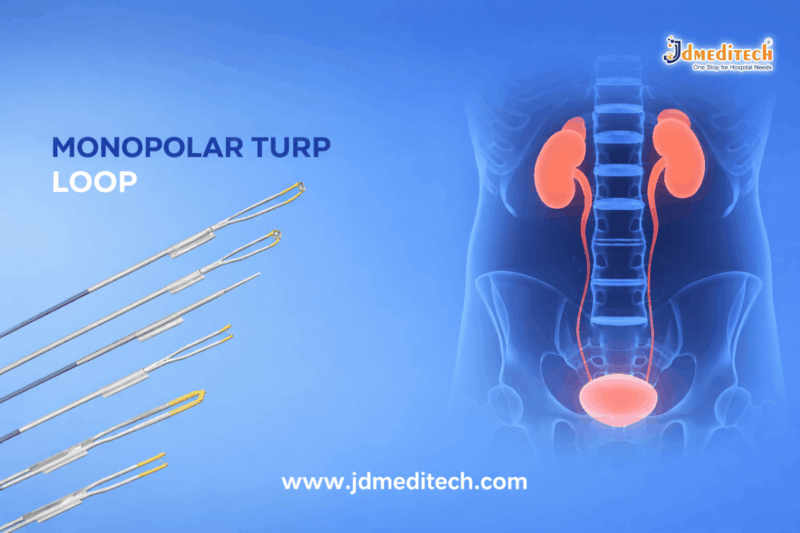
Precision Monopolar Loop Electrode for Prostate Surgery | TURP Guide

Introduction

The Precision Monopolar Loop Electrode for Prostate Surgery is a vital component in modern urological procedures, especially in Transurethral Resection of the Prostate (TURP). Designed for high precision and controlled tissue removal, this electrode plays a crucial role in ensuring safe and effective prostate surgery.

With advancements in surgical technology, the demand for reliable and high-performance monopolar loop electrodes has significantly increased. These instruments provide surgeons with excellent cutting efficiency and coagulation control, improving overall patient outcomes.

What is a Precision Monopolar Loop Electrode?

A Precision Monopolar Loop Electrode for Prostate Surgery is a specialized electrosurgical device used in TURP procedures. It is connected to an electrosurgical unit and is designed to cut and coagulate prostate tissue using high-frequency electrical current.

The loop-shaped design allows surgeons to precisely remove obstructive prostate tissue while maintaining visibility and control throughout the procedure.

Key Features of Precision Monopolar Loop Electrode for Prostate Surgery
1. High Precision Cutting

The electrode ensures smooth and accurate tissue resection, minimizing damage to surrounding structures.

2.High Electrical Conductivity

Manufactured with medical-grade materials, it provides efficient energy transmission for consistent performance.

3. Superior Hemostasis Control

It allows effective coagulation, reducing intraoperative bleeding and enhancing surgical safety.

4. Durable Medical-Grade Construction

Built using high-quality stainless steel or tungsten wire loops for long-lasting performance.

5. Compatibility with Standard Resectoscopes

The Precision Monopolar Loop Electrode for Prostate Surgery is designed to work seamlessly with most monopolar TURP systems.

Applications in Prostate Surgery

The Precision Monopolar Loop Electrode for Prostate Surgery is primarily used in:

  • Transurethral Resection of the Prostate (TURP)
  • Treatment of Benign Prostatic Hyperplasia (BPH)
  • Removal of obstructive prostate tissue
  • Electrosurgical cutting and coagulation procedures
Advantages of Using Precision Monopolar Loop Electrode
1. Enhanced Surgical Accuracy

Surgeons can perform precise resections with better control, reducing complications.

2. Improved Patient Safety

Efficient coagulation minimizes blood loss and lowers the risk of post-operative complications.

3. Cost-Effective Solution

Compared to advanced technologies, monopolar systems are widely available and economical.

4. Proven Clinical Performance

Monopolar TURP remains a gold-standard procedure in urology, supported by decades of successful outcomes.

How It Works

The Precision Monopolar Loop Electrode for Prostate Surgery works by delivering high-frequency electrical current through the loop. When the loop comes into contact with tissue:

  • The current generates heat
  • Tissue is cut or vaporized
  • Simultaneous coagulation occurs to control bleeding

A grounding pad is used to safely return the electrical current, completing the circuit.

Monopolar vs Bipolar Loop Electrode
Feature Monopolar Loop Electrode Bipolar Loop Electrode
Energy Flow Through body to grounding pad Localized between electrodes
Irrigation Fluid Non-conductive (glycine) Saline
Cost More affordable Higher
Usage Widely used in TURP Increasingly popular
Safety Considerations

When using a Precision Monopolar Loop Electrode for Prostate Surgery, it is important to:

  • Ensure proper grounding pad placement
  • Use compatible electrosurgical units
  • Monitor irrigation fluids carefully
  • Follow sterilization protocols
  • Avoid excessive thermal exposure
Choosing the Right Monopolar Loop Electrode

Selecting the right Precision Monopolar Loop Electrode for Prostate Surgery depends on:

  • Loop size and shape
  • Material quality
  • Compatibility with resectoscope
  • Surgeon preference
  • Procedure requirements

High-quality electrodes ensure consistent performance and reduce procedural risks.

Maintenance and Handling

Proper care of the Precision Monopolar Loop Electrode for Prostate Surgery enhances its lifespan:

  • Clean and sterilize after each use
  • Inspect for damage or wear
  • Store in sterile conditions
  • Follow manufacturer guidelines
Conclusion

The Precision Monopolar Loop Electrode for Prostate Surgery remains an essential tool in TURP procedures. Its ability to deliver precise cutting and effective coagulation makes it a trusted choice among urologists worldwide.

With its proven efficiency, cost-effectiveness, and reliability, this device continues to play a critical role in improving prostate surgery outcomes.


Explore More Product >>

Get Connected:

+91 79909 93062 | +91 63513 72032 | exports@jdmeditech.com
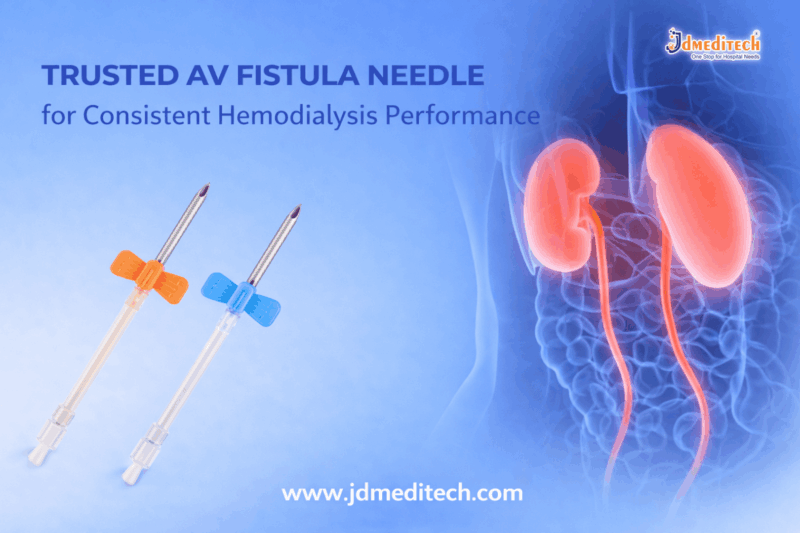


Trusted AV Fistula Needle for Consistent Hemodialysis Performance

Introduction

In hemodialysis, reliable vascular access is critical for effective treatment outcomes. One of the most essential components in this process is the AV fistula needle for hemodialysis, designed to ensure smooth blood flow and patient safety during dialysis sessions. A high-quality AV fistula needle plays a vital role in maintaining consistent performance, reducing complications, and improving overall treatment efficiency.

What is an AV Fistula Needle?

An AV fistula needle for hemodialysis is a specialized medical device used to access the arteriovenous fistula—an artificial connection between an artery and a vein created for dialysis. These needles allow blood to flow from the patient to the dialysis machine and back efficiently.

They are typically available in various gauges and lengths to suit different patient needs and vascular conditions.

Key Features of a Trusted AV Fistula Needle
1. Ultra-Sharp Needle Tip

Designed for smooth and painless insertion, minimizing trauma to the vascular access site.

2. High-Quality Medical-Grade Stainless Steel

Ensures durability, corrosion resistance, and safe clinical performance.

3. Thin-Wall Technology

Provides optimal blood flow rates, improving dialysis efficiency.

4. Flexible and Kink-Resistant Tubing

Maintains uninterrupted blood flow during the procedure.

5. Color-Coded Hubs

Allows easy identification of needle size for quick and accurate selection.

6. Secure Wing Design

Ensures stable positioning and reduces the risk of needle displacement.

Benefits of Using AV Fistula Needle for Hemodialysis
Consistent Blood Flow

The AV fistula needle for hemodialysis ensures steady blood circulation, which is essential for effective toxin removal.

Enhanced Patient Comfort

Advanced needle design reduces pain and discomfort during insertion.

Reduced Risk of Complications

Minimizes infiltration, infection, and vascular damage.

Improved Treatment Efficiency

Supports optimal dialysis performance with reliable vascular access.

Ease of Handling for Clinicians

Ergonomic design enables better control and precise placement.

Applications in Hemodialysis

The AV fistula needle for hemodialysis is widely used in:

  • Chronic kidney disease (CKD) treatment
  • Long-term dialysis procedures
  • Hospital and dialysis center settings
  • Home hemodialysis setups
How to Choose the Right AV Fistula Needle

Selecting the appropriate needle is crucial for patient safety and treatment success. Consider:

  • Gauge Size : Based on blood flow requirements
  • Needle Length : Suitable for fistula depth
  • Material Quality : Always choose medical-grade components
  • Safety Features : Look for secure wings and anti-kink tubing
Best Practices for Safe Use
  • Always follow aseptic techniques
  • Use proper insertion angle and technique
  • Rotate puncture sites to prevent damage
  • Monitor for signs of infection or infiltration
  • Dispose of needles safely after use
Why Quality Matters in AV Fistula Needles

Using a trusted and high-quality AV fistula needle for hemodialysis directly impacts patient outcomes. Inferior needles can lead to complications, poor blood flow, and increased discomfort. Investing in reliable products ensures consistent dialysis performance and enhances patient safety.

Conclusion

A trusted AV fistula needle for hemodialysis is essential for delivering safe, efficient, and consistent dialysis treatment. With advanced design features, superior materials, and patient-focused benefits, these needles play a critical role in modern renal care.

For healthcare providers, choosing the right AV fistula needle is not just a clinical decision—it’s a commitment to better patient outcomes and long-term treatment success.


Explore More Product >>

Get Connected:

+91 79909 93062 | +91 63513 72032 | exports@jdmeditech.com
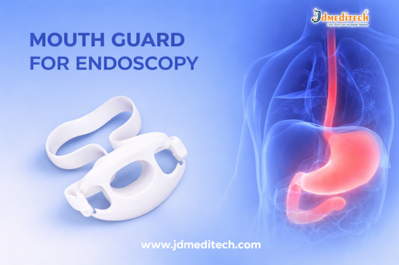


Mouth Guard for Endoscopy

Mouth Guard for Endoscopy – Protection, Comfort, and Precision

Introduction

Endoscopic procedures require precision, safety, and patient comfort. One essential yet often overlooked accessory is the mouth guard for endoscopy. This small but critical device plays a major role in protecting both the patient and the endoscope during procedures like upper GI endoscopy, ERCP, and bronchoscopy.

In this guide, we’ll explore everything you need to know about endoscopy mouth guards, including their purpose, benefits, types, and how to choose the right one.

What is a Mouth Guard for Endoscopy?

A mouth guard for endoscopy is a medical device placed in the patient’s mouth during endoscopic procedures. It helps keep the mouth open while protecting the teeth, lips, and the endoscope from damage.

These guards are typically made from medical-grade, soft yet durable materials, ensuring safety and comfort throughout the procedure.

Why is a Mouth Guard Important in Endoscopy?

Using a mouth guard in endoscopy is crucial for several reasons:

1. Protection of the Endoscope

Patients may involuntarily bite during procedures. A mouth guard prevents damage to expensive endoscopic equipment.

2. Patient Safety

It protects teeth, gums, and lips from injury, especially during long or complex procedures.

3. Improved Procedure Efficiency

By stabilizing the mouth, it allows smooth insertion and maneuverability of the endoscope.

4. Infection Control

Many modern endoscopy mouth guards are designed for single-use, reducing cross-contamination risks.

Key Features of Endoscopy Mouth Guards

A high-quality mouth guard for endoscopy offers:

  • Ergonomic Design for a secure and comfortable fit
  • Soft Bite Block to prevent dental injury
  • High Durability to withstand pressure
  • Adjustable Straps for stability during procedures
  • Latex-Free Material to avoid allergic reactions
Types of Mouth Guards for Endoscopy

Different procedures require specific designs. Common types include:

1. Standard Bite Block Mouth Guard

Used in routine upper GI endoscopy procedures.

2. Mouth Guard with Oxygen Port

Allows oxygen delivery during sedation, improving patient safety.

3. Adjustable Mouth Guard

Suitable for different patient sizes, including pediatric and adult use.

4. Disposable Mouth Guard

Single-use options that ensure hygiene and reduce infection risks.

Applications of Mouth Guard in Endoscopy

The endoscopy mouth guard is widely used in:

  • Upper GI Endoscopy
  • ERCP (Endoscopic Retrograde Cholangiopancreatography)
  • Bronchoscopy
  • Transesophageal Echocardiography (TEE)
How to Choose the Right Mouth Guard for Endoscopy

Selecting the right mouth guard for endoscopy depends on several factors:

  • Patient Comfort : Choose soft, anatomically designed guards
  • Procedure Type : Ensure compatibility with the procedure
  • Material Quality : Opt for medical-grade, non-toxic materials
  • Ease of Use : Quick placement and secure fit
  • Infection Control : Prefer disposable or serializable options
Benefits of Using a High-Quality Endoscopy Mouth Guard

Using a premium endoscopy mouth guard provides:

  • Enhanced patient comfort
  • Reduced risk of dental and soft tissue injury
  • Protection of costly endoscopic instruments
  • Improved procedural accuracy
  • Better clinical outcomes
Maintenance and Safety Tips
  • Use disposable mouth guards whenever possible
  • Ensure proper sterilization for reusable models
  • Check for damage before use
  • Use the correct size for each patient

Conclusion

The mouth guard for endoscopy is a small but indispensable tool that ensures safety, comfort, and precision during procedures. By protecting both the patient and the equipment, it contributes significantly to successful outcomes.

Healthcare providers should always prioritize high-quality endoscopy mouth guards to enhance efficiency and maintain the highest standards of care.


Explore More Product >>

Get Connected:

+91 79909 93062 | +91 63513 72032 | exports@jdmeditech.com
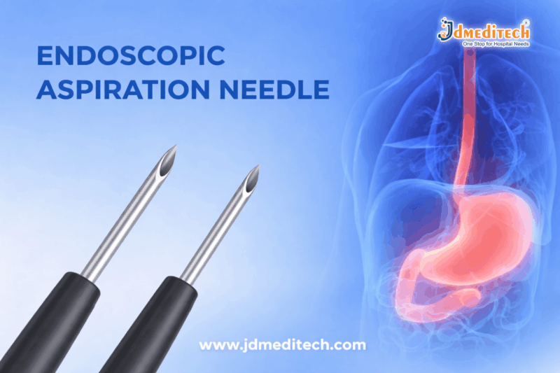
 

Endoscopic Aspiration Needle: Uses, Types, Benefits & Procedure

Endoscopic Aspiration Needle: A Complete Guide for Accurate Diagnosis & Minimally Invasive Procedures

Introduction

An Endoscopic Aspiration Needle is a vital medical device used in modern diagnostic and therapeutic procedures. It plays a crucial role in collecting tissue or fluid samples from internal organs through endoscopic guidance.

With increasing demand for minimally invasive procedures, this device has become essential in gastroenterology, pulmonology, and oncology.

What is an Endoscopic Aspiration Needle?

An Endoscopic Aspiration Needle is a specialized needle designed to pass through an endoscope to obtain tissue samples from lesions, lymph nodes, or masses inside the body.

Doctors commonly use it during procedures like:

  • Endoscopic Ultrasound (EUS)
  • Bronchoscopy
  • Fine Needle Aspiration (FNA)
Key Features of Endoscopic Aspiration Needle

Modern Endoscopic Aspiration Needle devices are designed for precision and safety. Key features include:

  • High-quality stainless steel needle
  • Echogenic tip for better visibility
  • Flexible catheter for easy navigation
  • Ergonomic handle for control
  • Multiple size options (19G, 22G, 25G)

These features ensure accurate targeting and effective sample collection.

Types of Endoscopic Aspiration Needles

Different procedures require different types of Endoscopic Aspiration Needle:

1. Fine Needle Aspiration (FNA) Needle

Used for collecting cytological samples.

2. Fine Needle Biopsy (FNB) Needle

Designed to obtain core tissue samples for histology.

3. EUS-Guided Aspiration Needle

Used with endoscopic ultrasound for precise targeting.

Applications in Medical Procedures

The Endoscopic Aspiration Needle is widely used across various specialties:

  • Gastroenterology– Sampling pancreatic lesions
  • Oncology– Tumor diagnosis and staging
  • Pulmonology– Lung mass biopsy
  • Hepatology– Liver lesion evaluation

This versatility makes it a critical diagnostic tool.

Benefits of Using Endoscopic Aspiration Needle

Using an Endoscopic Aspiration Needle offers several advantages:

  • Minimally invasive procedure
  • Reduced patient discomfort
  • Quick recovery time
  • High diagnostic accuracy
  • Lower risk of complications

As a result, it is preferred over traditional surgical biopsy methods.

Procedure Overview

The procedure using an Endoscopic Aspiration Needle typically involves:

  1. Insertion of the endoscope into the body
  2. Identification of the target area
  3. Needle advancement through the endoscope
  4. Sample collection via aspiration
  5. Withdrawal and lab analysis

The entire process is safe and efficient when performed by trained professionals.

Safety and Precautions

Although the Endoscopic Aspiration Needle is safe, certain precautions are necessary:

  • Proper sterilization
  • Use by trained specialists only
  • Monitoring for complications like bleeding or infection
  • Correct needle selection based on procedure

Following these ensures optimal patient outcomes.

How to Choose the Right Endoscopic Aspiration Needle

Selecting the right Endoscopic Aspiration Needle depends on:

  • Target tissue type
  • Required sample size
  • Procedure type (EUS, bronchoscopy)
  • Needle gauge

Consulting medical experts helps in making the best choice.

Conclusion

The Endoscopic Aspiration Needle is an indispensable tool in modern medicine. It enables accurate diagnosis while minimizing patient discomfort.

With advancements in technology, these needles continue to improve precision, safety, and efficiency in medical procedures.


Explore More Product >>

Get Connected:

+91 79909 93062 | +91 63513 72032 | exports@jdmeditech.com
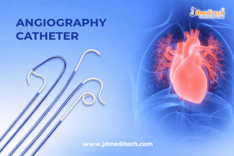
Angiography Diagnostic Catheter: Uses, Types, Procedure & Benefits

Angiography Diagnostic Catheter: Complete Guide for Accurate Vascular Imaging

Introduction

The Angiography Diagnostic Catheter is an essential tool in modern medical imaging. It helps doctors examine blood vessels with high precision. In fact, this device plays a key role in diagnosing cardiovascular and vascular diseases.

Today, healthcare professionals prefer the Angiography Diagnostic Catheter because it offers accurate results and supports minimally invasive procedures. Therefore, it has become a standard instrument in hospitals and diagnostic centers.

What is an Angiography Diagnostic Catheter?

An Angiography Diagnostic Catheter is a thin, flexible medical tube used to inject contrast dye into blood vessels. As a result, doctors can clearly view arteries and veins using imaging techniques like fluoroscopy.

Moreover, the Angiography Diagnostic Catheter is designed to move smoothly through blood vessels. This ensures both safety and efficiency during diagnostic procedures.

Key Features of Angiography Diagnostic Catheter
Flexible and Smooth Design

The Angiography Diagnostic Catheter offers excellent flexibility. Therefore, it easily navigates complex vascular pathways.

Radiopaque Visibility

Additionally, it contains radiopaque markers. These markers help doctors track the Angiography Diagnostic Catheter during imaging.

Multiple Shapes and Sizes

The Angiography Diagnostic Catheter is available in various shapes like Judkins, Amplatz, and Pigtail. Each type serves a specific diagnostic purpose.

Biocompatible Material

Furthermore, the materials used in the Angiography Diagnostic Catheter reduce the risk of infection and complications.

Types of Angiography Diagnostic Catheter
Coronary Angiography Catheter

This Angiography Diagnostic Catheter is used to examine coronary arteries.

Peripheral Angiography Catheter

It helps diagnose vascular issues in limbs and other body parts.

Neuro Angiography Catheter

Doctors use this Angiography Diagnostic Catheter for brain-related vascular imaging.

Pigtail Catheter

This type of Angiography Diagnostic Catheter ensures uniform dye distribution in large vessels.

Selective Catheter

It allows precise access to targeted blood vessels.

Procedure Using Angiography Diagnostic Catheter
Step 1: Patient Preparation

First, the patient is prepared, and local anesthesia is given.

Step 2: Catheter Insertion

Next, the Angiography Diagnostic Catheter is inserted into an artery, usually in the wrist or groin.

Step 3: Navigation

Then, doctors guide the Angiography Diagnostic Catheter to the target area using imaging.

Step 4: Contrast Injection

After that, contrast dye is injected through the Angiography Diagnostic Catheter.

Step 5: Imaging

Finally, images are captured for diagnosis.

Benefits of Angiography Diagnostic Catheter
  • Provides accurate diagnosis
  • Supports minimally invasive procedures
  • Ensures faster recovery
  • Offers real-time imaging
  • Improves treatment planning

Because of these advantages, the Angiography Diagnostic Catheter is widely used in modern healthcare.

Applications of Angiography Diagnostic Catheter

The Angiography Diagnostic Catheter is used in several medical fields:

  • Cardiology
  • Neurology
  • Vascular surgery
  • Interventional radiology

Thus, it plays a crucial role in diagnosing serious conditions like blockages and aneurysms.

Safety and Precautions

Although the Angiography Diagnostic Catheter is safe, some precautions are necessary:

  • Monitor for dye allergies
  • Maintain sterile conditions
  • Choose the correct catheter type
  • Observe the patient during the procedure
Conclusion

The Angiography Diagnostic Catheter is a vital device for accurate vascular diagnosis. It improves imaging quality while ensuring patient safety. In addition, its advanced design supports efficient and reliable procedures.

Therefore, healthcare providers trust the Angiography Diagnostic Catheter for precise and effective diagnostics.


Explore More Product >>

Get Connected:

+91 79909 93062 | +91 63513 72032 | exports@jdmeditech.com
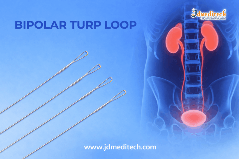


High-Precision Bipolar TURP Loop for Safe Prostate Surgery

High-Precision Bipolar TURP Loop for Safe & Effective Prostate Surgery

Introduction

The High-Precision Bipolar TURP Loop is an essential instrument in modern urology. It plays a vital role in treating benign prostatic hyperplasia (BPH). Today, surgeons prefer the High-Precision Bipolar TURP Loop because it offers better control, higher safety, and improved surgical outcomes.

Moreover, this advanced device ensures minimal complications. As a result, it has become a standard choice in TURP procedures worldwide.

What is a High-Precision Bipolar TURP Loop?

A High-Precision Bipolar TURP Loop is an electrosurgical device used during transurethral resection of the prostate. It uses bipolar energy to cut and coagulate tissue simultaneously.

Unlike traditional systems, the High-Precision Bipolar TURP Loop works with saline irrigation. Therefore, it significantly reduces the risk of complications and improves surgical safety.

Key Features of High-Precision Bipolar TURP Loop

The High-Precision Bipolar TURP Loop is designed to deliver superior performance. Key features include:

  • High precision cutting for accurate tissue removal
  • Smooth loop design for consistent performance
  • Efficient energy transmission
  • Minimal thermal damage to surrounding tissue
  • Excellent compatibility with bipolar systems
  • Ergonomic handling for better surgical control
Benefits of High-Precision Bipolar TURP Loop
1. Enhanced Surgical Precision

The High-Precision Bipolar TURP Loop allows surgeons to perform controlled and accurate resections. Consequently, it improves overall surgical results.

2. Improved Patient Safety

Because the High-Precision Bipolar TURP Loop uses saline, it reduces the risk of TUR syndrome. Therefore, it ensures safer procedures.

3. Better Hemostasis

This device provides excellent coagulation. As a result, bleeding during surgery is minimized.

4. Faster Recovery

Patients treated with a High-Precision Bipolar TURP Loop often recover faster due to reduced tissue trauma.

5. Reduced Complications

The High-Precision Bipolar TURP Loop lowers the chances of post-operative complications, ensuring better patient outcomes.

How Does High-Precision Bipolar TURP Loop Work?

The High-Precision Bipolar TURP Loop works by passing electrical energy between two electrodes on the loop. This design enables:

  • Precise cutting of prostate tissue
  • Simultaneous coagulation
  • Controlled energy flow

As a result, the High-Precision Bipolar TURP Loop ensures high efficiency and safety during procedures.

Applications of High-Precision Bipolar TURP Loop

The High-Precision Bipolar TURP Loop is widely used in:

  • Benign Prostatic Hyperplasia (BPH) treatment
  • Transurethral resection procedures
  • Advanced urology surgeries
  • Hospitals and specialized clinics
Bipolar vs Monopolar TURP Loop
Feature Bipolar TURP Loop Monopolar TURP Loop
Irrigation Saline Glycine
Safety High Moderate
TUR Syndrome Risk Very Low Higher
Precision High Moderate
Recovery Faster Slower

Clearly, the High-Precision Bipolar TURP Loop provides better safety and efficiency compared to traditional options.

Why Choose High-Precision Bipolar TURP Loop?

Choosing a High-Precision Bipolar TURP Loop offers multiple advantages. First, it improves surgical accuracy. Second, it enhances patient safety. Finally, it ensures reliable performance in every procedure.

Therefore, hospitals and surgeons increasingly rely on the High-Precision Bipolar TURP Loop for consistent and high-quality results.

Maintenance Tips

To maintain the performance of the High-Precision Bipolar TURP Loop, follow these steps:

  • Clean thoroughly after each use
  • Sterilize according to medical standards
  • Inspect before every procedure
  • Store in a sterile environment

Proper care ensures long-lasting efficiency of the High-Precision Bipolar TURP Loop.

Conclusion

The High-Precision Bipolar TURP Loop is a game-changing tool in urology. It combines precision, safety, and efficiency in one device. Additionally, it reduces complications and improves patient recovery.

In conclusion, adopting the High-Precision Bipolar TURP Loop is essential for modern surgical success.


Explore More Product >>

Get Connected:

+91 79909 93062 | +91 63513 72032 | exports@jdmeditech.com
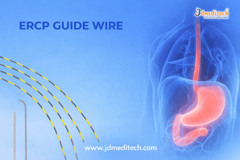
ERCP Guide Wire: Features, Benefits & Clinical Applications

ERCP Guide Wire: Precision Navigation for Advanced Biliary and Pancreatic Procedures

Introduction

An ERCP Guide Wire is a critical tool used in Endoscopic Retrograde Cholangiopancreatography (ERCP) procedures. It helps physicians navigate complex biliary and pancreatic ducts with precision and safety. As minimally invasive procedures continue to grow in popularity, the importance of a reliable ERCP guide wire becomes even more significant.

In this blog, we will explore the features, benefits, applications, and selection criteria of ERCP guide wires to help healthcare professionals make informed decisions.

What is an ERCP Guide Wire?

An ERCP Guide Wire is a flexible, steerable wire designed to assist in accessing and navigating the biliary and pancreatic ducts during ERCP procedures. It acts as a pathway for other devices such as catheters, stents, and dilation balloons.

Because of its design and material, the guide wire ensures smooth insertion and minimizes trauma to delicate ductal structures.

Key Features of ERCP Guide Wire
1. High Flexibility and Torque Control

ERCP guide wires offer excellent flexibility. At the same time, they provide superior torque control. As a result, clinicians can maneuver through complex anatomy with ease.

2. Hydrophilic Coating

Most ERCP guide wires come with a hydrophilic coating. Therefore, they reduce friction and allow smoother navigation through tight or tortuous ducts.

3. Radiopaque Tip

The radiopaque tip enhances visibility under fluoroscopy. This feature ensures accurate placement and improves procedural safety.

4. Optimal Stiffness

The shaft of the ERCP guide wire provides the right balance between stiffness and flexibility. Consequently, it supports device exchange without compromising control.

5. Kink Resistance

High-quality guide wires are designed to resist kinking. This ensures consistent performance during long or complex procedures.

Applications of ERCP Guide Wire

An ERCP Guide Wire is widely used in various therapeutic and diagnostic procedures:

  • Biliary cannulation
  • Pancreatic duct access
  • Stone extraction procedures
  • Stent placement
  • Stricture dilation
  • Drainage procedures

Because of its versatility, it is an essential component in every ERCP setup.

Benefits of Using ERCP Guide Wire
Improved Procedural Accuracy

The guide wire enhances precision. Therefore, clinicians can access target ducts more effectively.

Reduced Risk of Complications

Smooth navigation reduces trauma. As a result, the risk of perforation and other complications decreases.

Faster Procedure Time

Efficient access leads to shorter procedure times. This improves patient comfort and clinical workflow.

Enhanced Device Compatibility

ERCP guide wires are compatible with a wide range of endoscopic accessories. Thus, they support seamless device exchange.

Types of ERCP Guide Wires
Hydrophilic Guide Wires

These wires are coated for smooth navigation. They are ideal for difficult or tight strictures.

Hybrid Guide Wires

Hybrid wires combine hydrophilic tips with stiff shafts. Therefore, they provide both flexibility and support.

Stiff Guide Wires

These wires are used when strong support is needed, especially during stent placement.

How to Choose the Right ERCP Guide Wire

Selecting the right ERCP Guide Wire depends on several factors:

  • Procedure type: Diagnostic or therapeutic
  • Duct anatomy: Straight or tortuous
  • Required support level: Flexible vs stiff
  • Coating type: Hydrophilic or non-coated
  • Visibility needs: Radiopaque features

By considering these factors, healthcare professionals can improve procedural outcomes.

Best Practices for Using ERCP Guide Wire
  • Always confirm guide wire position under fluoroscopy
  • Avoid excessive force during insertion
  • Use appropriate wire type for each procedure
  • Maintain sterility to prevent infections
  • Replace damaged or kinked wires immediately

Following these practices ensures safety and efficiency.

Why Choose High-Quality ERCP Guide Wires?

High-quality ERCP Guide Wires offer consistent performance, durability, and enhanced safety. Moreover, they reduce procedural risks and improve clinical success rates.

For hospitals and endoscopy centers, investing in premium guide wires is essential for delivering reliable patient care.

Conclusion

An ERCP Guide Wire plays a vital role in modern gastroenterology procedures. It ensures precise navigation, supports device delivery, and enhances overall procedural success. Therefore, choosing the right guide wire is crucial for both patient safety and clinical efficiency.

As ERCP procedures continue to evolve, the demand for advanced, high-performance guide wires will only increase.


Explore More Product >>

Get Connected:

+91 79909 93062 | +91 63513 72032 | exports@jdmeditech.com
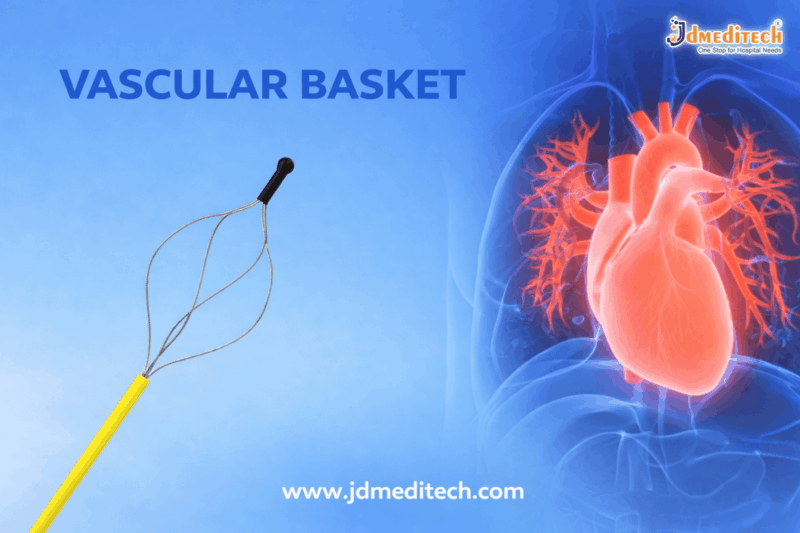
Vascular Basket: Safe & Effective Foreign Body Retrieval Device

Vascular Basket: A Precision Tool for Safe and Effective Foreign Body Retrieval

Introduction

A Vascular Basket is an advanced medical device widely used in interventional procedures to retrieve foreign bodies from blood vessels. It plays a crucial role in ensuring patient safety during minimally invasive vascular interventions.

In modern healthcare, precision and efficiency are essential. Therefore, a Vascular Basket helps clinicians remove clots, fragments, and other obstructions with accuracy and minimal trauma.

What is a Vascular Basket?

A Vascular Basket is a flexible, wire-based retrieval device designed to capture and remove foreign objects from the vascular system. Typically, it is inserted through a catheter and guided to the target location under imaging techniques like fluoroscopy.

Moreover, its basket-like structure expands inside the vessel, allowing secure capture of the object before retrieval.

Key Features of Vascular Basket

The Vascular Basket offers several features that enhance procedural success:

  • Flexible Design– Ensures smooth navigation through blood vessels
  • Shape Memory Alloy– Allows the basket to expand and contract effectively
  • High Precision Control– Enables accurate placement and retrieval
  • Minimally Invasive Use– Reduces patient recovery time
  • Radiopaque Markers– Improves visibility under imaging

These features make the Vascular Basket a reliable tool for complex vascular procedures.

Types of Vascular Basket

Different types of Vascular Basket devices are available depending on clinical requirements:

1. Helical Basket

Designed for efficient capture of elongated or irregular objects.

2. Four-Wire Basket

Provides balanced strength and flexibility for general retrieval.

3. Six-Wire Basket

Offers enhanced grip and stability during complex procedures.

4. Collapsible Basket

Allows easy insertion and controlled expansion inside vessels.

Applications of Vascular Basket

The Vascular Basket is used in various medical scenarios:

  • Retrieval of broken catheter fragments
  • Removal of embolized coils or devices
  • Extraction of blood clots or thrombus
  • Foreign body retrieval from veins and arteries
  • Support in interventional radiology procedures

Because of its versatility, the Vascular Basket is an essential tool in vascular surgery and radiology.

Advantages of Using Vascular Basket

Using a Vascular Basket provides multiple benefits:

  • High Success Rate in Retrieval Procedures
  • Reduced Risk of Vessel Damage
  • Shorter Procedure Time
  • Improved Patient Safety
  • Minimized Need for Open Surgery

As a result, healthcare professionals prefer the Vascular Basket for safe and effective outcomes.

How Does a Vascular Basket Work?

The working mechanism of a Vascular Basket is simple yet effective:

  1. The device is inserted through a catheter into the vessel
  2. It is guided to the target area using imaging techniques
  3. The basket expands to capture the foreign object
  4. The device is carefully withdrawn, securing the object

This process ensures precise and controlled retrieval.

Factors to Consider When Choosing a Vascular Basket

Before selecting a Vascular Basket, consider the following:

  • Basket Size and Diameter
  • Wire Configuration
  • Compatibility with Catheters
  • Material Quality
  • Ease of Handling

Choosing the right Vascular Basket improves procedural efficiency and safety.

Maintenance and Handling Tips

Proper care of a Vascular Basket is essential:

  • Store in a sterile environment
  • Handle with care to maintain structural integrity
  • Follow manufacturer guidelines for usage
  • Dispose of single-use devices properly
Conclusion

The Vascular Basket is a vital tool in modern vascular interventions. Its precision, flexibility, and safety make it indispensable for foreign body retrieval procedures.

As medical technology continues to advance, the role of the Vascular Basket will only grow, ensuring better patient outcomes and improved procedural success.


Explore More Product >>

Get Connected:

+91 79909 93062 | +91 63513 72032 | exports@jdmeditech.com
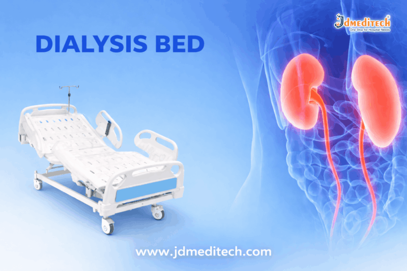


Dialysis Bed for Hemodialysis Procedures – Features, Benefits & Buying Guide

Dialysis Bed for Hemodialysis Procedures: Comfort, Safety & Efficiency

Introduction

A dialysis bed for hemodialysis procedures plays a vital role in ensuring patient comfort, clinical efficiency, and procedural safety. Hemodialysis sessions often last several hours, making it essential to provide patients with a stable, ergonomic, and adjustable surface.

Modern dialysis beds are designed with advanced features that enhance both patient experience and healthcare provider convenience. In this blog, we explore everything you need to know about dialysis beds, including features, benefits, types, and selection tips.

What is a Dialysis Bed for Hemodialysis Procedures?

A dialysis bed for hemodialysis procedures is a specialized medical bed designed to support patients undergoing dialysis treatment. Unlike standard hospital beds, dialysis beds are built with enhanced adjustability, mobility, and patient positioning capabilities.

These beds allow healthcare professionals to perform procedures efficiently while keeping patients comfortable throughout long sessions.

Key Features of Dialysis Beds
1. Adjustable Positioning

Dialysis beds offer multiple position adjustments, including:

  • Backrest elevation
  • Leg support adjustment
  • Trendelenburg and reverse Trendelenburg positions

This flexibility helps in improving blood circulation and patient comfort.

2. Ergonomic Design

The ergonomic structure reduces strain on both patients and caregivers. It supports proper body posture during extended dialysis sessions.

3. Electric or Hydraulic Controls

Modern dialysis beds are equipped with:

  • Electric motorized controls
  • Easy-to-use remote systems

These controls allow smooth and precise adjustments.

4. High-Quality Mattress

Comfortable, anti-bacterial, and easy-to-clean mattresses ensure hygiene and prevent pressure sores.

5. Mobility with Locking Wheels

Dialysis beds come with smooth-rolling castors and locking mechanisms for safe positioning and easy transportation.

6. Safety Features
  • Side rails for patient protection
  • Emergency CPR function
  • Stable frame structure
Benefits of Using a Dialysis Bed
Enhanced Patient Comfort

Since dialysis sessions last 3–5 hours, a comfortable bed reduces fatigue and improves patient compliance.

Improved Clinical Efficiency

Adjustable positions make it easier for healthcare professionals to access vascular sites and perform procedures.

Better Safety

Built-in safety mechanisms reduce the risk of falls and complications during treatment.

Reduced Caregiver Strain

Motorized adjustments minimize physical effort for nurses and technicians.

Types of Dialysis Beds
Manual Dialysis Bed
  • Operated using mechanical levers
  • Cost-effective option
  • Suitable for small clinics
Semi-Electric Dialysis Bed
  • Combination of manual and electric controls
  • Moderate pricing
  • Offers better convenience
Fully Electric Dialysis Bed
  • Complete motorized operation
  • Advanced features and maximum comfort
  • Ideal for modern hospitals and dialysis centers
How to Choose the Right Dialysis Bed
Consider Patient Comfort

Look for cushioned mattresses and multiple adjustment options.

Evaluate Build Quality

Choose beds made from durable materials with a stable frame.

Check Safety Features

Ensure the presence of side rails, locking wheels, and emergency controls.

Ease of Operation

Electric controls should be smooth and user-friendly.

Maintenance & Hygiene

Select beds with easy-to-clean surfaces and anti-bacterial materials.

Applications of Dialysis Beds

A dialysis bed for hemodialysis procedures is widely used in:

  • Hospitals
  • Dialysis centers
  • Nephrology clinics
  • Intensive care units (ICU)
Why Dialysis Beds are Essential in Hemodialysis

Dialysis is a life-saving treatment, and patient comfort directly affects treatment success. A high-quality dialysis bed ensures:

  • Stable patient positioning
  • Reduced complications
  • Enhanced treatment efficiency

Therefore, investing in the right dialysis bed is crucial for healthcare facilities.

Conclusion

A dialysis bed for hemodialysis procedures is more than just a piece of equipment—it is a critical component of patient care. With features like adjustable positioning, ergonomic design, and advanced safety mechanisms, these beds significantly improve both patient comfort and clinical workflow.

Healthcare providers should carefully select dialysis beds based on quality, functionality, and patient needs to ensure the best treatment outcomes.


Explore More Product >>

Get Connected:

+91 79909 93062 | +91 63513 72032 | exports@jdmeditech.com
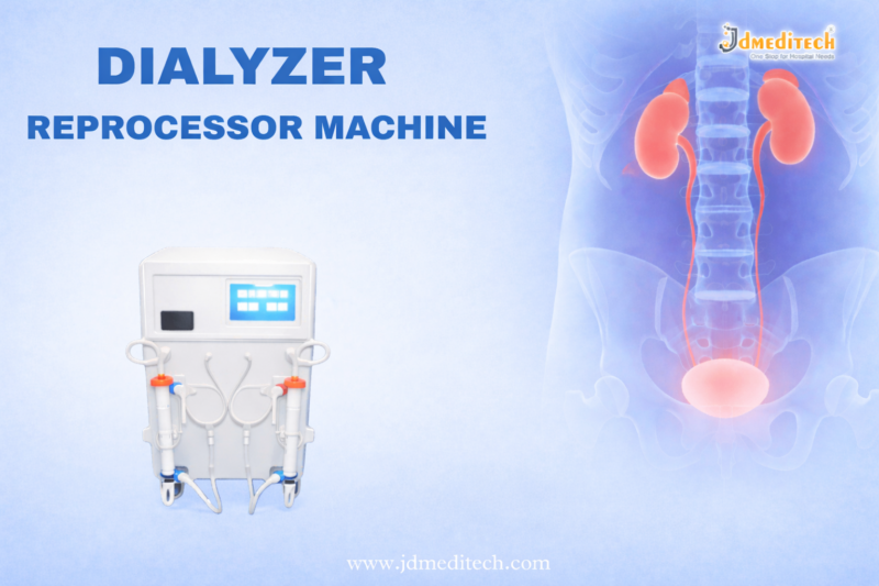


Dialyzer Reprocessing Machine: Benefits, Working & Features

Dialyzer Reprocessing Machine: Cost-Effective & Safe Dialysis Solution

Introduction

A Dialyzer Reprocessing Machine plays a crucial role in modern dialysis centers by enabling the safe reuse of dialyzers. As the demand for cost-effective healthcare grows, reprocessing technology helps hospitals reduce expenses while maintaining high standards of patient safety.

In this blog, we will explore how a dialyzer reprocessing machine works, its benefits, features, and why it is essential for dialysis units.

What is a Dialyzer Reprocessing Machine?

A Dialyzer Reprocessing Machine is a medical device designed to clean, disinfect, and test dialyzers for reuse in hemodialysis treatments. It ensures that each dialyzer is safe, sterile, and functioning properly before being reused for the same patient.

This process follows strict medical guidelines to prevent contamination and ensure patient safety.

How Does a Dialyzer Reprocessing Machine Work?

The working process of a dialyzer reprocessing machine involves several important steps:

1. Cleaning

First, the machine removes blood residues and impurities from the dialyzer using specialized cleaning solutions.

2. Performance Testing

Next, it checks the dialyzer’s efficiency, including fiber integrity and volume capacity.

3. Disinfection

After testing, the machine disinfects the dialyzer using approved chemicals to eliminate bacteria and pathogens.

4. Labeling & Storage

Finally, the dialyzer is labeled and stored for reuse by the same patient.

Key Features of Dialyzer Reprocessing Machines

Modern dialyzer reprocessing machines come with advanced features, including:

  • Automated cleaning and disinfection cycles
  • Accurate performance monitoring
  • User-friendly interface
  • Digital data recording and tracking
  • High safety standards and compliance
  • Compact and efficient design

These features help improve workflow efficiency and reduce manual errors.

Benefits of Using a Dialyzer Reprocessing Machine
1. Cost Efficiency

Dialyzer reuse significantly reduces the cost per dialysis session, making treatment more affordable.

2. Improved Resource Utilization

Hospitals can optimize resources by safely reusing dialyzers instead of discarding them after a single use.

3. Consistent Performance

The machine ensures that only dialyzers meeting quality standards are reused.

4. Enhanced Safety

Strict disinfection protocols reduce the risk of infection and contamination.

5. Eco-Friendly Approach

Reprocessing minimizes medical waste, supporting environmentally sustainable healthcare practices.

Applications in Healthcare

Dialyzer reprocessing machines are widely used in:

  • Dialysis centers
  • Hospitals and nephrology departments
  • Multi-specialty healthcare facilities

They are especially beneficial in high-volume dialysis units where cost control and efficiency are essential.

Safety Guidelines for Dialyzer Reprocessing

To ensure optimal performance and patient safety, follow these guidelines:

  • Use dialyzers for the same patient only
  • Follow proper disinfection protocols
  • Monitor dialyzer performance regularly
  • Maintain machine calibration and servicing
  • Train staff properly in reprocessing procedures
Why Choose a High-Quality Dialyzer Reprocessing Machine?

Choosing the right dialyzer reprocessing machine ensures:

  • Reliable performance
  • Long-term cost savings
  • Compliance with medical standards
  • Improved patient outcomes

Investing in a high-quality machine is essential for delivering safe and effective dialysis care.

Conclusion

A Dialyzer Reprocessing Machine is an indispensable tool in modern dialysis practice. It not only reduces treatment costs but also ensures safety, efficiency, and sustainability.

By adopting advanced reprocessing technology, healthcare providers can deliver high-quality dialysis care while optimizing operational efficiency.


Explore More Product >>

Get Connected:

+91 79909 93062 | +91 63513 72032 | exports@jdmeditech.com
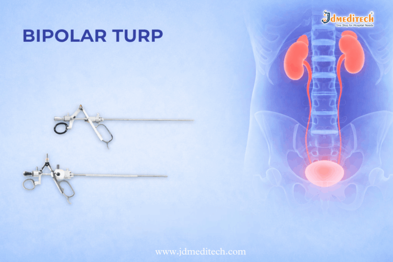
 

Bipolar TURP: Safe and Advanced Treatment for Enlarged Prostate

Bipolar TURP: Safe and Advanced Treatment for Enlarged Prostate

What Is Bipolar TURP?

Bipolar TURP is a modern surgical procedure used to treat an enlarged prostate. Doctors often recommend Bipolar TURP for men with moderate to severe urinary symptoms caused by benign prostatic hyperplasia (BPH). This procedure removes extra prostate tissue and helps improve urine flow.

Unlike older methods, Bipolar TURP uses bipolar energy with saline irrigation. As a result, it offers better safety and improved surgical control. Because of these benefits, many hospitals and urology specialists now prefer Bipolar TURP.

Why Is Bipolar TURP Needed?

An enlarged prostate can press against the urethra and block the normal flow of urine. Therefore, patients may face several uncomfortable symptoms. In many cases, Bipolar TURP is advised when medicines no longer provide enough relief.

Common symptoms include:

  • Frequent urination
  • Difficulty starting urination
  • Weak urine stream
  • Night-time urination
  • Incomplete bladder emptying
  • Sudden urge to urinate
  • Urinary retention

When these symptoms become severe, Bipolar TURP can provide long-term relief.

How Bipolar TURP Works

During Bipolar TURP, the surgeon inserts a resectoscope through the urethra. No external cut is needed. Then the surgeon uses bipolar electrical energy to remove the extra prostate tissue blocking the urinary passage.

At the same time, saline solution is used for irrigation. This improves visibility and also lowers the risk of complications linked with non-saline fluids. After tissue removal, the surgeon places a catheter for temporary drainage.

In most cases, Bipolar TURP is completed within 60 to 90 minutes.

Key Benefits of Bipolar TURP

There are many reasons why Bipolar TURP is considered a preferred option for BPH treatment.

1. Better Safety

Bipolar TURP uses saline solution, which reduces the risk of TUR syndrome. This makes the procedure safer than traditional monopolar methods.

2. Less Bleeding

Because bipolar energy offers better coagulation, Bipolar TURP helps control bleeding during surgery.

3. Faster Recovery

Most patients recover faster after Bipolar TURP. In addition, hospital stay is often shorter.

4. Improved Precision

The system allows accurate cutting and effective removal of obstructing tissue.

5. Suitable for Many Patients

Doctors may choose Bipolar TURP for elderly patients or for those who need a safer surgical option.

Bipolar TURP vs Monopolar TURP

Both procedures treat enlarged prostate, but Bipolar TURP offers several important advantages.

Feature Bipolar TURP Monopolar TURP
Energy system Bipolar Monopolar
Irrigation fluid Saline Non-saline fluid
TUR syndrome risk Very low Higher
Bleeding control Better Moderate
Recovery time Faster Slower
Safety profile Higher Lower

Because of these differences, many surgeons now prefer Bipolar TURP over monopolar TURP.

Who Can Benefit From Bipolar TURP?

Bipolar TURP is usually recommended for men who:

  • Have moderate to severe BPH symptoms
  • Do not respond well to medication
  • Experience repeated urinary retention
  • Need effective prostate tissue removal
  • Want a minimally invasive treatment option

However, the final decision depends on the patient’s condition, prostate size, and overall health.

Recovery After Bipolar TURP

Recovery after Bipolar TURP is generally smooth. Most patients stay in the hospital for a short period. The catheter is usually removed within one to two days.

After surgery, patients may notice:

  • Mild discomfort during urination
  • Small amounts of blood in urine
  • Frequent urge to urinate for a few days

These symptoms usually improve with time. Still, proper care is important after Bipolar TURP.

Recovery Tips
  • Drink enough water
  • Avoid heavy lifting
  • Follow medical advice carefully
  • Take prescribed medicines on time
  • Attend all follow-up visits

In most cases, patients return to normal daily activities within two to three weeks after Bipolar TURP.

Possible Risks of Bipolar TURP

Although Bipolar TURP is considered safe, every surgery carries some risk. Possible complications may include:

  • Urinary tract infection
  • Mild bleeding
  • Temporary difficulty in urination
  • Retrograde ejaculation
  • Temporary urinary incontinence

However, serious complications are uncommon, especially when Bipolar TURP is performed by experienced surgeons.

Why Bipolar TURP Is a Preferred Choice

Today, Bipolar TURP is widely used because it combines safety, efficiency, and reliable results. It offers better control during surgery and supports quicker recovery. Moreover, it reduces the risk of some complications seen in older TURP methods.

For these reasons, Bipolar TURP has become a trusted solution for the treatment of enlarged prostate.

Conclusion

Bipolar TURP is a safe, advanced, and effective treatment for men suffering from enlarged prostate symptoms. It improves urine flow, reduces discomfort, and supports faster recovery. In addition, it offers clear benefits over traditional monopolar techniques.

If you are looking for an advanced BPH treatment option, Bipolar TURP may be the right choice. Always consult a qualified urology specialist for proper diagnosis and treatment planning.


Explore More Product >>

Get Connected:

+91 79909 93062 | +91 63513 72032 | exports@jdmeditech.com
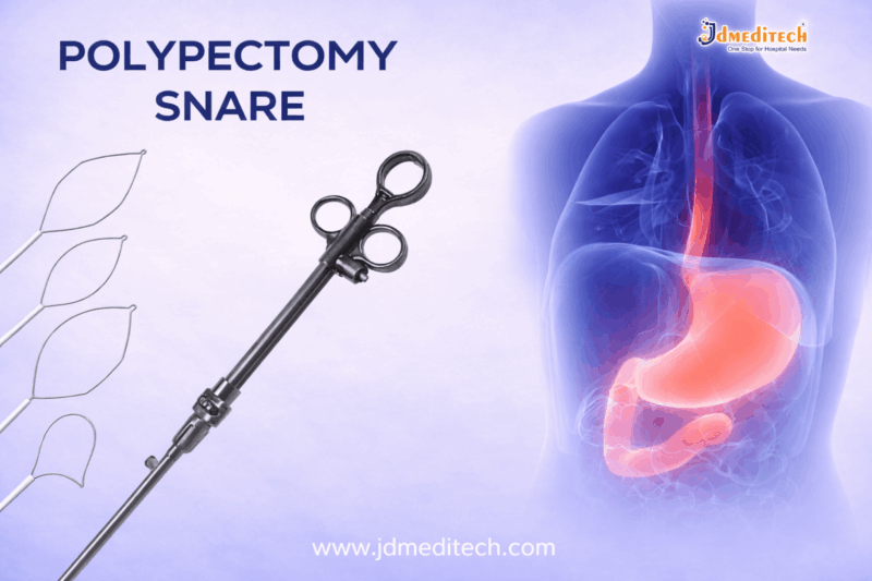
 

Polypectomy Snare – Safe & Effective Endoscopic Polyp Removal Tool

What is a Polypectomy Snare?

A polypectomy snare is a medical instrument with a thin wire loop attached to a flexible tube. Doctors pass the polypectomy snare through an endoscope to reach the polyp.

Then, the loop surrounds the polyp and tightens to remove it. In many cases, the polypectomy snare uses electrocautery to reduce bleeding and improve safety.

Types of Polypectomy Snare

There are different types of polypectomy snare devices designed for specific needs.

Cold Polypectomy Snare

A polypectomy snare without electric current is called a cold snare. It is best for small polyps and reduces tissue damage.

Hot Polypectomy Snare

This type of polypectomy snare uses electrocautery. It helps remove larger polyps and controls bleeding effectively.

Rotatable Polypectomy Snare

A rotatable polypectomy snare improves positioning. Therefore, it offers better precision in difficult areas.

Special Shape Polypectomy Snare

Some polypectomy snare designs come in oval or hexagonal shapes. These shapes improve grip and removal efficiency.

Key Features of Polypectomy Snare

A high-quality polypectomy snare offers several important features:

  • Flexible catheter for smooth insertion
  • Strong wire loop for clean cutting
  • Easy handle control for precision
  • Compatibility with standard endoscopes
  • Sterile design for safe use

Because of these features, the polypectomy snare ensures reliable performance.

Uses of Polypectomy Snare

The polypectomy snare is widely used in medical procedures such as:

  • Colonoscopy procedures
  • Gastrointestinal polyp removal
  • Diagnostic endoscopy
  • Cancer prevention treatments

Therefore, the polypectomy snare is essential for early disease management.

Benefits of Polypectomy Snare

Using a polypectomy snare provides many benefits:

  • Minimally invasive procedure
  • High precision and control
  • Reduced recovery time
  • Lower risk of complications
  • Effective polyp removal

In addition, the polypectomy snare improves patient outcomes.

How Does a Polypectomy Snare Work?

The polypectomy snare works in a simple and effective way:

  1. First, the endoscope is inserted
  2. Then, the polypectomy snare reaches the polyp
  3. Next, the loop surrounds the polyp
  4. After that, the snare tightens
  5. Finally, the polyp is removed

If required, the polypectomy snare uses electric current to stop bleeding.

Safety Tips for Using Polypectomy Snare

To ensure safe use of a polypectomy snare, doctors should follow these steps:

  • Choose the correct size of polypectomy snare
  • Use controlled electrocautery
  • Maintain clear visualization
  • Monitor for complications

Thus, proper handling of the polypectomy snare ensures better results.

Why Choose a Quality Polypectomy Snare?

A high-quality polypectomy snare improves both safety and efficiency. It allows doctors to perform procedures with confidence.

At JDMeditech, we provide reliable and advanced polypectomy snare devices designed for precision and durability.

Conclusion

The polypectomy snare is an essential tool in endoscopy. It allows safe, quick, and effective polyp removal.

Overall, the polypectomy snare supports early diagnosis and prevention of serious diseases. Therefore, it remains a vital device in modern healthcare.


Explore More Product >>

Get Connected:

+91 79909 93062 | +91 63513 72032 | exports@jdmeditech.com
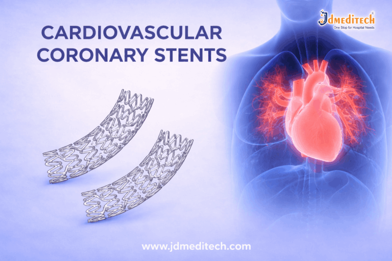


Cardiovascular Coronary Stent: Types, Procedure, Benefits & Recovery Guide

What is a Cardiovascular Coronary Stent?

A cardiovascular coronary stent is a small mesh tube placed inside a narrowed artery. It keeps the artery open and allows blood to flow normally.

Doctors insert the cardiovascular coronary stent during a minimally invasive procedure. In most cases, the stent stays in the artery permanently.

Why is a Cardiovascular Coronary Stent Used?

Doctors recommend a cardiovascular coronary stent to treat:

  • Blocked coronary arteries
  • Coronary artery disease
  • Chest pain (angina)
  • Heart attack

When plaque builds up, blood flow decreases. Therefore, a cardiovascular coronary stent helps restore circulation and improves heart function.

Types of Cardiovascular Coronary Stent
1. Bare-Metal Stent

A bare-metal cardiovascular coronary stent provides basic support to the artery. However, it may have a higher risk of re-narrowing.

2. Drug-Eluting Stent

A drug-eluting cardiovascular coronary stent releases medication. As a result, it prevents blockage from returning. This type is commonly used today.

3. Bioabsorbable Stent

A bioabsorbable cardiovascular coronary stent dissolves over time. Therefore, it reduces long-term complications.

Cardiovascular Coronary Stent Procedure

The cardiovascular coronary stent is placed using angioplasty.

Steps of the Procedure:
  1. First, a catheter is inserted into the artery
  2. Next, it reaches the blocked area
  3. Then, a balloon expands to open the artery
  4. After that, the cardiovascular coronary stentis placed
  5. Finally, the catheter is removed

This procedure is quick and safe. In most cases, it takes less than two hours.

Benefits of Cardiovascular Coronary Stent

A cardiovascular coronary stent offers several benefits:

  • Improves blood flow
  • Reduces chest pain
  • Prevents heart attacks
  • Requires minimal surgery
  • Ensures faster recovery

Moreover, the cardiovascular coronary stent improves overall quality of life.

Risks of Cardiovascular Coronary Stent

Although safe, a cardiovascular coronary stent may involve risks:

  • Blood clot formation
  • Re-blockage of artery
  • Minor bleeding
  • Rare infection

However, proper medication reduces these risks significantly.

Recovery After Cardiovascular Coronary Stent
Recovery Tips:
  • Take prescribed medicines regularly
  • Avoid heavy activity for a few days
  • Follow a healthy diet
  • Visit your doctor regularly

Most patients recover within a week after receiving a cardiovascular coronary stent.

Lifestyle Changes After Cardiovascular Coronary Stent

To maintain results of a cardiovascular coronary stent, follow these habits:

  • Eat heart-friendly food
  • Exercise daily
  • Quit smoking
  • Control cholesterol
  • Manage stress

As a result, the cardiovascular coronary stent works effectively for a long time.

Conclusion

A cardiovascular coronary stent plays a vital role in treating heart disease. It improves blood flow, reduces symptoms, and prevents serious complications.

Therefore, choosing the right treatment and following a healthy lifestyle ensures long-term success with a cardiovascular coronary stent.


Explore More Product >>

Get Connected:

+91 79909 93062 | +91 63513 72032 | exports@jdmeditech.com
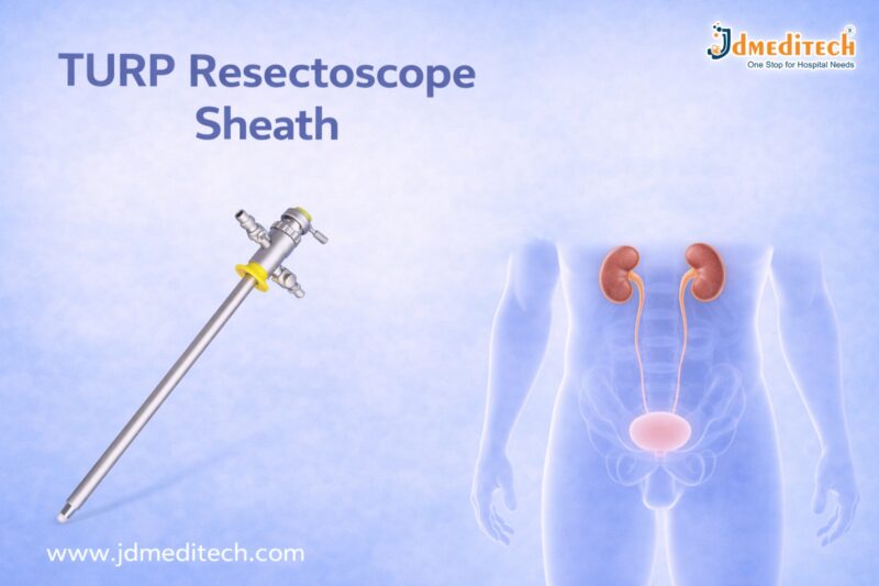
TURP Resectoscope Sheath: Features, Uses & Benefits in TURP Surgery

TURP Resectoscope Sheath: Essential Instrument for Safe and Effective TURP Surgery

Introduction

The TURP resectoscope sheath plays a critical role in modern urological procedures, especially in Transurethral Resection of the Prostate (TURP). As one of the most important components of a resectoscope system, the sheath ensures smooth instrument insertion, continuous irrigation, and enhanced surgical visibility.

In this blog, we will explore everything about the TURP resectoscope sheath, including its features, uses, benefits, and why it is essential for successful prostate surgery.

What is a TURP Resectoscope Sheath?

A TURP resectoscope sheath is a specialized outer tube used during TURP procedures. It houses the resectoscope and allows surgeons to safely access the prostate through the urethra.

The sheath is designed to:

  • Maintain a clear surgical field
  • Enable continuous irrigation
  • Protect surrounding tissues
  • Support precise instrument movement
Key Features of TURP Resectoscope Sheath
1. High-Quality Medical-Grade Material

Most TURP sheaths are made from durable stainless steel or high-grade materials, ensuring longevity and reliability.

2. Continuous Flow System

The sheath allows continuous inflow and outflow of irrigation fluid, which is crucial for clear visibility during surgery.

3. Ergonomic Design

Its user-friendly design ensures easy handling and precise control for surgeons.

4. Compatibility

The TURP resectoscope sheath is compatible with various resectoscope systems and working elements.

5. Smooth Surface Finish

A polished surface minimizes tissue trauma and ensures safe insertion.

Uses of TURP Resectoscope Sheath

The TURP resectoscope sheath is primarily used in:

  • Benign Prostatic Hyperplasia (BPH) treatment
  • Transurethral Resection of the Prostate (TURP)
  • Bladder tumor resections (in some cases)
  • Endoscopic urological procedures
Benefits of Using TURP Resectoscope Sheath
Improved Surgical Visibility

Continuous irrigation helps maintain a clear field, allowing surgeons to perform precise resections.

Enhanced Patient Safety

The sheath protects surrounding tissues and reduces the risk of injury.

Efficient Fluid Management

Controlled inflow and outflow prevent fluid accumulation and complications.

Reduced Procedure Time

Smooth instrument movement improves surgical efficiency.

Types of TURP Resectoscope Sheaths
1. Continuous Flow Sheath

Allows simultaneous inflow and outflow of irrigation fluid.

2. Non-Continuous Flow Sheath

Basic design with limited irrigation control.

3. Different Sizes

Available in various diameters to suit different patient needs and surgical requirements.

How to Choose the Right TURP Resectoscope Sheath

When selecting a TURP resectoscope sheath, consider:

  • Material quality and durability
  • Compatibility with existing equipment
  • Flow mechanism (continuous vs non-continuous)
  • Ease of sterilization
  • Manufacturer reliability
Maintenance and Sterilization

Proper maintenance of the TURP sheath ensures longevity and patient safety:

  • Clean immediately after use
  • Use approved sterilization methods
  • Inspect for damage before reuse
  • Store in a sterile environment
Why Choose JDMeditech TURP Resectoscope Sheath?

At JDMeditech, we provide high-quality TURP resectoscope sheaths designed for precision, durability, and safety.

Our Advantages:
  • Premium-grade materials
  • Advanced design for optimal flow
  • Strict quality control
  • Reliable performance in every procedure
Conclusion

The TURP resectoscope sheath is an indispensable component in urological surgeries. Its role in ensuring clear visibility, efficient fluid management, and patient safety makes it essential for successful TURP procedures.

Choosing a high-quality sheath can significantly enhance surgical outcomes and overall efficiency.


Explore More Product >>

Get Connected:

+91 79909 93062 | +91 63513 72032 | exports@jdmeditech.com
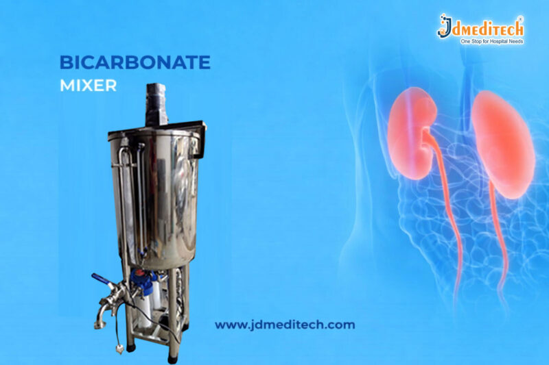


Bicarbonate Mixer: Accurate Dialysis Solution Preparation Equipment

What is a Bicarbonate Mixer?

A bicarbonate mixer is a medical device used to prepare bicarbonate concentrate solutions by mixing bicarbonate powder with purified water. These solutions are then used in dialysis machines to help maintain the patient’s acid-base balance during treatment.

It ensures:

  • Precise concentration levels
  • Safe and hygienic mixing
  • Consistent dialysis outcomes

How Does a Bicarbonate Mixer Work?

The working of a bicarbonate mixer is simple yet highly efficient:

  1. Water Input– Purified water is fed into the mixing chamber
  2. Powder Addition– Measured bicarbonate powder is added
  3. Automated Mixing– The system mixes the solution uniformly
  4. Solution Output– Ready-to-use bicarbonate solution is delivered

Because of automation, the process reduces manual effort and improves accuracy.

Key Features of a Bicarbonate Mixer
1. Automatic Mixing System

The device ensures uniform mixing without manual intervention, which improves efficiency.

2. High Accuracy

It maintains precise concentration levels required for safe dialysis.

3. User-Friendly Interface

Modern bicarbonate mixers come with simple controls and digital displays.

4. Hygienic Design

Made with medical-grade materials, ensuring contamination-free operation.

5. Compact and Durable

Designed for long-term use in busy dialysis centers.

Benefits of Using a Bicarbonate Mixer
Improved Patient Safety

Accurate solution preparation minimizes risks during dialysis treatment.

Time Efficiency

Automation reduces preparation time significantly.

Reduced Human Error

Eliminates manual mixing inconsistencies.

Cost-Effective

Minimizes wastage of bicarbonate powder and water.

Consistent Performance

Delivers reliable results for every dialysis session.

Applications of Bicarbonate Mixer

A bicarbonate mixer is widely used in:

  • Dialysis centers
  • Hospitals
  • Nephrology clinics
  • Medical research facilities

It is especially essential for hemodialysis procedures, where maintaining the correct chemical balance is crucial.

Why is a Bicarbonate Mixer Important in Dialysis?

During dialysis, maintaining the patient’s acid-base balance is vital. The bicarbonate solution acts as a buffer that neutralizes excess acid in the blood.

Without a reliable bicarbonate mixer:

  • Solution inconsistencies may occur
  • Patient safety could be compromised
  • Treatment efficiency may decrease

Therefore, investing in a high-quality bicarbonate mixer is essential for healthcare providers.

Choosing the Right Bicarbonate Mixer

When selecting a bicarbonate mixer, consider:

  • Mixing capacity
  • Accuracy and consistency
  • Ease of operation
  • Cleaning and maintenance
  • Compliance with medical standards

A good mixer improves both workflow and patient outcomes.

Maintenance
Tips for Bicarbonate Mixer

Proper maintenance ensures long-lasting performance:

  • Clean the mixing chamber regularly
  • Use only recommended bicarbonate powder
  • Check for leaks or blockages
  • Follow manufacturer guidelines
  • Schedule periodic servicing
Conclusion

A bicarbonate mixer is an indispensable device in dialysis treatment. It ensures accurate solution preparation, enhances patient safety, and improves overall efficiency in healthcare settings.

By investing in a reliable bicarbonate mixer, hospitals and dialysis centers can deliver safe, effective, and consistent dialysis care.


Explore More Product >>

Get Connected:

+91 79909 93062 | +91 63513 72032 | exports@jdmeditech.com
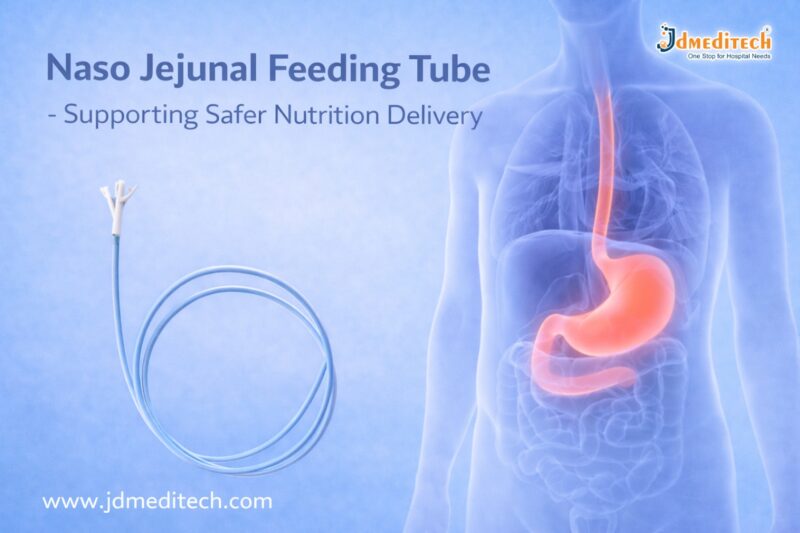


Struggling with Enteral Feeding? Try Naso Jejunal Feeding Tube

What is a Naso Jejunal Feeding Tube?

A naso jejunal feeding tube is a thin, flexible medical device inserted through the nose and guided into the jejunum (the middle section of the small intestine). Unlike nasogastric tubes, which end in the stomach, this tube bypasses the stomach entirely.

As a result, it is ideal for patients who cannot tolerate gastric feeding due to medical conditions such as severe reflux, pancreatitis, or gastric dysfunction.

Why is a Naso Jejunal Feeding Tube Used?

Doctors recommend a naso jejunal feeding tube when patients need long-term or specialized nutritional support. It is commonly used in the following situations:

  • Severe gastrointestinal disorders
  • Pancreatitis requiring minimal pancreatic stimulation
  • High risk of aspiration
  • Delayed gastric emptying (gastroparesis)
  • Post-surgical recovery
  • Critical care and ICU patients

Because it delivers nutrients directly to the intestine, it reduces the risk of vomiting and aspiration.

Key Benefits of Naso Jejunal Feeding Tube

Using a naso jejunal feeding tube offers several clinical advantages:

1. Safe Nutrition Delivery

It significantly reduces the risk of aspiration pneumonia by bypassing the stomach.

2. Improved Tolerance

Patients who cannot tolerate gastric feeding often respond better to jejunal feeding.

3. Effective Nutrient Absorption

The jejunum allows efficient absorption of nutrients, ensuring optimal patient recovery.

4. Minimally Invasive Procedure

The tube placement is non-surgical and can be performed with imaging guidance.

5. Reliable for Critical Care

It provides consistent and controlled nutrition for ICU and high-risk patients.

Procedure for Naso Jejunal Feeding Tube Placement

The placement of a naso jejunal feeding tube is a carefully monitored medical procedure:

  1. The tube is inserted through the patient’s nose
  2. It passes through the esophagus and stomach
  3. It is guided into the jejunum using fluoroscopy or endoscopy
  4. Proper placement is confirmed using imaging techniques

This procedure ensures accurate positioning for safe and effective feeding.

Care and Maintenance Tips

Proper care is essential to ensure the effectiveness of a naso jejunal feeding tube:

  • Flush the tube regularly to prevent blockages
  • Maintain hygiene around the nasal area
  • Monitor for signs of displacement
  • Use prescribed feeding formulas only
  • Follow healthcare provider instructions strictly

Good maintenance helps avoid complications and prolongs the usability of the tube.

Potential Risks and Complications

Although a naso jejunal feeding tube is generally safe, some risks may occur:

  • Tube displacement
  • Blockage or clogging
  • Nasal irritation
  • Diarrhea or intolerance to feeding formula

However, proper monitoring and care can significantly reduce these risks.

Naso Jejunal Feeding Tube vs Nasogastric Tube

Understanding the difference helps in choosing the right feeding method:

Feature Naso Jejunal Feeding Tube Nasogastric Tube
Placement Jejunum (small intestine) Stomach
Aspiration Risk Low Higher
Feeding Tolerance Better Moderate
Use Case Critical patients General feeding
Conclusion

The naso jejunal feeding tube is a vital medical device that ensures safe, effective, and reliable nutrition delivery for patients who cannot eat normally. Its ability to bypass the stomach makes it particularly useful in critical care and complex medical conditions.

With proper placement, care, and monitoring, this feeding solution significantly improves patient outcomes and recovery.


Explore More Product >>

+91 79909 93062 | +91 63513 72032 | exports@jdmeditech.com

 

Safe and Effective PTCA Balloon Catheter for Cardiology Experts

What is a PTCA Balloon Catheter?

A PTCA Balloon Catheter is a minimally invasive medical device used to open narrowed coronary arteries caused by plaque buildup. It consists of a small balloon attached to the tip of a catheter.

During the procedure:

  • The catheter is inserted into a blood vessel
  • Guided to the blocked coronary artery
  • The balloon is inflated to compress plaque
  • Blood flow is restored efficiently

This procedure is commonly known as angioplasty.

Why PTCA Balloon Catheters Are Essential in Cardiology

Cardiologists rely on PTCA balloon catheters because they offer:

  • Minimally invasive treatment
  • Quick restoration of blood flow
  • Reduced need for open-heart surgery
  • Faster patient recovery time

As a result, these catheters have become a cornerstone in treating coronary artery disease (CAD).

Key Features of Safe and Effective PTCA Balloon Catheters
1. High-Pressure Balloon Capability

Modern PTCA balloon catheters can withstand high pressure, allowing precise dilation of hardened plaques without damaging the artery.

2. Smooth Navigation

These catheters are designed for easy navigation through complex vascular pathways, ensuring accurate placement.

3. Excellent Crossability

They can cross tight lesions efficiently, making them ideal for challenging cases.

4. Controlled Balloon Inflation

Precise inflation and deflation provide better control, reducing procedural risks.

5. Biocompatible Materials

Manufactured using advanced materials, they minimize the risk of adverse reactions.

Types of PTCA Balloon Catheters
1. Semi-Compliant Balloons
  • Flexible and adaptable
  • Suitable for general angioplasty procedures
2. Non-Compliant Balloons
  • Maintain shape under high pressure
  • Ideal for precise dilation and post-stent expansion
3. Cutting Balloons
  • Equipped with micro blades
  • Used for resistant or calcified lesions
4. Drug-Coated Balloons
  • Release medication to prevent restenosis
  • Improve long-term outcomes
Benefits of Using PTCA Balloon Catheters

Using a safe and effective PTCA balloon catheter offers multiple benefits:

  • ✔ Minimally invasive procedure
  • ✔ Reduced hospital stay
  • ✔ Lower complication rates
  • ✔ Faster recovery
  • ✔ Improved patient comfort
  • ✔ High procedural success rate
Safety Considerations in PTCA Procedures

While PTCA is generally safe, proper technique and equipment selection are essential.

Important Safety Measures:
  • Use appropriate balloon size
  • Monitor inflation pressure carefully
  • Ensure proper imaging guidance
  • Follow sterilization protocols
  • Choose high-quality, certified devices

By adhering to these practices, cardiology experts can significantly reduce risks.

Applications of PTCA Balloon Catheters

PTCA balloon catheters are widely used in:

  • Coronary artery disease treatment
  • Pre-stent dilation
  • Post-stent expansion
  • Treatment of restenosis
  • Peripheral angioplasty procedures
How to Choose the Right PTCA Balloon Catheter

Selecting the right catheter is crucial for optimal results.

Consider the following factors:
  • Lesion type and severity
  • Vessel size
  • Balloon compliance type
  • Trackability and flexibility
  • Manufacturer quality and certification

Choosing the right device ensures both safety and effectiveness.

Future Innovations in PTCA Balloon Technology

Advancements in medical technology continue to improve PTCA outcomes.

Emerging trends include:

  • Drug-eluting balloon technologies
  • Enhanced catheter flexibility
  • Improved imaging compatibility
  • AI-assisted procedural planning

These innovations are shaping the future of interventional cardiology.

Conclusion

The PTCA Balloon Catheter remains an indispensable tool for cardiology experts. Its ability to safely and effectively restore blood flow makes it a preferred choice in modern cardiac care.

With continuous advancements in design and technology, PTCA balloon catheters are becoming even more efficient, ensuring better patient outcomes and procedural success.


Explore More Product >>

Get Connected:

+91 79909 93062 | +91 63513 72032 | exports@jdmeditech.com
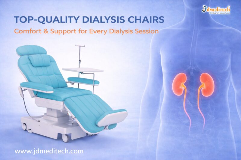
 

Top-Quality Dialysis Chairs – Comfort & Support for Every Dialysis Session

Introduction

Dialysis is a life-saving procedure for patients with kidney failure. However, long treatment sessions can be physically and mentally exhausting. This is where top-quality dialysis chairs play a crucial role. These specialized chairs are designed to provide maximum comfort, safety, and support throughout every dialysis session.

In this blog, we will explore the importance, features, benefits, and selection criteria for dialysis chairs, helping healthcare providers make informed decisions.

What Are Top-Quality Dialysis Chairs?

Top-quality dialysis chairs are ergonomically designed medical chairs used during dialysis treatments. Patients often remain seated for several hours, so these chairs are built to ensure comfort, proper posture, and easy accessibility for healthcare professionals.

They are commonly used in:

  • Hospitals
  • Dialysis centers
  • Specialty clinics
Why Comfort Matters in Dialysis Sessions

Dialysis sessions typically last 3–5 hours. During this time, patients must remain still. Poor seating can lead to:

  • Back pain and discomfort
  • Poor blood circulation
  • Increased anxiety or restlessness

By using top-quality dialysis chairs, healthcare facilities can significantly enhance patient comfort and overall treatment experience.

Key Features of Top-Quality Dialysis Chairs
1. Ergonomic Design for Patient Comfort

Modern dialysis chairs are designed to support the natural body posture. Adjustable backrests, armrests, and footrests ensure optimal comfort.

2. Electric Adjustment Mechanism

High-end chairs come with motorized controls that allow smooth adjustments. This helps both patients and caregivers.

3. Durable and Hygienic Materials

These chairs are made with easy-to-clean, antimicrobial upholstery, ensuring hygiene and infection control.

4. Safety Features

Top-quality dialysis chairs include:

  • Lockable wheels
  • Stable base structure
  • Emergency positioning
5. Accessibility for Medical Procedures

The chair design allows easy access for healthcare professionals during dialysis procedures.

Benefits of Using Top-Quality Dialysis Chairs
Enhanced Patient Comfort

Patients experience less fatigue during long sessions, improving their overall well-being.

Improved Clinical Efficiency

Adjustable features help medical staff perform procedures more efficiently.

Better Patient Compliance

Comfortable patients are more likely to complete their sessions without interruption.

Increased Safety

Advanced safety mechanisms reduce the risk of accidents.

How to Choose the Best Dialysis Chair

Selecting the right dialysis chair is essential for both patient satisfaction and clinical performance.

Consider Patient Comfort

Look for chairs with cushioning, adjustability, and ergonomic support.

Evaluate Build Quality

Choose durable materials that withstand frequent use.

Check for Advanced Features

Electric controls, memory positions, and safety locks are important.

Ensure Easy Maintenance

Hygienic surfaces and easy cleaning are critical in healthcare settings.

Applications of Dialysis Chairs in Healthcare

Top-quality dialysis chairs are widely used in:

  • Hemodialysis units
  • Oncology treatment centers
  • Blood transfusion units
  • Daycare surgical centers

Their versatility makes them a valuable asset in modern healthcare facilities.

Maintenance Tips for Long-Term Use

To ensure longevity and performance:

  • Clean the chair regularly with approved disinfectants
  • Inspect mechanical and electrical components
  • Perform routine servicing
  • Train staff on proper usage
Why Choose JDMeditech Dialysis Chairs?

At JDMeditech, we provide top-quality dialysis chairs designed to meet international healthcare standards. Our chairs offer:

  • Superior comfort and ergonomic design
  • Advanced safety features
  • Durable and hygienic materials
  • Reliable performance for long-term use
Conclusion

Investing in top-quality dialysis chairs is essential for improving patient comfort, safety, and treatment efficiency. With the right chair, healthcare providers can deliver better care while enhancing operational performance.

If you are looking for reliable and high-performance dialysis chairs, JDMeditech offers solutions tailored to your needs.


Explore More Product >>

Get Connected:

+91 79909 93062 | +91 63513 72032 | exports@jdmeditech.com
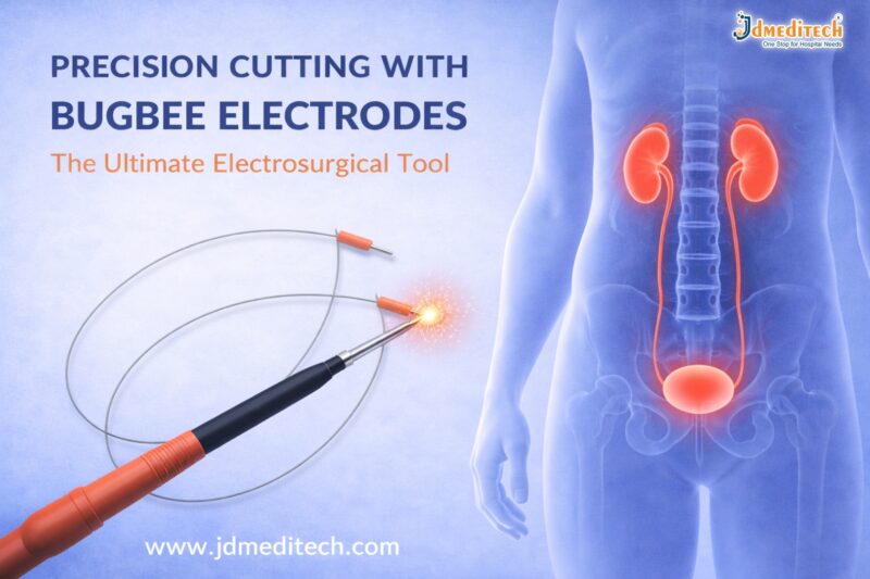


Precision Cutting with Bugbee Electrodes – Ultimate Electrosurgical Tool

Precision Cutting with Bugbee Electrodes – The Ultimate Electrosurgical Tool

Introduction

Precision cutting with Bugbee electrodes has transformed modern electrosurgical procedures. Surgeons rely on this advanced tool for accurate tissue cutting and effective coagulation, especially in urology and endoscopy.

Bugbee electrodes provide excellent control, minimal tissue damage, and improved surgical outcomes. As a result, they have become an essential instrument in operating rooms worldwide.

What Are Bugbee Electrodes?

Bugbee electrodes are flexible, monopolar electrosurgical instruments designed for precise cutting and coagulation of soft tissues. They are commonly used in minimally invasive procedures, particularly in urology.

These electrodes consist of a thin wire tip that delivers controlled electrical energy. This energy allows surgeons to cut tissue while simultaneously controlling bleeding.

Why Precision Cutting with Bugbee Electrodes Matters

Precision cutting with Bugbee electrodes ensures clean incisions and reduces unnecessary tissue damage. This improves patient recovery and minimizes complications.

Key Benefits:
  • High precision during surgery
  • Minimal thermal spread
  • Reduced bleeding
  • Enhanced visibility in the surgical field
  • Faster healing time

Because of these advantages, surgeons prefer Bugbee electrodes for delicate procedures.

Applications of Bugbee Electrodes in Medical Procedures

Bugbee electrodes are widely used across various medical specialties. However, they are most popular in urology and endoscopic surgeries.

1. Urological Procedures

Surgeons use Bugbee electrodes during cystoscopy to remove small tumors, coagulate bleeding areas, and treat bladder conditions.

2. Endoscopic Surgeries

These electrodes allow precise cutting in minimally invasive procedures, reducing trauma to surrounding tissues.

3. Tumor Resection

Bugbee electrodes help in removing small growths with accuracy while maintaining hemostasis.

4. Coagulation of Bleeding Vessels

They effectively control bleeding by coagulating blood vessels during surgery.

How Bugbee Electrodes Work

Bugbee electrodes operate using monopolar electrosurgical energy. The electrical current passes through the electrode tip to the targeted tissue.

This process creates heat, which enables:

  • Cutting of tissue
  • Coagulation of blood vessels
  • Controlled surgical precision

The surgeon can adjust the energy level depending on the procedure, ensuring safe and effective outcomes.

Advantages of Using Bugbee Electrodes

Precision cutting with Bugbee electrodes offers several advantages over traditional surgical tools.

Improved Surgical Accuracy

The fine tip allows surgeons to perform delicate procedures with maximum control.

Reduced Tissue Damage

Minimal thermal spread protects surrounding tissues.

Enhanced Efficiency

Surgeries become quicker due to simultaneous cutting and coagulation.

Cost-Effective Solution

Reusable options make Bugbee electrodes a budget-friendly choice for hospitals.

Safety Considerations

Although Bugbee electrodes are highly effective, proper usage is essential for safety.

  • Always use appropriate power settings
  • Ensure proper grounding during monopolar procedures
  • Regularly inspect the electrode for damage
  • Follow sterilization protocols

By following these guidelines, healthcare professionals can ensure safe and successful procedures.

Choosing the Right Bugbee Electrode

Selecting the correct Bugbee electrode depends on the surgical requirement.

Factors to Consider:
  • Electrode length and flexibility
  • Tip size and shape
  • Compatibility with electrosurgical units
  • Reusable or disposable type

Choosing the right instrument enhances both precision and safety.

Future of Electrosurgical Tools

The demand for precision cutting with Bugbee electrodes continues to grow. Innovations in electrosurgical technology are improving efficiency, safety, and ease of use.

Modern designs now focus on better insulation, enhanced flexibility, and improved energy control. These advancements will further improve surgical outcomes in the future.

Conclusion

Precision cutting with Bugbee electrodes has become a cornerstone of modern electrosurgery. Their ability to deliver accurate cuts with minimal tissue damage makes them the ultimate electrosurgical tool.

From urology to endoscopic procedures, Bugbee electrodes provide unmatched performance and reliability. As technology advances, their role in surgery will only continue to expand.


Explore More Product >>

Get Connected:

+91 79909 93062 | +91 63513 72032 | exports@jdmeditech.com
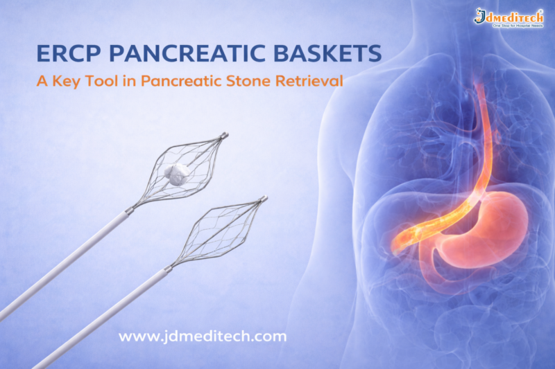


ERCP Pancreatic Baskets: Uses, Types, Procedure & Benefits

What Are ERCP Pancreatic Baskets?

ERCP pancreatic baskets are small wire devices. Doctors use them to catch and remove stones from the pancreatic duct.

They pass through an endoscope during the ERCP procedure. These baskets are strong, flexible, and easy to control.

Key Features:
  • Flexible design for narrow ducts
  • Strong wires for firm grip
  • Smooth opening and closing system
  • Available in different sizes
Why Are ERCP Pancreatic Baskets Used?

Pancreatic stones can block the duct. Because of this, patients may feel severe pain. In some cases, it can lead to pancreatitis.

ERCP pancreatic baskets help to:
  • Remove stones safely
  • Improve enzyme flow
  • Reduce pain quickly
  • Prevent further complications
Types of ERCP Pancreatic Baskets

Different baskets are used for different cases. Therefore, doctors choose them based on the situation.

1. Four-Wire Basket

This is the most common type. It works well for medium-sized stones.

2. Eight-Wire Basket

It offers a stronger grip. So, it is useful for multiple or uneven stones.

3. Helical Basket

This basket moves easily in curved ducts. Hence, it is ideal for complex cases.

4. Nitinol (Memory) Basket

This basket keeps its shape. As a result, it works well even after repeated use.

ERCP Procedure Using Pancreatic Baskets

The procedure is simple and minimally invasive.

Steps:
  1. The doctor inserts an endoscope through the mouth
  2. The pancreatic duct is located
  3. A dye is used to view the duct clearly
  4. The basket is passed into the duct
  5. The stone is captured
  6. The basket is pulled out with the stone

This process is quick and effective.

Benefits of ERCP Pancreatic Baskets

These devices offer many benefits.

1. Minimally Invasive

No major surgery is needed. Therefore, recovery is faster.

2. High Success Rate

Most stones can be removed easily.

3. Better Control

Doctors can handle stones with precision.

4. Fewer Complications

There is less risk compared to open surgery.

5. Cost-Effective

Short hospital stays reduce overall cost.

Applications in Medical Practice

Doctors use ERCP pancreatic baskets in many situations.

  • Pancreatic stone removal
  • Chronic pancreatitis treatment
  • ERCP procedures
  • Endoscopic stone extraction
Risks and Considerations

The procedure is safe. However, some risks may occur.

Possible Risks:
  • Pancreatitis
  • Infection
  • Minor duct injury
  • Rare basket trapping

Even so, trained doctors can manage these risks well.

How to Choose the Right Basket

Choosing the right basket is important.

Doctors consider:

  • Stone size
  • Stone shape
  • Duct structure
  • Material type

Therefore, high-quality baskets give better results.

Future Developments

Medical tools are improving every year. As a result, ERCP baskets are becoming more advanced.

New Improvements:
  • Better flexibility
  • Stronger materials
  • Single-use options
  • Easy handling design

These changes help improve safety and performance.

Conclusion

ERCP Pancreatic Baskets are key tools for removing pancreatic stones. They are safe, effective, and easy to use.

Because they are minimally invasive, patients recover faster. In addition, new designs continue to improve results. Therefore, these baskets remain a top choice in modern ERCP procedures.


Explore More Product >>

Get Connected:

+91 79909 93062 | +91 63513 72032 | exports@jdmeditech.com
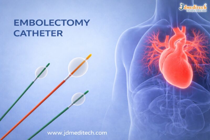
 

Embolectomy Catheter – Uses, Procedure & Benefits Explained

An embolectomy catheter is a medical device used to remove blood clots from blood vessels. These clots can block blood flow and cause serious health problems. Therefore, doctors must remove them quickly.

In many cases, this device helps restore circulation within minutes. As a result, it reduces the risk of stroke, tissue damage, and organ failure.

Today, the embolectomy catheter is widely used in cardiovascular procedures. It offers a fast, safe, and minimally invasive solution.

What is an Embolectomy Catheter?

An embolectomy catheter is a thin and flexible tube. Doctors insert it into a blocked artery or vein.

At the tip, there is a small balloon. First, the catheter reaches the clot. Then, the balloon inflates. After that, the doctor pulls the catheter back. As a result, the clot is removed from the vessel.

Because of this simple method, blood flow returns quickly.

Why is an Embolectomy Catheter Used?

Blood clots can be dangerous. In some cases, they can be life-threatening. Therefore, quick treatment is important.

Doctors use an embolectomy catheter in the following conditions:

  • Acute arterial blockage
  • Deep vein thrombosis (DVT)
  • Pulmonary embolism
  • Stroke-related clots
  • Peripheral artery disease

In addition, this device is useful in emergency situations. It helps save time and improves survival.

Types of Embolectomy Catheters

There are different types of embolectomy catheter devices. Each type serves a specific purpose.

Balloon Embolectomy Catheter

This is the most common type. It uses a balloon to capture and remove the clot.

Fogarty Catheter

Doctors often use this catheter in vascular surgery. It is reliable and easy to use.

Aspiration Catheter

This type removes clots using suction instead of a balloon.

Mechanical Devices

These devices break the clot into small pieces. Then, they remove it from the vessel.

Embolectomy Procedure

The procedure is simple and quick. Moreover, it is usually safe.

Step 1: Preparation

First, the patient receives anesthesia.

Step 2: Insertion

Next, the doctor inserts the embolectomy catheter into the blood vessel.

Step 3: Positioning

Then, imaging helps guide the catheter to the clot.

Step 4: Removal

After that, the balloon inflates and captures the clot. The doctor removes it carefully.

Step 5: Completion

Finally, blood flow returns to normal.

Benefits of Embolectomy Catheter

The embolectomy catheter offers many benefits.

  • Fast clot removal
  • Minimally invasive procedure
  • Quick recovery time
  • Lower risk of complications
  • High success rate

Most importantly, it helps prevent serious damage to tissues and organs.

Risks and Complications

Like any procedure, there are some risks. However, they are usually rare.

  • Bleeding
  • Infection
  • Vessel injury
  • Partial clot removal

Even so, experienced doctors manage these risks effectively.

Role in Cardiovascular Procedures

The embolectomy catheter plays a key role in modern medicine.

Doctors use it in:

  • Emergency vascular treatment
  • Stroke management
  • Cardiac procedures
  • Peripheral artery care

Therefore, it is an important tool in saving lives.

How to Choose the Right Embolectomy Catheter

Choosing the correct device is important. Doctors consider several factors.

  • Size of the vessel
  • Location of the clot
  • Type of blockage
  • Patient condition

As a result, the right choice improves treatment success.

Future of Embolectomy Catheters

Medical technology is improving every year. Therefore, newer devices are more advanced.

For example:

  • Better flexibility
  • Improved precision
  • Smart balloon systems
  • Combined clot removal techniques

In the future, these devices will become even safer and more effective.

Conclusion

The embolectomy catheter is a powerful tool for clot removal. It helps restore blood flow quickly and safely.

Because it is minimally invasive, patients recover faster. In addition, it reduces the risk of serious complications.

Overall, this device plays a vital role in cardiovascular procedures.


Explore More Product >>

Get Connected:

+91 79909 93062 | +91 63513 72032 | exports@jdmeditech.com
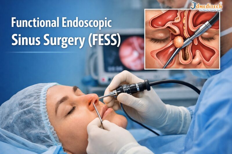
Functional Endoscopic Sinus Surgery (FESS)

Functional Endoscopic Sinus Surgery (FESS) is a minimally invasive surgical procedure used to treat chronic sinus problems. It helps restore normal drainage of the sinuses and improves breathing.

Today, FESS is one of the most advanced and commonly performed procedures for patients suffering from chronic sinusitis, nasal blockages, and sinus infections.

In this blog, we will explain everything about Functional Endoscopic Sinus Surgery, including its procedure, benefits, risks, and recovery.

What is Functional Endoscopic Sinus Surgery (FESS)?

Functional Endoscopic Sinus Surgery (FESS) is a modern surgical technique that uses a thin, flexible endoscope inserted through the nose to treat sinus issues.

Unlike traditional surgery, FESS:

  • Does not require external cuts
  • Is less painful
  • Offers faster recovery

This procedure focuses on restoring the natural function of the sinuses rather than removing large amounts of tissue.

Why is FESS Needed?

Doctors recommend Functional Endoscopic Sinus Surgery when medications fail to treat sinus conditions.

Common Conditions Treated:
  • Chronic sinusitis
  • Nasal polyps
  • Deviated nasal septum (in some cases)
  • Sinus infections
  • Blocked sinus drainage pathways
  • Fungal sinusitis

If you experience persistent nasal congestion, facial pain, or breathing difficulty, FESS may be the right solution.

Functional Endoscopic Sinus Surgery Procedure

The FESS procedure is safe and usually performed under general anesthesia.

Step-by-Step Procedure:
1. Preparation

The patient is given anesthesia to ensure comfort.

2. Endoscope Insertion

A small camera (endoscope) is inserted through the nostrils.

3. Identifying Blockages

The surgeon carefully examines sinus passages.

4. Removing Obstructions

Blocked tissues, polyps, or bone are removed to open sinus pathways.

5. Restoring Drainage

Normal airflow and sinus drainage are restored.

The entire procedure typically takes 1 to 3 hours, depending on the condition.

Benefits of Functional Endoscopic Sinus Surgery

FESS offers several advantages compared to traditional sinus surgery.

Key Benefits:
  • Minimally invasive procedure
  • No external scars
  • Faster recovery time
  • Reduced pain and bleeding
  • Improved breathing
  • Long-term relief from sinus symptoms

As a result, patients often experience a significant improvement in quality of life.

Risks and Complications of FESS

Although Functional Endoscopic Sinus Surgery is generally safe, some risks may occur.

Possible Risks:
  • Bleeding
  • Infection
  • Recurrence of sinus problems
  • Damage to surrounding structures (rare)
  • Vision-related complications (very rare)

However, choosing an experienced surgeon greatly reduces these risks.

Recovery After Functional Endoscopic Sinus Surgery

Recovery from FESS is usually quick and smooth.

What to Expect:
  • Mild discomfort for a few days
  • Nasal congestion or slight bleeding
  • Return to normal activities within 1–2 weeks
Post-Surgery Care Tips:
  • Avoid blowing your nose forcefully
  • Use prescribed nasal sprays
  • Attend follow-up appointments
  • Keep your nasal passages clean

Following proper care ensures faster healing and better results.

Who is a Good Candidate for FESS?

You may be a suitable candidate for Functional Endoscopic Sinus Surgery if:

  • You have chronic sinusitis lasting more than 12 weeks
  • Medications are not effective
  • You have nasal polyps or structural blockage
  • You frequently experience sinus infections

Consult an ENT specialist for proper evaluation.

Conclusion

Functional Endoscopic Sinus Surgery (FESS) is a safe, effective, and minimally invasive solution for chronic sinus problems. It helps restore normal sinus function, improves breathing, and enhances overall quality of life.

If you are struggling with long-term sinus issues, consult your doctor to see if FESS is the right option for you.

Explore More Product >>


Get Connected:

+91 79909 93062 | +91 63513 72032 | exports@jdmeditech.com

Pathology: Types, Importance & Role in Disease Diagnosis

Pathology is a vital branch of medical science that focuses on the study of diseases. It helps doctors understand the causes, development, and effects of various illnesses. Without pathology, accurate diagnosis and effective treatment would not be possible.

In modern healthcare, pathology plays a crucial role in detecting diseases early. As a result, it improves patient outcomes and supports life-saving treatments.

What is Pathology?

Pathology is the scientific study of disease through the examination of organs, tissues, blood, and bodily fluids. Pathologists analyze samples to identify abnormalities and determine the exact nature of a disease.

In simple terms, pathology answers key questions like:

  • What disease is present?
  • How severe is it?
  • What is causing it?

Therefore, it acts as the foundation of medical diagnosis.

Types of Pathology

Pathology is divided into several specialized branches. Each type focuses on a different aspect of disease analysis.

1. Clinical Pathology

Clinical pathology involves the analysis of blood, urine, and other body fluids. It includes:

  • Hematology (study of blood)
  • Microbiology (study of infections)
  • Biochemistry (study of chemical processes in the body)
2. Anatomical Pathology

Anatomical pathology focuses on examining tissues and organs. It includes:

  • Histopathology (study of tissues under a microscope)
  • Cytopathology (study of individual cells)
3. Molecular Pathology

Molecular pathology studies diseases at a genetic and molecular level. It is widely used in cancer diagnosis and personalized medicine.

4. Forensic Pathology

Forensic pathology determines the cause of death, especially in cases of accidents or criminal investigations.

Importance of Pathology in Healthcare

Pathology is essential in every stage of healthcare. It not only helps in diagnosis but also guides treatment decisions.

1. Accurate Disease Diagnosis

Pathology provides precise information about diseases. As a result, doctors can make correct diagnoses and avoid unnecessary treatments.

2. Early Detection of Diseases

Many diseases, including cancer, can be detected early through pathology tests. Early detection significantly improves survival rates.

3. Treatment Planning

Pathology reports help doctors choose the best treatment options. For example, cancer treatments often depend on biopsy results.

4. Monitoring Disease Progress

Pathology tests are used to track how a disease is progressing or responding to treatment.

Common Pathology Tests

Pathology laboratories perform a wide range of tests. Some of the most common include:

  • Blood tests (CBC, glucose levels)
  • Urine analysis
  • Biopsy tests
  • Pap smear tests
  • Culture tests for infections

These tests provide essential insights into a patient’s health.

Role of Pathologists

Pathologists are highly trained medical professionals who specialize in diagnosing diseases. Although they usually work behind the scenes, their role is critical.

They:

  • Analyze lab results
  • Interpret biopsy samples
  • Provide diagnostic reports
  • Support clinicians in treatment decisions

Therefore, pathologists are key contributors to patient care.

How Pathology Supports Modern Medicine

Pathology is the backbone of modern medicine. It supports advanced medical practices such as:

  • Cancer diagnosis and staging
  • Genetic testing and personalized medicine
  • Infectious disease detection
  • Organ transplant compatibility testing

As technology advances, pathology continues to evolve with digital pathology and AI-based diagnostics.

Future of Pathology

The future of pathology is rapidly advancing. New technologies are improving accuracy and efficiency.

Some key trends include:

  • Digital pathology systems
  • Artificial intelligence in diagnostics
  • Automated laboratory testing
  • Precision medicine

These innovations will make diagnosis faster and more reliable.

Conclusion

Pathology is a fundamental part of healthcare that ensures accurate diagnosis and effective treatment. From routine blood tests to advanced molecular analysis, pathology provides critical insights into diseases.

As healthcare continues to evolve, pathology will remain at the center of medical innovation and patient care.

Explore More Product >>


Get Connected:

+91 79909 93062 | +91 63513 72032 | exports@jdmeditech.com
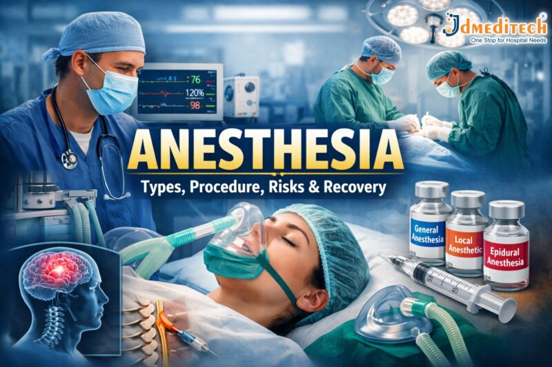


Anesthesia: Types, Procedure, Risks, Benefits & Recovery Guide

Anaesthesia is a medical treatment that prevents pain during surgery or medical procedures. It works by blocking nerve signals in the body or inducing a temporary loss of sensation or consciousness.

A trained specialist called an anaesthesiologist administers anaesthesia and monitors the patient throughout the procedure.

Types of Anaesthesia

Different types of anaesthesia are used depending on the procedure, patient condition, and duration of surgery.

1. General Anaesthesia

General anaesthesia makes the patient completely unconscious.

Used for:

  • Major surgeries (heart surgery, brain surgery)
  • Long-duration procedures

Effects:

  • No awareness
  • No pain sensation
  • Temporary loss of reflexes
2. Regional Anaesthesia

Regional anaesthesia blocks pain in a specific part of the body.

Examples:

  • Spinal anaesthesia
  • Epidural anaesthesia

Used for:

  • Childbirth (C-section)
  • Orthopedic surgeries
3. Local Anaesthesia

Local anaesthesia numbs a small area of the body.

Used for:

  • Dental procedures
  • Minor surgeries

Benefits:

  • Patient remains awake
  • Quick recovery
4. Sedation (Conscious Sedation)

Sedation helps patients relax while remaining partially awake.

Used for:

  • Endoscopy
  • Minor procedures
How Does Anaesthesia Work?

Anaesthesia works by interrupting nerve signals in the brain and body. As a result, the patient does not feel pain during the procedure.

There are three main mechanisms:

  • Blocking pain signals
  • Inducing unconsciousness
  • Relaxing muscles
Anaesthesia Procedure: Step-by-Step
1. Pre-Anaesthesia Evaluation

Before surgery, the anaesthesiologist:

  • Reviews medical history
  • Checks allergies
  • Evaluates vital signs
2. Administration of Anaesthesia

Anaesthesia is given through:

  • Injection
  • Inhalation (gas)
  • Intravenous (IV)
3. Monitoring During Surgery

Doctors continuously monitor:

  • Heart rate
  • Blood pressure
  • Oxygen levels
4. Recovery Phase

After surgery, the patient is moved to a recovery room where medical staff monitor vital signs until the effects wear off.

Benefits of Anaesthesia

Anaesthesia offers several advantages:

  • Pain-free procedures
  • Improved surgical precision
  • Reduced anxiety
  • Controlled muscle relaxation
  • Safer complex surgeries
Risks and Side Effects of Anaesthesia

Although anaesthesia is generally safe, some risks may occur.

Common Side Effects
  • Nausea and vomiting
  • Drowsiness
  • Sore throat
  • Dizziness
Rare Risks
  • Allergic reactions
  • Breathing problems
  • Nerve damage (very rare)

However, modern techniques and monitoring systems significantly reduce these risks.

Recovery After Anaesthesia

Recovery depends on the type of anaesthesia used.

General Recovery Tips:
  • Follow doctor’s instructions
  • Avoid driving for 24 hours
  • Stay hydrated
  • Rest properly

Most patients recover quickly, especially after local or regional anesthesia.

Who Administers Anaesthesia?

Anaesthesia is administered by trained professionals such as:

  • Anaesthesiologists
  • Nurse anaesthetists
  • Anaesthesia assistants

They ensure patient safety before, during, and after the procedure.

When is Anaesthesia Required?

Anaesthesia is used in various medical procedures, including:

  • Surgical operations
  • Dental treatments
  • Diagnostic procedures
  • Cosmetic surgeries
Conclusion

Anaesthesia is a cornerstone of modern healthcare, making surgeries safe, painless, and effective. With advancements in medical technology, anaesthesia has become more precise and safer than ever before.

If you are scheduled for a procedure, understanding anaesthesia can help reduce anxiety and prepare you for a smooth experience.

Explore More Product >>


Get Connected:

+91 79909 93062 | +91 63513 72032 | exports@jdmeditech.com
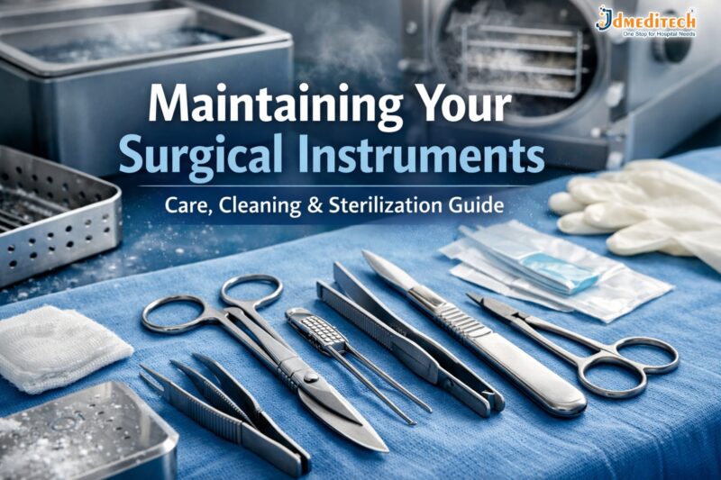
 

Maintaining Your Surgical Instruments

Surgical instruments are the backbone of any healthcare facility. Therefore, maintaining your surgical instruments is essential for ensuring patient safety, improving surgical outcomes, and extending the lifespan of expensive tools.

Proper care not only reduces the risk of infection but also prevents instrument damage. In this guide, you will learn the best practices, step-by-step cleaning methods, and expert tips to maintain surgical instruments effectively.

Why Maintaining Your Surgical Instruments is Important

First and foremost, well-maintained instruments ensure precision during surgery. On the other hand, poorly maintained tools can lead to complications.

Key Benefits:
  • Enhances patient safety
  • Prevents surgical site infections (SSIs)
  • Increases instrument lifespan
  • Reduces replacement costs
  • Ensures compliance with healthcare standards
Types of Surgical Instruments That Require Maintenance

Different instruments need different care methods. Therefore, understanding categories is crucial.

1. Cutting Instruments
  • Scalpels
  • Scissors
  • Bone cutters
2. Grasping Instruments
  • Forceps
  • Clamps
3. Retracting Instruments
  • Surgical retractors
4. Specialized Instruments
  • Endoscopic tools
  • Microsurgical instruments
Step-by-Step Guide to Maintaining Surgical Instruments
1. Immediate Cleaning After Use

After surgery, instruments should be cleaned immediately. This step prevents blood and tissue from drying.

Tips:

  • Use lukewarm water
  • Avoid saline (it causes corrosion)
  • Wipe visible debris
2. Proper Cleaning Techniques

Cleaning is the most critical step. Therefore, it must be done carefully.

Manual Cleaning
  • Use a soft brush
  • Clean joints and hinges thoroughly
  • Use neutral pH detergents
Ultrasonic Cleaning
  • Removes microscopic debris
  • Ideal for complex instruments
  • Saves time and effort
3. Rinsing and Drying

After cleaning, rinse instruments with distilled water. Then, dry them completely.

Why this matters:
  • Prevents rust formation
  • Avoids water stains
4. Inspection and Function Testing

Next, inspect each instrument carefully.

Check for:
  • Cracks or corrosion
  • Loose joints
  • Sharpness of cutting tools

Replace or repair damaged instruments immediately.

5. Lubrication

Lubrication ensures smooth movement.

Best practices:
  • Use water-based lubricants
  • Apply to hinges and joints
  • Avoid oil-based products
6. Sterilization

Sterilization is essential to eliminate microorganisms.

Common Methods:
  • Autoclaving (steam sterilization)
  • Ethylene oxide (ETO)
  • Plasma sterilization

Always follow manufacturer guidelines for best results.

7. Proper Storage

Finally, store instruments correctly.

Storage tips:
  • Use dry, clean environments
  • Avoid overcrowding
  • Use protective trays
Common Mistakes to Avoid

Even small errors can damage instruments. Therefore, avoid these common mistakes:

  • Delaying cleaning after surgery
  • Using harsh chemicals
  • Skipping inspection
  • Improper sterilization techniques
  • Storing instruments while wet
Best Practices for Long-Term Maintenance

To maintain surgical instruments effectively, consistency is key.

Follow These Practices:
  • Train staff regularly
  • Use instrument tracking systems
  • Follow manufacturer instructions
  • Schedule routine maintenance checks
How Often Should You Maintain Surgical Instruments?

Maintenance should be a continuous process.

  • After every use: Cleaning and sterilization
  • Daily: Inspection and drying
  • Weekly: Deep cleaning and lubrication
  • Monthly: Detailed inspection and servicing
Latest Innovations in Instrument Maintenance

Modern technology has improved maintenance processes significantly.

New Trends:
  • Automated washer-disinfectors
  • Smart tracking systems
  • Anti-corrosion coatings
  • AI-based instrument monitoring

These innovations help reduce human error and improve efficiency.

Conclusion

Maintaining your surgical instruments is not just a routine task—it is a critical responsibility. Proper cleaning, sterilization, and storage ensure safety, efficiency, and cost savings.

By following the steps and best practices in this guide, healthcare facilities can significantly improve surgical outcomes while extending the life of their instruments.

Explore More Product >>


Get Connected:

+91 79909 93062 | +91 63513 72032 | exports@jdmeditech.com