
Biliary Stenting: Fast Relief for Blocked Bile Ducts

Biliary stenting is a modern medical procedure that helps restore normal bile flow when the bile duct becomes blocked or narrowed. The biliary system plays an important role in digestion, as it carries bile from the liver to the small intestine. When this flow is interrupted, patients may develop symptoms such as jaundice, abdominal discomfort, and infection.

This is where biliary stenting becomes essential. Doctors place a small tube, called a stent, inside the bile duct to keep it open. As a result, bile can flow normally again. Because the procedure is minimally invasive, patients usually recover quickly and avoid major surgery.

Understanding the Biliary System
What is the Biliary System?

The biliary system includes the liver, gallbladder, and bile ducts. Together, these organs produce, store, and transport bile. This digestive fluid helps break down fats and supports proper digestion.

Why Does Bile Flow Get Blocked?

Several conditions can block or narrow the bile ducts. The most common causes include:

  • Gallstones
  • Tumors in the pancreas or bile duct
  • Benign or malignant strictures
  • Infections or inflammation

When any of these conditions occur, bile cannot flow properly. Therefore, timely treatment such as biliary stenting becomes necessary.

What is Biliary Stenting?
Definition and Purpose

Biliary stenting is an endoscopic technique used to open blocked bile ducts. The main purpose is to restore the natural flow of bile and reduce pressure within the biliary system.

Types of Biliary Stents

Doctors choose the type of stent based on the patient’s condition:

  • Plastic stents are suitable for short-term use
  • Metal stents are more durable and ideal for long-term placement

Each type offers specific benefits, depending on the severity and cause of the blockage.

Indications for Biliary Stenting
When is Biliary Stenting Required?

Doctors recommend biliary stenting in several situations, including:

  • Obstructive jaundice
  • Bile duct blockage caused by cancer
  • Benign biliary strictures
  • Complications after surgery
  • Pancreatic conditions affecting bile flow

In these cases, biliary stenting helps relieve symptoms and improves overall health.

Procedure of Biliary Stenting
How is Biliary Stenting Performed?

Biliary stenting is usually performed using a technique called Endoscopic Retrograde Cholangiopancreatography (ERCP). The procedure follows these steps:

  1. The patient receives sedation for comfort
  2. The doctor inserts an endoscope through the mouth into the digestive tract
  3. A contrast dye helps visualize the bile ducts
  4. The blockage is identified
  5. A stent is placed to open the duct

Because this approach is minimally invasive, the procedure is generally quick and effective.

Benefits of Biliary Stenting
Advantages of the Procedure

Biliary stenting offers several important benefits:

  • Quick relief from jaundice and itching
  • Restoration of normal bile flow
  • Faster recovery compared to surgery
  • Minimal discomfort during and after the procedure
  • Improved digestion and liver function

For these reasons, biliary stenting is widely preferred by both doctors and patients.

Risks and Complications
Possible Side Effects

Although biliary stenting is safe, some risks may occur in rare cases:

  • Infection
  • Stent blockage or movement
  • Pancreatitis
  • Mild bleeding

However, regular follow-up and proper care can help manage and reduce these risks effectively.

Post-Procedure Care and Recovery
What to Expect After Biliary Stenting

After the procedure, most patients stay under observation for a short time. In many cases, they can return home the same day or the next day.

Doctors may advise dietary changes, medications, and follow-up visits. In some situations, the stent may need replacement or removal after a certain period.

Biliary Stenting vs Surgical Alternatives
Why Choose Endoscopic Solutions?

Compared to traditional surgery, biliary stenting offers clear advantages:

  • Less invasive procedure
  • Shorter hospital stay
  • Lower risk of complications
  • Faster recovery time

As a result, it is often the first choice, especially for elderly or high-risk patients.

Future Advancements in Biliary Stenting
Innovations in Endoscopic Technology

Medical technology continues to improve biliary stenting techniques. Newer stents are designed to last longer and reduce the risk of blockage.

Researchers are also developing advanced options such as drug-eluting and biodegradable stents. These innovations aim to provide better long-term outcomes for patients.


Conclusion
Restoring Health with Biliary Stenting

Biliary stenting is a reliable and effective method for restoring bile flow in patients with blocked bile ducts. It not only relieves symptoms but also prevents serious complications.

With its minimally invasive approach and high success rate, biliary stenting has become a key procedure in modern gastroenterology. It improves patient comfort, supports faster recovery, and enhances overall quality of life.


Explore More Product >>

Get Connected:
+91 79909 93062 | +91 63513 72032 | exports@jdmeditech.com 


Plastibell Circumcision Device – Safe Infant Circumcision Solution

The Plastibell Circumcision Device is a widely used medical tool designed for safe and controlled infant circumcision. Doctors prefer the Plastibell Circumcision Device because it ensures minimal bleeding, reduced complications, and faster healing compared to traditional surgical methods.

In modern pediatric surgery, the Plastibell Circumcision Device has become a trusted solution due to its simplicity, effectiveness, and excellent clinical outcomes.

What is a Plastibell Circumcision Device?

The Plastibell Circumcision Device is a small, sterile plastic ring placed over the glans penis during circumcision. It helps guide the removal of the foreskin while protecting sensitive tissues.

After the procedure, the Plastibell Circumcision Device remains in place for a few days and naturally falls off as the tissue heals.

How the Plastibell Circumcision Device Works

The procedure using the Plastibell Circumcision Device is straightforward and highly controlled:

  1. The foreskin is gently separated from the glans
  2. The Plastibell Circumcision Deviceis positioned over the glans
  3. A surgical thread is tied around the foreskin
  4. Excess foreskin is removed safely
  5. The Plastibell Circumcision Devicestays in place until healing begins

This method reduces surgical trauma and ensures precision.

Benefits of Plastibell Circumcision Device
1. Minimal Bleeding

The Plastibell Circumcision Device applies controlled pressure, significantly reducing bleeding during the procedure.

2. Reduced Risk of Infection

Since the Plastibell Circumcision Device protects the surgical site, it lowers infection chances.

3. Faster Healing

Infants recover quickly because the Plastibell Circumcision Device promotes natural healing.

4. No Sutures Required

Unlike traditional methods, the Plastibell Circumcision Device eliminates the need for stitches.

5. Safe and Reliable

The Plastibell Circumcision Device is clinically proven and widely accepted by pediatric surgeons.

Plastibell Circumcision Device in Pediatric Care

The Plastibell Circumcision Device is commonly used for newborns and infants. Pediatricians recommend the Plastibell Circumcision Device due to its predictable outcomes and ease of use.

It is especially beneficial in hospital and clinical settings where safety and efficiency are priorities.

Aftercare Following Plastibell Circumcision Device Procedure

Proper care ensures smooth recovery after using the Plastibell Circumcision Device:

  • Keep the area clean and dry
  • Avoid unnecessary handling
  • Monitor for redness or swelling
  • Allow the Plastibell Circumcision Deviceto fall off naturally

Parents should consult a doctor if healing appears delayed.

Possible Risks and Considerations

Although the Plastibell Circumcision Device is safe, some minor risks may include:

  • Temporary swelling
  • Mild discomfort
  • Delayed ring separation

These issues are rare and usually resolve without complications.

Why Choose Plastibell Circumcision Device?

The Plastibell Circumcision Device offers a modern approach to infant circumcision. Its advantages over traditional methods make it a preferred choice for healthcare providers worldwide.

With consistent results and improved patient comfort, the Plastibell Circumcision Device continues to be a leading solution in pediatric surgery.

Conclusion

The Plastibell Circumcision Device is a safe, effective, and widely trusted method for infant circumcision. Its ability to minimize complications, ensure faster healing, and provide reliable results makes it an essential tool in modern medical practice.


Explore More Product >>

Get Connected:

+91 79909 93062 | +91 63513 72032 | exports@jdmeditech.com
Transcatheter Aortic Valve Replacement (TAVR) for Aortic Stenosis

Transcatheter Aortic Valve Replacement is an advanced cardiac procedure used to treat severe aortic stenosis without open-heart surgery. Doctors use Transcatheter Aortic Valve Replacement to replace a damaged valve through a catheter, which reduces surgical trauma and recovery time.

Moreover, Transcatheter Aortic Valve Replacement improves blood flow and helps patients regain normal heart function. As a result, many cardiologists now prefer Transcatheter Aortic Valve Replacement for high-risk and elderly patients.

What is Transcatheter Aortic Valve Replacement?

Transcatheter Aortic Valve Replacement is a minimally invasive technique in which doctors insert a new valve inside the diseased aortic valve. During Transcatheter Aortic Valve Replacement, the medical team guides a catheter through a blood vessel to reach the heart.

Once positioned, the new valve expands and starts working immediately. Therefore, Transcatheter Aortic Valve Replacement restores proper blood circulation and reduces symptoms quickly.

Why is Transcatheter Aortic Valve Replacement Needed?

Doctors recommend Transcatheter Aortic Valve Replacement for patients with severe narrowing of the aortic valve. This condition restricts blood flow and leads to fatigue, chest pain, and breathlessness.

In such cases, Transcatheter Aortic Valve Replacement offers a safer alternative to traditional surgery. Additionally, Transcatheter Aortic Valve Replacement benefits patients who cannot tolerate open-heart procedures.

Benefits of Transcatheter Aortic Valve Replacement

Transcatheter Aortic Valve Replacement provides several advantages that make it a preferred treatment option. Compared to surgery, Transcatheter Aortic Valve Replacement ensures faster recovery and fewer complications.

Key Benefits of Transcatheter Aortic Valve Replacement
  • Minimally invasive approach with small incisions
  • Faster recovery after Transcatheter Aortic Valve Replacement
  • Shorter hospital stay and reduced discomfort
  • Improved heart function after Transcatheter Aortic Valve Replacement
  • Better quality of life for patients
Procedure of Transcatheter Aortic Valve Replacement

Doctors perform Transcatheter Aortic Valve Replacement in a specialized cardiac lab using advanced imaging techniques. The team carefully plans every step of Transcatheter Aortic Valve Replacement to ensure safety and accuracy.

Step-by-Step Procedure
  1. Doctors insert a catheter into a blood vessel
  2. They guide the catheter to the heart
  3. They position the new valve inside the damaged valve
  4. The new valve expands during Transcatheter Aortic Valve Replacement
  5. Doctors confirm proper function and remove the catheter

This structured process makes Transcatheter Aortic Valve Replacement efficient and reliable.

Risks and Considerations

Although Transcatheter Aortic Valve Replacement is safe, some risks still exist. However, proper planning reduces complications during Transcatheter Aortic Valve Replacement.

Possible Risks
  • Bleeding at the catheter site
  • Stroke or irregular heart rhythm
  • Valve leakage after Transcatheter Aortic Valve Replacement
  • Infection or vascular complications
Recovery After Transcatheter Aortic Valve Replacement

Recovery after Transcatheter Aortic Valve Replacement is usually quick. Most patients feel better within a few days after Transcatheter Aortic Valve Replacement.

Recovery Guidelines
  • Follow medications after Transcatheter Aortic Valve Replacement
  • Attend regular check-ups
  • Maintain a healthy diet
  • Start light exercise as advised
  • Avoid heavy physical activity initially
Who is a Suitable Candidate?

Transcatheter Aortic Valve Replacement suits patients with severe aortic stenosis who face high surgical risk. Doctors also consider Transcatheter Aortic Valve Replacement for moderate-risk patients.

A detailed evaluation helps determine whether Transcatheter Aortic Valve Replacement is the right option.

Future of Transcatheter Aortic Valve Replacement

The future of Transcatheter Aortic Valve Replacement looks promising due to continuous innovation. New valve designs and improved techniques will make Transcatheter Aortic Valve Replacement even safer.

As technology advances, Transcatheter Aortic Valve Replacement will expand to a broader patient population.

Conclusion

Transcatheter Aortic Valve Replacement has transformed the treatment of aortic valve disease. This minimally invasive procedure improves outcomes and reduces recovery time.

Today, Transcatheter Aortic Valve Replacement stands as a reliable and effective solution for patients with aortic stenosis.

Explore More Product >>

Get Connected:
+91 79909 93062 | +91 63513 72032 | exports@jdmeditech.com 
Path Finder Ureteral Navigation Tool for Safe and Precise Access

Path Finder ureteral navigation plays a critical role in modern urology by enabling safe and accurate access to the urinary tract. Urologists rely on this advanced tool to navigate complex ureteral pathways during procedures such as ureteroscopy and stone management. With increasing demand for minimally invasive solutions, Path Finder ureteral navigation improves procedural efficiency while reducing complications.

The design of Path Finder ureteral navigation focuses on precision, flexibility, and control. These features allow clinicians to access difficult anatomical areas with confidence. As a result, patient outcomes improve and recovery times become shorter.

What is Path Finder Ureteral Navigation

Path Finder ureteral navigation is a specialized medical device designed to guide instruments safely through the ureter. It supports urologists during diagnostic and therapeutic procedures by providing a stable and controlled pathway.

This tool integrates advanced materials and engineering to ensure smooth navigation. Its atraumatic tip and flexible shaft help minimize tissue injury while maintaining directional accuracy. Therefore, Path Finder ureteral navigation becomes essential in delicate urological interventions.

Key Features of Path Finder Ureteral Navigation
High Flexibility and Trackability

Path Finder ureteral navigation offers excellent flexibility, allowing it to pass through curved and narrow ureteral structures. This feature ensures smooth advancement even in challenging cases.

Atraumatic Tip Design

The soft and atraumatic tip reduces the risk of ureteral injury. It enhances patient safety while maintaining procedural efficiency.

Excellent Control and Stability
Path Finder ureteral navigation provides superior control during insertion and manipulation. This stability allows precise positioning of devices.
Radiopaque Visibility

The device includes radiopaque elements that improve visibility under fluoroscopy. This helps clinicians monitor movement in real time.

Durable Construction

High-quality materials ensure durability and consistent performance during procedures. Path Finder ureteral navigation maintains integrity even under demanding conditions.

Clinical Applications of Path Finder Ureteral Navigation
Ureteroscopy Procedures

Path Finder ureteral navigation assists in guiding ureteroscopes through the urinary tract. It enables accurate access to the target site.

Kidney Stone Management

During stone removal procedures, Path Finder ureteral navigation ensures smooth instrument passage. It supports effective treatment outcomes.

Stricture Management

Path Finder ureteral navigation helps in navigating through ureteral strictures. It allows precise placement of dilators or stents.

Stent Placement

The tool provides reliable support during ureteral stent insertion. It ensures correct positioning and reduces procedural complications.

Benefits of Path Finder Ureteral Navigation
Enhanced Safety

Path Finder ureteral navigation minimizes trauma to the ureter. Its design protects delicate tissues during procedures.

Improved Precision

The tool enables accurate navigation, which improves procedural success rates.

Reduced Procedure Time

Efficient navigation reduces overall procedure time. This benefits both clinicians and patients.

Better Patient Outcomes

Path Finder ureteral navigation contributes to faster recovery and reduced post-operative discomfort.

Why Choose Path Finder Ureteral Navigation

Path Finder ureteral navigation stands out due to its advanced design and clinical reliability. It supports modern urological practices that prioritize minimally invasive techniques.

Healthcare professionals prefer Path Finder ureteral navigation because it offers consistent performance and ease of use. It also aligns with global standards in urology equipment.

For more insights into urological procedures, visit
https://www.urologyhealth.org/urology-a-z

Maintenance and Handling Guidelines

Proper handling of Path Finder ureteral navigation ensures optimal performance. Clinicians should follow sterilization protocols and manufacturer guidelines.

Storage in a clean and controlled environment maintains product quality. Regular inspection helps identify any defects before use.

Future of Ureteral Navigation Technology

Advancements in medical technology continue to improve Path Finder ureteral navigation. Future innovations may include enhanced imaging compatibility and smarter navigation systems.

These developments will further increase safety and precision in urological procedures. Path Finder ureteral navigation will remain an essential tool in modern healthcare.

Conclusion

Path Finder ureteral navigation has transformed the way urologists approach complex procedures. Its precision, flexibility, and safety features make it an indispensable tool in clinical practice.

As demand for minimally invasive solutions grows, Path Finder ureteral navigation will continue to play a vital role in improving patient care and surgical outcomes.


Explore More Product >>

Get Connected:
+91 79909 93062 | +91 63513 72032 | exports@jdmeditech.com 
Endoscopic Submucosal Dissection (ESD) for Early GI Cancer

Endoscopic Submucosal Dissection is transforming how doctors treat early-stage gastrointestinal cancers. Unlike traditional surgery, Endoscopic Submucosal Dissection allows precise removal of cancerous tissue without major incisions. As a result, patients experience faster recovery, minimal pain, and improved outcomes.

Today, Endoscopic Submucosal Dissection is widely used in advanced gastroenterology centers for treating early cancers of the esophagus, stomach, and colon.

What is Endoscopic Submucosal Dissection?

Endoscopic Submucosal Dissection is a minimally invasive endoscopic procedure used to remove early-stage tumors from the gastrointestinal (GI) tract. During Endoscopic Submucosal Dissection, a flexible endoscope equipped with specialized instruments is inserted through the mouth or rectum.

Then, the surgeon carefully dissects the submucosal layer to remove the lesion in one piece (en bloc resection). Because of this precision, Endoscopic Submucosal Dissection ensures accurate diagnosis and reduces recurrence risk.

Why Endoscopic Submucosal Dissection is Important

There are several reasons why Endoscopic Submucosal Dissection is considered a breakthrough:

  • Enables early cancer removal without open surgery
  • Preserves healthy surrounding tissue
  • Provides higher curative rates
  • Reduces hospital stay and recovery time
  • Minimizes complications compared to traditional surgery

Therefore, Endoscopic Submucosal Dissection plays a crucial role in improving survival rates in GI cancer patients.

Indications for Endoscopic Submucosal Dissection

Doctors recommend Endoscopic Submucosal Dissection for:

  • Early gastric cancer
  • Esophageal cancer (superficial lesions)
  • Colorectal tumors and polyps
  • Precancerous lesions (dysplasia)
  • Large or flat lesions unsuitable for conventional removal

In these cases, Endoscopic Submucosal Dissection offers a safe and effective treatment alternative.

Procedure of Endoscopic Submucosal Dissection

The Endoscopic Submucosal Dissection procedure involves several key steps:

1. Lesion Marking

First, the doctor marks the boundaries of the lesion.

2. Injection

A solution is injected beneath the lesion to lift it away from deeper layers.

3. Incision

Next, a precise incision is made around the lesion.

4. Submucosal Dissection

The lesion is carefully dissected and removed in one piece.

5. Hemostasis

Finally, bleeding is controlled using advanced endoscopic tools.

Because of these steps, Endoscopic Submucosal Dissection ensures precise and complete tumor removal.

Advantages of Endoscopic Submucosal Dissection

Compared to traditional methods, Endoscopic Submucosal Dissection offers:

  • Minimally invasive approach
  • No external scars
  • Shorter hospital stay
  • Faster recovery time
  • Lower recurrence rates
  • Accurate pathological evaluation

Thus, Endoscopic Submucosal Dissection significantly improves patient quality of life.

Risks and Complications

Although Endoscopic Submucosal Dissection is safe, some risks may include:

  • Bleeding
  • Perforation of the GI tract
  • Infection
  • Stricture formation

However, with experienced specialists, Endoscopic Submucosal Dissection complications are rare and manageable.

Recovery After Endoscopic Submucosal Dissection

Recovery after Endoscopic Submucosal Dissection is usually quick. Patients may:

  • Resume normal activities within a few days
  • Follow a soft or liquid diet initially
  • Attend regular follow-ups

Because it is minimally invasive, Endoscopic Submucosal Dissection ensures faster healing compared to conventional surgery.

Future of Endoscopic Submucosal Dissection

The future of Endoscopic Submucosal Dissection is promising. With continuous advancements in endoscopic tools and techniques, Endoscopic Submucosal Dissection is becoming more accessible worldwide.

Moreover, innovations in imaging and AI-assisted diagnosis are enhancing the accuracy and safety of Endoscopic Submucosal Dissection procedures.

Conclusion

In conclusion, Endoscopic Submucosal Dissection is a revolutionary technique in the treatment of early GI cancers. By combining precision, safety, and effectiveness, Endoscopic Submucosal Dissection has become a preferred choice for both patients and healthcare professionals.

If detected early, GI cancers can be successfully treated using Endoscopic Submucosal Dissection, making it a true breakthrough in modern medicine.


Explore More Product >>

Get Connected:
+91 79909 93062 | +91 63513 72032 | exports@jdmeditech.com 
ICD Heart Implant Surgery: Lifesaving Treatment Guide

ICD surgery is a lifesaving cardiac procedure that helps prevent sudden cardiac arrest. In simple terms, ICD surgery involves placing a small device called an implantable cardioverter defibrillator inside the chest. This device continuously monitors heart rhythms and delivers immediate correction when needed.

Moreover, ICD surgery has become essential for patients with serious arrhythmias. As technology advances, doctors now perform ICD surgery with high precision and safety, ensuring better outcomes.

What is ICD Surgery?

ICD surgery is a procedure used to implant a device that detects abnormal heart rhythms. When the heart beats dangerously fast or irregularly, the ICD delivers electrical shocks to restore normal rhythm.

The device used in ICD surgery includes:

  • Pulse generator
  • Leads inserted into the heart
  • Electrodes for rhythm detection

Because of this mechanism, ICD surgery provides continuous protection against life-threatening cardiac events.

Who Needs ICD Surgery?

Doctors recommend ICD surgery for patients at high risk of sudden cardiac arrest. Typically, ICD surgery is advised in the following conditions:

  • Previous cardiac arrest
  • Severe heart failure
  • Cardiomyopathy
  • Ventricular tachycardia
  • Genetic arrhythmia disorders

Therefore, early consultation for ICD surgery can significantly improve survival chances.

Procedure of ICD Surgery

ICD surgery is minimally invasive and usually completed within 1–2 hours. During ICD surgery, doctors follow these steps:

  1. Make a small incision below the collarbone
  2. Insert leads into the heart through veins
  3. Connect the device and position it under the skin
  4. Test the system

After ICD surgery, patients are monitored briefly before discharge.

Benefits of ICD Surgery

There are several important benefits of ICD surgery, which make it a preferred treatment:

  • Prevents sudden cardiac death
  • Provides continuous monitoring
  • Delivers instant therapy
  • Improves long-term survival
  • Enhances patient confidence

As a result, ICD surgery is widely considered a gold standard in cardiac care.

Risks of ICD Surgery

Although ICD surgery is safe, some risks may occur. For example, ICD surgery complications include:

  • Infection
  • Bleeding
  • Lead displacement
  • Device malfunction

However, with modern techniques, risks related to ICD surgery remain low.

Recovery After ICD Surgery

Recovery after ICD surgery is generally quick. Patients undergoing ICD surgery should follow these guidelines:

  • Avoid heavy lifting
  • Keep the incision clean
  • Attend follow-ups
  • Limit arm movement initially

Consequently, most people return to normal life soon after ICD surgery.

Lifestyle After ICD Surgery

Living after ICD surgery requires minor adjustments. Still, after ICD surgery, patients can maintain an active lifestyle by:

  • Carrying an ID card
  • Avoiding strong electromagnetic fields
  • Following a heart-healthy diet
  • Attending regular check-ups

Thus, ICD surgery supports a longer and healthier life.

Advancements in ICD Surgery Technology

Recent innovations have improved ICD surgery outcomes significantly. Modern ICD surgery devices are:

  • Smaller and lightweight
  • MRI-compatible
  • Equipped with remote monitoring
  • Designed for longer battery life

Because of these advancements, ICD surgery continues to evolve rapidly.

Why ICD Surgery is Lifesaving

The main purpose of ICD surgery is to prevent sudden cardiac death. Without warning, dangerous arrhythmias can occur. However, ICD surgery ensures immediate correction.

According to the American Heart Association (https://www.heart.org), ICD devices significantly improve survival rates in high-risk patients. Therefore, ICD surgery remains one of the most effective cardiac interventions.

Conclusion

In conclusion, ICD surgery is a powerful and reliable solution for managing serious heart rhythm disorders. By offering continuous monitoring and instant treatment, ICD surgery protects patients from life-threatening conditions.

Ultimately, choosing ICD surgery can make a critical difference in long-term heart health and survival.


Explore More Product >>

Get Connected:
+91 79909 93062 | +91 63513 72032 | exports@jdmeditech.com
Hemicolectomy Surgery: Colon Resection Guide

Hemicolectomy surgery is a commonly performed colon resection procedure used to remove a diseased portion of the large intestine. Doctors recommend hemicolectomy surgery for conditions such as colon cancer, severe infections, inflammatory bowel disease, and intestinal blockages.

In modern healthcare, hemicolectomy surgery plays a vital role in restoring digestive health while preventing the spread of serious diseases. With advancements in minimally invasive techniques, hemicolectomy surgery has become safer and more effective.

What is Hemicolectomy Surgery?

Hemicolectomy surgery involves removing either the right or left half of the colon. After removing the affected section, surgeons reconnect the remaining parts to restore normal bowel function.

There are two main types of hemicolectomy surgery:

  • Right Hemicolectomy Surgery : Removes the ascending colon
  • Left Hemicolectomy Surgery : Removes the descending colon

In both cases, hemicolectomy surgery ensures that diseased tissues are eliminated while preserving as much healthy colon as possible.

When is Hemicolectomy Surgery Required?

Doctors recommend hemicolectomy surgery in several medical conditions, including:

  • Colon cancer
  • Crohn’s disease
  • Ulcerative colitis
  • Diverticulitis
  • Intestinal obstruction
  • Severe gastrointestinal bleeding

In these cases, hemicolectomy surgery becomes essential to prevent complications and improve patient outcomes.

Procedure of Hemicolectomy Surgery

The hemicolectomy surgery procedure typically includes the following steps:

  1. Anesthesia Administration
    The patient is placed under general anesthesia before hemicolectomy surgery begins.
  2. Surgical Approach
  • Open hemicolectomy surgery involves a large abdominal incision
  • Laparoscopic hemicolectomy surgery uses small incisions and a camera
    1. Colon Resection
      The surgeon removes the diseased portion during hemicolectomy surgery.
    2. Anastomosis
      The remaining colon sections are reconnected.
    3. Closure
      The incision is closed carefully to complete hemicolectomy surgery.
Benefits of Hemicolectomy Surgery

Hemicolectomy surgery offers several advantages:

  • Removes cancerous or diseased tissue
  • Prevents disease progression
  • Restores bowel function
  • Improves overall quality of life
  • Minimally invasive options reduce recovery time

With proper care, hemicolectomy surgery significantly enhances patient outcomes.

Risks and Complications of Hemicolectomy Surgery

Like any major procedure, hemicolectomy surgery carries some risks:

  • Infection
  • Bleeding
  • Anastomotic leakage
  • Blood clots
  • Temporary bowel dysfunction

However, with experienced surgeons and advanced techniques, hemicolectomy surgery risks are well-managed.

Recovery After Hemicolectomy Surgery

Recovery from hemicolectomy surgery depends on the surgical method and patient health.

Post-Surgery Care:
  • Hospital stay of 3–7 days
  • Gradual return to normal diet
  • Pain management
  • Physical activity monitoring

Most patients recover within a few weeks after hemicolectomy surgery, especially with laparoscopic methods.

Medical Devices Used in Hemicolectomy Surgery

Several specialized instruments are used during hemicolectomy surgery, including:

  • Surgical staplers
  • Laparoscopic instruments
  • Energy devices (electrocautery)
  • Anastomosis devices

At JDMeditech, we provide high-quality surgical instruments designed to support procedures like hemicolectomy surgery with precision and reliability.

Why Choose Advanced Hemicolectomy Surgery Techniques?

Modern hemicolectomy surgery techniques offer:

  • Smaller incisions
  • Faster healing
  • Reduced pain
  • Lower complication rates

These advancements make hemicolectomy surgery a preferred choice for both patients and surgeons.

Conclusion

Hemicolectomy surgery is a critical colon resection procedure that helps treat serious gastrointestinal conditions effectively. With modern surgical advancements and high-quality medical devices, hemicolectomy surgery ensures better patient safety, faster recovery, and improved outcomes.

If you are looking for reliable surgical solutions, hemicolectomy surgery remains one of the most trusted approaches in colorectal treatment.


Explore More Product >>

Get Connected:

+91 79909 93062 | +91 63513 72032 | exports@jdmeditech.com
Cardiac Catheterization: Heart Diagnosis & Treatment Guide

The cardiac catheterization procedure is one of the most advanced techniques used in modern cardiology. Doctors rely on the cardiac catheterization procedure to diagnose and treat a wide range of heart conditions with precision. Because it is minimally invasive, the cardiac catheterization procedure has become a preferred choice for both patients and healthcare professionals.

What is Cardiac Catheterization?

The cardiac catheterization procedure involves inserting a thin, flexible catheter into a blood vessel and guiding it toward the heart. During the cardiac catheterization procedure, doctors use imaging techniques to examine the heart and blood vessels.

This allows the cardiac catheterization procedure to provide detailed insights into heart function, blood flow, and arterial blockages.

Why is the Cardiac Catheterization Procedure Performed?

The cardiac catheterization procedure is performed for both diagnostic and treatment purposes.

Diagnosis of Heart Conditions

Doctors use the cardiac catheterization procedure to detect coronary artery disease, valve disorders, and congenital abnormalities.

Evaluation of Chest Pain

When symptoms are unclear, the cardiac catheterization procedure helps identify the exact cause.

Treatment Planning

The cardiac catheterization procedure supports procedures like angioplasty and stent placement.

Monitoring Heart Function

Doctors assess heart performance using the cardiac catheterization procedure in critical cases.

Types of Cardiac Catheterization

There are several variations of the cardiac catheterization procedure depending on clinical needs.

Diagnostic Procedure

The cardiac catheterization procedure evaluates heart structure and function.

Interventional Procedure

In this type, the cardiac catheterization procedure treats blocked arteries.

Right Heart Catheterization

The cardiac catheterization procedure measures pressure in the lungs and right heart.

Left Heart Catheterization

This cardiac catheterization procedure focuses on coronary arteries.

Step-by-Step Cardiac Catheterization Procedure

Understanding the cardiac catheterization procedure helps reduce patient anxiety.

  1. Preparation
    Before the cardiac catheterization procedure, the patient receives anesthesia.
  2. Insertion
    The cardiac catheterization procedurebegins with catheter insertion through the wrist or groin.
  3. Navigation
    Imaging guides the cardiac catheterization proceduretoward the heart.
  4. Imaging
    During the cardiac catheterization procedure, contrast dye highlights arteries.
  5. Treatment
    If needed, the cardiac catheterization procedureincludes interventions like stenting.
  6. Completion
    The cardiac catheterization procedureends with catheter removal.
Benefits of Cardiac Catheterization

The cardiac catheterization procedure offers several advantages:

  • Minimally invasive technique
  • High diagnostic accuracy
  • Immediate treatment capability
  • Faster recovery
  • Reduced hospital stay

Because of these benefits, the cardiac catheterization procedure is widely recommended.

Risks and Complications

Although safe, the cardiac catheterization procedure may involve minor risks:

  • Bleeding
  • Infection
  • Allergic reaction
  • Vessel damage

However, the cardiac catheterization procedure is generally low-risk when performed by experts.

Recovery After Cardiac Catheterization

Recovery after the cardiac catheterization procedure is usually quick. Most patients recover within a few days after the cardiac catheterization procedure.

Patients should:

  • Rest after the cardiac catheterization procedure
  • Avoid heavy lifting
  • Follow medical advice carefully
Technological Advancements

Modern innovations have improved the cardiac catheterization procedure significantly. Advanced imaging systems make the cardiac catheterization procedure safer and more accurate.

Importance of Quality Medical Devices

The success of the cardiac catheterization procedure depends heavily on high-quality equipment. At JDMeditech, we provide reliable solutions that support every cardiac catheterization procedure with precision and safety.

Conclusion

The cardiac catheterization procedure is a cornerstone in modern heart care. From diagnosis to treatment, the cardiac catheterization procedure ensures accurate results and better patient outcomes. As technology advances, the cardiac catheterization procedure continues to play a critical role in saving lives.


Explore More Product >>

Get Connected:

+91 79909 93062 | +91 63513 72032 | exports@jdmeditech.com
 

Stripped Guide Wire for Accurate Placement & Radiopacity

In modern minimally invasive procedures, precision and visibility are critical. A Stripped Guide Wire for Accurate Placement plays a vital role in ensuring successful navigation through complex anatomical pathways. Designed with enhanced radiopacity, this specialized guide wire allows clinicians to achieve superior visualization under fluoroscopy, resulting in improved procedural outcomes.

Whether used in urology, cardiology, or interventional radiology, a Stripped Guide Wire for Accurate Placement ensures safe device delivery and precise positioning. As medical technologies continue to evolve, the demand for high-performance guide wires is increasing rapidly.

What is a Stripped Guide Wire?

A Stripped Guide Wire for Accurate Placement is a medical device engineered with a partially exposed or “stripped” section that enhances flexibility and radiographic visibility. This design allows healthcare professionals to guide catheters and other instruments with maximum control.

The stripped section improves maneuverability while maintaining the structural integrity required for safe navigation. Additionally, the enhanced radiopacity ensures that the Stripped Guide Wire for Accurate Placement remains clearly visible during imaging procedures.

Key Features of Stripped Guide Wire for Accurate Placement
1. Enhanced Radiopacity

One of the most important advantages of a Stripped Guide Wire for Accurate Placement is its superior radiopacity. This feature allows precise visualization under fluoroscopy, ensuring accurate placement even in challenging anatomical regions.

2. Excellent Flexibility and Control

The stripped design provides excellent flexibility and control, enabling smooth navigation through tortuous pathways. A Stripped Guide Wire for Accurate Placement reduces the risk of trauma while maintaining directional accuracy.

3. Smooth Surface for Easy Navigation

A highly polished surface ensures minimal friction, allowing the Stripped Guide Wire for Accurate Placement to glide effortlessly through vessels or urinary tracts.

4. Strong Core Construction

Designed with a durable core, the Stripped Guide Wire for Accurate Placement offers excellent pushability and torque control, making it suitable for complex interventions.

5. Versatile Compatibility

The Stripped Guide Wire for Accurate Placement is compatible with a wide range of catheters, sheaths, and endoscopic instruments, enhancing its utility across multiple specialties.

Applications of Stripped Guide Wire for Accurate Placement

The Stripped Guide Wire for Accurate Placement is widely used in various medical fields:

Urology
  • Ureteral access procedures
  • Stone management and retrieval
  • Placement of stents and catheters
Cardiology
  • Coronary interventions
  • Angioplasty procedures
  • Vascular access support
Interventional Radiology
  • Image-guided procedures
  • Drainage catheter placement
  • Embolization techniques

Its enhanced radiopacity ensures that the Stripped Guide Wire for Accurate Placement delivers reliable performance in all these applications.

Benefits of Using Stripped Guide Wire for Accurate Placement
Improved Procedural Accuracy

The enhanced visibility of a Stripped Guide Wire for Accurate Placement allows clinicians to position devices with exceptional precision.

Reduced Risk of Complications

Better control and visualization minimize the chances of vessel injury or incorrect placement.

Time Efficiency

The Stripped Guide Wire for Accurate Placement helps reduce procedure time by enabling quick and accurate navigation.

Enhanced Safety

With superior design and materials, the Stripped Guide Wire for Accurate Placement ensures patient safety during complex procedures.

Why Choose JDMeditech Stripped Guide Wire?

At JDMeditech, we specialize in manufacturing high-quality medical devices designed for precision and reliability. Our Stripped Guide Wire for Accurate Placement is engineered to meet international standards and deliver consistent performance.

Our Advantages:
  • Advanced manufacturing technology
  • Strict quality control processes
  • High radiopacity for better imaging
  • Customizable specifications
  • Trusted by healthcare professionals
Best Practices for Using Stripped Guide Wire

To maximize the efficiency of a Stripped Guide Wire for Accurate Placement, consider the following tips:

  • Always verify compatibility with the intended device
  • Use fluoroscopic guidance for precise placement
  • Avoid excessive force during insertion
  • Maintain sterile handling conditions
Conclusion

The Stripped Guide Wire for Accurate Placement is an essential tool in modern medical procedures, offering unmatched precision, flexibility, and visibility. Its enhanced radiopacity ensures accurate positioning, making it a preferred choice for healthcare professionals worldwide.

As minimally invasive techniques continue to advance, the importance of reliable tools like the Stripped Guide Wire for Accurate Placement cannot be overstated. Choosing a high-quality product from trusted manufacturers like JDMeditech ensures optimal performance and patient safety.


Explore More Product >>

Get Connected:

+91 79909 93062 | +91 63513 72032 | exports@jdmeditech.com
Upper GI Endoscopy (EGD) – Diagnosis of Digestive Disorders

Upper GI Endoscopy (EGD) is a widely used diagnostic procedure that allows doctors to examine the upper digestive tract. With Upper GI Endoscopy (EGD), specialists can view the esophagus, stomach, and duodenum in real time. As a result, Upper GI Endoscopy (EGD) helps detect abnormalities early and improves patient outcomes.

Moreover, Upper GI Endoscopy (EGD) is minimally invasive, making it a preferred choice in modern gastroenterology.

What is Upper GI Endoscopy (EGD)?

Upper GI Endoscopy (EGD) involves inserting a flexible endoscope through the mouth to visualize internal digestive structures. The device used in Upper GI Endoscopy (EGD) contains a high-resolution camera and light source.

Because of its precision, Upper GI Endoscopy (EGD) enables accurate diagnosis of ulcers, inflammation, and tumors. Therefore, many healthcare providers rely on Upper GI Endoscopy (EGD) for effective evaluation.

Why is Upper GI Endoscopy (EGD) Performed?

Doctors recommend Upper GI Endoscopy (EGD) when patients show symptoms related to digestive disorders. In addition, Upper GI Endoscopy (EGD) is useful for both diagnosis and treatment.

Common Indications
  • Chronic acid reflux (GERD)
  • Difficulty swallowing
  • Persistent abdominal pain
  • Unexplained vomiting
  • Gastrointestinal bleeding

In such cases, Upper GI Endoscopy (EGD) helps identify the root cause quickly.

Procedure of Upper GI Endoscopy (EGD)

Understanding Upper GI Endoscopy (EGD) helps patients feel more comfortable before undergoing the procedure.

Before the Procedure

Before Upper GI Endoscopy (EGD), patients must fast for several hours. Additionally, doctors review medications to ensure safety.

During the Procedure

During Upper GI Endoscopy (EGD), the doctor inserts the endoscope gently through the mouth. The camera used in Upper GI Endoscopy (EGD) displays clear images on a monitor.

After the Procedure

After Upper GI Endoscopy (EGD), patients are monitored until sedation wears off. Most individuals recover quickly after Upper GI Endoscopy (EGD).

Benefits of Upper GI Endoscopy (EGD)

There are several reasons why Upper GI Endoscopy (EGD) is considered highly effective.

  • Accurate Diagnosis:Upper GI Endoscopy (EGD) provides direct visualization
  • Early Detection:Upper GI Endoscopy (EGD) identifies diseases early
  • Minimally Invasive:Upper GI Endoscopy (EGD) avoids major surgery
  • Biopsy Capability:Upper GI Endoscopy (EGD) allows tissue sampling

Because of these advantages, Upper GI Endoscopy (EGD) remains a gold standard procedure.

Risks of Upper GI Endoscopy (EGD)

Although Upper GI Endoscopy (EGD) is generally safe, minor risks may occur. For example, Upper GI Endoscopy (EGD) may cause temporary throat discomfort.

However, serious complications from Upper GI Endoscopy (EGD) are rare. Therefore, doctors consider Upper GI Endoscopy (EGD) a safe diagnostic option.

Technology in Upper GI Endoscopy (EGD)

Modern advancements have improved Upper GI Endoscopy (EGD) significantly. High-definition imaging enhances the accuracy of Upper GI Endoscopy (EGD).

Furthermore, innovations in Upper GI Endoscopy (EGD) devices ensure better patient comfort and improved clinical outcomes.

Role of Devices in Upper GI Endoscopy (EGD)

The success of Upper GI Endoscopy (EGD) depends on high-quality medical devices.

Key tools used in Upper GI Endoscopy (EGD) include:

  • Endoscopes
  • Biopsy forceps
  • Cytology brushes
  • Polypectomy snares

Reliable instruments enhance the performance of Upper GI Endoscopy (EGD) procedures.

Recovery After Upper GI Endoscopy (EGD)

Recovery after Upper GI Endoscopy (EGD) is usually quick. Most patients resume daily activities soon after Upper GI Endoscopy (EGD).

Additionally, doctors may recommend dietary adjustments following Upper GI Endoscopy (EGD).

Conclusion

Upper GI Endoscopy (EGD) plays a crucial role in diagnosing digestive disorders. With its accuracy and safety, Upper GI Endoscopy (EGD) continues to be an essential tool in modern healthcare.

As technology advances, Upper GI Endoscopy (EGD) will become even more effective in improving patient care.


Explore More Product >>

Get Connected:

+91 79909 93062 | +91 63513 72032 | exports@jdmeditech.com
Aortic Coarctation Treatment – Restore Healthy Blood Flow

Aortic Coarctation Treatment plays a critical role in managing a serious congenital heart defect that affects blood flow through the aorta. Aortic coarctation refers to the narrowing of the aorta, which can lead to high blood pressure, heart damage, and reduced circulation to vital organs.

With modern advancements, aortic coarctation treatment has become safer and more effective. Today, patients benefit from minimally invasive procedures and surgical innovations that restore normal blood flow and improve quality of life.

What is Aortic Coarctation?

Aortic coarctation is a condition where a section of the aorta becomes narrowed. This narrowing restricts blood flow, forcing the heart to work harder.

In many cases, the condition is present at birth (congenital). However, symptoms may not appear until later in life.

Without proper aortic coarctation treatment, complications can include:

  • Severe hypertension
  • Heart failure
  • Stroke
  • Aortic rupture
Symptoms That Indicate the Need for Aortic Coarctation Treatment

The symptoms vary depending on the severity of the narrowing. Early diagnosis ensures timely aortic coarctation treatment.

Common symptoms include:
  • High blood pressure in the arms
  • Weak or delayed pulse in the legs
  • Shortness of breath
  • Chest pain
  • Fatigue
  • Headaches

Infants may show more severe signs, including difficulty feeding and poor growth.

Diagnosis of Aortic Coarctation

Accurate diagnosis is essential before starting aortic coarctation treatment. Doctors use several imaging techniques to assess the condition.

Diagnostic methods include:
  • Echocardiography
  • MRI or CT angiography
  • Chest X-ray
  • Cardiac catheterization

These tests help determine the severity and guide the best aortic coarctation treatment approach.

Advanced Aortic Coarctation Treatment Options

Modern aortic coarctation treatment focuses on restoring normal blood flow while minimizing complications. Treatment choice depends on patient age, severity, and overall health.

1. Balloon Angioplasty

Balloon angioplasty is a minimally invasive aortic coarctation treatment. A catheter with a balloon is inserted and inflated at the narrowed section to widen the artery.

Benefits:
  • Less invasive
  • Short recovery time
  • Effective for selected patients
2. Stent Placement

Stenting is often combined with angioplasty as an advanced aortic coarctation treatment. A metal stent is placed to keep the artery open.

Advantages:
  • Long-term stability
  • Reduced risk of re-narrowing
  • Suitable for adolescents and adults
3. Surgical Repair

Surgery remains a reliable aortic coarctation treatment, especially in infants and complex cases.

Common surgical techniques include:

  • Resection with end-to-end anastomosis
  • Subclavian flap aortoplasty
  • Patch aortoplasty

Surgical aortic coarctation treatment provides durable results and is often preferred in severe cases.

Recovery After Aortic Coarctation Treatment

Recovery depends on the type of aortic coarctation treatment performed.

Post-treatment care includes:
  • Regular blood pressure monitoring
  • Follow-up imaging tests
  • Lifestyle modifications
  • Medications if required

Most patients experience significant improvement after successful aortic coarctation treatment.

Risks and Complications

Although modern aortic coarctation treatment is safe, some risks may occur:

  • Re-narrowing of the aorta
  • Aneurysm formation
  • Infection
  • Bleeding

Choosing experienced medical professionals reduces these risks.

Why Early Aortic Coarctation Treatment is Important

Timely aortic coarctation treatment prevents serious complications and ensures better long-term outcomes.

Early intervention helps:

  • Normalize blood pressure
  • Reduce heart strain
  • Improve life expectancy
  • Enhance overall well-being
Latest Innovations in Aortic Coarctation Treatment

Advancements in technology continue to improve aortic coarctation treatment outcomes.

Innovations include:
  • Covered stents for better safety
  • Hybrid surgical-interventional techniques
  • Advanced imaging guidance
  • Personalized treatment planning

These innovations make aortic coarctation treatment more precise and effective.

Conclusion

Aortic Coarctation Treatment has evolved significantly with advanced medical technologies and surgical expertise. From minimally invasive procedures to complex surgical repairs, modern solutions ensure effective restoration of circulatory balance.

If diagnosed early and treated properly, patients can lead healthy and active lives. Investing in the right aortic coarctation treatment approach is essential for long-term heart health.


Explore More Product >>

Get Connected:

+91 79909 93062 | +91 63513 72032 | exports@jdmeditech.com
Nitinol Guide Wire – Durable & High Trackability Medical Guidewire

In modern minimally invasive procedures, precision and control are essential. The Nitinol Guide Wire plays a critical role in ensuring smooth navigation through complex anatomical pathways. Thanks to its unique material properties, this advanced medical device combines durability with excellent trackability, making it a preferred choice for urologists and interventional specialists.

Moreover, the growing demand for reliable and flexible guidewires has increased the importance of nitinol-based solutions in today’s healthcare environment.

What is a Nitinol Guide Wire?

A Nitinol Guide Wire is a medical device made from a nickel-titanium alloy known as nitinol. This material is widely recognized for its shape memory and superelasticity, which allow the guidewire to return to its original shape even after bending.

As a result, the Nitinol Guide Wire provides superior maneuverability and consistent performance during complex procedures.

Key Features of Nitinol Guide Wire
1. Exceptional Flexibility and Shape Memory

One of the standout features of the Nitinol Guide Wire is its ability to bend without breaking. It easily adapts to curved anatomical structures while maintaining its original shape.

2. Superior Durability

Unlike traditional guidewires, the Nitinol Guide Wire resists kinking and deformation. Therefore, it ensures long-lasting performance even in demanding clinical environments.

3. Excellent Trackability

The Nitinol Guide Wire is designed for smooth advancement through narrow and tortuous pathways. Consequently, it enhances procedural efficiency and reduces complications.

4. Enhanced Torque Control

Physicians benefit from precise control, as the Nitinol Guide Wire transmits rotational movements effectively from the proximal to distal end.

5. Hydrophilic Coating Compatibility

Many variants of the Nitinol Guide Wire come with hydrophilic coatings. This feature reduces friction and ensures effortless navigation.

Applications of Nitinol Guide Wire

The Nitinol Guide Wire is widely used across multiple medical specialties due to its versatility.

Urology

It is commonly used in procedures such as:

  • Ureteroscopy
  • Stone management
  • Catheter placement
Cardiology

In cardiovascular interventions, the Nitinol Guide Wire assists in navigating blood vessels during angioplasty and stent placement.

Gastroenterology

It is also utilized in endoscopic procedures for accessing bile ducts and other gastrointestinal pathways.

Advantages Over Conventional Guidewires

Compared to stainless steel guidewires, the Nitinol Guide Wire offers several advantages:

  • Greater flexibility without permanent deformation
  • Improved patient safety due to reduced trauma
  • Better navigation in complex anatomy
  • Longer lifespan and reliability

Because of these benefits, healthcare professionals increasingly prefer the Nitinol Guide Wire for advanced procedures.

How to Choose the Right Nitinol Guide Wire

Selecting the appropriate Nitinol Guide Wire depends on several factors:

  • Procedure type – Urological, cardiac, or GI
  • Wire diameter and length
  • Coating type – Hydrophilic or PTFE
  • Tip configuration – Straight or J-tip

Therefore, understanding clinical requirements helps in choosing the most suitable guide wire.

Safety and Handling Considerations

Although the Nitinol Guide Wire is highly durable, proper handling is essential:

  • Avoid excessive force during insertion
  • Maintain sterility at all times
  • Inspect the wire before use
  • Follow manufacturer guidelines

These precautions ensure optimal performance and patient safety.

Future of Nitinol Guide Wire Technology

With ongoing advancements in medical technology, the Nitinol Guide Wire continues to evolve. Innovations such as improved coatings, enhanced tip designs, and hybrid materials are further enhancing its performance.

As a result, the Nitinol Guide Wire is expected to remain a cornerstone of minimally invasive procedures.

Conclusion

The Nitinol Guide Wire represents a perfect combination of durability, flexibility, and excellent trackability. Its advanced design supports safe and efficient navigation across various medical specialties.

For healthcare professionals seeking precision and reliability, the Nitinol Guide Wire is undoubtedly an essential tool in modern clinical practice.


Explore More Product >>

Get Connected:

+91 79909 93062 | +91 63513 72032 | exports@jdmeditech.com
 

Pancreatic Necrosectomy Surgery for Pancreatic Necrosis

Pancreatic necrosectomy is a critical surgical procedure used to remove dead or infected pancreatic tissue caused by severe pancreatitis. When pancreatic necrosis develops, timely intervention becomes essential to prevent life-threatening complications such as infection, organ failure, and sepsis.

In modern healthcare, pancreatic necrosectomy plays a vital role in improving survival rates and restoring patient health. This blog explores the procedure, indications, techniques, benefits, and recovery aspects in detail.

What is Pancreatic Necrosis?

Pancreatic necrosis occurs when pancreatic tissue dies due to inflammation, infection, or lack of blood supply. It is commonly associated with acute pancreatitis, especially in severe cases.

Key Causes Include:
  • Severe acute pancreatitis
  • Gallstones
  • Chronic alcohol consumption
  • Pancreatic trauma
  • Infections

When necrotic tissue becomes infected, pancreatic necrosectomy becomes necessary to remove the damaged areas and control infection.

What is Pancreatic Necrosectomy?

Pancreatic necrosectomy is a surgical procedure designed to remove necrotic (dead) pancreatic tissue while preserving as much healthy tissue as possible.

Surgeons perform pancreatic necrosectomy using minimally invasive or open surgical techniques depending on the patient’s condition.

Indications for Pancreatic Necrosectomy

Doctors recommend pancreatic necrosectomy in the following situations:

  • Infected pancreatic necrosis
  • Persistent organ failure
  • Severe abdominal pain not responding to treatment
  • Sepsis or systemic infection
  • Failure of conservative or minimally invasive therapies
Types of Pancreatic Necrosectomy Procedures
1. Open Pancreatic Necrosectomy

This traditional method involves a large incision to access and remove necrotic tissue. It is used in complex or severe cases.

2. Minimally Invasive Necrosectomy

Modern techniques have made pancreatic necrosectomy safer and more effective.

a. Endoscopic Necrosectomy
  • Performed using an endoscope through the stomach
  • Less invasive with faster recovery
b. Video-Assisted Retroperitoneal Debridement (VARD)
  • Access through small incisions
  • Reduced complications
Procedure of Pancreatic Necrosectomy

The pancreatic necrosectomy procedure typically includes:

  1. Administration of anesthesia
  2. Access to the pancreas (open or minimally invasive)
  3. Removal of necrotic tissue
  4. Drainage of infected fluids
  5. Placement of drainage tubes

The goal of pancreatic necrosectomy is to eliminate infection and promote healing.

Benefits of Pancreatic Necrosectomy
  • Removes infected and dead tissue
  • Prevents sepsis and organ failure
  • Improves survival rates
  • Reduces inflammation and pain
  • Supports faster recovery with minimally invasive techniques
Risks and Complications

Although pancreatic necrosectomy is life-saving, it may involve risks such as:

  • Bleeding
  • Infection
  • Pancreatic fistula
  • Organ dysfunction
  • Delayed recovery in severe cases

Proper surgical expertise significantly reduces these risks.

Recovery After Pancreatic Necrosectomy

Recovery after pancreatic necrosectomy depends on the severity of the condition and surgical approach.

Postoperative Care Includes:
  • ICU monitoring (in severe cases)
  • Antibiotic therapy
  • Nutritional support
  • Drain management

Most patients gradually recover over weeks with proper medical care.

Why Choose Advanced Surgical Solutions?

Modern pancreatic necrosectomy techniques focus on minimally invasive approaches, ensuring:

  • Reduced hospital stay
  • Less postoperative pain
  • Faster healing
  • Better patient outcomes
Medical Equipment Used in Pancreatic Necrosectomy

High-quality surgical instruments play a crucial role in successful pancreatic necrosectomy procedures.

Conclusion

Pancreatic necrosectomy is a vital surgical intervention for managing pancreatic necrosis. With advancements in minimally invasive techniques, the procedure has become safer and more effective. Early diagnosis and timely pancreatic necrosectomy significantly improve patient survival and recovery outcomes.


Explore More Product >>

Get Connected:

+91 79909 93062 | +91 63513 72032 | exports@jdmeditech.com
 

Pacemaker Implantation for Irregular Heart Rhythms

Pacemaker implantation for irregular heart rhythms plays a crucial role in modern cardiac care. When the heart beats too slowly, too quickly, or irregularly, it can lead to serious health issues. A pacemaker is a small but powerful medical device that helps regulate heart rhythm effectively.

Moreover, this life-saving procedure improves both longevity and quality of life. As cardiac technology advances, pacemaker implantation has become safer, more precise, and widely accessible.

What is Pacemaker Implantation for Irregular Heart Rhythms?

Pacemaker implantation for irregular heart rhythms is a minimally invasive surgical procedure. It involves placing a small electronic device under the skin near the chest. This device sends electrical signals to the heart to maintain a normal rhythm.

Typically, doctors recommend this procedure for patients with:

  • Bradycardia (slow heart rate)
  • Heart block
  • Arrhythmias
  • Atrial fibrillation (in some cases)

Therefore, pacemakers help ensure consistent and controlled heart function.

Types of Pacemakers Used in Cardiac Care
1. Single-Chamber Pacemaker

This type stimulates one chamber of the heart, usually the right ventricle.

2. Dual-Chamber Pacemaker

It controls both the right atrium and right ventricle for better coordination.

3. Biventricular Pacemaker (CRT)

Also known as cardiac resynchronization therapy, it improves heart efficiency in heart failure patients.

Each type of pacemaker implantation for irregular heart rhythms is selected based on the patient’s condition.

How the Pacemaker Implantation Procedure Works

The procedure for pacemaker implantation for irregular heart rhythms is straightforward and usually completed within 1–2 hours.

Step-by-Step Process:
  1. Local anesthesia is administered
  2. A small incision is made near the collarbone
  3. Leads (wires) are inserted into the heart via veins
  4. The pacemaker device is connected and placed under the skin
  5. The system is tested and incision is closed

As a result, patients often recover quickly and can return home within a day.

Benefits of Pacemaker Implantation for Irregular Heart Rhythms
  • Restores normal heart rhythm
  • Reduces symptoms like dizziness and fatigue
  • Prevents fainting episodes
  • Improves overall quality of life
  • Supports long-term cardiac health

In addition, pacemaker implantation for irregular heart rhythms significantly reduces the risk of life-threatening complications.

Risks and Considerations

Although the procedure is safe, some risks may include:

  • Infection at the implantation site
  • Bleeding or swelling
  • Lead displacement
  • Rare device malfunction

However, with modern techniques and proper care, complications remain minimal.

Recovery After Pacemaker Implantation

Recovery after pacemaker implantation for irregular heart rhythms is generally smooth.

Post-Procedure Care Tips:
  • Avoid heavy lifting for a few weeks
  • Keep the incision area clean and dry
  • Attend regular follow-ups
  • Avoid strong electromagnetic interference

Most patients resume normal activities within a short time.

Who Needs Pacemaker Implantation?

Doctors may recommend pacemaker implantation for irregular heart rhythms for individuals experiencing:

  • Persistent fatigue
  • Shortness of breath
  • Chest discomfort
  • Fainting spells
  • Irregular heartbeat

Early diagnosis and treatment can prevent serious cardiac events.

Advanced Technology in Pacemaker Systems

Modern pacemakers offer:

  • MRI compatibility
  • Wireless monitoring
  • Longer battery life
  • Adaptive pacing technology

These advancements make pacemaker implantation for irregular heart rhythms more efficient and patient-friendly.

Why Choose Quality Medical Devices for Pacemaker Procedures?

Using high-quality components ensures:

  • Better performance
  • Increased safety
  • Long-lasting results

At JDMeditech, we support healthcare providers with reliable medical solutions for cardiac procedures.

Conclusion

Pacemaker implantation for irregular heart rhythms is a vital and effective solution for managing heart rhythm disorders. With advancements in technology and skilled medical care, patients can lead healthier, more active lives.

If you or your patients experience symptoms of irregular heart rhythms, timely consultation and intervention can make a life-saving difference.


Explore More Product >>

Get Connected:

+91 79909 93062 | +91 63513 72032 | exports@jdmeditech.com
Stripped Hydrophilic Guide Wire – Smooth Urological Access

The Stripped Hydrophilic Guide Wire is an essential medical device designed to provide smooth navigation for urological access during minimally invasive procedures. Urologists rely on the stripped hydrophilic guide wire to achieve precise entry into the urinary tract while minimizing trauma and resistance.

With advanced coating technology and a flexible core, the stripped hydrophilic guide wire ensures seamless movement through complex anatomical pathways. As a result, it plays a crucial role in improving procedural success rates and patient outcomes.

What is a Stripped Hydrophilic Guide Wire?

A stripped hydrophilic guide wire is a specialized wire used in urology to guide catheters, stents, and other instruments into the urinary system. It features a hydrophilic coating that becomes slippery when activated with fluid, allowing smooth insertion.

Unlike conventional wires, the stripped hydrophilic guide wire has a partially uncoated (stripped) section, which enhances grip and control during handling. Therefore, it offers the perfect balance between lubricity and maneuverability.

Key Features of Stripped Hydrophilic Guide Wire
1. Hydrophilic Coating for Smooth Navigation

The hydrophilic surface of the stripped hydrophilic guide wire reduces friction significantly. Consequently, it enables easy passage through narrow or obstructed urinary tracts.

2. Stripped Section for Better Control

The stripped portion of the stripped hydrophilic guide wire provides improved handling. Moreover, it allows clinicians to maintain a firm grip during delicate procedures.

3. Excellent Flexibility and Control

The stripped hydrophilic guide wire offers excellent flexibility and control, making it suitable for navigating complex anatomies.

4. Kink Resistance

Designed with a strong core, the stripped hydrophilic guide wire resists kinking. Thus, it maintains stability even in challenging situations.

5. Radiopaque Tip

The stripped hydrophilic guide wire often includes a radiopaque tip. Hence, it ensures clear visibility under fluoroscopy for accurate placement.

Applications of Stripped Hydrophilic Guide Wire in Urology

The stripped hydrophilic guide wire is widely used in various urological procedures:

  • Ureteroscopy procedures
  • Stone removal surgeries
  • Stent placement
  • Catheter insertion
  • Percutaneous nephrolithotomy (PCNL)

Because of its versatility, the stripped hydrophilic guide wire is a preferred choice for both routine and complex interventions.

Advantages of Using Stripped Hydrophilic Guide Wire
1. Reduced Patient Discomfort

The smooth coating of the stripped hydrophilic guide wire minimizes friction. Therefore, patients experience less discomfort during insertion.

2. Enhanced Procedural Efficiency

The stripped hydrophilic guide wire allows faster and more accurate navigation, improving overall procedural efficiency.

3. Lower Risk of Trauma

Due to its atraumatic design, the stripped hydrophilic guide wire reduces the risk of tissue injury.

4. Superior Control for Surgeons

The combination of hydrophilic coating and stripped section gives surgeons better control when using the stripped hydrophilic guide wire.

Why Choose JDMeditech Stripped Hydrophilic Guide Wire?

At JDMeditech, we manufacture high-quality stripped hydrophilic guide wire products that meet international medical standards. Our guide wires are designed with precision to ensure optimal performance in every procedure.

Key Benefits:
  • High-quality medical-grade materials
  • Advanced hydrophilic coating technology
  • Consistent performance and reliability
  • Designed for smooth urological access
  • Competitive pricing for global markets
Best Practices for Using Stripped Hydrophilic Guide Wire

To achieve optimal results with a stripped hydrophilic guide wire, follow these best practices:

  • Always activate the hydrophilic coating with sterile fluid
  • Handle the stripped section carefully for better grip
  • Use imaging guidance for precise placement
  • Avoid excessive force during insertion

By following these guidelines, clinicians can maximize the benefits of the stripped hydrophilic guide wire.

Conclusion

The Stripped Hydrophilic Guide Wire is a vital tool for modern urological procedures. Its unique design ensures smooth navigation for urological access, improved control, and enhanced patient safety.

As minimally invasive techniques continue to evolve, the stripped hydrophilic guide wire remains indispensable for achieving precise and efficient outcomes. Choosing a high-quality guide wire from trusted manufacturers like JDMeditech ensures reliability and superior performance in every procedure.


Explore More Product >>

Get Connected:

+91 79909 93062 | +91 63513 72032 | exports@jdmeditech.com
 

Colectomy Surgery – Safe Colon Removal Guide

Colectomy surgery is a life-saving procedure that involves removing part or all of the colon (large intestine). Doctors recommend colectomy surgery when diseases like colon cancer, inflammatory bowel disease, or severe infections damage the colon.

With advances in modern surgical techniques, colectomy surgery has become safer and more effective. As a result, patients experience improved digestive function and a better quality of life after recovery.

What is Colectomy Surgery?

Colectomy surgery refers to the surgical removal of the colon, either partially or completely. Surgeons perform colectomy surgery to eliminate diseased sections and restore normal bowel function.

Depending on the condition, doctors may perform:

  • Partial Colectomy – Removes only the affected section
  • Total Colectomy – Removes the entire colon
  • Hemicolectomy – Removes either the left or right side of the colon
  • Proctocolectomy – Removes both colon and rectum

Each type of colectomy surgery depends on the severity and location of the disease.

When is Colectomy Surgery Required?

Doctors recommend colectomy surgery in several medical conditions:

1. Colon Cancer

One of the most common reasons for colectomy surgery is the removal of cancerous tumors in the colon.

2. Ulcerative Colitis

Chronic inflammation may require colectomy surgery when medications fail.

3. Crohn’s Disease

Severe complications can make colectomy surgery necessary.

4. Diverticulitis

Recurrent infections may lead to the need for colectomy surgery.

5. Bowel Obstruction

Blocked intestines sometimes require emergency colectomy surgery.

Types of Colectomy Surgery Procedures
Open Colectomy Surgery

In this traditional approach, surgeons make a large incision in the abdomen to perform colectomy surgery.

Laparoscopic Colectomy Surgery

This minimally invasive technique uses small incisions and a camera, making colectomy surgery less painful with faster recovery.

Robotic Colectomy Surgery

Advanced robotic systems improve precision during colectomy surgery, offering better outcomes.

Step-by-Step Colectomy Surgery Procedure

Understanding the process of colectomy surgery helps reduce patient anxiety:

  1. Preoperative Preparation – Bowel cleansing and fasting
  2. Anesthesia Administration – General anesthesia is given
  3. Colon Removal – Diseased section is removed during colectomy surgery
  4. Reconnection (Anastomosis) – Healthy parts are joined
  5. Closure – Incisions are closed carefully

In some cases, surgeons may create a temporary or permanent colostomy after colectomy surgery.

Benefits of Colectomy Surgery

Colectomy surgery provides several important benefits:

  • Removes cancerous or diseased tissue
  • Relieves severe abdominal pain
  • Improves digestive function
  • Prevents disease progression
  • Enhances overall quality of life

Because of these advantages, colectomy surgery remains a highly effective treatment option.

Risks and Complications of Colectomy Surgery

Although colectomy surgery is generally safe, some risks include:

  • Infection
  • Bleeding
  • Anastomotic leakage
  • Blood clots
  • Temporary digestive issues

However, experienced surgeons minimize complications in colectomy surgery.

Recovery After Colectomy Surgery

Recovery from colectomy surgery depends on the surgical method:

  • Hospital Stay : 3–7 days
  • Diet : Gradual transition from liquids to solids
  • Activity : Light activity after a few weeks
  • Full Recovery : 4–8 weeks

Following medical advice ensures faster healing after colectomy surgery.

Lifestyle After Colectomy Surgery

Patients can lead a normal life after colectomy surgery by:

  • Eating a balanced diet
  • Staying hydrated
  • Avoiding heavy lifting initially
  • Following regular medical check-ups

Proper care helps maintain long-term success after colectomy surgery.

Latest Advances in Colectomy Surgery

Modern innovations have improved colectomy surgery, including:

  • Minimally invasive laparoscopic techniques
  • Robotic-assisted precision surgery
  • Enhanced recovery protocols
  • Better post-operative care

These advancements make colectomy surgery safer and more effective than ever.

Conclusion

Colectomy surgery plays a crucial role in treating serious colon diseases. By removing the damaged portion of the colon, colectomy surgery restores digestive health and improves patient outcomes.

With modern surgical techniques and proper post-operative care, colectomy surgery offers a reliable path to recovery and long-term wellness.


Explore More Product >>

Get Connected:

+91 79909 93062 | +91 63513 72032 | exports@jdmeditech.com
 

Left Atrial Appendage Closure Procedure for Stroke Prevention

The Left Atrial Appendage Closure Procedure has emerged as a modern cardiac solution for preventing stroke in patients with atrial fibrillation. This advanced technique offers a safe and minimally invasive alternative to long-term blood thinner therapy.

Atrial fibrillation increases the risk of blood clot formation in the heart. Most clots originate in the left atrial appendage. Therefore, the Left Atrial Appendage Closure Procedure plays a critical role in reducing stroke risk effectively.

What is the Left Atrial Appendage Closure Procedure?

The Left Atrial Appendage Closure Procedure is a minimally invasive cardiac intervention that seals off the left atrial appendage. This prevents blood clots from entering the bloodstream and causing stroke.

Doctors typically recommend this procedure for patients who cannot tolerate anticoagulant medications. The Left Atrial Appendage Closure Procedure ensures long-term protection without the risks of bleeding associated with blood thinners.

Why is the Left Atrial Appendage Closure Procedure Important?
Left Atrial Appendage Closure Procedure for Stroke Prevention

The Left Atrial Appendage Closure Procedure significantly lowers stroke risk in patients with atrial fibrillation. Since the appendage is the main source of clot formation, closing it provides direct protection.

Reduced Dependency on Blood Thinners

Patients undergoing the Left Atrial Appendage Closure Procedure often reduce or eliminate the need for anticoagulants. This minimizes complications like internal bleeding.

Improved Quality of Life

With the Left Atrial Appendage Closure Procedure, patients experience fewer restrictions and better long-term cardiac health outcomes.

How the Left Atrial Appendage Closure Procedure Works

The Left Atrial Appendage Closure Procedure involves inserting a catheter through a vein in the groin. The device is guided to the heart and placed in the left atrial appendage.

Once deployed, the device seals the appendage completely. Over time, heart tissue grows over the device, ensuring permanent closure after the Left Atrial Appendage Closure Procedure.

Step-by-Step Left Atrial Appendage Closure Procedure
  1. Patient preparation and anesthesia
  2. Catheter insertion via femoral vein
  3. Guided imaging using fluoroscopy and echocardiography
  4. Device placement in the appendage
  5. Confirmation of proper sealing
  6. Removal of catheter and recovery

Each step in the Left Atrial Appendage Closure Procedure is performed with precision to ensure safety and effectiveness.

Benefits of the Left Atrial Appendage Closure Procedure
  • Minimally invasive approach
  • Reduced stroke risk
  • Lower bleeding risk compared to anticoagulants
  • Short recovery time
  • Long-term protection

The Left Atrial Appendage Closure Procedure offers a reliable solution for patients seeking safer stroke prevention methods.

Risks and Considerations

Although the Left Atrial Appendage Closure Procedure is generally safe, some risks may include:

  • Device-related complications
  • Infection
  • Bleeding at the insertion site
  • Rare procedural complications

However, experienced cardiologists significantly reduce these risks during the Left Atrial Appendage Closure Procedure.

Who Should Consider the Left Atrial Appendage Closure Procedure?

The Left Atrial Appendage Closure Procedure is ideal for:

  • Patients with atrial fibrillation
  • Individuals at high risk of stroke
  • Patients intolerant to blood thinners
  • Those with a history of bleeding complications

Consultation with a specialist determines suitability for the Left Atrial Appendage Closure Procedure.

Recovery After the Left Atrial Appendage Closure Procedure

Recovery after the Left Atrial Appendage Closure Procedure is typically quick. Most patients return home within a day or two.

Doctors recommend follow-up imaging to ensure proper device placement. Patients may take short-term medication after the Left Atrial Appendage Closure Procedure.

Conclusion

The Left Atrial Appendage Closure Procedure represents a breakthrough in modern cardiac care. It provides an effective, minimally invasive solution for stroke prevention in atrial fibrillation patients.

With its proven safety and long-term benefits, the Left Atrial Appendage Closure Procedure continues to transform how cardiologists manage stroke risk.


Explore More Product >>

Get Connected:

+91 79909 93062 | +91 63513 72032 | exports@jdmeditech.com
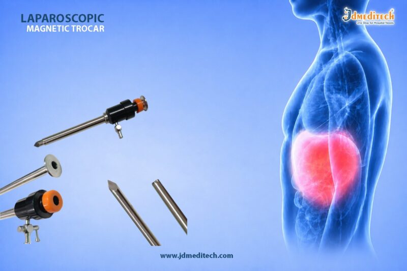
PTFE Guide Wire for Urology – Smooth & Reliable Access

The PTFE Guide Wire plays a crucial role in modern urological procedures. It ensures smooth navigation and reliable access through the urinary tract. Doctors depend on the PTFE Guide Wire for precision, flexibility, and patient safety.

In minimally invasive urology, the right guide wire improves procedural success. Therefore, the PTFE Guide Wire has become a preferred choice for many specialists worldwide.

What is a PTFE Guide Wire?

A PTFE Guide Wire is a medical device coated with Polytetrafluoroethylene (PTFE). This coating reduces friction and allows the wire to glide smoothly through anatomical pathways.

Because of its unique properties, the PTFE Guide Wire offers excellent control during procedures such as:

  • Ureteroscopy
  • Catheter placement
  • Stone removal procedures
  • Percutaneous nephrolithotomy (PCNL)
Key Features of PTFE Guide Wire
1. Smooth Surface for Easy Navigation

The PTFE coating ensures minimal resistance. As a result, the PTFE Guide Wire moves easily through narrow and complex pathways.

2. Excellent Flexibility

Flexibility allows the PTFE Guide Wire to adapt to anatomical curves. This reduces trauma during insertion.

3. High Strength and Durability

Despite its flexibility, the PTFE Guide Wire maintains strong structural integrity.

4. Hydrophobic Coating

Unlike hydrophilic wires, the PTFE Guide Wire provides controlled movement without excessive slipperiness.

5. Kink Resistance

The design prevents bending or kinking, ensuring consistent performance.

Applications of PTFE Guide Wire in Urology

The PTFE Guide Wire is widely used in various urological procedures. Its versatility makes it an essential tool.

Common Uses Include:
  • Assisting catheter insertion
  • Guiding ureteral stents
  • Supporting endoscopic procedures
  • Facilitating access during kidney stone removal

Because of its reliability, the PTFE Guide Wire improves procedural efficiency.

Advantages of Using PTFE Guide Wire
1. Enhanced Procedural Control

The PTFE Guide Wire provides better tactile feedback. Surgeons can guide it accurately.

2. Reduced Tissue Trauma

Its smooth surface minimizes friction. Therefore, it reduces irritation and injury.

3. Cost-Effective Solution

Compared to advanced coated wires, the PTFE Guide Wire offers a balance between performance and cost.

4. Reliable Performance

The consistent behavior of the PTFE Guide Wire makes it suitable for routine procedures.

PTFE Guide Wire vs Hydrophilic Guide Wire
Feature PTFE Guide Wire Hydrophilic Guide Wire
Surface Smooth (low friction) Extremely slippery when wet
Control High control Less tactile feedback
Use Case Routine procedures Complex or tight pathways
Cost More economical Higher cost

While hydrophilic wires excel in complex cases, the PTFE Guide Wire remains ideal for standard procedures.

How to Choose the Right PTFE Guide Wire

When selecting a PTFE Guide Wire, consider the following factors:

  • Diameter and length based on procedure
  • Tip design (straight or J-tip)
  • Stiffness level required
  • Compatibility with other instruments

Choosing the right PTFE Guide Wire ensures better outcomes and safety.

Safety and Handling Tips

Proper handling of the PTFE Guide Wire is essential. Follow these guidelines:

  • Always maintain sterility
  • Avoid excessive force during insertion
  • Use imaging guidance when necessary
  • Inspect for damage before use

These practices enhance patient safety and device performance.

Why Choose PTFE Guide Wire from JDMeditech?

At JDMeditech, we offer high-quality PTFE Guide Wire solutions designed for precision and reliability.

Our Key Benefits:
  • Premium-grade materials
  • Strict quality control
  • Designed for smooth navigation
  • Suitable for various urological procedures
Conclusion

The PTFE Guide Wire is an essential tool in urological procedures. It provides smooth access, reliable control, and enhanced safety. Because of its performance and affordability, the PTFE Guide Wire remains a trusted choice among medical professionals.

If you aim for precision and efficiency in urology, the PTFE Guide Wire is the right solution.


Explore More Product >>

Get Connected:

+91 79909 93062 | +91 63513 72032 | exports@jdmeditech.com
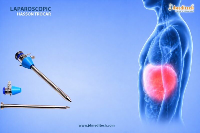
Pancreatic Drainage Procedure for Fluid Build-Up Relief

Introduction to Ross ProcedureThe Pancreatic Drainage Procedure is a specialized surgical technique used to treat pancreatic fluid accumulation caused by conditions such as pancreatitis or pancreatic cysts. This Pancreatic Drainage Procedure helps relieve pressure, reduce pain, and prevent serious complications.

When pancreatic fluid builds up in or around the pancreas, it can lead to infection, inflammation, and organ dysfunction. Therefore, doctors recommend the Pancreatic Drainage Procedure to restore normal pancreatic function and improve patient outcomes.

What is a Pancreatic Drainage Procedure?

A Pancreatic Drainage Procedure involves creating a pathway to drain excess pancreatic fluid from cysts, pseudocysts, or blocked ducts. Surgeons perform this Pancreatic Drainage Procedure using minimally invasive or open surgical techniques.

In many cases, the Pancreatic Drainage Procedure connects the fluid collection to the stomach or small intestine, allowing safe internal drainage.

Why is Pancreatic Drainage Procedure Needed?

Doctors recommend a Pancreatic Drainage Procedure when fluid collections cause symptoms or complications. Common indications include:

  • Pancreatic pseudocysts
  • Chronic pancreatitis
  • Pancreatic duct obstruction
  • Infected fluid collections
  • Persistent abdominal pain

Without timely treatment, these conditions may worsen. Therefore, the Pancreatic Drainage Procedure plays a crucial role in preventing further damage.

Types of Pancreatic Drainage Procedure
1. Endoscopic Pancreatic Drainage Procedure

This Pancreatic Drainage Procedure uses an endoscope to access the pancreas through the digestive tract. It is minimally invasive and offers faster recovery.

2. Percutaneous Pancreatic Drainage Procedure

In this Pancreatic Drainage Procedure, a catheter is inserted through the skin under imaging guidance to drain fluid externally.

3. Surgical Pancreatic Drainage Procedure

Surgeons perform this Pancreatic Drainage Procedure when other methods are not effective. It includes procedures like cystogastrostomy or pancreaticojejunostomy.

Procedure Steps of Pancreatic Drainage Procedure

The Pancreatic Drainage Procedure typically follows these steps:

  1. Patient evaluation and imaging (CT scan or MRI)
  2. Administration of anesthesia
  3. Accessing the fluid collection
  4. Creating a drainage pathway
  5. Placement of stents or drains
  6. Monitoring and post-procedure care

Each Pancreatic Drainage Procedure is tailored to the patient’s condition and severity.

Benefits of Pancreatic Drainage Procedure

The Pancreatic Drainage Procedure offers several advantages:

  • Relieves abdominal pain and pressure
  • Prevents infection and complications
  • Improves pancreatic function
  • Minimally invasive options available
  • Faster recovery time

Because of these benefits, the Pancreatic Drainage Procedure is widely used in modern gastroenterology.

Risks and Complications

Although the Pancreatic Drainage Procedure is generally safe, some risks may include:

  • Bleeding
  • Infection
  • Leakage of pancreatic fluid
  • Damage to surrounding organs

However, experienced surgeons minimize these risks during the Pancreatic Drainage Procedure.

Recovery After Pancreatic Drainage Procedure

Recovery from a Pancreatic Drainage Procedure depends on the technique used. Most patients experience:

  • Short hospital stay
  • Gradual return to normal diet
  • Pain relief within days
  • Regular follow-up imaging

Proper post-operative care ensures successful outcomes after the Pancreatic Drainage Procedure.

Advanced Technologies in Pancreatic Drainage Procedure

Modern advancements have improved the success of the Pancreatic Drainage Procedure:

  • Endoscopic ultrasound (EUS)-guided drainage
  • Biodegradable stents
  • High-resolution imaging systems
  • Minimally invasive surgical tools

These innovations make the Pancreatic Drainage Procedure safer and more effective.

Conclusion

The Pancreatic Drainage Procedure is a highly effective solution for managing pancreatic fluid build-up. It relieves symptoms, prevents complications, and restores normal pancreatic function.

With advanced techniques and improved safety, the Pancreatic Drainage Procedure continues to play a vital role in modern surgical care. Early diagnosis and timely intervention can significantly improve patient outcomes.


Explore More Product >>

Get Connected:

+91 79909 93062 | +91 63513 72032 | exports@jdmeditech.com 
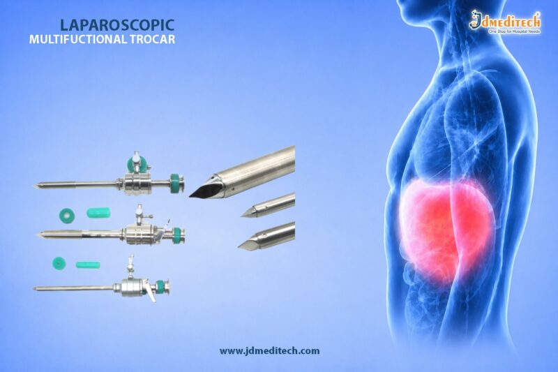
Hydrophilic Guide wire for Urology – Smooth & Precise Navigation

In modern urology, precision and control play a critical role in achieving successful outcomes. The Hydrophilic Guide wire stands out as an essential tool that enables smooth and efficient navigation through complex anatomical pathways. Due to its advanced coating and flexibility, this guide wire reduces friction and enhances access during minimally invasive procedures.

In this blog, we explore the features, benefits, applications, and importance of the Hydrophilic Guide wire in urological interventions.

What is a Hydrophilic Guide wire?

A Hydrophilic Guide wire is a specialized medical device designed to assist physicians in navigating through the urinary tract during diagnostic and therapeutic procedures. It features a hydrophilic (water-attracting) coating that becomes extremely slippery when activated by fluids.

As a result, the Hydrophilic Guide wire easily glides through narrow, tortuous, or obstructed pathways, making it ideal for challenging cases.

Key Features of Hydrophilic Guide wire
1. Hydrophilic Coating for Smooth Glide

The defining feature of a Hydrophilic Guide wire is its lubricious coating. When exposed to saline or bodily fluids, it significantly reduces friction, allowing effortless insertion and movement.

2.Excellent Flexibility and Control

The Hydrophilic Guide wire offers excellent flexibility while maintaining sufficient stiffness for control. This balance ensures precise navigation without compromising safety.

3. Atraumatic Tip Design

Most Hydrophilic Guide wire models come with a soft, atraumatic tip. Therefore, they minimize the risk of tissue injury during insertion.

4. Radiopacity for Visibility

The Hydrophilic Guide wire is visible under fluoroscopy, allowing clinicians to monitor its position in real-time for enhanced accuracy.

5. Kink Resistance

High-quality materials provide durability and kink resistance, ensuring consistent performance throughout the procedure.

Applications of Hydrophilic Guide wire in Urology

The Hydrophilic Guide wire plays a vital role in various urological procedures. Its smooth navigation capabilities make it indispensable in complex cases.

1. Ureteroscopy

During ureteroscopy, the Hydrophilic Guide wire helps access the ureter and kidney with ease, especially in tight or obstructed passages.

2. PCNL (Percutaneous Nephrolithotomy)

The Hydrophilic Guide wire assists in establishing access to the renal collecting system, ensuring safe and efficient tract creation.

3. Stone Management Procedures

In stone removal procedures, the Hydrophilic Guide wire facilitates device placement and improves procedural success rates.

4. Stent Placement

The Hydrophilic Guide wire is widely used for guiding ureteral stents into the correct position.

5. Difficult Anatomy Navigation

In cases involving strictures or anatomical variations, the Hydrophilic Guide wire provides reliable access where standard guide wires may fail.

Benefits of Using Hydrophilic Guide wire
Enhanced Maneuverability

The Hydrophilic Guide wire allows smooth passage through complex pathways, reducing procedural difficulty.

Reduced Trauma

Its lubricious surface and soft tip minimize tissue damage, ensuring patient safety.

Improved Procedural Efficiency

By enabling faster and easier navigation, the Hydrophilic Guide wire reduces procedure time.

Higher Success Rates

Clinicians achieve better outcomes in challenging cases with the help of a Hydrophilic Guide wire.

Versatility

The Hydrophilic Guide wire adapts to multiple urological applications, making it a valuable addition to any surgical setup.

How to Choose the Right Hydrophilic Guide wire

Selecting the appropriate Hydrophilic Guide wire depends on several factors:

  • Length and Diameter : Choose based on procedural requirements
  • Tip Type : Straight or angled depending on anatomy
  • Stiffness Level : Balance between flexibility and support
  • Coating Quality : Ensure consistent hydrophilic performance
Why Hydrophilic Guide wire is Essential in Modern Urology

The growing demand for minimally invasive procedures has increased the importance of advanced tools like the Hydrophilic Guide wire. Its ability to provide smooth, controlled, and safe navigation makes it indispensable for urologists.

Moreover, as procedural complexity increases, reliance on high-performance devices like the Hydrophilic Guide wire continues to grow.

Conclusion

The Hydrophilic Guide wire has transformed urological procedures by offering unmatched smoothness, flexibility, and control. Whether dealing with complex anatomy or routine interventions, this device ensures safe and efficient access.

For healthcare providers aiming to enhance precision and patient outcomes, investing in a high-quality Hydrophilic Guide wire is a smart and necessary choice.


Explore More Product >>

Get Connected:

+91 79909 93062 | +91 63513 72032 | exports@jdmeditech.com
Ross Procedure: Natural Valve Replacement for Heart Function

The Ross Procedure is a highly advanced cardiac surgery used to treat aortic valve disease. In this technique, surgeons replace the damaged valve with the patient’s own pulmonary valve. As a result, the Ross Procedure restores natural heart function more effectively than artificial alternatives.

Moreover, the Ross Procedure reduces dependency on synthetic valves. Because it uses living tissue, it improves long-term performance. Therefore, many specialists consider the Ross Procedure a superior option for selected patients.

What is Ross Procedure?

The Ross Procedure is a surgical method that replaces the diseased aortic valve using the patient’s pulmonary valve. Then, surgeons insert a donor valve into the pulmonary position. This unique approach allows the Ross Procedure to maintain natural valve dynamics.

In addition, the Ross Procedure ensures better blood flow and pressure control. Consequently, patients experience improved cardiovascular efficiency.

Why Ross Procedure is Performed

Doctors recommend the Ross Procedure for several conditions:

  • Aortic stenosis
  • Aortic regurgitation
  • Congenital valve defects
  • Pediatric valve disorders

Furthermore, the Ross Procedure is ideal for patients who want to avoid long-term anticoagulants. Because of this advantage, the Ross Procedure is widely used in younger patients.

Key Benefits of Ross Procedure

The Ross Procedure provides multiple clinical benefits.

Natural Valve Function

The Ross Procedure uses a living valve, which adapts naturally inside the body.

No Lifelong Blood Thinners

Unlike mechanical valves, the Ross Procedure does not require permanent anticoagulation.

Growth Compatibility

Importantly, the Ross Procedure supports growth in children and young adults.

Long-Term Performance

With proper care, the Ross Procedure delivers excellent durability.

Improved Lifestyle

Patients undergoing the Ross Procedure often return to active lifestyles quickly.

Step-by-Step Ross Procedure

The Ross Procedure involves precise surgical steps:

  1. Surgeons access the heart through open surgery
  2. They remove the damaged aortic valve
  3. They transfer the pulmonary valve
  4. They implant it in the aortic position
  5. They place a donor valve in the pulmonary area
  6. They restore and test heart function

Each stage of the Ross Procedure requires expert surgical skill.

Risks of Ross Procedure

Although the Ross Procedure is effective, some risks exist:

  • Infection
  • Bleeding
  • Valve degeneration
  • Reoperation in rare cases

However, with experienced surgeons, the success rate of the Ross Procedure remains high.

Recovery After Ross Procedure

Recovery after the Ross Procedure is generally smooth.

Initially, patients stay in the hospital for about a week. Then, they gradually resume daily activities. In addition, regular follow-ups help monitor the success of the Ross Procedure.

Most importantly, patients report better heart function after the Ross Procedure.

Ross Procedure vs Other Valve Surgeries

The Ross Procedure differs significantly from other options:

Feature Ross Procedure Mechanical Valve Tissue Valve
Natural Function Yes No Partial
Anticoagulants Not required Required Not required
Durability High Very high Moderate
Growth Potential Yes No No

Clearly, the Ross Procedure offers a balanced and natural solution.

Who Should Consider Ross Procedure

The Ross Procedure is best suited for:

  • Children and young adults
  • Active individuals
  • Patients avoiding anticoagulants
  • Those seeking natural valve replacement

Before choosing the Ross Procedure, doctors perform detailed evaluations.

Future of Ross Procedure

Modern advancements continue to improve the Ross Procedure. Surgeons now use refined techniques for better outcomes. As a result, the Ross Procedure is gaining popularity worldwide.

Conclusion

The Ross Procedure stands as one of the most advanced solutions for aortic valve replacement. By using the patient’s own valve, the Ross Procedure restores natural heart function effectively.

Ultimately, the Ross Procedure offers durability, safety, and improved quality of life. Therefore, it remains a preferred choice for long-term cardiac care.


Explore More Product >>

Get Connected:

+91 79909 93062 | +91 63513 72032 | exports@jdmeditech.com
Hepaticojejunostomy Surgery for Biliary Flow Restoration

Hepaticojejunostomy surgery plays a vital role in restoring normal bile flow when the bile ducts become blocked or damaged. Surgeons perform hepaticojejunostomy surgery to create a direct connection between the hepatic duct and the jejunum. As a result, bile bypasses the obstruction and flows smoothly into the intestine.

This procedure is widely used in hepatobiliary surgery because it offers a reliable and long-term solution for bile duct injuries and strictures.

What is Hepaticojejunostomy Surgery?

Hepaticojejunostomy surgery is a reconstructive surgical procedure that connects the hepatic duct to a segment of the small intestine known as the jejunum. Doctors recommend hepaticojejunostomy surgery when bile cannot flow naturally from the liver to the intestine.

In many cases, hepaticojejunostomy surgery becomes necessary after bile duct injury, tumor removal, or chronic inflammation. Therefore, it ensures proper digestion and prevents complications such as jaundice and infection.

Why is Hepaticojejunostomy Surgery Performed?

Doctors perform hepaticojejunostomy surgery to treat various hepatobiliary conditions. Some of the most common indications include:

  • Bile duct injury during gallbladder surgery
  • Biliary strictures or narrowing
  • Cholangiocarcinoma (bile duct cancer)
  • Congenital bile duct abnormalities
  • Chronic pancreatitis affecting bile flow

Because these conditions disrupt bile drainage, hepaticojejunostomy surgery provides an effective bypass solution.

Hepaticojejunostomy Surgery Procedure

Hepaticojejunostomy surgery involves precise surgical steps to ensure long-term success.

1. Preparation

Surgeons evaluate the patient using imaging tests such as MRI, CT scans, or ERCP. Proper planning helps achieve accurate reconstruction.

2. Surgical Approach

Surgeons may perform hepaticojejunostomy surgery using:

  • Open surgery
  • Laparoscopic techniques
  • Robotic-assisted surgery

Minimally invasive methods often reduce recovery time.

3. Creating the Connection

The surgeon connects the hepatic duct to a loop of the jejunum. This connection, known as an anastomosis, allows bile to flow directly into the intestine.

4. Ensuring Bile Flow

Surgeons check the connection carefully to prevent leakage or blockage. Proper alignment is essential for long-term success.

Benefits of Hepaticojejunostomy Surgery

Hepaticojejunostomy surgery offers several advantages:

  • Restores normal bile flow
  • Prevents bile leakage and infections
  • Reduces jaundice and liver damage
  • Provides a long-term solution for bile duct obstruction
  • Improves digestion and nutrient absorption

Because of these benefits, hepaticojejunostomy surgery remains a preferred treatment option.

Risks and Complications

Although hepaticojejunostomy surgery is generally safe, some risks may occur:

  • Infection at the surgical site
  • Bile leakage
  • Anastomotic stricture (narrowing of the connection)
  • Bleeding
  • Delayed gastric emptying

However, experienced surgeons minimize these risks with proper technique and postoperative care.

Recovery After Hepaticojejunostomy Surgery

Recovery after hepaticojejunostomy surgery depends on the surgical approach and patient condition.

  • Hospital stay usually lasts 5–10 days
  • Patients gradually resume normal diet
  • Regular follow-up ensures proper bile flow
  • Full recovery may take several weeks

Following medical advice plays a key role in successful healing.

Hepaticojejunostomy Surgery in Modern Healthcare

With advancements in surgical technology, hepaticojejunostomy surgery has become safer and more effective. Laparoscopic and robotic techniques improve precision and reduce complications.

Medical device innovations also support better outcomes. High-quality surgical instruments ensure accurate anastomosis and reduce procedural risks.

Conclusion

Hepaticojejunostomy surgery stands as a highly effective solution for restoring biliary flow in patients with bile duct obstruction. By creating a direct connection between the liver and intestine, hepaticojejunostomy surgery ensures proper digestion and prevents serious complications.

With modern surgical precision and advanced medical tools, hepaticojejunostomy surgery continues to improve patient outcomes and quality of life.


Explore More Product >>

Get Connected:

+91 79909 93062 | +91 63513 72032 | exports@jdmeditech.com
ICD Implantation – Lifesaving Heart Procedure for Arrhythmia

ICD implantation is a critical medical procedure designed to prevent sudden cardiac death in patients with life-threatening heart rhythm disorders. An Implantable Cardioverter Defibrillator (ICD) continuously monitors heart rhythms and delivers electrical therapy when dangerous arrhythmias occur.

Cardiovascular diseases remain a leading cause of mortality worldwide. Therefore, ICD implantation has become a trusted and effective solution for patients at high risk of sudden cardiac arrest.

What is ICD Implantation?

ICD implantation refers to the surgical placement of a small electronic device inside the chest. This device monitors the heart’s electrical activity and responds instantly to abnormal rhythms such as ventricular tachycardia or ventricular fibrillation.

The ICD system consists of:

  • A pulse generator (battery and circuitry)
  • Leads (thin wires placed in the heart)
  • Electrodes to deliver therapy

When the device detects an irregular heartbeat, it delivers a shock to restore normal rhythm.

Why is ICD Implantation Necessary?

Doctors recommend ICD implantation for patients who have a high risk of sudden cardiac arrest or severe arrhythmias.

Common Indications Include:
  • History of cardiac arrest
  • Ventricular tachycardia or fibrillation
  • Heart failure with reduced ejection fraction
  • Cardiomyopathy (dilated or hypertrophic)
  • Genetic heart rhythm disorders

By performing ICD implantation, healthcare providers significantly reduce mortality rates and improve patient survival.

How ICD Implantation Works

The primary function of ICD implantation is to detect and correct abnormal heart rhythms automatically.

Device Functions:
  • Continuous heart rhythm monitoring
  • Anti-tachycardia pacing (ATP)
  • Cardioversion (low-energy shock)
  • Defibrillation (high-energy shock)

The device acts within seconds, making ICD implantation one of the most reliable emergency interventions inside the body.

Procedure of ICD Implantation

ICD implantation is a minimally invasive procedure usually performed under local anesthesia with sedation.

Step-by-Step Process:
  1. A small incision is made below the collarbone
  2. Leads are guided through a vein into the heart
  3. The ICD device is positioned under the skin
  4. Leads are connected to the device
  5. Device testing ensures proper functioning

The entire ICD implantation procedure typically takes 1–2 hours.

Benefits of ICD Implantation

There are numerous advantages of ICD implantation, especially for high-risk cardiac patients.

Key Benefits:
  • Prevents sudden cardiac death
  • Provides continuous monitoring
  • Immediate response to arrhythmias
  • Improves long-term survival
  • Enhances quality of life

Because of these benefits, ICD implantation is widely recommended in modern cardiology.

Risks and Complications of ICD Implantation

Although ICD implantation is generally safe, some risks may occur.

Possible Complications:
  • Infection at the implant site
  • Bleeding or swelling
  • Lead displacement
  • Device malfunction
  • Rare allergic reactions

Proper post-operative care reduces the likelihood of complications after ICD implantation.

Recovery After ICD Implantation

Recovery following ICD implantation is usually quick and smooth.

Post-Procedure Care:
  • Avoid heavy lifting for a few weeks
  • Keep the incision area clean and dry
  • Attend regular follow-up visits
  • Avoid strong electromagnetic fields

Most patients return to normal activities within a short period after ICD implantation.

ICD Implantation vs Pacemaker

While both devices regulate heart rhythm, ICD implantation serves a more advanced purpose.

Feature ICD Implantation Pacemaker
Function Corrects life-threatening arrhythmias Controls slow heart rate
Shock Delivery Yes No
Emergency Response Immediate Limited

Thus, ICD implantation is essential for patients at risk of sudden cardiac arrest.

Latest Advancements in ICD Implantation

Modern ICD implantation technology has evolved significantly.

Innovations Include:
  • Subcutaneous ICDs (no leads in the heart)
  • MRI-compatible devices
  • Remote monitoring systems
  • Longer battery life

These advancements have made ICD implantation safer, more efficient, and patient-friendly.

Conclusion

ICD implantation is a lifesaving heart procedure that plays a crucial role in preventing sudden cardiac death. With advanced technology and proven effectiveness, it offers patients a reliable safeguard against dangerous arrhythmias.

If you or your patients are at risk of severe heart rhythm disorders, ICD implantation could be the key to long-term cardiac safety and improved quality of life.


Explore More Product >>

Get Connected:

+91 79909 93062 | +91 63513 72032 | exports@jdmeditech.com
PCNL Basket – Advanced Tool for Kidney Stone Extraction

Kidney stones can cause severe discomfort and complications if not treated effectively. Modern urology offers advanced minimally invasive solutions like Percutaneous Nephrolithotomy (PCNL). One of the most essential tools used during this procedure is the PCNL Basket.

A PCNL Basket plays a critical role in ensuring safe, precise, and efficient stone extraction. In this blog, we will explore how the PCNL Basket enhances nephrolithotomy surgery and why it remains a preferred choice among urologists.

What is a PCNL Basket?

A PCNL Basket is a specialized urological instrument designed to capture, hold, and remove kidney stones during PCNL procedures. It is typically introduced through a nephroscope and allows surgeons to retrieve stone fragments with high accuracy.

The design of a PCNL Basket ensures flexibility, durability, and controlled manipulation inside the kidney.

Role of PCNL Basket in Nephrolithotomy Surgery

During Percutaneous Nephrolithotomy, surgeons create a small tract to access the kidney. Once the stones are fragmented, the PCNL Basket is used to retrieve them.

Key Functions:
  • Captures fragmented stones effectively
  • Prevents stone migration
  • Ensures complete clearance
  • Minimizes procedure time

The use of a PCNL Basket significantly improves surgical outcomes and reduces the risk of residual stones.

Types of PCNL Basket

Different types of PCNL Basket devices are available to suit various clinical needs:

1. Nitinol PCNL Basket
  • Highly flexible
  • Shape memory for better control
  • Ideal for complex anatomies
2. Stainless Steel PCNL Basket
  • Strong and durable
  • Suitable for larger stones
  • Cost-effective option
3. Tipless PCNL Basket
  • Reduces trauma risk
  • Allows safer navigation
  • Preferred for delicate procedures

Each PCNL Basket type offers specific advantages depending on the surgical requirement.

Key Features of PCNL Basket

A high-quality PCNL Basket includes several advanced features:

  • Excellent flexibility for smooth navigation
  • Strong grip for secure stone capture
  • Corrosion-resistant materials
  • Ergonomic handle for better control
  • Radiopaque design for visibility

These features make the PCNL Basket an indispensable tool in modern urology.

Advantages of Using PCNL Basket

The PCNL Basket offers multiple benefits during nephrolithotomy surgery:

1. High Precision

It allows accurate stone capture, reducing the chances of incomplete removal.

2. Improved Efficiency

Surgeons can quickly retrieve stone fragments, shortening procedure time.

3. Minimally Invasive Support

It complements the minimally invasive nature of PCNL.

4. Reduced Complications

Using a PCNL Basket lowers the risk of residual stones and repeat procedures.

How to Choose the Right PCNL Basket

Selecting the right PCNL Basket depends on several factors:

  • Size and type of kidney stones
  • Patient anatomy
  • Surgeon preference
  • Material (Nitinol vs Stainless Steel)
  • Basket design (tipless or standard)

Choosing the appropriate PCNL Basket ensures better surgical outcomes and patient safety.

Applications of PCNL Basket

The PCNL Basket is widely used in:

  • Kidney stone removal procedures
  • Percutaneous nephrolithotomy surgeries
  • Retrieval of stone fragments
  • Complex urological interventions

Its versatility makes the PCNL Basket a standard instrument in urology setups.

Best Practices for Using PCNL Basket

To achieve optimal results, surgeons should:

  • Use gentle manipulation techniques
  • Avoid excessive force
  • Select the correct basket size
  • Ensure proper visualization during use

Following these practices enhances the performance of the PCNL Basket.

Why Choose JDMeditech PCNL Basket

At JDMeditech, we provide high-quality PCNL Basket solutions designed for precision and reliability.

Our Key Highlights:
  • Premium-grade materials
  • Advanced ergonomic design
  • High flexibility and durability
  • Strict quality standards
  • Competitive pricing

Our PCNL Basket products are trusted by healthcare professionals for delivering consistent performance.

Conclusion

The PCNL Basket is a crucial instrument in nephrolithotomy surgery, enabling efficient and safe stone extraction. With advancements in urological technology, the demand for high-quality PCNL Basket devices continues to grow.

By choosing the right PCNL Basket, surgeons can improve procedural success, reduce complications, and ensure better patient outcomes.


Explore More Product >>

Get Connected:

+91 79909 93062 | +91 63513 72032 | exports@jdmeditech.com
Arterial Switch Operation for TGA – Best Surgical Solution for TGA

The Arterial Switch Operation for TGA is a life-saving cardiac surgery performed to correct Transposition of the Great Arteries (TGA), a serious congenital heart defect. In this condition, the positions of the main arteries are reversed, which disrupts normal blood circulation.

Without timely intervention, TGA can lead to severe oxygen deficiency in the body. However, the Arterial Switch Operation for TGA restores proper blood flow and significantly improves survival rates in newborns.

What is Transposition of the Great Arteries (TGA)?

Transposition of the Great Arteries is a condition where:

  • The aorta arises from the right ventricle
  • The pulmonary artery arises from the left ventricle

As a result:

  • Oxygen-poor blood circulates through the body
  • Oxygen-rich blood cycles back to the lungs

This abnormal circulation makes early surgical correction essential. The Arterial Switch Operation for TGA corrects this issue effectively.

Why is Arterial Switch Operation for TGA Necessary?

Doctors recommend the Arterial Switch Operation for TGA because it:

  • Restores normal blood circulation
  • Ensures proper oxygen supply to vital organs
  • Prevents life-threatening complications
  • Offers long-term survival and improved quality of life

Early surgery, usually within the first few weeks of life, delivers the best outcomes.

Procedure of Arterial Switch Operation for TGA

The Arterial Switch Operation for TGA involves precise surgical steps:

1. Open Heart Access

Surgeons open the chest and connect the baby to a heart-lung machine.

2. Artery Repositioning

They detach and switch the positions of the aorta and pulmonary artery.

3. Coronary Artery Transfer

Surgeons carefully reattach the coronary arteries to the new aorta.

4. Restoration of Blood Flow

They reconstruct the vessels to establish normal blood circulation.

This procedure ensures that oxygen-rich blood reaches the body efficiently.

Benefits of Arterial Switch Operation for TGA

The Arterial Switch Operation for TGA offers several advantages:

  •  Restores normal heart anatomy
  •  Provides near-normal life expectancy
  •  Improves oxygen levels instantly
  •  Reduces long-term cardiac complications
  •  Supports healthy growth and development
Risks and Complications

Although the Arterial Switch Operation for TGA is highly successful, some risks may include:

  • Bleeding or infection
  • Coronary artery complications
  • Arrhythmias
  • Narrowing of arteries (stenosis)

However, with modern surgical techniques, complications are minimal and manageable.

Recovery After Arterial Switch Operation for TGA

Recovery after the Arterial Switch Operation for TGA requires careful monitoring:

  • Initial stay in the neonatal intensive care unit (NICU)
  • Gradual return to normal feeding and activity
  • Regular follow-ups with a pediatric cardiologist

Most infants recover well and lead active, healthy lives.

Long-Term Outcomes

The long-term success of the Arterial Switch Operation for TGA is excellent:

  • High survival rates
  • Normal physical development
  • Minimal restrictions in daily activities

Regular check-ups ensure ongoing heart health.

Medical Equipment Used in Arterial Switch Operation for TGA

High-quality medical devices play a crucial role in successful outcomes. These include:

  • Cardiac surgical instruments
  • Heart-lung machines
  • Vascular clamps and forceps
  • Surgical sutures and monitoring systems

At JDMeditech, we provide advanced medical solutions designed to support complex cardiac procedures like the Arterial Switch Operation for TGA.

Conclusion

The Arterial Switch Operation for TGA remains the gold standard for correcting Transposition of the Great Arteries. It restores normal heart function and offers newborns a chance at a healthy life.

With timely diagnosis, skilled surgeons, and advanced medical equipment, this procedure delivers outstanding results and long-term success.


Explore More Product >>

Get Connected:

+91 79909 93062 | +91 63513 72032 | exports@jdmeditech.com