
Ureteral Access Sheath for Safe and Smooth Urology Procedures

The ureteral access sheath is a crucial tool in modern urology. It provides smooth, repeated access to the urinary tract during procedures like ureteroscopy and stone management. Because of its efficiency and safety, the ureteral access sheath has become an essential device for urologists worldwide.

In this blog, we will explore how a ureteral access sheath improves procedural outcomes, enhances safety, and supports advanced urological interventions.

What is a Ureteral Access Sheath?

A ureteral access sheath is a flexible, hollow tube inserted into the ureter. It creates a continuous working channel that allows easy passage of instruments such as ureteroscopes, laser fibers, and retrieval devices.

As a result, the sheath minimizes repeated trauma to the ureter and ensures smoother instrument exchange.

Why is Ureteral Access Sheath Important?

The ureteral access sheath plays a vital role in improving both efficiency and patient safety.

Key Advantages:
  • Reduces ureteral trauma during repeated instrument insertion
  • Maintains low intrarenal pressure
  • Improves visualization during procedures
  • Facilitates faster and smoother device exchange
  • Enhances overall surgical efficiency

Therefore, many urologists prefer using a ureteral access sheath in complex procedures.

Key Features of Ureteral Access Sheath
1. Smooth Hydrophilic Coating

The hydrophilic coating reduces friction. Consequently, it allows easy insertion and removal without damaging the ureter.

2. Flexible Yet Durable Design

The sheath offers excellent flexibility. At the same time, it maintains enough strength to prevent kinking.

3. Radiopaque Markers

Radiopaque markers ensure accurate placement under fluoroscopy. This improves procedural safety.

4. Dual-Layer Construction

Many ureteral access sheaths feature a dual-layer design. This combination provides both flexibility and structural support.

5. Wide Range of Sizes

Available in different diameters and lengths, the sheath suits various patient anatomies and procedural needs.

Applications of Ureteral Access Sheath

The ureteral access sheath is widely used in:

  • Ureteroscopy (URS)
  • Kidney stone removal procedures
  • Laser lithotripsy
  • Retrograde intrarenal surgery (RIRS)
  • Diagnostic urological procedures

Because of its versatility, it is a standard tool in modern urology.

Benefits of Using Ureteral Access Sheath
Improved Procedure Efficiency

The sheath allows quick instrument exchanges. Therefore, it significantly reduces operation time.

Enhanced Patient Safety

By minimizing ureteral trauma and maintaining low pressure, the sheath reduces complications.

Better Visualization

Continuous irrigation flow improves visibility, which helps surgeons perform precise procedures.

Reduced Infection Risk

Lower intrarenal pressure helps prevent bacterial migration, thereby reducing infection risk.

How to Use a Ureteral Access Sheath?
  1. Insert a guidewire into the ureter
  2. Advance the ureteral access sheath over the guidewire
  3. Position it correctly under imaging guidance
  4. Introduce the ureteroscope and other instruments
  5. Perform the procedure efficiently

Proper technique ensures safe and effective outcomes.

Choosing the Right Ureteral Access Sheath

When selecting a ureteral access sheath, consider:

  • Size and length compatibility
  • Coating quality for smooth insertion
  • Flexibility and strength
  • Compatibility with instruments
  • Patient anatomy

Choosing the right sheath improves both performance and safety.

Why Choose JDMeditech Ureteral Access Sheath?

At JDMeditech, we provide high-quality ureteral access sheaths designed for reliability and precision.

Our Advantages:
  • Premium-grade materials
  • Smooth hydrophilic coating
  • Excellent flexibility and durability
  • Sterile and ready-to-use packaging
  • Competitive pricing with superior quality

Explore more products at: https://www.jdmeditech.com

Conclusion

The ureteral access sheath is an indispensable device in urological procedures. It ensures smooth access, improved safety, and better surgical outcomes. As urology continues to advance, the importance of reliable tools like the ureteral access sheath will only grow.


Explore More Product >>

Get Connected:

+91 79909 93062 | +91 63513 72032 | exports@jdmeditech.com
Endoscopy LED Light Source for Precision Imaging

In modern endoscopic procedures, clear visualization is critical for accurate diagnosis and treatment. The Endoscopy LED Light Source plays a vital role by delivering bright, consistent, and energy-efficient illumination. As a result, it enhances image clarity and supports precision during minimally invasive procedures.

Today, healthcare professionals prefer LED technology over traditional light sources because it offers longer lifespan, lower heat generation, and superior illumination quality. Therefore, investing in a high-quality Endoscopy LED Light Source ensures better clinical outcomes and improved patient safety.

What is an Endoscopy LED Light Source?

An Endoscopy LED Light Source is a medical device that provides high-intensity light to illuminate internal body structures during endoscopic procedures. It connects to an endoscope via a fiber optic cable, ensuring optimal visibility inside cavities such as the gastrointestinal tract, urinary system, and respiratory pathways.

Unlike conventional halogen or xenon light sources, LED technology offers stable brightness, reduced maintenance, and energy efficiency.

Key Features of Endoscopy LED Light Source
1. High-Intensity Illumination

The Endoscopy LED Light Source produces bright and uniform light, which improves visualization during procedures. Consequently, clinicians can identify even minor abnormalities with ease.

2. Energy Efficiency

LED technology consumes less power compared to traditional systems. Therefore, hospitals can reduce operational costs while maintaining high performance.

3. Long Lifespan

One of the biggest advantages of an Endoscopy LED Light Source is its durability. It offers thousands of hours of operation, reducing the need for frequent replacements.

4. Low Heat Generation

Unlike xenon light sources, LED systems generate minimal heat. As a result, they improve patient safety and reduce the risk of tissue damage.

5. Compact and User-Friendly Design

Modern systems are designed for easy integration with endoscopy units. Additionally, they feature intuitive controls for brightness adjustment.

Applications of Endoscopy LED Light Source

The Endoscopy LED Light Source is widely used across various medical specialties, including:

  • Gastroenterology – for procedures like gastroscopy, colonoscopy, and ERCP
  • Urology – for cystoscopy and ureteroscopy
  • Pulmonology – for bronchoscopy
  • Gynecology – for hysteroscopy
  • ENT procedures – for nasal and throat examinations

Because of its versatility, it has become an essential component in modern endoscopy systems.

Benefits of Using Endoscopy LED Light Source
Improved Visualization

The Endoscopy LED Light Source delivers consistent brightness, which helps in accurate diagnosis and treatment.

Enhanced Procedure Efficiency

Better illumination allows clinicians to perform procedures faster and with greater confidence.

Cost-Effective Solution

Since LED lights last longer and consume less energy, healthcare facilities can save significantly on maintenance and electricity.

Increased Patient Safety

Lower heat emission reduces the risk of burns or discomfort during procedures.

Why Choose JDMeditech Endoscopy LED Light Source?

At JDMeditech, we provide high-quality Endoscopy LED Light Source solutions designed for precision and reliability. Our products are manufactured using advanced technology to meet international medical standards.

Key Advantages:
  • Superior brightness and clarity
  • Durable and long-lasting performance
  • Energy-efficient design
  • Easy compatibility with various endoscopy systems
  • Competitive pricing with assured quality
Maintenance Tips for Endoscopy LED Light Source

To ensure optimal performance of your Endoscopy LED Light Source, follow these simple tips:

  • Regularly clean the external surfaces
  • Check fiber optic cables for damage
  • Avoid overloading the system
  • Ensure proper ventilation during operation
  • Schedule routine servicing

Proper maintenance extends the life of the device and ensures consistent performance.

Future of LED Technology in Endoscopy

The future of Endoscopy LED Light Source technology looks promising. With continuous advancements, newer models are becoming more efficient, compact, and intelligent. Additionally, integration with digital imaging systems will further enhance diagnostic accuracy.

Conclusion

The Endoscopy LED Light Source is an essential component in modern medical procedures. Its ability to provide bright, efficient, and reliable illumination significantly improves the accuracy and safety of endoscopic interventions.

By choosing a high-quality solution from JDMeditech, healthcare providers can ensure superior performance, long-term reliability, and better patient outcomes.


Explore More Product >>

Get Connected:

+91 79909 93062 | +91 63513 72032 | exports@jdmeditech.com
Foley Balloon Catheter for Long-Term Urinary Drainage

The Foley Balloon Catheter plays a crucial role in modern urology by providing a safe, effective, and reliable solution for long-term urinary drainage. Widely used in hospitals, ICUs, and home care settings, this catheter ensures continuous bladder emptying in patients who cannot urinate naturally.

Because of its secure retention mechanism and ease of use, the Foley Balloon Catheter has become the preferred choice among healthcare professionals worldwide.

What is a Foley Balloon Catheter?

A Foley Balloon Catheter is a flexible, sterile medical device inserted into the bladder through the urethra. It features an inflatable balloon at the tip, which holds the catheter securely in place once positioned inside the bladder.

As a result, it allows continuous urine drainage without frequent reinsertion, making it ideal for long-term use.

Key Features of Foley Balloon Catheter
1. Inflatable Balloon for Secure Placement

The catheter includes a balloon that inflates after insertion, ensuring stable positioning and preventing accidental removal.

2. High-Quality Biocompatible Material

Manufactured using latex or silicone, the Foley Balloon Catheter minimizes irritation and enhances patient comfort during prolonged use.

3. Smooth Surface for Easy Insertion

Its low-friction surface ensures painless insertion and reduces trauma to the urethral lining.

4. Multiple Lumen Design

Typically available in 2-way and 3-way configurations, it supports:

  • Urine drainage
  • Balloon inflation
  • Bladder irrigation (in 3-way catheters)
5. Sterile and Disposable

Each Foley Balloon Catheter is individually packed and sterilized, reducing the risk of infection.

Types of Foley Balloon Catheters
2-Way Foley Balloon Catheter
  • Most commonly used
  • One lumen for drainage and one for balloon inflation
  • Ideal for general urinary drainage
3-Way Foley Balloon Catheter
  • Includes an additional lumen for irrigation
  • Commonly used in post-surgical bladder care, especially after TURP
Applications of Foley Balloon Catheter

The Foley Balloon Catheter is widely used in various medical scenarios:

  • Urinary retention management
  • Post-operative bladder drainage
  • Critical care and ICU patients
  • Neurological disorders affecting bladder control
  • Monitoring urine output in hospitalized patients
  • Long-term bedridden patient care
Advantages of Foley Balloon Catheter

The popularity of the Foley Balloon Catheter comes from its multiple advantages:

  • Ensures continuous and controlled urine drainage
  • Reduces the need for repeated catheterization
  • Enhances patient comfort and convenience
  • Offers secure retention with minimal displacement risk
  • Supports accurate urine output monitoring
Procedure for Foley Balloon Catheter Insertion

Healthcare professionals follow a sterile technique during insertion:

  1. Clean and disinfect the urethral area
  2. Gently insert the catheter into the bladder
  3. Inflate the balloon with sterile water
  4. Connect the catheter to a urine drainage bag

Proper placement ensures safe and effective drainage.

Care and Maintenance Tips

To ensure optimal performance and reduce complications:

  • Maintain proper hygiene around the insertion site
  • Keep the drainage bag below bladder level
  • Avoid kinking or pulling of the catheter
  • Monitor for signs of infection such as fever or discomfort
  • Replace the catheter as recommended by healthcare professionals
Why Choose Foley Balloon Catheter from JDMeditech?

At JDMeditech, we provide high-quality Foley Balloon Catheters designed for safety, comfort, and reliability.

Our Key Benefits:
  • Premium-grade materials
  • Strict quality control standards
  • Wide range of sizes and configurations
  • Competitive pricing
  • Trusted by healthcare providers
Conclusion

The Foley Balloon Catheter remains a trusted solution for long-term urinary drainage, thanks to its secure design, ease of use, and versatility. Whether used in hospitals or home care, it ensures effective bladder management and improved patient comfort.

For healthcare providers seeking dependable urology solutions, the Foley Balloon Catheter continues to be an essential medical device.


Explore More Product >>

Get Connected:

+91 79909 93062 | +91 63513 72032 | exports@jdmeditech.com
Biliary Dilator in Endoscopy for Effective Bile Duct Dilation

A Biliary Dilator in Endoscopy plays a crucial role in modern gastrointestinal procedures. It helps physicians safely and effectively dilate strictures within the bile ducts, thereby improving access and treatment outcomes. As endoscopic techniques continue to evolve, the demand for reliable and precise biliary dilators has increased significantly.

In this blog, we will explore the features, benefits, applications, and importance of a Biliary Dilator in Endoscopy, ensuring optimized readability and SEO performance.

What is a Biliary Dilator in Endoscopy?

A Biliary Dilator in Endoscopy is a specialized medical device used to widen narrowed bile ducts during procedures such as ERCP (Endoscopic Retrograde Cholangiopancreatography). These dilators allow smooth passage of stents, catheters, and other therapeutic devices.

Moreover, physicians use biliary dilators to manage strictures caused by stones, tumors, or inflammation. As a result, they ensure better bile flow and reduce complications.

Why is Biliary Dilation Important?

Biliary strictures can severely impact bile flow, leading to complications such as jaundice, infection, and liver dysfunction. Therefore, using a Biliary Dilator in Endoscopy becomes essential.

Key reasons include:
  • Improves bile drainage
  • Facilitates stent placement
  • Enhances procedural success rate
  • Reduces patient discomfort
  • Prevents long-term complications

Consequently, proper dilation ensures smoother and safer interventions.

Types of Biliary Dilators

Different types of Biliary Dilator in Endoscopy are available based on clinical needs:

1. Balloon Dilators

These dilators use controlled pressure to expand strictures gradually. They provide uniform dilation and are widely used in ERCP procedures.

2. Bougie Dilators

Bougie dilators offer mechanical dilation and are ideal for progressive widening.

3. Tapered Tip Dilators

These devices feature a tapered design that allows easy entry into tight strictures.

Each type of Biliary Dilator in Endoscopy ensures precision and efficiency in different clinical scenarios.

Key Features of Biliary Dilator in Endoscopy

Modern Biliary Dilator in Endoscopy devices are designed with advanced features to enhance performance:

  • High flexibility for smooth navigation
  • Atraumatic tip design to minimize tissue injury
  • Radiopaque markers for better visualization under fluoroscopy
  • Graduated sizing for controlled dilation
  • Strong pushability and torque control

Additionally, these features ensure both safety and procedural efficiency.

Applications in Clinical Practice

A Biliary Dilator in Endoscopy is widely used in various procedures:

  • ERCP procedures
  • Bile duct stricture management
  • Pre-stent dilation
  • Post-surgical narrowing treatment
  • Management of malignant and benign obstructions

Furthermore, it plays a vital role in both diagnostic and therapeutic endoscopy.

Advantages of Using Biliary Dilator in Endoscopy

Using a Biliary Dilator in Endoscopy offers multiple advantages:

  • Minimally invasive treatment
  • Faster recovery time
  • Reduced risk of complications
  • Improved patient outcomes
  • Enhanced procedural accuracy

Therefore, it has become an essential tool in gastroenterology.

How to Choose the Right Biliary Dilator?

Selecting the appropriate Biliary Dilator in Endoscopy depends on several factors:

  • Type and severity of stricture
  • Patient anatomy
  • Procedure type
  • Compatibility with guidewires and endoscopes

Hence, choosing the right device ensures optimal performance and safety.

Best Practices for Safe Use

To achieve the best results with a Biliary Dilator in Endoscopy, clinicians should follow these practices:

  • Use imaging guidance during dilation
  • Apply gradual pressure to avoid injury
  • Select appropriate size increments
  • Maintain sterile technique

As a result, these steps help reduce complications and improve outcomes.

Future Trends in Biliary Dilation

Technological advancements are shaping the future of Biliary Dilator in Endoscopy:

  • Improved material technology
  • Enhanced flexibility and control
  • Integration with advanced imaging systems
  • Development of single-use devices

Consequently, these innovations will further improve patient safety and procedural success.

Conclusion

A Biliary Dilator in Endoscopy is an indispensable tool for managing bile duct strictures effectively. With its advanced design and clinical benefits, it ensures safer procedures and better patient outcomes.

As endoscopic technology continues to advance, the importance of high-quality biliary dilators will only grow. Therefore, choosing a reliable solution is essential for achieving consistent and successful results.


Explore More Product >>

Get Connected:

+91 79909 93062 | +91 63513 72032 | exports@jdmeditech.com
Tiemann Catheter for Urethral Strictures – Precision Urinary Access

The Tiemann catheter is a specially designed urinary catheter used in urology for patients with difficult urethral anatomy. Unlike standard catheters, the Tiemann catheter features a curved, tapered tip that allows smoother navigation through urethral strictures and obstructions.

Because urethral strictures can make catheterization challenging, healthcare professionals rely on the Tiemann catheter to ensure safe, controlled, and minimally traumatic insertion. As a result, it plays a vital role in modern urological practice.

What is a Tiemann Catheter?

A Tiemann catheter is a type of Foley or intermittent catheter with a slightly angled, firm tip. This design helps guide the catheter through narrowed or obstructed areas of the urethra.

Key Design Features
  • Curved (Coudé) Tip : Improves directional control
  • Firm Structure : Prevents bending during insertion
  • Smooth Surface Finish : Reduces friction and trauma
  • Multiple Size Options : Suitable for different patient needs

Therefore, the Tiemann catheter provides a reliable solution when standard catheters fail.

Why Use a Tiemann Catheter for Urethral Strictures?

Urethral strictures cause narrowing of the urethra, which can block urine flow and complicate catheterization. In such cases, the Tiemann catheter offers several advantages.

1. Enhanced Navigation

The angled tip allows the Tiemann catheter to bypass obstructions more effectively. Consequently, insertion becomes easier even in complex cases.

2. Reduced Risk of Trauma

Because of its smooth and guided entry, the Tiemann catheter minimizes urethral injury. This feature is especially important for sensitive patients.

3. Better Patient Comfort

With improved insertion control, patients experience less discomfort during catheterization.

4. High Success Rate

Healthcare providers often achieve higher success rates using a Tiemann catheter compared to standard straight-tip catheters.

Clinical Applications of Tiemann Catheter

The Tiemann catheter is widely used in various urological conditions, including:

  • Urethral strictures
  • Benign Prostatic Hyperplasia (BPH)
  • Difficult male catheterization
  • Urinary retention cases
  • Post-surgical urological care

Additionally, the Tiemann catheter is commonly used in emergency settings where rapid bladder drainage is required.

How to Use a Tiemann Catheter Safely

Proper technique is essential when using a Tiemann catheter. Follow these general steps:

  1. Prepare Sterile Equipment : Maintain aseptic conditions
  2. Lubricate Generously : Reduce friction during insertion
  3. Align the Tip Correctly : The curved tip should face upward
  4. Insert Gently : Avoid force to prevent injury
  5. Confirm Placement : Ensure proper urine flow

However, only trained professionals should perform catheterization using a Tiemann catheter.

Tiemann Catheter vs Standard Catheter
Feature Tiemann Catheter Standard Catheter
Tip Design Curved Straight
Navigation Excellent for strictures Limited
Trauma Risk Lower Higher in difficult cases
Use Case Complex catheterization Routine use

Clearly, the Tiemann catheter offers superior performance in challenging urological situations.

Material and Variants

The Tiemann catheter is available in different materials, such as:

  • Latex : Flexible and cost-effective
  • Silicone : Biocompatible and suitable for long-term use
  • PVC : Durable and widely used

Moreover, it comes in both intermittent and Foley variants, making the Tiemann catheter adaptable to various clinical needs.

Advantages of Tiemann Catheter
  • Precise navigation through strictures
  • Reduced insertion force required
  • Lower complication rates
  • Suitable for difficult male catheterization
  • Widely accepted in urology practice

Therefore, the Tiemann catheter is an essential tool for safe and effective urinary management.

Best Practices for Handling and Storage

To maintain the quality of the Tiemann catheter, follow these guidelines:

  • Store in a cool, dry place
  • Keep in sterile packaging until use
  • Avoid exposure to direct sunlight
  • Check expiry before use

These practices ensure optimal performance and patient safety.

Conclusion

The Tiemann catheter stands out as a specialized and reliable solution for navigating urethral strictures. Thanks to its unique curved tip and firm design, the Tiemann catheter enables safe, precise, and comfortable catheterization in difficult cases.

As urological challenges continue to evolve, the Tiemann catheter remains an indispensable tool for healthcare professionals seeking better outcomes and improved patient care.


Explore More Product >>

Get Connected:

+91 79909 93062 | +91 63513 72032 | exports@jdmeditech.com

ERCP Guiding Catheter for Biliary Interventions | Complete Guide

Endoscopic Retrograde Cholangiopancreatography (ERCP) has transformed the diagnosis and treatment of biliary and pancreatic disorders. At the heart of this advanced procedure lies a critical device—the ERCP Guiding Catheter. This specialized instrument plays a key role in enabling precise access, navigation, and therapeutic intervention within the biliary system.

In this blog, we explore how an ERCP guiding catheter enhances procedural efficiency, improves patient outcomes, and supports complex biliary interventions.

What is an ERCP Guiding Catheter?

An ERCP Guiding Catheter is a flexible, thin, and highly maneuverable medical device used during ERCP procedures. It helps clinicians access the bile duct or pancreatic duct through the duodenum, acting as a pathway for guidewires and other therapeutic instruments.

Because of its design and functionality, the catheter ensures smooth navigation through complex anatomical structures while minimizing trauma.

Importance in Biliary Interventions

Biliary procedures demand accuracy and control. The ERCP guiding catheter plays a crucial role by:

  • Facilitating selective cannulation of bile and pancreatic ducts
  • Providing a stable channel for guidewire insertion
  • Enhancing access in difficult or tortuous anatomy
  • Supporting therapeutic procedures such as stent placement and stone removal

As a result, it significantly reduces procedure time and improves success rates.

Key Features of ERCP Guiding Catheter
Excellent Flexibility and Torque Control

The catheter is designed to navigate complex biliary anatomy with ease. Its flexibility allows smooth advancement, while torque control ensures precise positioning.

2. Atraumatic Tip Design

A soft, rounded tip minimizes mucosal injury, enhancing patient safety during cannulation.

3. Radiopaque Markers

These markers improve visualization under fluoroscopy, allowing accurate placement within the biliary tree.

4. Multiple Tip Configurations

Different tip shapes help clinicians select the most suitable catheter based on patient anatomy and procedural requirements.

5. Smooth Lumen for Guidewire Compatibility

The inner lumen supports seamless guidewire passage, enabling efficient device exchange.

Applications of ERCP Guiding Catheter

The ERCP guiding catheter is widely used in various biliary and pancreatic procedures, including:

  • Biliary drainage procedures
  • Common bile duct (CBD) stone removal
  • Biliary stricture management
  • Stent placement and exchange
  • Pancreatic duct interventions

Its versatility makes it an essential tool in modern endoscopic practice.

Advantages of Using ERCP Guiding Catheter

Using a high-quality ERCP guiding catheter offers several benefits:

  • Improves procedural precision and control
  • Reduces procedure time
  • Enhances success rate of cannulation
  • Minimizes risk of complications
  • Provides smooth device delivery

Consequently, it supports both clinicians and patients by ensuring safer and more efficient interventions.

How to Choose the Right ERCP Guiding Catheter

Selecting the right catheter depends on multiple factors:

  • Tip design suited to patient anatomy
  • Length and diameter compatibility
  • Guidewire size compatibility
  • Radiopacity for imaging clarity
  • Material quality and flexibility

Careful selection ensures optimal performance and better clinical outcomes.

Best Practices for Use

To maximize effectiveness:

  • Always use with compatible guidewires and accessories
  • Maintain proper sterility and handling techniques
  • Use fluoroscopic guidance for accurate placement
  • Avoid excessive force during cannulation
  • Follow institutional protocols and clinical guidelines

These practices help reduce complications and improve procedural success.

Why Quality Matters in ERCP Procedures

In ERCP, precision is everything. A poorly designed catheter can increase procedure time, cause complications, or lead to failed cannulation. Therefore, choosing a high-quality ERCP guiding catheter ensures:

  • Reliable performance
  • Consistent results
  • Enhanced patient safety
Conclusion

The ERCP Guiding Catheter is a vital tool that streamlines biliary interventions by enabling precise access, smooth navigation, and efficient therapeutic delivery. Its advanced design and versatility make it indispensable in modern gastroenterology.

As ERCP procedures continue to evolve, the importance of high-quality guiding catheters will only grow—helping clinicians achieve better outcomes with greater confidence.


Explore More Product >>

Get Connected:

+91 79909 93062 | +91 63513 72032 | exports@jdmeditech.com
IUI Catheter for Fertility Procedures | Precision Fertility Tool

An IUI catheter is an essential device in assisted reproductive treatment. Doctors use it during intrauterine insemination to place prepared sperm directly into the uterus. This method improves sperm delivery and supports a more controlled fertility procedure.

Today, many fertility specialists prefer a high-quality IUI catheter for fertility procedures because it offers accuracy, flexibility, and patient comfort. In addition, it helps reduce trauma during insertion and supports smoother clinical performance. As fertility care continues to advance, the demand for a reliable IUI catheter also continues to grow.

What Is an IUI Catheter?

An IUI catheter is a thin, sterile, flexible tube designed for intrauterine insemination. It allows the clinician to introduce washed sperm into the uterine cavity at the right time in the ovulation cycle.

This device plays a direct role in the success of insemination. Because the catheter delivers sperm beyond the cervix, it helps place motile sperm closer to the fallopian tubes. As a result, an IUI catheter for fertility procedures supports better precision and a more efficient treatment process.

Why an IUI Catheter Matters in Fertility Procedures

An IUI catheter for fertility procedures does much more than carry sperm. It helps doctors perform insemination with better control, reduced resistance, and improved patient comfort.

Here is why it matters:

  • It supports precise sperm placement inside the uterus
  • It helps bypass cervical barriers
  • It reduces sperm loss during delivery
  • It improves procedural efficiency
  • It supports a gentle and minimally invasive approach

Therefore, choosing the right IUI catheter can make a meaningful difference in fertility treatment quality.

Key Features of an IUI Catheter

A well-designed IUI catheter for fertility procedures should provide safety, ease of use, and dependable performance. Below are the most important features.

Soft and Flexible Body

The catheter should bend easily and move smoothly through the cervical canal. This flexibility helps reduce discomfort and supports atraumatic insertion.

Rounded Atraumatic Tip

A smooth distal tip helps the clinician place the catheter gently. This design lowers the risk of irritation or injury.

Sterile Disposable Construction

A sterile, single-use IUI catheter helps maintain hygiene and reduces the risk of contamination.

Medical-Grade Material

High-quality biocompatible material improves safety and supports reliable clinical use.

Suitable Length and Diameter

A properly designed catheter offers better reach and handling during insemination.

Smooth Surface Finish

A smooth surface reduces friction and helps the catheter pass more easily through the cervix.

How an IUI Catheter Is Used

The use of an IUI catheter for fertility procedures follows a simple and controlled clinical process.

1. Ovulation Timing

First, the fertility team monitors ovulation to identify the best insemination window.

2. Semen Preparation

Next, the semen sample is processed to separate motile and healthy sperm from unwanted material.

3. Catheter Loading

Then, the prepared sperm sample is drawn into a syringe connected to the IUI catheter.

4. Catheter Insertion

The doctor gently inserts the catheter through the cervix and into the uterus.

5. Sperm Delivery

After correct placement, the doctor slowly injects the sperm into the uterine cavity.

6. Catheter Removal

Finally, the catheter is removed carefully, and the procedure ends within a few minutes.

Because the process is simple and minimally invasive, most patients tolerate it well.

Benefits of an IUI Catheter for Fertility Procedures

Using a high-quality IUI catheter for fertility procedures offers many practical and clinical benefits.

Improved Precision

The catheter helps place sperm exactly where it is needed.

Better Patient Comfort

Its soft and flexible structure supports gentle insertion and lower discomfort.

Minimal Trauma

An atraumatic tip and smooth finish help protect sensitive tissue.

Quick Procedure

Doctors can complete insemination in a short time, which improves workflow in busy clinics.

Reduced Infection Risk

A sterile disposable IUI catheter supports clean and safe single-use application.

Better Clinical Confidence

Reliable design gives the clinician improved control during the procedure.

Clinical Applications of an IUI Catheter

Doctors use an IUI catheter for fertility procedures in many reproductive treatment settings. These include:

  • Unexplained infertility
  • Mild male factor infertility
  • Cervical factor infertility
  • Ejaculatory dysfunction
  • Ovulation-related fertility support
  • Donor sperm insemination

In each of these cases, the IUI catheter helps deliver sperm with accuracy and care.

How to Choose the Right IUI Catheter

Selecting the right IUI catheter for fertility procedures is important for both performance and patient safety. Clinics should look for the following qualities:

  • Soft and flexible tubing
  • Atraumatic rounded tip
  • Sterile individual packing
  • Consistent product quality
  • Easy handling during insertion
  • High-grade biocompatible material

In addition, a trusted manufacturer helps ensure product consistency across every procedure.

Importance of Quality in an IUI Catheter

Quality affects every step of insemination. A poorly designed catheter may create resistance, discomfort, or inaccurate placement. On the other hand, a premium IUI catheter for fertility procedures supports smooth insertion, better control, and dependable results.

For that reason, clinics should never compromise on device quality. A good IUI catheter supports both clinical efficiency and patient trust.

Conclusion

An IUI catheter for fertility procedures is a precision device that plays a major role in assisted reproduction. It supports accurate sperm placement, smooth handling, and better patient comfort. Moreover, it helps fertility specialists perform intrauterine insemination with greater confidence and consistency.

As fertility treatments continue to expand, the need for a safe, sterile, and reliable IUI catheter will remain strong. For clinics that value precision and patient care, choosing the right catheter is an important step toward better reproductive outcomes.


Explore More Product >>

Get Connected:

+91 79909 93062 | +91 63513 72032 | exports@jdmeditech.com
Endoscopy Irrigation Pump – Advanced Fluid Management for Endoscopic Procedures

In modern endoscopic procedures, clear visualization is critical for accurate diagnosis and effective treatment. An Endoscopy Irrigation Pump plays a vital role by delivering controlled fluid flow to clean the operative field, remove debris, and improve visibility.

As endoscopy techniques continue to advance, the demand for reliable and efficient fluid management systems has increased significantly. This is where an advanced irrigation pump becomes indispensable.

What is an Endoscopy Irrigation Pump?

An Endoscopy Irrigation Pump is a specialized medical device designed to deliver sterile fluid at controlled pressure and flow rates during endoscopic procedures. It helps maintain a clear field of view by flushing blood, mucus, and tissue debris.

These pumps are widely used in:

  • Gastrointestinal (GI) endoscopy
  • Urology procedures
  • Arthroscopy
  • Laparoscopy
Importance of Fluid Management in Endoscopy

Effective fluid management directly impacts procedural success. Without proper irrigation:

  • Visibility becomes compromised
  • Procedure time increases
  • Risk of complications rises

Therefore, an endoscopy irrigation pump ensures consistent and precise fluid delivery, enabling clinicians to perform procedures with confidence.

Key Features of Endoscopy Irrigation Pump
1. Controlled Flow Rate

Modern irrigation pumps offer adjustable flow settings, allowing clinicians to tailor fluid delivery according to procedural needs.

2. Consistent Pressure Regulation

Maintains steady pressure, preventing tissue damage while ensuring effective cleaning.

3. User-Friendly Interface

Most systems include intuitive controls and digital displays for easy operation during procedures.

4. Compatibility with Endoscopes

Designed to work seamlessly with various endoscopic instruments, ensuring flexibility.

5. Compact and Portable Design

Lightweight and space-efficient, making them ideal for operating rooms and endoscopy suites.

Benefits of Using an Endoscopy Irrigation Pump
Enhanced Visualization

Continuous irrigation ensures a clear and unobstructed view, improving diagnostic accuracy.

Increased Procedure Efficiency

Reduces interruptions caused by manual flushing, leading to faster procedures.

Improved Patient Safety

Controlled pressure minimizes the risk of tissue trauma and complications.

Better Clinical Outcomes

Accurate visualization leads to precise interventions and improved treatment results.

Applications in Medical Procedures
Gastrointestinal Endoscopy

Used to clear blood and debris during procedures like:

  • Colonoscopy
  • Gastroscopy
  • Polypectomy
Urology Procedures

Supports procedures such as:

  • Ureteroscopy
  • Cystoscopy
Surgical Endoscopy

Assists in maintaining clarity during:

  • Minimally invasive surgeries
  • Therapeutic endoscopic interventions
Why Choose an Advanced Irrigation Pump?

An advanced Endoscopy Irrigation Pump offers:

  • High precision fluid control
  • Reduced manual workload
  • Enhanced procedural safety
  • Reliable performance in critical situations

Healthcare providers benefit from consistent results and improved workflow efficiency.

Maintenance and Safety Considerations

To ensure optimal performance:

  • Use sterile irrigation fluids
  • Regularly check tubing and connections
  • Follow manufacturer guidelines for cleaning
  • Perform routine maintenance

Proper care extends the lifespan of the device and ensures patient safety.

Future Trends in Endoscopy Irrigation Systems

The future of irrigation pumps includes:

  • Smart flow automation
  • Integration with endoscopy systems
  • Real-time pressure monitoring
  • Improved ergonomic designs

These advancements will further enhance precision and efficiency in endoscopic procedures.

Conclusion

The Endoscopy Irrigation Pump is an essential component in modern medical practice, ensuring advanced fluid management, enhanced visibility, and improved procedural outcomes. Its role in supporting clinicians during complex procedures cannot be overstated.

Investing in a high-quality irrigation pump not only improves workflow efficiency but also significantly enhances patient care.


Explore More Product >>

Get Connected:

+91 79909 93062 | +91 63513 72032 | exports@jdmeditech.com
Clean Intermittent Catheter (CIC) – Safe Bladder Emptying Solution

Bladder dysfunction can significantly affect a patient’s quality of life, leading to discomfort, infections, and long-term complications. Fortunately, Clean Intermittent Catheterization (CIC) offers a safe, effective, and convenient solution for individuals who cannot empty their bladder naturally.

Widely recommended by urologists, Clean Intermittent Catheter (CIC) is a proven method that allows controlled bladder drainage while maintaining independence and reducing infection risks.

What is a Clean Intermittent Catheter (CIC)?

A Clean Intermittent Catheter (CIC) is a flexible, sterile tube inserted into the bladder through the urethra to drain urine at regular intervals. Unlike indwelling catheters, CIC is removed after each use, making it a safer and more hygienic option.

This technique is commonly used by patients with:

  • Neurogenic bladder
  • Spinal cord injuries
  • Urinary retention
  • Post-surgical bladder dysfunction
  • Multiple sclerosis or other neurological disorders
How Clean Intermittent Catheterization Works

The process of Clean Intermittent Catheterization (CIC) involves inserting a catheter into the bladder several times a day to empty urine completely.

Step-by-Step Overview:
  1. Wash hands thoroughly
  2. Clean the urethral area
  3. Lubricate the catheter (if not pre-lubricated)
  4. Gently insert the catheter into the bladder
  5. Allow urine to drain completely
  6. Slowly remove the catheter
  7. Dispose of or clean the catheter as per guidelines

This method helps maintain bladder health and prevents urine buildup.

Key Benefits of Clean Intermittent Catheter (CIC)
1. Reduced Risk of Infection

Compared to indwelling catheters, CIC significantly lowers the risk of urinary tract infections (UTIs) when performed correctly.

2. Improved Patient Independence

Patients can perform CIC themselves, giving them greater control over their daily routine.

3. Better Bladder Function

Regular emptying helps maintain bladder tone and reduces complications such as overdistension.

4. Convenience and Comfort

Modern CIC catheters are designed for smooth insertion, minimizing discomfort.

5. Discreet and Portable

Compact designs allow easy use at home or while traveling.

Types of Clean Intermittent Catheters

There are several types of Clean Intermittent Catheters (CIC) available to suit different patient needs:

  • Hydrophilic Catheters – Pre-lubricated for smoother insertion
  • Uncoated Catheters – Require external lubrication
  • Closed System Catheters – Include a collection bag for sterile use
  • Antibacterial Catheters – Designed to reduce infection risk
Who Needs Clean Intermittent Catheterization?

Doctors may recommend CIC for patients who experience:

  • Incomplete bladder emptying
  • Urinary retention
  • Weak bladder muscles
  • Obstructed urinary flow
  • Chronic neurological conditions

It is also widely used in both hospital and home care settings.

Safety Tips for Using CIC

To ensure safe and effective use of Clean Intermittent Catheter (CIC):

  • Always maintain proper hygiene
  • Use sterile or clean catheters as advised
  • Follow a regular catheterization schedule
  • Stay hydrated
  • Watch for signs of infection (burning, fever, cloudy urine)
  • Consult a healthcare professional if discomfort occurs
CIC vs Indwelling Catheters
Feature Clean Intermittent Catheter (CIC) Indwelling Catheter
Usage Temporary (insert & remove) Continuous
Infection Risk Lower Higher
Comfort More comfortable Less comfortable long-term
Mobility High Limited
Maintenance Simple Requires ongoing care
Why Choose High-Quality CIC Products?

Using a high-quality Clean Intermittent Catheter (CIC) ensures:

  • Smooth and painless insertion
  • Reduced trauma to the urethra
  • Better patient compliance
  • Enhanced safety and reliability

Premium medical-grade materials and advanced designs make a significant difference in patient outcomes.

Conclusion

The Clean Intermittent Catheter (CIC) is a safe, reliable, and patient-friendly solution for managing bladder emptying issues. With proper technique and hygiene, it empowers patients to lead a more independent and comfortable life.

Whether used in hospitals or at home, CIC remains a gold standard in urological care due to its effectiveness and reduced risk profile.


Explore More Product >>

Get Connected:

+91 79909 93062 | +91 63513 72032 | exports@jdmeditech.com
Biliary Metal Stent for Long-Term Bile Duct Patency

Biliary obstruction is a serious condition that disrupts the normal flow of bile from the liver to the intestine. It can result from malignancies, strictures, or gallstones, leading to complications such as jaundice, infection, and liver dysfunction.

A biliary metal stent offers an advanced, long-term solution for maintaining bile duct patency. Designed for durability and efficiency, these stents are widely used in endoscopic and interventional procedures to ensure continuous bile drainage.

What is a Biliary Metal Stent?

A biliary metal stent is a self-expanding tubular device made from biocompatible metal alloys such as nitinol. It is placed within the bile duct to relieve obstruction and restore bile flow.

Unlike plastic stents, metal stents expand after placement and provide a larger internal diameter, which significantly improves drainage and reduces the risk of blockage.

Key Features of Biliary Metal Stents
1. Self-Expanding Design

Biliary metal stents automatically expand once deployed, ensuring optimal lumen size and effective bile drainage.

2. Superior Patency

They offer longer patency compared to plastic stents, reducing the need for frequent replacements.

3. High Radial Force

The strong radial force helps maintain ductal opening even in cases of severe strictures or external compression.

4. Biocompatible Material

Manufactured using materials like nitinol, these stents are corrosion-resistant and safe for long-term implantation.

5. Covered and Uncovered Options
  • Covered stents : Prevent tumor ingrowth
  • Uncovered stents : Allow tissue integration for stability
Types of Biliary Metal Stents
1. Fully Covered Metal Stents (FCSEMS)
  • Prevent tissue and tumor ingrowth
  • Easy to remove or reposition
  • Suitable for benign strictures
2. Partially Covered Metal Stents
  • Balance between fixation and protection
  • Reduced migration risk
3. Uncovered Metal Stents (USEMS)
  • Promote tissue embedding
  • Commonly used in malignant obstructions
  • Lower migration risk
Clinical Applications

Biliary metal stents are widely used in various medical conditions:

  • Malignant biliary obstruction (e.g., pancreatic or bile duct cancer)
  • Benign biliary strictures
  • Post-surgical bile duct narrowing
  • Palliative care to relieve jaundice
  • Chronic pancreatitis-related strictures
Advantages Over Plastic Stents
Feature Metal Stents Plastic Stents
Patency Duration Long-term Short-term
Diameter Larger Smaller
Replacement Frequency Less frequent More frequent
Cost Efficiency Higher initial cost but fewer procedures Lower cost but frequent replacements

Overall, biliary metal stents provide better long-term outcomes, especially in patients requiring prolonged biliary drainage.

Procedure Overview

Biliary metal stents are typically placed using ERCP (Endoscopic Retrograde Cholangiopancreatography) or percutaneous techniques.

Steps Involved:
  1. Access the bile duct using an endoscope
  2. Identify the site of obstruction
  3. Advance a guidewire across the stricture
  4. Deploy the stent under imaging guidance
  5. Confirm proper expansion and bile flow

The procedure is minimally invasive and usually performed under sedation.

Benefits of Biliary Metal Stents
  • Ensures continuous bile drainage
  • Reduces symptoms like jaundice and itching
  • Minimizes hospital visits due to fewer replacements
  • Enhances patient comfort and quality of life
  • Provides effective palliative care in cancer patients
Potential Risks and Considerations

Although generally safe, some risks include:

  • Stent migration
  • Blockage due to sludge or tumor overgrowth
  • Infection (cholangitis)
  • Procedure-related complications

Proper patient selection and follow-up care help minimize these risks.

Why Choose High-Quality Biliary Metal Stents?

Selecting a reliable manufacturer ensures:

  • Consistent expansion performance
  • Optimal flexibility and strength
  • Reduced complication rates
  • Compliance with international medical standards

High-quality stents are crucial for achieving successful clinical outcomes.

Conclusion

A biliary metal stent is a highly effective solution for managing bile duct obstruction, offering long-term patency, improved drainage, and reduced need for repeat interventions.

With advancements in stent design and materials, these devices have become a preferred choice for both malignant and benign biliary conditions, significantly improving patient care and clinical success rates.


Explore More Product >>

Get Connected:

+91 79909 93062 | +91 63513 72032 | exports@jdmeditech.com
Urodynamic Catheter for Pressure-Flow Studies & Urological Assessment

Accurate diagnosis is the foundation of effective urological treatment. A urodynamic catheter plays a vital role in evaluating bladder and urethral function through detailed pressure-flow studies. Clinicians rely on this specialized device to measure internal pressures, assess urinary flow, and diagnose complex lower urinary tract disorders.

In this blog, we will explore the importance, features, applications, and benefits of the urodynamic catheter, making it easier to understand why it is indispensable in modern urology.

What is a Urodynamic Catheter?

A urodynamic catheter is a thin, flexible medical device designed to measure pressure within the bladder and sometimes the rectum during urodynamic testing. It helps clinicians evaluate how well the bladder stores and releases urine.

These catheters are typically connected to pressure transducers and monitoring systems, allowing real-time data collection during diagnostic procedures.

Key Features of Urodynamic Catheters
1. High-Precision Pressure Measurement

Urodynamic catheters are engineered to provide accurate and consistent pressure readings, ensuring reliable diagnostic outcomes.

2. Dual or Multi-Lumen Design

Many urodynamic catheters feature dual lumens. This design allows simultaneous bladder filling and pressure monitoring, improving procedural efficiency.

3. Flexible and Patient-Friendly Material

Manufactured from biocompatible materials, these catheters ensure patient comfort while minimizing irritation during insertion.

4. Smooth Surface for Easy Insertion

A smooth, lubricated surface reduces friction, making catheter placement easier and safer.

5. Compatibility with Urodynamic Systems

These catheters integrate seamlessly with modern urodynamic machines for precise data recording and analysis.

Applications of Urodynamic Catheter

The urodynamic catheter is widely used in diagnostic procedures, including:

  • Cystometry (CMG)– Measures bladder pressure during filling
  • Pressure-flow studies– Evaluates bladder outlet obstruction
  • Uroflowmetry support– Assesses urinary flow rate
  • Leak point pressure measurement– Diagnoses urinary incontinence
  • Neurogenic bladder assessment– Evaluates nerve-related dysfunction
Benefits of Using Urodynamic Catheters
Accurate Diagnosis

Provides precise pressure readings for identifying bladder dysfunctions.

Improved Treatment Planning

Helps clinicians develop targeted treatment strategies based on reliable data.

Minimally Invasive Procedure

Ensures patient safety with minimal discomfort.

Real-Time Monitoring

Allows continuous data recording during bladder filling and voiding phases.

Enhanced Clinical Efficiency

Supports faster and more effective urological assessments.

Why Choose a High-Quality Urodynamic Catheter?

Selecting a high-quality urodynamic catheter is crucial for achieving accurate results and ensuring patient comfort. Premium-grade catheters offer:

  • Superior material quality
  • Consistent performance
  • Reduced risk of complications
  • Better compatibility with diagnostic systems

At JDMeditech, we focus on delivering reliable and precision-engineered urological devices designed to meet global healthcare standards.

Best Practices for Use

To ensure optimal performance and patient safety:

  • Always use sterile catheters
  • Follow proper insertion techniques
  • Ensure correct calibration with urodynamic equipment
  • Monitor patient comfort throughout the procedure
  • Dispose of single-use catheters appropriately
Conclusion

The urodynamic catheter is an essential tool for accurate pressure-flow studies and comprehensive urological assessment. Its ability to provide real-time, precise data makes it indispensable for diagnosing bladder dysfunctions and guiding effective treatment.

As urology continues to advance, the demand for reliable diagnostic tools like urodynamic catheters will only grow. Investing in high-quality devices ensures better outcomes for both clinicians and patients.


Explore More Product >>

Get Connected:

+91 79909 93062 | +91 63513 72032 | exports@jdmeditech.com
CBD Stone Retrieval Basket for Effective Bile Duct Clearance

The CBD Stone Retrieval Basket is an essential tool in modern endoscopy. Doctors use it to safely remove stones from the common bile duct (CBD) during ERCP procedures.

Bile duct stones can block bile flow and cause pain, infection, or serious complications. Therefore, clinicians need a device that offers precision, control, and reliability. This is exactly where a high-quality retrieval basket makes a difference.

What is a CBD Stone Retrieval Basket?

A CBD Stone Retrieval Basket is a specialized medical device designed to capture and remove stones from the bile duct.

Doctors pass the basket through an endoscope and guide it into the bile duct. Once inside, the basket opens, surrounds the stone, and securely holds it for removal.

Its flexible wire structure allows smooth movement even in narrow or complex anatomy.

Key Features
Strong and Durable Wires

The basket uses high-strength wires that provide a firm grip on stones without breaking.

Flexible Design

It easily navigates through the endoscope and adapts to the bile duct’s shape.

Secure Stone Capture

The basket opens widely and closes tightly, ensuring the stone does not slip during removal.

Multiple Size Options

Different basket sizes and designs help doctors handle small, large, or multiple stones.

Easy-to-Control Handle

The ergonomic handle allows precise opening, closing, and rotation of the basket.

Clinical Applications

Doctors commonly use the CBD Stone Retrieval Basket for:

  • Removing bile duct stones
  • Treating biliary obstruction
  • Supporting ERCP procedures
  • Retrieving foreign bodies from the bile duct
Benefits
Minimally Invasive

The procedure avoids open surgery, reducing patient discomfort.

High Efficiency

The basket captures stones quickly, improving procedural success.

Lower Risk

Controlled movement reduces injury to the bile duct.

Time-Saving

Fast deployment and removal shorten procedure time.

Faster Recovery

Patients recover quicker compared to surgical methods.

How It Works

The procedure is simple and effective:

  1. The doctor inserts the basket through the endoscope.
  2. The device reaches the bile duct under imaging guidance.
  3. The basket opens to capture the stone.
  4. It closes securely around the stone.
  5. The doctor gently pulls it out, removing the stone.
Types of CBD Stone Retrieval Baskets
  • Standard Basket – Used for routine stone removal
  • Tipless Basket – Reduces risk of injury in delicate areas
  • Rotatable Basket – Offers better positioning and control
  • Lithotripsy Basket – Crushes large stones before removal
Why Choosing the Right Basket Matters

A high-quality CBD Stone Retrieval Basket ensures:

  • Better stone capture
  • Smooth and safe procedures
  • Reduced risk of complications
  • Reliable performance every time

Low-quality devices may lead to stone slippage, basket damage, or incomplete removal, which can complicate the procedure.

Best Practices

To achieve the best results:

  • Choose the correct basket size
  • Use imaging guidance at all times
  • Avoid applying excessive force
  • Use sterile or properly sterilized devices
Conclusion

The CBD Stone Retrieval Basket provides a safe, effective, and minimally invasive solution for bile duct stone removal. Its smart design helps doctors perform procedures with confidence and precision.

As a result, it remains a trusted choice for achieving successful bile duct clearance and better patient outcomes.


Explore More Product >>

Get Connected:

+91 79909 93062 | +91 63513 72032 | exports@jdmeditech.com

Dual Lumen Catheter for Advanced Urological Care

Modern urological procedures demand precision, efficiency, and patient safety. The Dual Lumen Catheter meets these needs by offering simultaneous dual-channel access for fluid management, drainage, and therapeutic interventions.

Unlike single-lumen devices, this advanced catheter improves workflow, reduces procedure time, and enhances clinical outcomes. As a result, it has become an essential tool in urology, critical care, and interventional procedures.

What is a Dual Lumen Catheter?

A Dual Lumen Catheter is a specialized medical device designed with two separate internal channels (lumens). Each lumen serves a distinct purpose, allowing healthcare professionals to perform multiple functions through a single catheter.

Key Functions:
  • One lumen for drainage of urine or fluids
  • Second lumen for irrigation, medication delivery, or guidewire access

This dual capability eliminates the need for multiple insertions, thereby minimizing patient discomfort and risk.

Key Features of Dual Lumen Catheter
1. Dual-Channel Design

The catheter includes two independent lumens, enabling simultaneous fluid inflow and outflow without interference.

2. High-Quality Biocompatible Material

Manufactured using medical-grade materials, the catheter ensures safety, flexibility, and reduced irritation.

3. Smooth Surface Finish

A polished surface allows easy insertion and removal, minimizing trauma to the urethral lining.

4. Kink-Resistant Construction

The catheter maintains consistent flow performance, even during prolonged use.

5. Radiopaque Marking

Radiopaque lines improve visibility under imaging, ensuring accurate placement.

6. Multiple Size Options

Available in various sizes to suit different clinical requirements and patient anatomies.

Clinical Applications

The Dual Lumen Catheter plays a critical role in several medical scenarios:

 Urological Drainage

Used for continuous urine drainage in patients with urinary retention or obstruction.

 Bladder Irrigation

Allows simultaneous irrigation and drainage, particularly useful after urological surgeries.

 Medication Administration

Facilitates direct delivery of medications into the urinary tract.

 Diagnostic Procedures

Supports contrast media injection for imaging studies.

 Critical Care Use

Commonly used in ICU settings where efficient fluid management is essential.

Benefits of Using Dual Lumen Catheter
 Improved Efficiency

Two functions in one device reduce the need for multiple catheters.

 Reduced Patient Discomfort

Minimizes repeated insertions, leading to better patient experience.

 Enhanced Safety

Lower risk of infection and trauma due to fewer interventions.

 Time-Saving for Clinicians

Streamlines procedures and improves workflow efficiency.

 Better Clinical Outcomes

Ensures consistent drainage and controlled irrigation.

Why Choose a High-Quality Dual Lumen Catheter?

Choosing the right catheter directly impacts patient safety and treatment success. A premium Dual Lumen Catheter offers:

  • Reliable performance during long-term use
  • Reduced risk of blockage or kinking
  • Superior patient comfort
  • Compatibility with modern urological procedures

Healthcare providers should always prioritize quality, sterility, and compliance with medical standards.

Best Practices for Use

To maximize the effectiveness of the Dual Lumen Catheter:

  • Ensure proper size selectionbased on patient needs
  • Maintain strict sterile technique during insertion
  • Monitor flow in both lumens regularly
  • Avoid excessive force during placement
  • Follow institutional guidelines for catheter care
Conclusion

The Dual Lumen Catheter represents a significant advancement in urological care by combining dual functionality with patient-centric design. Its ability to perform multiple tasks simultaneously makes it an indispensable tool in modern healthcare settings.

By improving efficiency, reducing complications, and enhancing patient comfort, this catheter continues to play a vital role in advanced urological and critical care procedures.


Explore More Product >>

Get Connected:

+91 79909 93062 | +91 63513 72032 | exports@jdmeditech.com
Biliary Cytology Brush for Accurate Biliary Diagnostics

The Biliary Cytology Brush is a vital tool in modern gastroenterology. It helps doctors collect precise cell samples from the bile ducts, making it easier to diagnose serious conditions such as strictures and cancers.

As biliary disorders continue to rise, accurate and early diagnosis becomes essential. This is where the biliary cytology brush plays a key role by supporting safe, reliable, and minimally invasive procedures.

What is a Biliary Cytology Brush?

A Biliary Cytology Brush is a specialized device used during ERCP (Endoscopic Retrograde Cholangiopancreatography) procedures. It allows clinicians to collect cells from the bile duct lining for laboratory analysis.

The device includes:

  • A flexible catheter for smooth navigation
  • A soft brush tip for effective cell collection
  • A control handle for precise movement

This design ensures both ease of use and patient safety.

Key Features of Biliary Cytology Brush
High-Efficiency Brush Tip

The soft bristles collect a sufficient number of cells while minimizing trauma to the bile duct.

Flexible and Smooth Catheter

The catheter passes easily through the endoscope, even in complex anatomies.

Radiopaque Markers

Markers help doctors see the exact position of the brush under imaging guidance.

User-Friendly Handle

The handle offers better control, allowing accurate and targeted sampling.

Sterile and Disposable Design

Single-use construction reduces infection risk and ensures hygiene.

Clinical Applications

The biliary cytology brush supports diagnosis in several conditions:

  • Biliary strictures
  • Cholangiocarcinoma (bile duct cancer)
  • Suspicious bile duct lesions
  • Pancreaticobiliary disorders
  • Abnormal findings in imaging studies

It is especially useful when imaging alone cannot confirm a diagnosis.

Benefits of Using a Biliary Cytology Brush
Improved Diagnostic Accuracy

It provides high-quality cellular samples for better laboratory results.

Minimally Invasive Approach

Doctors perform sampling during ERCP without requiring surgery.

Quick and Efficient

The procedure takes only a few minutes during endoscopy.

Enhanced Patient Safety

The design reduces the risk of injury and complications.

Cost-Effective Solution

It lowers the need for additional diagnostic procedures.

Procedure Overview

The process is simple and effective:

  1. The doctor inserts an endoscope into the duodenum.
  2. The biliary cytology brush is passed through the scope.
  3. The brush is positioned at the target area.
  4. Gentle brushing collects cells from the duct lining.
  5. The sample is sent for cytological examination.

This method ensures accurate and reliable results.

Why Quality Matters

Choosing a high-quality biliary cytology brush makes a clear difference. It improves sample collection, reduces complications, and enhances diagnostic confidence.

On the other hand, low-quality devices may lead to poor samples and repeat procedures. Therefore, investing in a reliable product is always the better choice.

Growing Importance in Modern Healthcare

The use of the biliary cytology brush continues to grow due to:

  • Increasing cases of biliary cancers
  • Rising demand for minimally invasive diagnostics
  • Continuous advancements in endoscopic technology

Healthcare providers now prefer tools that deliver precision, safety, and consistency.

Conclusion

The Biliary Cytology Brush is an essential device for accurate biliary diagnostics. It allows clinicians to collect reliable cell samples quickly and safely during ERCP procedures.

With its proven effectiveness and ease of use, it has become a trusted choice in gastroenterology. For better diagnostic outcomes, choosing a high-quality biliary cytology brush is a smart decision.


Explore More Product >>

Get Connected:

+91 79909 93062 | +91 63513 72032 | exports@jdmeditech.com
Ureteric Catheter for Temporary Drainage and Ureteral Access

A Ureteric Catheter is a vital medical device widely used in urology for temporary ureteral drainage and access. It plays a crucial role in diagnostic and therapeutic procedures, ensuring smooth urine flow from the kidney to the bladder while allowing clinicians to perform interventions with precision.

Because of its versatility and reliability, the ureteric catheter has become an indispensable tool in modern urological practice.

What is a Ureteric Catheter?

A ureteric catheter is a thin, flexible tube inserted into the ureter to:

  • Facilitate urine drainage
  • Provide access to the upper urinary tract
  • Assist in diagnostic imaging procedures

Typically made from biocompatible materials like polyurethane or PVC, these catheters ensure patient safety and procedural efficiency.

Key Features of Ureteric Catheter
1. Flexible and Kink-Resistant Design

The catheter offers excellent flexibility, which allows easy navigation through the ureter. At the same time, it resists kinking to maintain uninterrupted urine flow.

2. Smooth Surface Finish

Its smooth surface reduces friction, ensuring atraumatic insertion and enhanced patient comfort.

3. Radiopaque Markings

Radiopaque lines help clinicians visualize the catheter under imaging techniques such as X-ray or fluoroscopy, improving placement accuracy.

4. Multiple Size Options

Available in various lengths and diameters, the ureteric catheter meets diverse procedural needs.

5. Sterile and Single-Use

Each catheter is supplied sterile, reducing the risk of infection and ensuring safe usage.

Applications of Ureteric Catheter

The ureteric catheter is widely used in both diagnostic and therapeutic urology procedures:

Diagnostic Uses
  • Retrograde pyelography
  • Ureteral imaging
  • Identification of ureteral obstruction
Therapeutic Uses
  • Temporary urine drainage
  • Relief of ureteral obstruction
  • Support during endourological procedures
Surgical Assistance
  • Placement guidance for stents
  • Access during ureteroscopy
  • Dye injection for visualization
Benefits of Using Ureteric Catheter

Using a ureteric catheter offers several advantages:

  • Ensures effective temporary drainage
  • Enables easy access to the urinary tract
  • Reduces risk of ureteral trauma
  • Enhances procedural accuracy
  • Supports minimally invasive interventions

As a result, it improves both clinical outcomes and patient safety.

Why Choose High-Quality Ureteric Catheters?

Not all catheters deliver the same performance. A high-quality ureteric catheter ensures:

  • Better flexibility and durability
  • Reduced risk of complications
  • Accurate placement during procedures
  • Consistent and reliable drainage

Therefore, healthcare providers should always prioritize quality over low pricing, as inferior products may compromise patient safety.

How to Use a Ureteric Catheter Safely

To ensure optimal results:

  1. Maintain strict aseptic technique
  2. Select the appropriate size and length
  3. Use imaging guidance for accurate placement
  4. Monitor for signs of obstruction or infection
  5. Dispose of the catheter after single use

Following these steps helps maximize safety and effectiveness.

Conclusion

The Ureteric Catheter remains an essential tool for temporary ureteral drainage and access in modern urology. Its flexibility, safety features, and wide range of applications make it indispensable in both diagnostic and therapeutic procedures.

By choosing a high-quality ureteric catheter, healthcare professionals can ensure better patient outcomes, procedural efficiency, and long-term reliability.


Explore More Product >>

Get Connected:

+91 79909 93062 | +91 63513 72032 | exports@jdmeditech.com

Suprapubic Catheter Set Latex for Long-Term Bladder Drainage

The suprapubic catheter set latex provides a safe and reliable solution for long-term bladder drainage. Doctors often prefer this method because it improves patient comfort and reduces urethral complications.

Unlike traditional catheters, this device is inserted directly into the bladder through the abdominal wall. As a result, it minimizes irritation and supports better long-term care.

What is a Suprapubic Catheter Set Latex?

A suprapubic catheter set latex is a sterile medical kit used to drain urine from the bladder through a suprapubic route. It allows controlled catheter placement in patients who cannot use urethral catheters.

Typically, the set includes:

  • Latex catheter
  • Trocar or introducer needle
  • Guidewire (optional)
  • Dilators
  • Drainage connector

Therefore, it offers a complete solution for efficient catheterization.

Key Features of Suprapubic Catheter Set Latex
Sterile and Ready to Use

Each suprapubic catheter set latex comes in sterile packaging. Consequently, it helps reduce infection risk during insertion.

Flexible Latex Material

The catheter uses high-quality latex. As a result, it ensures smooth insertion and better adaptability inside the bladder.

Secure Placement Design

The design supports firm positioning. Therefore, it lowers the chances of accidental displacement.

Complete Procedure Kit

Since all components are included, clinicians can perform the procedure quickly and safely.

Consistent Drainage Flow

The catheter supports uninterrupted urine flow. Thus, it is ideal for long-term use.

Benefits of Suprapubic Catheter Set Latex

The suprapubic catheter set latex offers several important benefits:

  • Reduces urethral trauma
  • Improves patient comfort
  • Lowers infection risk compared to urethral catheters
  • Allows easier hygiene maintenance
  • Supports better patient mobility

Because of these advantages, it is widely used in urology practice.

Clinical Applications of Suprapubic Catheter Set Latex

Healthcare professionals use the suprapubic catheter set latex in many conditions:

  • Chronic urinary retention
  • Neurogenic bladder
  • Postoperative bladder drainage
  • Urethral obstruction
  • Long-term catheterization

In these cases, it ensures reliable and safe drainage.

Procedure Using Suprapubic Catheter Set Latex

The procedure is simple and effective when performed by trained professionals:

  1. First, the lower abdomen is cleaned and sterilized
  2. Then, local anesthesia is applied
  3. Next, a trocar is inserted into the bladder
  4. After that, the catheter is placed carefully
  5. Finally, the catheter is secured and connected

Thus, the process ensures safe catheter placement.

Why Choose Suprapubic Catheter Set Latex?

The suprapubic catheter set latex stands out due to its practical advantages. For instance, latex offers excellent flexibility and cost-effectiveness. Moreover, it delivers reliable drainage performance.

However, clinicians should always check for latex allergies before use.

Safety and Care Tips

To ensure the best results, follow these precautions:

  • Always use sterile techniques
  • Monitor for infection signs
  • Replace the catheter as advised
  • Secure the catheter properly

By following these steps, you can improve patient safety and outcomes.

Conclusion

The suprapubic catheter set latex remains a trusted choice for long-term bladder drainage. It combines safety, comfort, and efficiency in one complete solution.

Therefore, it is an essential device for modern urological care.


Explore More Product >>

Get Connected:

+91 79909 93062 | +91 63513 72032 | exports@jdmeditech.com
Sengstaken Esophageal Balloon Dilator for GI Bleeding

The Sengstaken Esophageal Balloon Dilator is a critical device used in emergency settings to control life-threatening upper gastrointestinal bleeding. In severe cases, especially esophageal varices, rapid intervention becomes essential. Therefore, this device provides immediate tamponade and stabilizes patients until definitive treatment is available.

What is a Sengstaken Esophageal Balloon Dilator?

The Sengstaken Esophageal Balloon Dilator is a multi-lumen tube designed to stop bleeding in the esophagus and stomach. Doctors commonly use it in patients with portal hypertension and variceal hemorrhage.

It includes:

  • A gastric balloon for anchoring
  • An esophageal balloon for compression
  • Multiple channels for suction and inflation

As a result, it enables both stabilization and monitoring during emergencies.

How the Sengstaken Esophageal Balloon Dilator Works

The Sengstaken Esophageal Balloon Dilator controls bleeding through balloon tamponade. First, clinicians insert the tube into the stomach. Then, they inflate the gastric balloon to secure placement. After that, they gently pull the tube upward.

Next, they inflate the esophageal balloon. Consequently, the balloon compresses bleeding vessels and stops hemorrhage. At the same time, suction removes blood and secretions, which improves visibility and airway safety.

Key Features of Sengstaken Esophageal Balloon Dilator
Rapid Hemorrhage Control

The device immediately compresses bleeding vessels. Therefore, it becomes highly effective in critical conditions.

Dual Balloon Mechanism

Both balloons work together to control bleeding in the stomach and esophagus.

Multi-Lumen Design

It allows suction, monitoring, and inflation simultaneously. As a result, clinicians manage patients more efficiently.

Emergency Life-Saving Tool

It acts as a temporary solution until endoscopic or surgical treatment begins.

Clinical Uses of Sengstaken Esophageal Balloon Dilator

Healthcare professionals use the Sengstaken Esophageal Balloon Dilator in several emergency conditions:

  • Esophageal variceal bleeding
  • Massive upper GI hemorrhage
  • Portal hypertension complications
  • Pre-endoscopic stabilization

Although modern treatments are available, this device remains essential in urgent scenarios.

Safety and Precautions

Clinicians must use the Sengstaken Esophageal Balloon Dilator carefully. Proper handling reduces complications and improves outcomes.

Key precautions:
  • Use only by trained professionals
  • Monitor the patient continuously
  • Control balloon pressure carefully
  • Limit usage duration
Possible complications:
  • Esophageal injury
  • Aspiration
  • Mucosal damage
  • Airway obstruction

Therefore, careful monitoring remains crucial during use.

Advantages of Sengstaken Esophageal Balloon Dilator

The Sengstaken Esophageal Balloon Dilator offers several advantages in emergencies:

  • Provides immediate bleeding control
  • Requires minimal setup time
  • Reduces the need for urgent surgery
  • Stabilizes patients effectively

In addition, it serves as a reliable backup when advanced tools are unavailable.

Why Sengstaken Esophageal Balloon Dilator is Still Important

Even today, the Sengstaken Esophageal Balloon Dilator remains relevant. While endoscopic techniques dominate modern care, this device still plays a key role.

For example:

  • It works in resource-limited settings
  • It provides instant bleeding control
  • It supports ICU and emergency care
  • It acts as a bridge to definitive treatment

Thus, it continues to save lives in critical situations.

Conclusion

The Sengstaken Esophageal Balloon Dilator remains a vital emergency device for controlling severe GI bleeding. Because of its rapid action and effectiveness, it helps stabilize patients during life-threatening conditions.

Although it is not a permanent solution, it provides critical support when time matters most.


Explore More Product >>

Get Connected:

+91 79909 93062 | +91 63513 72032 | exports@jdmeditech.com
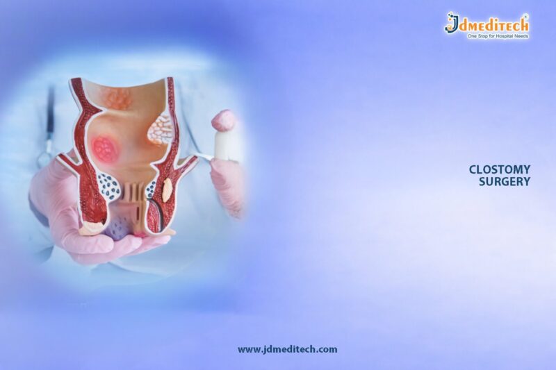
Prostatic Stent for BPH – Simple & Minimally Invasive Relief

A prostatic stent helps patients manage Benign Prostatic Hyperplasia (BPH) and urinary obstruction effectively. When the prostate enlarges, it presses on the urethra and blocks urine flow. This condition causes discomfort, weak stream, and incomplete emptying.

Doctors use a prostatic stent to restore urine flow without performing major surgery.

What is a Prostatic Stent?

A prostatic stent is a small tubular device that doctors place inside the urethra to keep it open. It supports the narrowed passage and allows urine to pass freely.

Manufacturers design each prostatic stent using biocompatible materials to ensure safety and flexibility.

How Does a Prostatic Stent Work?

The prostatic stent expands inside the urethra and creates a clear pathway for urine flow. It:

  • Keeps the urethra open
  • Relieves blockage caused by BPH
  • Improves urine flow immediately
  • Reduces pressure on the bladder

This action gives patients fast and noticeable relief.

Key Features of Prostatic Stent

The prostatic stent offers:

  • Endoscopic placement with minimal invasion
  • Self-expanding structure for secure fit
  • Flexible design for patient comfort
  • Biocompatible material to reduce irritation
  • Radiopaque markers for accurate positioning
  • Options for temporary and permanent use
Clinical Applications of Prostatic Stent

Doctors use the prostatic stent to treat:

  • Benign Prostatic Hyperplasia (BPH)
  • Urinary obstruction
  • Acute and chronic urinary retention
  • Patients who cannot undergo surgery
  • Temporary urinary management
Benefits of Prostatic Stent
1. Minimally Invasive Approach

Doctors place the prostatic stent without making major incisions.

2. Immediate Relief

Patients notice better urine flow soon after placement.

3. Quick Procedure

Doctors complete the prostatic stent procedure within 15–30 minutes.

4. Suitable for High-Risk Patients

Doctors recommend it for elderly patients and those with medical conditions.

5. Reversible Option

Doctors can remove temporary prostatic stent devices when needed.

Procedure Overview

Doctors follow a simple process:

  1. They administer local or regional anesthesia
  2. They insert a cystoscope through the urethra
  3. They position the prostatic stent carefully
  4. The stent expands and keeps the passage open

Most patients return home the same day.

Post-Procedure Care

Patients recover quickly after receiving a prostatic stent. Some may experience:

  • Mild discomfort
  • Urinary urgency
  • Slight irritation

These symptoms improve within a few days. Doctors schedule follow-ups to monitor stent performance.

Risks and Considerations

The prostatic stent remains safe, but some risks include:

  • Stent migration
  • Encrustation
  • Urinary tract infection
  • Temporary discomfort

Doctors reduce these risks through proper monitoring and care.

Why Choose a Prostatic Stent?

A prostatic stent provides a strong alternative to surgery. It delivers quick relief, reduces recovery time, and lowers complication risks. Therefore, many urologists prefer this solution.

Conclusion

The prostatic stent offers an effective and patient-friendly way to manage BPH and urinary obstruction. It combines safety, speed, and comfort in one solution.


Explore More Product >>

Get Connected:

+91 79909 93062 | +91 63513 72032 | exports@jdmeditech.com
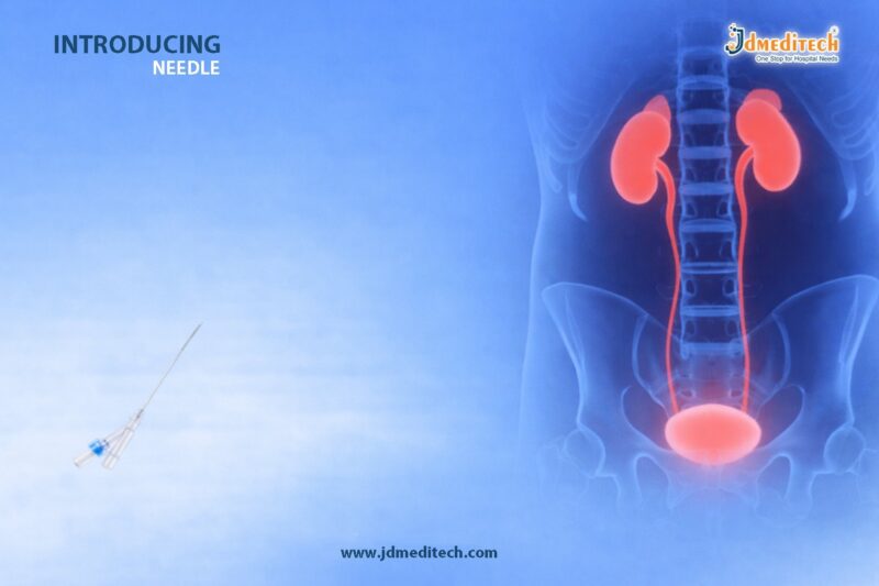
Endoscopy Hot Biopsy Forceps for Safe GI Tissue Sampling

Endoscopic procedures play a vital role in diagnosing and treating gastrointestinal (GI) disorders. Among the many tools used, Endoscopy Hot Biopsy Forceps stand out due to their dual functionality—allowing both tissue sampling and coagulation in a single device. This makes them highly efficient, especially in procedures involving bleeding control and precise biopsy.

In this blog, we explore the features, benefits, applications, and advantages of hot biopsy forceps in modern endoscopy.

What Are Endoscopy Hot Biopsy Forceps?

Endoscopy Hot Biopsy Forceps are specialized instruments used during GI endoscopy to grasp tissue and apply electrocautery simultaneously. Unlike standard biopsy forceps, these devices are connected to an electrosurgical unit, enabling controlled thermal energy delivery.

This dual-action capability helps clinicians perform biopsy and hemostasis at the same time, improving procedural efficiency and patient safety.

Key Features of Endoscopy Hot Biopsy Forceps
1. Dual Functionality (Biopsy + Coagulation)

Hot biopsy forceps combine mechanical tissue sampling with electrocautery, reducing the need for multiple instruments.

2. Insulated Shaft for Safety

The shaft is designed with high-quality insulation, ensuring targeted energy delivery and minimizing damage to surrounding tissues.

3. Precision Jaw Design

Sharp-edged jaws provide secure tissue grip and accurate sampling, even in delicate areas.

4. Compatibility with Electrosurgical Units

These forceps integrate seamlessly with standard electrosurgical generators, allowing controlled coagulation.

5. Ergonomic Handle

The user-friendly handle ensures better control and comfort, reducing operator fatigue during long procedures.

Clinical Applications

Endoscopy Hot Biopsy Forceps are widely used in various gastrointestinal procedures, including:

  • Polypectomy of small polyps
  • Biopsy of suspicious GI lesions
  • Control of minor bleeding (hemostasis)
  • Removal of superficial mucosal tissue
  • Therapeutic endoscopic interventions

Their ability to cut and coagulate simultaneously makes them especially useful in procedures where bleeding risk is present.

Benefits of Using Hot Biopsy Forceps
Enhanced Procedure Efficiency

Since tissue removal and coagulation happen together, procedure time is significantly reduced.

Improved Hemostasis

Electrocautery helps in immediate bleeding control, lowering complication risks.

Reduced Instrument Exchange

The dual-purpose design eliminates the need to switch between tools, improving workflow.

Increased Safety

Insulation and controlled energy delivery reduce the risk of thermal injury to surrounding tissues.

Cost-Effective Solution

By combining two functions in one device, healthcare facilities can optimize costs and inventory.

Why Choose High-Quality Hot Biopsy Forceps?

Selecting a premium-quality device ensures:

  • Consistent performance
  • Reliable energy transmission
  • Durability during repeated use
  • Patient safety and procedural success

Manufacturers like JDMeditech focus on delivering precision-engineered instruments that meet international quality standards.

Best Practices for Safe Use

To maximize safety and effectiveness:

  • Use appropriate electrosurgical settings
  • Avoid excessive energy application
  • Ensure proper grounding and insulation checks
  • Follow standard endoscopic guidelines
  • Train staff for optimal handling techniques
Comparison: Hot vs Cold Biopsy Forceps
Feature Hot Biopsy Forceps Cold Biopsy Forceps
Energy Use Electrocautery No energy
Bleeding Control Immediate Limited
Procedure Speed Faster Slower
Tissue Damage Minimal (controlled) Minimal
Use Case Biopsy + Hemostasis Diagnostic biopsy only
Conclusion

Endoscopy Hot Biopsy Forceps are an essential tool in modern GI endoscopy, offering a dual-function solution for tissue sampling and bleeding control. Their efficiency, safety, and versatility make them a preferred choice for gastroenterologists worldwide.

By integrating advanced design with reliable performance, these forceps enhance both clinical outcomes and procedural efficiency.


Explore More Product >>

Get Connected:

+91 79909 93062 | +91 63513 72032 | exports@jdmeditech.com
Re-Entry Malecot Nephrostomy Catheter for Drainage

The Re-Entry Malecot Nephrostomy Catheter offers a smart and efficient solution for renal drainage and repeat kidney access. It is specially designed for urology procedures where clinicians need both secure catheter placement and easy re-entry into the nephrostomy tract.

Unlike standard catheters, this device simplifies repeat interventions. It reduces the need for additional punctures, which improves patient comfort and saves valuable procedure time.

What is a Re-Entry Malecot Nephrostomy Catheter?

A Re-Entry Malecot Nephrostomy Catheter is a drainage catheter that combines:

  • A Malecot wing retention systemfor firm placement
  • A central lumen for guidewire access

This design allows doctors to maintain continuous drainage while also re-accessing the kidney tract whenever needed. As a result, it becomes highly useful in staged or repeat procedures.

Key Features
Secure Malecot Wing Retention

The catheter features expandable Malecot wings that:

  • Anchor the catheter securely in the renal pelvis
  • Reduce the risk of accidental dislodgement
  • Maintain stable positioning over time
Easy Re-Entry Access

The built-in re-entry feature allows:

  • Smooth guidewire insertion
  • Quick tract re-access
  • Fewer repeat punctures

This makes follow-up procedures faster and safer.

High-Quality Biocompatible Material

The catheter is made from medical-grade material that:

  • Minimizes tissue irritation
  • Enhances patient safety
  • Supports long-term use
Efficient Drainage Design

Multiple side holes:

  • Ensure continuous urine flow
  • Help prevent blockage
  • Improve overall drainage performance
Flexible and Kink-Resistant

The catheter remains flexible yet strong, which:

  • Prevents kinking
  • Maintains lumen patency
  • Adapts well to patient anatomy
Clinical Applications

The Re-Entry Malecot Nephrostomy Catheter is ideal for:

  • Percutaneous nephrostomy (PCN)
  • Obstructive uropathy management
  • Kidney stone procedures (PCNL)
  • Post-operative drainage
  • Staged urological procedures

It is especially useful when multiple interventions are planned, as it allows repeated access without creating a new tract.

Advantages Over Standard Catheters

This catheter provides clear benefits over conventional options:

  • Enables easy tract re-access
  • Reduces patient discomfort
  • Saves procedure time
  • Lowers the risk of complications
  • Improves overall procedural efficiency

Because of these advantages, it is widely preferred in modern urology practice.

Procedure Overview

Doctors typically place the catheter under imaging guidance:

  1. Access the renal pelvis
  2. Insert a guidewire
  3. Dilate the tract
  4. Position the catheter
  5. Deploy the Malecot wings for secure fixation

If another procedure is required, clinicians can reuse the same tract through the catheter lumen, making the process much simpler.

Why Choose This Catheter?

The Re-Entry Malecot Nephrostomy Catheter helps healthcare providers:

  • Maintain reliable drainage
  • Perform repeat procedures easily
  • Improve patient outcomes
  • Enhance workflow efficiency

It combines performance, safety, and convenience in one device.

Maintenance and Care

Proper care ensures long-lasting performance:

  • Flush regularly to prevent blockage
  • Monitor for infection signs
  • Secure the catheter properly
  • Replace as per clinical guidelines
Conclusion

The Re-Entry Malecot Nephrostomy Catheter is a practical and advanced solution for renal drainage. Its ability to provide secure placement and easy re-access makes it highly valuable in complex urological procedures.

For clinicians looking to improve efficiency and patient comfort, this catheter is a reliable choice.


Explore More Product >>

Get Connected:

+91 79909 93062 | +91 63513 72032 | exports@jdmeditech.com

Biliary Stent Introducer Set for Precise GI Stent Placement

In modern gastroenterology, precision and control are critical when managing biliary obstructions. The Biliary Stent Introducer Set plays a vital role in ensuring accurate and safe placement of stents during complex GI procedures such as ERCP (Endoscopic Retrograde Cholangiopancreatography).

Designed for efficiency, flexibility, and reliability, this advanced device helps clinicians navigate challenging anatomies while maintaining optimal procedural outcomes.

What is a Biliary Stent Introducer Set?

A Biliary Stent Introducer Set is a specialized medical device used to facilitate the controlled insertion of biliary stents into the bile duct. It typically includes:

  • Introducer catheter
  • Guidewire compatibility system
  • Pusher mechanism
  • Radiopaque markers for visualization

This set ensures smooth delivery and precise positioning of the stent in both simple and complex biliary interventions.

Key Features of Biliary Stent Introducer Set
1. High Precision Placement

The device enables accurate stent positioning within the biliary tract, minimizing procedural errors and improving clinical outcomes.

2. Smooth Trackability

Its flexible yet durable design allows easy navigation through tortuous or narrowed bile ducts.

3. Radiopaque Markers

Integrated markers enhance fluoroscopic visibility, ensuring correct placement under imaging guidance.

4. Guidewire Compatibility

Compatible with standard guidewires, making it versatile for various endoscopic procedures.

5. Controlled Deployment

The pusher system allows gradual and controlled stent release, reducing the risk of misplacement.

Clinical Applications

The Biliary Stent Introducer Set is widely used in:

  • Biliary obstruction management (benign & malignant)
  • ERCP procedures
  • Cholangiocarcinoma treatment
  • Gallstone-related bile duct blockage
  • Post-surgical biliary strictures

Its reliability makes it an essential tool in both routine and advanced GI interventions.

Benefits of Using Biliary Stent Introducer Set
  •  Ensures precise and stable stent placement
  •  Reduces procedure time
  •  Minimizes complications and trauma
  •  Enhances clinician control during deployment
  •  Improves patient safety and recovery
Why Choose a High-Quality Biliary Stent Introducer Set?

Choosing a premium-quality introducer set significantly impacts procedural success. High-grade materials and advanced engineering ensure:

  • Better flexibility and durability
  • Reduced friction during insertion
  • Consistent performance in complex anatomies

Healthcare professionals rely on trusted manufacturers like JDMeditech for reliable and high-performance GI devices.

Procedure Overview

During an ERCP procedure:

  1. A guidewire is first placed into the bile duct.
  2. The introducer catheter is advanced over the guidewire.
  3. The stent is loaded and positioned using fluoroscopic guidance.
  4. The pusher system deploys the stent precisely at the target site.

This step-by-step process ensures effective biliary drainage and restoration of bile flow.

Maintenance and Handling

Proper handling is essential for optimal performance:

  • Store in a sterile, dry environment
  • Use single-use devices as recommended
  • Follow standard sterilization protocols
  • Inspect packaging before use
Conclusion

The Biliary Stent Introducer Set is an indispensable tool in advanced GI endoscopy. With its precision, flexibility, and ease of use, it enhances the success rate of biliary stenting procedures while ensuring patient safety.

For healthcare providers aiming to achieve reliable outcomes in complex interventions, investing in a high-quality introducer set is essential.


Explore More Product >>

Get Connected:

+91 79909 93062 | +91 63513 72032 | exports@jdmeditech.com
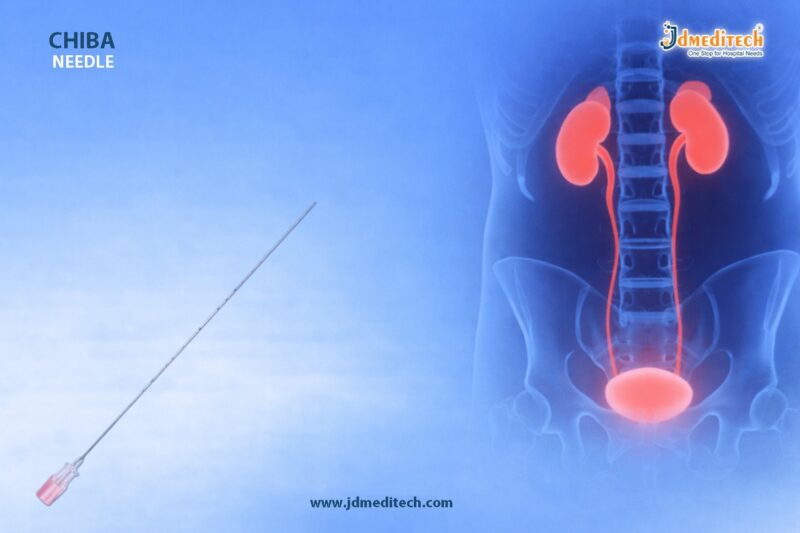
Malecot Catheter Kit for Safe Nephrostomy Drainage

The Malecot Catheter Kit plays a crucial role in modern urology, especially for effective nephrostomy drainage. Designed with a unique winged retention mechanism, this catheter ensures secure placement and continuous urine drainage from the kidney. As a trusted solution in percutaneous nephrostomy (PCN) procedures, it offers both reliability and ease of use for healthcare professionals.

What is a Malecot Catheter Kit?

A Malecot Catheter Kit is a comprehensive set used for establishing nephrostomy access and ensuring efficient renal drainage. The key component, the Malecot catheter, features expandable wings that anchor securely inside the renal pelvis.

This design minimizes the risk of dislodgement and provides consistent drainage, even in challenging clinical conditions.

Key Features of Malecot Catheter Kit
1. Winged Retention Design

The catheter features flexible Malecot wings that expand داخل the kidney, ensuring firm anchorage and preventing accidental displacement.

2. Smooth and Flexible Material

Made from high-quality medical-grade materials, the catheter remains flexible yet strong, preventing kinking and ensuring uninterrupted urine flow.

3. Complete Kit for Convenience

The kit typically includes:

  • Malecot catheter
  • Guidewire
  • Dilators
  • Introducer needle
  • Connecting tube

This all-in-one setup simplifies the nephrostomy procedure.

4. Radiopaque Visibility

Radiopaque lines allow precise placement under imaging guidance, improving procedural accuracy and patient safety.

5. Multiple Size Options

Available in various sizes to accommodate different patient anatomies and clinical requirements.

Clinical Applications
  • Percutaneous Nephrostomy (PCN)
  • Obstructive uropathy management
  • Hydronephrosis drainage
  • Post-surgical urinary diversion
  • Emergency kidney decompression
Advantages of Using Malecot Catheter Kit
Secure Placement

The winged structure ensures that the catheter stays firmly in place داخل the renal pelvis.

Efficient Drainage

Its wide lumen supports continuous and effective urine drainage, reducing the risk of blockage.

Reduced Complications

Minimizes risks such as catheter migration, leakage, and repeated interventions.

Ease of Insertion

The complete kit design streamlines the procedure, saving valuable time for clinicians.

Procedure Overview

During a percutaneous nephrostomy:

  1. The kidney is accessed using imaging guidance.
  2. A guidewire is inserted into the renal pelvis.
  3. Dilators are used to create a tract.
  4. The Malecot catheter is placed and its wings expand to secure positioning.
  5. The catheter is connected to a drainage system.
Why Choose a Malecot Catheter Kit?

Compared to other nephrostomy catheters, the Malecot Catheter Kit offers superior anchorage and reliability. Its winged design provides better retention than straight or pigtail catheters in certain clinical situations.

Maintenance and Care
  • Regularly monitor drainage output
  • Ensure proper fixation externally
  • Maintain sterile handling to prevent infection
  • Replace the catheter as per clinical guidelines
Conclusion

The Malecot Catheter Kit stands out as a complete and dependable solution for nephrostomy drainage. With its advanced retention mechanism, ease of use, and reliable performance, it remains a preferred choice among urologists and healthcare providers.

For high-quality Malecot Catheter Kits, visit www.jdmeditech.com and explore a wide range of advanced urology solutions.


Explore More Product >>

+91 79909 93062 | +91 63513 72032 | exports@jdmeditech.com
Malecot Nephrostomy Catheter – Secure Kidney Drainage Solution

The Malecot Nephrostomy Catheter plays a crucial role in modern urology by providing safe, stable, and efficient renal drainage. Designed with a unique winged (Malecot) retention tip, this catheter ensures secure placement within the renal pelvis, reducing the risk of displacement.

Healthcare professionals rely on the Malecot catheter in various nephrostomy procedures, especially when long-term drainage and stability are essential.

What is a Malecot Nephrostomy Catheter?

A Malecot Nephrostomy Catheter is a specialized drainage tube used to remove urine directly from the kidney when normal urinary flow is obstructed. Its defining feature is the expandable winged tip, which anchors the catheter securely inside the renal pelvis.

Unlike standard catheters, the Malecot design provides enhanced retention and reduced migration, making it ideal for critical drainage cases.

Key Features of Malecot Nephrostomy Catheter
1. Winged Retention Design

The Malecot wings expand inside the kidney, ensuring firm anchorage and minimizing accidental dislodgement.

2. Efficient Drainage Lumen

A wide internal lumen allows smooth and continuous urine drainage, preventing blockage.

3. Medical-Grade Biocompatible Material

Manufactured using high-quality, non-toxic materials, the catheter ensures patient safety and comfort.

4. Radiopaque Line for Visibility

The radiopaque marker enables accurate placement under imaging guidance.

5. Kink-Resistant Construction

Flexible yet strong design prevents kinking, ensuring uninterrupted flow.

Clinical Applications

The Malecot Nephrostomy Catheter is widely used in:

  • Percutaneous Nephrostomy (PCN) procedures
  • Obstructive uropathy management
  • Hydronephrosis relief
  • Post-surgical kidney drainage
  • Urinary diversion in complex cases
Advantages of Malecot Nephrostomy Catheter
Secure Positioning

The winged tip ensures strong internal fixation, reducing catheter migration.

Reliable Long-Term Drainage

Ideal for patients requiring extended nephrostomy support.

Reduced Risk of Leakage

Tight retention minimizes urine leakage around the catheter.

Easy Placement & Removal

Despite strong anchoring, the catheter allows controlled insertion and removal.

Procedure Overview

During a percutaneous nephrostomy, the Malecot catheter is inserted through the skin into the kidney under imaging guidance.

Steps include:

  1. Local anesthesia administration
  2. Needle puncture into renal pelvis
  3. Guidewire insertion
  4. Tract dilation
  5. Placement of Malecot catheter
  6. Expansion of wings for secure fixation
Why Choose Malecot Over Other Catheters?

Compared to pigtail catheters, the Malecot Nephrostomy Catheter offers stronger retention due to its winged design.

Feature Malecot Catheter Pigtail Catheter
Retention Strength High Moderate
Stability Excellent Good
Migration Risk Low Slightly Higher
Ideal Use Long-term drainage Short-term drainage
Care and Maintenance Tips

To ensure optimal performance:

  • Maintain proper catheter hygiene
  • Regularly monitor urine flow
  • Flush the catheter if advised by a clinician
  • Check for signs of infection or blockage
Conclusion

The Malecot Nephrostomy Catheter stands out as a reliable and secure solution for renal drainage, thanks to its innovative winged retention design. Its stability, efficiency, and safety make it a preferred choice among urologists worldwide.

For healthcare providers seeking dependable nephrostomy solutions, this catheter offers the perfect balance of performance and patient comfort.


Explore More Product >>

Get Connected:

+91 79909 93062 | +91 63513 72032 | exports@jdmeditech.com

 

Endoscopy Foreign Body Forceps for Safe Retrieval

Endoscopy foreign body forceps play a crucial role in gastrointestinal procedures where safe retrieval of ingested or lodged objects is required. These precision-designed instruments help clinicians grasp, hold, and remove foreign bodies effectively without causing mucosal injury.

With increasing cases of accidental ingestion and complex endoscopic interventions, the demand for reliable and high-performance foreign body forceps has grown significantly. Their ergonomic design and strong gripping capability make them essential tools in modern endoscopy suites.

What Are Endoscopy Foreign Body Forceps?

Endoscopy foreign body forceps are specialized devices used during endoscopic procedures to retrieve foreign objects from the gastrointestinal tract. These may include:

  • Coins
  • Food boluses
  • Bones
  • Dental prosthetics
  • Sharp or irregular objects

They are inserted through the working channel of an endoscope, allowing controlled and precise manipulation inside the GI tract.

Key Features of Endoscopy Foreign Body Forceps
1. Strong Gripping Strength

These forceps provide a firm hold on objects, reducing the risk of slippage during retrieval.

2. Precision-Engineered Jaws

Different jaw designs such as alligator, rat-tooth, or tripod enable handling of various object shapes and sizes.

3. Flexible and Durable Shaft

The flexible shaft allows easy navigation through the endoscope while maintaining structural integrity.

4. Smooth Operation

Ergonomic handles ensure precise control and smooth opening and closing of the jaws.

5. Atraumatic Design

Designed to minimize tissue damage during insertion and extraction.

Types of Foreign Body Forceps

Depending on the clinical need, several types are available:

  • Alligator Forceps : Ideal for grasping soft or flat objects
  • Rat-Tooth Forceps : Suitable for firm gripping of tough objects
  • Tripod Forceps : Provides multi-point grip for irregular shapes
  • Basket-Type Forceps : Used for capturing round or loose objects
Clinical Applications

Endoscopy foreign body forceps are widely used in:

  • Gastroenterology : Removal of ingested objects from esophagus, stomach, or intestines
  • Emergency Endoscopy : Managing accidental ingestion cases
  • Pediatric Endoscopy : Safe retrieval in children
  • ENT Procedures : Removal of foreign bodies in upper GI and airway
Benefits of Using Endoscopy Foreign Body Forceps
  •  Ensures safe and controlled removal
  •  Reduces risk of complications or perforation
  •  Provides high precision and maneuverability
  •  Compatible with most standard endoscopes
  •  Improves procedure efficiency and success rate
How to Choose the Right Forceps

Selecting the right instrument depends on:

  • Type and size of foreign body
  • Location within the GI tract
  • Required gripping strength
  • Endoscope compatibility

Clinicians should always opt for high-quality, medical-grade instruments to ensure optimal outcomes.

Why Choose JDMeditech Endoscopy Foreign Body Forceps?

At JDMeditech, we design endoscopy foreign body forceps with a focus on safety, precision, and durability. Our products offer:

  • Superior grip and control
  • High-quality biocompatible materials
  • Smooth functionality for easy handling
  • Reliable performance in critical procedures
Best Practices for Safe Use
  • Always visualize the object clearly before grasping
  • Use appropriate jaw type for the object
  • Avoid excessive force to prevent tissue injury
  • Ensure proper sterilization before use
Conclusion

Endoscopy foreign body forceps are indispensable tools for safe and effective retrieval of foreign objects during endoscopic procedures. Their advanced design, strong grip, and precision handling significantly enhance procedural success while ensuring patient safety.

Healthcare professionals rely on these instruments for efficient and complication-free outcomes, making them a vital component in modern endoscopy.


Explore More Product >>

Get Connected:

+91 79909 93062 | +91 63513 72032 | exports@jdmeditech.com
 

Endopyelotomy Stent for UPJ Repair – Reliable Ureteral Support

The Endopyelotomy Stent plays a crucial role in modern urological procedures, particularly in the management of ureteropelvic junction (UPJ) obstruction. Designed to provide internal drainage and structural support after endopyelotomy, this specialized stent ensures proper healing and maintains patency of the urinary tract.

With advancements in minimally invasive urology, endopyelotomy stents have become indispensable tools for urologists seeking reliable postoperative outcomes.

What is an Endopyelotomy Stent?

An Endopyelotomy Stent is a specially designed ureteral stent used after performing an endopyelotomy, a minimally invasive procedure to treat UPJ obstruction.

Unlike conventional stents, this device features:

  • A tailored design for precise placement
  • Enhanced lumen for efficient urine drainage
  • Structural support to aid healing of the incision site

It helps maintain the opening between the renal pelvis and ureter, preventing restenosis during recovery.

Understanding UPJ Obstruction and Its Treatment
What is UPJ Obstruction?

UPJ obstruction occurs when there is a blockage at the junction where the kidney meets the ureter. This condition can lead to:

  • Hydronephrosis (kidney swelling)
  • Flank pain
  • Recurrent infections
  • Impaired kidney function
Role of Endopyelotomy

Endopyelotomy involves making an incision at the obstructed segment to restore urine flow. However, maintaining the patency of this incision is critical—this is where the Endopyelotomy Stent for UPJ Repair becomes essential.

Key Features of Endopyelotomy Stents
1. Optimized Length and Design

The stent is longer and specifically shaped to span the renal pelvis and ureter, ensuring complete support across the UPJ.

2. Multiple Drainage Holes

Strategically placed side holes allow continuous urine drainage, reducing pressure buildup.

3. High-Quality Biocompatible Material

Typically made from polyurethane or silicone, these stents offer:

  • Flexibility
  • Reduced irritation
  • Long-term compatibility
4. Radiopaque Markers

These enable precise placement and easy visualization under fluoroscopy.

5. Anti-Migration Design

Pigtail or coil ends prevent displacement during the healing phase.

Benefits of Using an Endopyelotomy Stent for UPJ Repair

Using an Endopyelotomy Stent for UPJ Repair provides several clinical advantages:

  •  Maintains ureteral patency after incision
  •  Promotes faster and safer healing
  •  Reduces risk of restenosis
  •  Ensures continuous urine drainage
  •  Minimizes postoperative complications
Procedure Overview
Step-by-Step Use
  1. Diagnosis and Imaging
    UPJ obstruction is confirmed via ultrasound, CT scan, or IVP.
  2. Endopyelotomy Procedure
    The obstructed segment is incised using endoscopic tools.
  3. Stent Placement
    The endopyelotomy stent is inserted across the UPJ.
  4. Postoperative Monitoring
    The stent remains in place for several weeks to support healing.
  5. Stent Removal
    After adequate healing, the stent is removed endoscopically.
Clinical Applications

The Endopyelotomy Stent is widely used in:

  • Treatment of primary UPJ obstruction
  • Management of recurrent UPJ strictures
  • Postoperative support following endopyelotomy procedures
  • Selected cases of failed pyeloplasty
Why Choose High-Quality Endopyelotomy Stents?

Choosing a high-quality stent ensures:

  • Better patient comfort
  • Reduced encrustation risk
  • Improved procedural success rates
  • Reliable long-term outcomes

At JDMeditech, we focus on delivering precision-engineered urology devices that meet global standards.

Best Practices for Stent Management

To achieve optimal results:

  • Monitor patients regularly for complications
  • Ensure timely stent removal
  • Use appropriate stent size and length
  • Maintain strict sterile protocols
Conclusion

The Endopyelotomy Stent for UPJ Repair is a vital component in the successful management of ureteropelvic junction obstruction. By providing consistent drainage and structural support, it significantly enhances healing outcomes after endopyelotomy.

As minimally invasive techniques continue to evolve, the role of advanced stenting solutions will only become more critical in ensuring patient safety and procedural success.


Explore More Product >>

Get Connected:

+91 79909 93062 | +91 63513 72032 | exports@jdmeditech.com