Urometer for Accurate Urine Output Measurement in Critical Care

A urometer for accurate urine output measurement is an essential medical device in modern healthcare. In critical care settings, continuous monitoring of urine output is not just important, but also life-saving. Therefore, healthcare professionals rely on precise devices like urometers to assess kidney function and fluid balance effectively. Moreover, accurate urine measurement helps in early diagnosis and timely clinical intervention.

What is a Urometer?

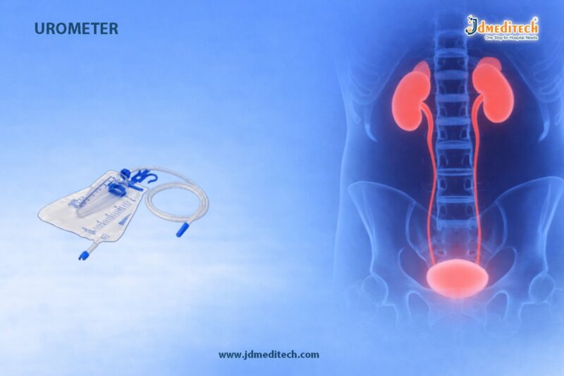
A urometer is a specialized urine collection system equipped with a calibrated measuring chamber. Unlike standard urine bags, it allows precise measurement over short intervals. As a result, it becomes highly suitable for ICU patients and postoperative care. In addition, its design ensures continuous monitoring without interrupting urine flow.

Why Accurate Urine Output Monitoring Matters

Urine output is a key indicator of a patient’s health status. For instance, reduced urine output may signal kidney dysfunction, while excessive output may indicate fluid imbalance. Therefore, using a urometer for accurate urine output measurement becomes critical.

Furthermore, proper monitoring helps in:

  • Assessing kidney function
  • Managing fluid therapy
  • Detecting dehydration early
  • Preventing fluid overload
  • Identifying acute kidney injury (AKI)

Consequently, clinicians can make faster and more informed decisions.

Key Features of a Urometer
Calibrated Measuring Chamber

First of all, the urometer includes a precisely graduated chamber, usually up to 500 ml. This ensures accurate hourly readings.

Anti-Reflux Valve

In addition, the anti-reflux valve prevents backflow of urine. As a result, it reduces infection risk and maintains hygiene.

Transparent Design

Moreover, the clear body allows easy observation of urine color and volume. Therefore, clinicians can quickly detect abnormalities.

Needle Sampling Port

Similarly, the sampling port enables safe and contamination-free urine collection. This feature is especially useful for lab testing.

Universal Connector

Furthermore, the device connects easily with standard Foley catheters. Hence, it offers wide compatibility.

Large Drainage Bag

Finally, the attached urine bag allows continuous drainage. Consequently, it reduces the need for frequent emptying.

Applications in Critical Care

A urometer is widely used in several medical environments. For example, it is essential in ICUs where patients require constant monitoring. Likewise, it is commonly used in postoperative recovery units and emergency departments.

Additionally, it plays a vital role in:

  • Nephrology and urology wards
  • Trauma care units
  • Burn management units

Thus, it supports accurate patient assessment across multiple specialties.

How a Urometer Works

The working mechanism of a urometer is simple yet efficient. Initially, urine flows from the catheter into the calibrated chamber. Then, healthcare providers measure the output at regular intervals, usually hourly. After that, the chamber automatically drains into the collection bag. As a result, continuous monitoring becomes easy and uninterrupted.

Benefits of Using a Urometer

Using a urometer for accurate urine output measurement offers multiple advantages.

  • First, it provides high precision in measurement
  • Additionally, it enables early detection of complications
  • Moreover, it improves patient safety
  • Furthermore, it reduces infection risk due to a closed system
  • Lastly, it is easy to use and dispose of

Therefore, it significantly enhances clinical efficiency.

Urometer vs Standard Urine Collection Bag

Although both devices collect urine, their purposes differ. A standard urine bag is suitable for general use. However, a urometer is specifically designed for critical care. Hence, it provides more accurate and reliable measurements.

Quality and Manufacturing Standards

At JDMeditech, we manufacture high-quality urometers using medical-grade materials. Moreover, our products comply with international quality standards. As a result, healthcare providers can rely on our devices for safety, durability, and performance.

Conclusion

In conclusion, a urometer for accurate urine output measurement in critical care is indispensable in modern healthcare. Not only does it provide precise monitoring, but it also helps improve patient outcomes. Therefore, choosing a reliable and high-quality urometer is essential for effective clinical management


Explore More Product >>

+91 79909 93062 | +91 63513 72032 | exports@jdmeditech.com


Ileostomy Surgery – Effective Treatment for GI Conditions

Ileostomy surgery is a life-saving procedure used to manage severe gastrointestinal conditions when normal bowel function is no longer possible or safe. In this procedure, surgeons create an opening (stoma) in the abdominal wall to divert waste from the small intestine (ileum) to an external pouch. As a result, patients can safely eliminate waste without passing it through the colon.

Moreover, ileostomy surgery plays a critical role in improving the quality of life for patients suffering from chronic digestive disorders, cancers, or traumatic injuries.

What is Ileostomy Surgery?

Ileostomy surgery is a surgical procedure in which the ileum is brought through the abdominal wall to form a stoma. This stoma allows intestinal waste to exit the body into a specially designed ostomy bag.

Unlike colostomy, where waste passes through part of the colon, ileostomy bypasses the entire large intestine. Therefore, the output is usually more liquid and requires careful management.

Why is Ileostomy Surgery Performed?

Doctors recommend ileostomy surgery when the colon or rectum cannot function properly. In many cases, it becomes the most effective treatment for severe gastrointestinal conditions.

Common Indications:
  • Inflammatory bowel diseases such as Crohn’s disease and ulcerative colitis
  • Colorectal cancer
  • Bowel obstruction or perforation
  • Familial adenomatous polyposis (FAP)
  • Severe infections or trauma affecting the colon
  • Congenital abnormalities in children

Additionally, ileostomy may be temporary or permanent depending on the patient’s condition and recovery plan.

Types of Ileostomy

Understanding the types of ileostomy helps patients and healthcare providers choose the most suitable approach.

1. End Ileostomy

In this type, the end of the ileum is brought out through the abdomen. It is often permanent and commonly performed after removal of the colon.

2. Loop Ileostomy

Here, a loop of the small intestine is brought to the surface, and an opening is created. This type is usually temporary and used to divert waste while the lower bowel heals.

3. Continent Ileostomy (Kock Pouch)

In this advanced technique, an internal pouch is created, and the patient drains waste using a catheter. Although less common, it offers an alternative to external bags.

Procedure of Ileostomy Surgery

Ileostomy surgery is performed under general anesthesia by experienced surgeons.

First, the surgeon makes an incision in the abdomen. Then, the ileum is carefully separated and brought out through the abdominal wall. After that, the stoma is formed and secured in place. Finally, an ostomy pouch is attached to collect waste.

In many cases, surgeons may use minimally invasive (laparoscopic) techniques. Consequently, patients may experience faster recovery and reduced postoperative pain.

Benefits of Ileostomy Surgery

Ileostomy surgery offers several advantages, especially for patients with severe gastrointestinal disorders.

  • Relieves symptoms such as pain, bleeding, and obstruction
  • Prevents life-threatening complications
  • Improves overall quality of life
  • Allows healing of the affected bowel
  • Enables better disease management

Furthermore, patients often regain the ability to perform daily activities with proper stoma care.

Risks and Complications

Like any surgical procedure, ileostomy surgery carries certain risks. However, proper medical care significantly reduces complications.

Possible Risks:
  • Infection at the surgical site
  • Stoma irritation or skin problems
  • Dehydration due to fluid loss
  • Blockage of the stoma
  • Hernia near the stoma

Therefore, regular follow-ups and correct stoma management are essential for long-term success.

Recovery and Postoperative Care

Recovery after ileostomy surgery varies from patient to patient. Typically, patients stay in the hospital for several days.

Initially, patients receive guidance on how to care for the stoma. Then, they gradually adapt to using ostomy bags and managing their diet.

Key Care Tips:
  • Maintain proper hygiene around the stoma
  • Stay hydrated to prevent dehydration
  • Follow a balanced and low-residue diet initially
  • Monitor stoma output regularly
  • Consult healthcare professionals for any concerns

In addition, support from trained stoma care nurses plays a crucial role in recovery.

Living with an Ileostomy

Adjusting to life with an ileostomy can feel challenging at first. However, with the right support and education, most patients lead active and fulfilling lives.

Patients can return to work, travel, and participate in social activities. Modern ostomy products provide comfort, discretion, and reliability, which further enhances confidence.

Conclusion

Ileostomy surgery is an effective treatment for severe gastrointestinal conditions that cannot be managed through conventional therapies. It not only saves lives but also significantly improves patient comfort and well-being.

With advancements in surgical techniques and improved ostomy care products, patients today can enjoy a better quality of life after surgery. Therefore, understanding the procedure, benefits, and care requirements is essential for successful outcomes.


Explore More Product >>

Get Connected:
+91 79909 93062 | +91 63513 72032 | exports@jdmeditech.com 

Stone Collector for Safe Stone Retrieval in Urology

A Stone Collector plays a key role in urology by helping doctors remove urinary stones safely during procedures like ureteroscopy and cystoscopy. As minimally invasive treatments continue to grow, surgeons rely more on precise and efficient stone retrieval tools.

This device allows doctors to capture and remove stones or fragments from the kidney, ureter, or bladder. It also helps achieve complete stone clearance and lowers the risk of recurrence.

What is a Stone Collector?

A Stone Collector is a specially designed instrument that retrieves stones from the urinary tract. It includes a flexible wire basket or grasping mechanism that fits through an endoscope.

During the procedure, the surgeon locates the stone and opens the basket to capture it securely. Then, the surgeon carefully withdraws the device while protecting surrounding tissues. This method ensures safe and controlled stone removal.

Key Features of a Stone Collector

A high-quality Stone Collector offers features that improve performance and ease of use:

  • Flexible basket design allows smooth navigation
  • Atraumatic structure reduces the risk of tissue injury
  • Strong grip secures the stone during retrieval
  • Smooth mechanism provides better control
  • Compatibility supports use with standard endoscopes
  • Multiple sizes meet different clinical needs

These features help surgeons perform procedures with greater confidence and accuracy.

Applications in Urology

Surgeons use Stone Collectors in various urology procedures. They remove ureteral stones during ureteroscopy and clear kidney stones in PCNL procedures. They also retrieve bladder stones during cystoscopy.

In addition, surgeons use these devices after lithotripsy to collect fragmented stones. This approach ensures complete removal and improves treatment success.

Benefits of Using a Stone Collector

A Stone Collector offers several important benefits. It helps doctors remove stones completely and reduces the chance of recurrence. It also protects the urinary tract by minimizing trauma.

Furthermore, it improves procedural efficiency and shortens surgery time. Surgeons gain better control and precision during stone retrieval, which leads to better patient outcomes.

Types of Stone Collectors

Different types of Stone Collectors support various clinical needs:

  • Basket type captures stones effectively
  • Tipless design reduces trauma during insertion
  • Helical or spiral basket grips irregular stones better
  • Nitinol basket provides flexibility and durability

Surgeons choose the right type based on stone size, shape, and location.

Why Choose JDMeditech Stone Collector?

JDMeditech manufactures and exports high-quality urology instruments that meet international standards. Our Stone Collectors deliver reliable performance in demanding procedures.

We design our products with precision and focus on durability and ease of use. Our strict quality checks ensure safety and consistency in every device.

Best Practices for Use

Follow proper techniques to achieve the best results. Choose the correct size and type based on the stone. Ensure clear visualization before capturing it.

Handle the device gently during insertion and retrieval to avoid injury. Always follow proper sterilization protocols for reusable instruments.

Conclusion

A Stone Collector remains an essential tool in modern urology. It helps surgeons perform safe, precise, and efficient stone retrieval procedures.

With proper use and high-quality design, this device improves surgical outcomes and supports faster patient recovery. Choosing a trusted manufacturer like JDMeditech ensures consistent quality and performance.

Explore More Product >>

+91 79909 93062 | +91 63513 72032 | exports@jdmeditech.com
 

TUR Set for Transurethral Resection Procedures | Complete Guide

A TUR Set (Transurethral Resection Set) is an essential collection of surgical instruments used in urology for performing minimally invasive procedures such as Transurethral Resection of the Prostate (TURP) and bladder tumor resection. These procedures are widely preferred due to their precision, reduced recovery time, and minimal complications.

In modern urological practice, a high-quality TUR Set plays a critical role in ensuring efficiency, safety, and optimal surgical outcomes.

What is a TUR Set?

A TUR Set is a comprehensive kit that includes all the necessary instruments required to perform transurethral resections. It allows surgeons to access the urinary tract through the urethra without making external incisions.

As a result, the procedure becomes less invasive, reduces hospital stay, and improves patient comfort.

Components of a TUR Set

A standard TUR Set typically includes:

  • Resectoscope Sheath (Inner & Outer)
  • Working Element (Monopolar or Bipolar)
  • Electrodes/Loops (Cutting Loop, Roller Ball, Vaporization Electrode)
  • Optical Telescope (0°, 12°, or 30°)
  • Obturator
  • Irrigation Tubing System
  • Light Source Cable
  • Camera System (optional for advanced setups)

Each component is carefully designed to provide precision, durability, and ease of use during surgical procedures.

Key Features of a High-Quality TUR Set
1. Precision Engineering

The instruments are designed for accurate tissue resection, ensuring better surgical control.

2. High-Grade Materials

Most components are made from medical-grade stainless steel, ensuring durability and resistance to corrosion.

3. Compatibility

A good TUR Set is compatible with standard endoscopic systems, including cameras and light sources.

4. Ergonomic Design

The working element and handles are designed to reduce surgeon fatigue during long procedures.

5. Reusable & Sterilizable

These sets are easy to clean and sterilize, making them cost-effective for hospitals.

Applications of TUR Set

The TUR Set is widely used in various urological procedures, including:

  • Transurethral Resection of the Prostate (TURP)
  • Bladder Tumor Resection (TURBT)
  • Management of urethral strictures
  • Removal of abnormal tissue growths

Because of its versatility, it is a must-have instrument in any urology department.

Advantages of Using a TUR Set

Using a high-quality TUR Set offers several benefits:

  • Minimally invasive procedure
  • Reduced bleeding and complications
  • Faster recovery time
  • Shorter hospital stay
  • Improved surgical precision

Therefore, it has become the gold standard for many urological treatments.

Monopolar vs Bipolar TUR Set
Monopolar TUR Set
  • Requires non-conductive irrigation fluid (glycine)
  • Widely used and cost-effective
  • Suitable for standard procedures
Bipolar TUR Set
  • Uses saline as irrigation fluid
  • Lower risk of TUR syndrome
  • Better safety and control

Choosing between the two depends on the surgeon’s preference and clinical requirements.

How to Choose the Right TUR Set

When selecting a TUR Set, consider the following factors:

  • Quality and certification standards
  • Compatibility with existing equipment
  • Type (monopolar or bipolar)
  • Ease of sterilization
  • Manufacturer reliability

A trusted manufacturer ensures consistent performance and long-term reliability.

Why Choose JDMeditech TUR Set?

At JDMeditech, we specialize in manufacturing and exporting high-quality TUR Sets that meet international standards.

Our Advantages:
  • Premium-quality materials
  • Precision manufacturing
  • Global supply capability
  • Competitive pricing
  • Strict quality control
Conclusion

The TUR Set is a complete and indispensable solution for transurethral resection procedures. With its advanced design, precision, and versatility, it significantly improves surgical outcomes while ensuring patient safety.

As minimally invasive urology continues to evolve, investing in a high-quality TUR Set is essential for modern healthcare facilities.


Explore More Product >>

+91 79909 93062 | +91 63513 72032 | exports@jdmeditech.com 

Hybrid Coronary Revascularization – Combined Treatment

Hybrid Coronary Revascularization is an advanced cardiac treatment that combines the strengths of coronary artery bypass surgery and percutaneous coronary intervention (PCI). This innovative approach offers an effective solution for patients with complex coronary artery disease, especially when traditional methods alone may not provide optimal results. By integrating surgical precision with minimally invasive stent placement, Hybrid Coronary Revascularization improves patient outcomes while reducing recovery time.

What is Hybrid Coronary Revascularization?

Hybrid Coronary Revascularization is a treatment strategy that involves performing a minimally invasive coronary artery bypass graft (CABG) on critical arteries, typically the left anterior descending artery (LAD), along with stenting of other blocked coronary arteries using PCI. This combination allows cardiologists and cardiac surgeons to tailor treatment based on the patient’s specific condition.

Unlike conventional open-heart surgery, this approach avoids full sternotomy in many cases. As a result, patients benefit from less trauma, reduced hospital stay, and faster recovery.

Why Hybrid Coronary Revascularization is Needed

Coronary artery disease can affect multiple vessels with varying levels of blockage. In many cases, one artery may require surgical bypass for long-term durability, while others can be effectively treated with stents. Therefore, Hybrid Coronary Revascularization offers a balanced and patient-specific solution.

Moreover, this method is particularly useful for:

  • Patients with multi-vessel coronary artery disease
  • Individuals at high risk for traditional open-heart surgery
  • Patients requiring faster recovery due to lifestyle or medical conditions
  • Cases where minimally invasive options are preferred
Procedure Overview

The Hybrid Coronary Revascularization procedure is typically performed in two stages or sometimes in a single hybrid operating room.

First, a cardiac surgeon performs a minimally invasive bypass graft, usually using the internal mammary artery to bypass the LAD artery. This step ensures long-term patency and excellent blood flow.

Next, an interventional cardiologist performs angioplasty and places stents in other affected coronary arteries. These stents help restore blood flow without the need for extensive surgery.

Because both techniques are used, the procedure maximizes the advantages of each method while minimizing their limitations.

Benefits of Hybrid Coronary Revascularization

Hybrid Coronary Revascularization offers several advantages over traditional treatments. Firstly, it reduces surgical trauma since it avoids full chest opening in many cases. Secondly, patients experience shorter hospital stays and quicker return to normal activities.

In addition, this approach provides:

  • Improved long-term outcomes for critical arteries like the LAD
  • Lower risk of complications compared to full CABG
  • Reduced blood loss and infection risk
  • Enhanced cosmetic results due to smaller incisions
  • Faster recovery and rehabilitation
Risks and Considerations

Although Hybrid Coronary Revascularization is highly effective, it still carries some risks. These may include bleeding, infection, or complications related to stent placement. However, with experienced medical teams and proper patient selection, these risks remain minimal.

Furthermore, not all patients are suitable candidates. Therefore, thorough evaluation by a multidisciplinary heart team is essential before proceeding with this treatment.

Recovery and Post-Procedure Care

Recovery after Hybrid Coronary Revascularization is generally faster compared to traditional open-heart surgery. Most patients can return to daily activities within a few weeks.

However, proper post-procedure care is crucial. Patients are advised to follow prescribed medications, maintain a heart-healthy diet, and engage in regular physical activity. In addition, routine follow-ups with cardiologists help ensure long-term success.

Advanced Technology Supporting Hybrid Procedures

Modern cardiac procedures rely on advanced medical equipment for precision and safety. Devices such as Electrosurgical Units play a vital role in minimally invasive surgeries by enabling precise tissue cutting and coagulation.

Future of Hybrid Coronary Revascularization

Hybrid Coronary Revascularization represents the future of cardiac care. As technology continues to evolve, more hospitals are adopting hybrid operating rooms that allow seamless collaboration between surgeons and cardiologists.

Furthermore, advancements in imaging, robotic surgery, and stent technology will continue to enhance the effectiveness of this approach. Consequently, patients can expect even better outcomes and safer procedures in the coming years.

Conclusion

Hybrid Coronary Revascularization is a breakthrough in the treatment of coronary artery disease. By combining surgical bypass with stent-based intervention, it offers a personalized, less invasive, and highly effective solution.

As a result, patients benefit from improved recovery times, reduced risks, and long-term cardiovascular health. Therefore, this combined approach is becoming an increasingly preferred option in modern cardiac care.

Explore More Product >>

Get Connected:
+91 79909 93062 | +91 63513 72032 | exports@jdmeditech.com 

Penile Clamp – Effective Solution for Male Urinary Incontinence

Penile Clamp is a simple yet highly effective device designed to manage male urinary incontinence. This condition, which involves involuntary leakage of urine, can significantly affect a patient’s confidence and quality of life. However, with the use of a Penile Clamp, patients can regain better control over urinary leakage in a non-invasive and convenient way.

Male urinary incontinence may occur due to prostate surgery, neurological disorders, aging, or bladder dysfunction. Therefore, finding a reliable management solution becomes essential. The Penile Clamp offers a practical approach by applying gentle pressure to the urethra, thereby preventing unwanted urine flow while allowing controlled release when needed.

What is a Penile Clamp?

A Penile Clamp is a small external medical device that fits around the penis to control urine leakage. It works by compressing the urethra, which stops urine from passing involuntarily. Importantly, the device is adjustable, comfortable, and reusable, making it suitable for long-term use under medical guidance.

Moreover, modern penile clamps are made using soft medical-grade materials to ensure patient safety and comfort. They are lightweight, discreet, and easy to use, which makes them a preferred choice for many patients dealing with urinary incontinence.

How Does a Penile Clamp Work?

The Penile Clamp functions by applying controlled pressure to the urethra. As a result, it effectively blocks the flow of urine. However, when the patient needs to urinate, the clamp can be easily released, allowing normal urination.

In addition, the device is designed to distribute pressure evenly. This prevents discomfort or tissue damage when used correctly. Most importantly, proper usage and regular adjustments are necessary to maintain blood circulation and avoid complications.

Key Features of Penile Clamp
  • Adjustable pressure control for personalized comfort
  • Soft padding for enhanced safety and reduced irritation
  • Lightweight and discreet design for daily use
  • Reusable and cost-effective solution
  • Easy to apply and remove without assistance
Benefits of Using a Penile Clamp

Using a Penile Clamp provides several advantages for patients suffering from male urinary incontinence.

Firstly, it offers immediate control over urine leakage, which improves confidence in social and professional settings. Secondly, it eliminates the constant need for absorbent pads or diapers, reducing long-term costs. Furthermore, it allows patients to maintain an active lifestyle without fear of embarrassment.

Additionally, the device is non-invasive, meaning it does not require surgery or medication. Therefore, it is an excellent option for patients looking for a simple and effective management solution.

Applications of Penile Clamp

The Penile Clamp is widely used in various clinical and home care settings.

  • Post-prostate surgery incontinence
  • Stress urinary incontinence in men
  • Neurological conditions affecting bladder control
  • Temporary urine control during rehabilitation
Safety and Usage Guidelines

While the Penile Clamp is highly effective, proper usage is essential to ensure safety.

Patients should not wear the clamp continuously for extended periods. Instead, it is recommended to release the clamp every 1–2 hours to restore normal blood flow. Additionally, the device should be positioned correctly to avoid excessive pressure.

Furthermore, patients must consult a healthcare professional before using a Penile Clamp, especially if they have sensitive skin or circulatory issues.

Why Choose JDMeditech Penile Clamp?

JDMeditech offers high-quality Penile Clamp solutions designed with precision and patient comfort in mind. Our products are manufactured using advanced materials to ensure durability, safety, and optimal performance.

With a focus on innovation and reliability, JDMeditech provides medical devices that meet international quality standards. As a result, healthcare professionals and patients trust our solutions for effective urinary incontinence management.

Conclusion

Penile Clamp is a practical and effective solution for managing male urinary incontinence. It provides a non-invasive, affordable, and reliable way to control urine leakage while improving quality of life. With proper usage and medical guidance, patients can achieve better comfort, confidence, and independence in their daily lives.

Explore More Product >>

Colostomy Surgery – Effective Treatment for Intestinal Conditions

Colostomy Surgery is a life-saving medical procedure used to treat severe intestinal conditions when normal bowel function becomes impossible. In this procedure, surgeons create an opening (stoma) in the abdominal wall to divert waste into a colostomy bag. As a result, patients can safely eliminate waste while protecting the affected portion of the colon.

Moreover, Colostomy Surgery plays a crucial role in managing conditions such as colorectal cancer, bowel obstruction, trauma, and inflammatory bowel disease. Therefore, understanding this procedure helps patients and caregivers make informed decisions.

What is Colostomy Surgery?

Colostomy Surgery involves bringing one end of the large intestine (colon) through the abdominal wall to form a stoma. This opening allows waste to exit the body into a disposable pouch.

Additionally, the surgery may be temporary or permanent depending on the underlying condition. While temporary colostomies allow the colon to heal, permanent ones are necessary when part of the colon is removed.

Types of Colostomy Surgery

There are different types of Colostomy Surgery, depending on the location of the stoma:

  • Ascending Colostomy– Located on the right side of the abdomen
  • Transverse Colostomy– Positioned across the upper abdomen
  • Descending Colostomy– Found on the left side
  • Sigmoid Colostomy– Most common type, near the sigmoid colon

Each type of Colostomy Surgery affects stool consistency and output frequency.

Indications for Colostomy Surgery

Doctors recommend Colostomy Surgery in several serious conditions. For example:

  • Colorectal cancer
  • Severe intestinal injury or trauma
  • Bowel obstruction
  • Diverticulitis complications
  • Inflammatory bowel diseases (Crohn’s disease, ulcerative colitis)
  • Congenital defects

Therefore, Colostomy Surgery ensures safe waste elimination when the colon cannot function normally.

Procedure of Colostomy Surgery

The process of Colostomy Surgery involves several steps:

  1. Administration of general anesthesia
  2. Making an incision in the abdomen
  3. Identifying and preparing the colon
  4. Creating a stoma on the abdominal wall
  5. Attaching a colostomy bag

Furthermore, surgeons carefully position the stoma to ensure comfort and easy management for the patient.

Benefits of Colostomy Surgery

Colostomy Surgery offers multiple advantages:

  • Relieves severe intestinal blockage
  • Prevents infection and complications
  • Improves quality of life
  • Supports healing of damaged intestines
  • Provides a safe and controlled waste removal system

As a result, patients experience significant relief and improved health outcomes.

Risks and Complications

Although Colostomy Surgery is generally safe, some risks may occur:

  • Infection
  • Stoma irritation or swelling
  • Bleeding
  • Hernia near the stoma
  • Blockage of the stoma

However, proper post-operative care reduces these risks significantly.

Recovery and Post-Surgical Care

Recovery after Colostomy Surgery requires careful attention. Initially, patients stay in the hospital for monitoring. After discharge:

  • Learn proper stoma care
  • Maintain hygiene around the stoma
  • Follow a balanced diet
  • Avoid heavy lifting
  • Attend regular follow-ups

In addition, healthcare providers guide patients on using and changing colostomy bags effectively.

Lifestyle After Colostomy Surgery

Living with a colostomy may require adjustments. However, most patients return to normal activities after recovery.

  • Maintain a healthy diet
  • Stay physically active
  • Manage stress effectively
  • Use appropriate colostomy products

Consequently, Colostomy Surgery does not prevent individuals from leading a fulfilling life.

Conclusion

Colostomy Surgery – Effective Treatment for Severe Intestinal Conditions is a vital procedure that saves lives and improves patient well-being. By diverting waste safely, it allows the colon to heal or compensates for lost function.

Therefore, with proper care, guidance, and support, patients undergoing Colostomy Surgery can achieve a comfortable and healthy lifestyle.


Explore More Product >>

Get Connected:
+91 79909 93062 | +91 63513 72032 | exports@jdmeditech.com 

Introducing Needle for Guidewire Placement – Clinical Use

Introduction to Introducing Needle for Guidewire Placement

The introducing needle for guidewire placement plays a crucial role in modern interventional procedures. It enables clinicians to achieve accurate vascular or cavity access before advancing a guidewire or catheter. Because precision matters in every step, the introducing needle for guidewire placement ensures smooth entry while minimizing tissue trauma.

Moreover, this specialized needle is widely used in cardiology, radiology, urology, and critical care. As a result, healthcare professionals rely on the introducing needle for guidewire placement to improve procedural success rates and patient safety.

What is an Introducing Needle?

An introducing needle for guidewire placement is a hollow, sharp medical instrument designed to puncture tissue and create an access pathway. After insertion, a guidewire passes through the needle lumen, allowing subsequent placement of catheters or other devices.

In addition, the introducing needle for guidewire placement features a beveled tip for easy penetration. It also includes a hub that ensures secure handling during delicate procedures. Therefore, it provides both control and accuracy in clinical settings.

Key Features of Introducing Needle for Guidewire Placement

The introducing needle for guidewire placement offers several advanced features that enhance its performance:

  • Sharp beveled tip for smooth and precise entry
  • Optimal lumen size for easy guidewire passage
  • Ergonomic hub for better grip and control
  • Medical-grade stainless steel construction
  • High resistance to bending and deformation
  • Available in multiple gauges and lengths

Furthermore, these features ensure that the introducing needle for guidewire placement performs efficiently across various procedures.

Applications in Medical Procedures

The introducing needle for guidewire placement is essential in a wide range of procedures. For example:

  • Central venous catheter insertion
  • Arterial access procedures
  • Interventional cardiology techniques
  • Urological interventions
  • Drainage procedures
  • Dialysis catheter placement

Because of its versatility, the introducing needle for guidewire placement becomes a standard tool in many operating rooms and diagnostic centers.

Benefits of Using Introducing Needle for Guidewire Placement

There are several advantages to using the introducing needle for guidewire placement:

Firstly, it ensures accurate initial access, which reduces procedural complications. Secondly, it supports smooth guidewire insertion, improving efficiency. Additionally, it minimizes patient discomfort due to its sharp and refined design.

As a result, the introducing needle for guidewire placement enhances both clinical outcomes and practitioner confidence.

Safety and Handling Considerations

Proper handling of the introducing needle for guidewire placement is essential. Clinicians must always maintain sterility during use. In addition, they should confirm correct positioning before advancing the guidewire.

Furthermore, using imaging guidance such as ultrasound or fluoroscopy improves accuracy. Therefore, combining technique and technology maximizes the effectiveness of the introducing needle for guidewire placement.

Why Choose JDMeditech Introducing Needle?

At JDMeditech, we focus on quality, precision, and reliability. Our introducing needle for guidewire placement is manufactured using advanced technology and strict quality standards.

Moreover, we offer a wide range of sizes to meet diverse clinical needs. Because we prioritize performance and safety, our products deliver consistent results in demanding environments.

Conclusion

The introducing needle for guidewire placement is an essential component in modern medical procedures. It provides precise access, supports smooth device insertion, and enhances procedural safety. Therefore, choosing a high-quality introducing needle for guidewire placement is critical for achieving optimal outcomes.

In conclusion, as medical technology continues to evolve, the role of the introducing needle for guidewire placement remains fundamental in ensuring accuracy, efficiency, and patient care excellence.


Explore More Product >>

Get Connected:
+91 79909 93062 | +91 63513 72032 | exports@jdmeditech.com

Endoscopic Ultrasound for GI Evaluation

Endoscopic Ultrasound is a highly advanced diagnostic procedure that combines endoscopy and ultrasound imaging to evaluate the gastrointestinal (GI) tract and surrounding organs. This modern technique allows doctors to obtain highly detailed images of internal structures, including the esophagus, stomach, pancreas, bile ducts, and nearby lymph nodes.

Unlike traditional imaging methods, Endoscopic Ultrasound provides real-time, high-resolution visualization, which significantly improves diagnostic accuracy. As a result, it has become an essential tool in gastroenterology for both diagnosis and staging of various diseases.

What is Endoscopic Ultrasound?

Endoscopic Ultrasound is a minimally invasive procedure that uses a flexible endoscope equipped with an ultrasound probe. The device is inserted through the mouth or rectum to capture detailed images of the digestive tract walls and adjacent organs.

Because the ultrasound probe is placed close to the target organs, Endoscopic Ultrasound produces clearer and more precise images than external ultrasound techniques. Therefore, it is widely preferred for evaluating complex GI conditions.

How Endoscopic Ultrasound Works

During an Endoscopic Ultrasound procedure, the physician gently guides the endoscope into the digestive tract. The ultrasound component emits sound waves that create detailed images of internal tissues.

Additionally, Endoscopic Ultrasound allows for fine needle aspiration (FNA), enabling tissue sampling for biopsy. This feature makes it highly valuable for diagnosing tumors, cysts, and other abnormalities.

Key Advantages of Endoscopic Ultrasound

Endoscopic Ultrasound offers several benefits that make it a preferred diagnostic tool:

  • High-resolution imaging of GI organs
  • Accurate detection of tumors and lesions
  • Ability to perform guided biopsies
  • Minimally invasive with low risk
  • Real-time visualization for precise diagnosis

Moreover, Endoscopic Ultrasound helps physicians detect diseases at an early stage, which improves treatment outcomes.

Applications of Endoscopic Ultrasound

Endoscopic Ultrasound is widely used in various medical conditions, including:

  • Evaluation of pancreatic diseases such as pancreatitis and pancreatic cancer
  • Assessment of esophageal and gastric tumors
  • Diagnosis of bile duct disorders
  • Staging of gastrointestinal cancers
  • Detection of submucosal lesions

In addition, Endoscopic Ultrasound plays a crucial role in guiding therapeutic procedures, such as cyst drainage and targeted drug delivery.

Procedure and Patient Experience

The Endoscopic Ultrasound procedure is usually performed under sedation to ensure patient comfort. The process typically takes 30 to 60 minutes.

Before the procedure, patients may need to fast for several hours. Afterward, mild throat discomfort or bloating may occur, but these symptoms usually resolve quickly.

Because Endoscopic Ultrasound is minimally invasive, recovery time is short, and most patients can return to normal activities within a day.

Safety and Risks

Endoscopic Ultrasound is considered a safe procedure when performed by trained professionals. However, like any medical procedure, it carries minimal risks such as:

  • Mild bleeding
  • Infection (rare)
  • Reaction to sedation

Nevertheless, complications are uncommon, and the benefits of Endoscopic Ultrasound far outweigh the risks.

Why Endoscopic Ultrasound is Important in Modern Medicine

Endoscopic Ultrasound has revolutionized the way gastrointestinal diseases are diagnosed and managed. It bridges the gap between imaging and intervention, providing both diagnostic clarity and therapeutic capability.

Furthermore, its ability to combine imaging with biopsy makes Endoscopic Ultrasound a powerful tool in early disease detection and cancer staging.

Conclusion

Endoscopic Ultrasound stands as a modern, reliable, and highly effective technique for GI evaluation. With its superior imaging capabilities and minimally invasive approach, it continues to play a vital role in improving patient care and clinical outcomes.

As medical technology advances, Endoscopic Ultrasound will remain at the forefront of gastrointestinal diagnostics, offering precision, safety, and efficiency.

Explore More Product >>

Get Connected:
+91 79909 93062 | +91 63513 72032 | exports@jdmeditech.com


Cardiac Biopsy – Accurate Heart Diagnosis Procedure

What is Cardiac Biopsy?

Cardiac Biopsy is a specialized procedure that helps doctors diagnose heart conditions by examining a small tissue sample. In this process, doctors collect heart tissue and analyze it under a microscope. As a result, Cardiac Biopsy reveals conditions that imaging tests may miss.

Moreover, Cardiac Biopsy provides direct and reliable information about heart health. Therefore, doctors often use Cardiac Biopsy when they need a clear and accurate diagnosis. In addition, this procedure supports better clinical decision-making.

Importance of Cardiac Biopsy in Diagnosis

Cardiac Biopsy plays a crucial role in identifying serious heart conditions. Because it allows direct tissue evaluation, doctors can confirm diagnoses with greater confidence. Consequently, Cardiac Biopsy improves treatment accuracy.

Doctors commonly use Cardiac Biopsy to:

  • Diagnose myocarditis
  • Detect cardiomyopathy
  • Monitor heart transplant rejection
  • Identify infections in heart tissue
  • Evaluate unexplained heart failure

Furthermore, Cardiac Biopsy helps doctors choose the most effective treatment plan. As a result, patients receive more targeted care.

Procedure of Cardiac Biopsy

Doctors perform Cardiac Biopsy in a controlled clinical setting. First, they prepare the patient and apply local anesthesia. Then, they insert a catheter with a biopsy tool through a vein.

Next, doctors guide the catheter into the heart using imaging support. After reaching the target area, they collect small tissue samples. Finally, specialists analyze these samples in a laboratory.

Steps involved in Cardiac Biopsy:

  1. Prepare the patient and apply anesthesia
  2. Insert the catheter
  3. Guide it to the heart
  4. Collect tissue samples
  5. Analyze the samples

Because doctors use a minimally invasive approach, Cardiac Biopsy allows quick recovery.

Benefits of Cardiac Biopsy

Cardiac Biopsy offers multiple advantages in heart disease diagnosis. Most importantly, it delivers precise and reliable results.

Key benefits include:

  • Provides accurate diagnosis
  • Detects diseases at an early stage
  • Supports better treatment planning
  • Helps monitor transplant patients
  • Reduces the need for major surgery

Additionally, Cardiac Biopsy improves diagnostic confidence. Therefore, many specialists prefer Cardiac Biopsy in complex cases.

Risks and Safety of Cardiac Biopsy

Cardiac Biopsy remains a safe procedure when trained professionals perform it. However, like any medical procedure, it involves minor risks.

Possible risks include:

  • Bleeding at the insertion site
  • Temporary irregular heartbeat
  • Infection
  • Rare complications such as perforation

Even so, modern techniques significantly reduce these risks. Therefore, Cardiac Biopsy continues to be a reliable diagnostic method.

Applications of Cardiac Biopsy in Modern Medicine

Cardiac Biopsy supports many areas of cardiology. For example, doctors use it in both diagnosis and research.

Common applications include:

  • Monitoring heart transplants
  • Diagnosing inflammatory heart diseases
  • Evaluating drug-related heart damage
  • Supporting clinical research

As technology advances, Cardiac Biopsy continues to expand its role. Consequently, it remains essential in modern healthcare.

Advanced Equipment for Cardiac Biopsy

High-quality instruments play a key role in Cardiac Biopsy procedures. Precision-designed biopsy tools allow doctors to collect accurate samples with minimal discomfort.

At JDMeditech, we provide advanced medical devices that support Cardiac Biopsy procedures. Moreover, our products meet international standards for safety and performance. Therefore, healthcare professionals can rely on our solutions.

Conclusion

Cardiac Biopsy provides a powerful way to diagnose heart conditions accurately. Because it offers direct tissue analysis, Cardiac Biopsy helps doctors make informed decisions.

In conclusion, Cardiac Biopsy remains an essential tool in modern cardiology. As a result, it improves diagnosis, enhances treatment outcomes, and supports better patient care.


Explore More Product >>

Get Connected:
+91 79909 93062 | +91 63513 72032 | exports@jdmeditech.com