Malecot Nephrostomy Catheter – Secure Kidney Drainage Solution

The Malecot Nephrostomy Catheter plays a crucial role in modern urology by providing safe, stable, and efficient renal drainage. Designed with a unique winged (Malecot) retention tip, this catheter ensures secure placement within the renal pelvis, reducing the risk of displacement.

Healthcare professionals rely on the Malecot catheter in various nephrostomy procedures, especially when long-term drainage and stability are essential.

What is a Malecot Nephrostomy Catheter?

A Malecot Nephrostomy Catheter is a specialized drainage tube used to remove urine directly from the kidney when normal urinary flow is obstructed. Its defining feature is the expandable winged tip, which anchors the catheter securely inside the renal pelvis.

Unlike standard catheters, the Malecot design provides enhanced retention and reduced migration, making it ideal for critical drainage cases.

Key Features of Malecot Nephrostomy Catheter
1. Winged Retention Design

The Malecot wings expand inside the kidney, ensuring firm anchorage and minimizing accidental dislodgement.

2. Efficient Drainage Lumen

A wide internal lumen allows smooth and continuous urine drainage, preventing blockage.

3. Medical-Grade Biocompatible Material

Manufactured using high-quality, non-toxic materials, the catheter ensures patient safety and comfort.

4. Radiopaque Line for Visibility

The radiopaque marker enables accurate placement under imaging guidance.

5. Kink-Resistant Construction

Flexible yet strong design prevents kinking, ensuring uninterrupted flow.

Clinical Applications

The Malecot Nephrostomy Catheter is widely used in:

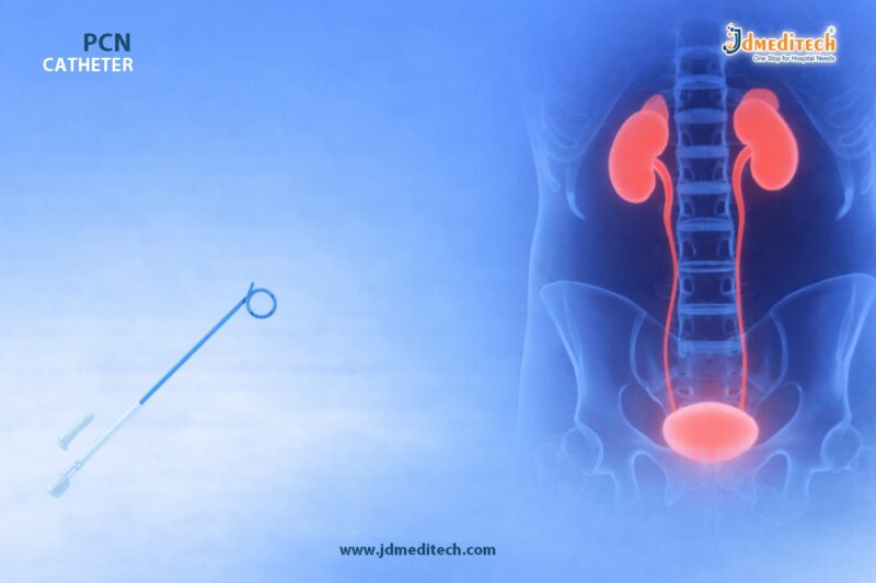
  • Percutaneous Nephrostomy (PCN) procedures
  • Obstructive uropathy management
  • Hydronephrosis relief
  • Post-surgical kidney drainage
  • Urinary diversion in complex cases
Advantages of Malecot Nephrostomy Catheter
Secure Positioning

The winged tip ensures strong internal fixation, reducing catheter migration.

Reliable Long-Term Drainage

Ideal for patients requiring extended nephrostomy support.

Reduced Risk of Leakage

Tight retention minimizes urine leakage around the catheter.

Easy Placement & Removal

Despite strong anchoring, the catheter allows controlled insertion and removal.

Procedure Overview

During a percutaneous nephrostomy, the Malecot catheter is inserted through the skin into the kidney under imaging guidance.

Steps include:

  1. Local anesthesia administration
  2. Needle puncture into renal pelvis
  3. Guidewire insertion
  4. Tract dilation
  5. Placement of Malecot catheter
  6. Expansion of wings for secure fixation
Why Choose Malecot Over Other Catheters?

Compared to pigtail catheters, the Malecot Nephrostomy Catheter offers stronger retention due to its winged design.

Feature Malecot Catheter Pigtail Catheter
Retention Strength High Moderate
Stability Excellent Good
Migration Risk Low Slightly Higher
Ideal Use Long-term drainage Short-term drainage
Care and Maintenance Tips

To ensure optimal performance:

  • Maintain proper catheter hygiene
  • Regularly monitor urine flow
  • Flush the catheter if advised by a clinician
  • Check for signs of infection or blockage
Conclusion

The Malecot Nephrostomy Catheter stands out as a reliable and secure solution for renal drainage, thanks to its innovative winged retention design. Its stability, efficiency, and safety make it a preferred choice among urologists worldwide.

For healthcare providers seeking dependable nephrostomy solutions, this catheter offers the perfect balance of performance and patient comfort.


Explore More Product >>

Get Connected:

+91 79909 93062 | +91 63513 72032 | exports@jdmeditech.com


Endoscopy Foreign Body Forceps for Safe Retrieval

Endoscopy foreign body forceps play a crucial role in gastrointestinal procedures where safe retrieval of ingested or lodged objects is required. These precision-designed instruments help clinicians grasp, hold, and remove foreign bodies effectively without causing mucosal injury.

With increasing cases of accidental ingestion and complex endoscopic interventions, the demand for reliable and high-performance foreign body forceps has grown significantly. Their ergonomic design and strong gripping capability make them essential tools in modern endoscopy suites.

What Are Endoscopy Foreign Body Forceps?

Endoscopy foreign body forceps are specialized devices used during endoscopic procedures to retrieve foreign objects from the gastrointestinal tract. These may include:

  • Coins
  • Food boluses
  • Bones
  • Dental prosthetics
  • Sharp or irregular objects

They are inserted through the working channel of an endoscope, allowing controlled and precise manipulation inside the GI tract.

Key Features of Endoscopy Foreign Body Forceps
1. Strong Gripping Strength

These forceps provide a firm hold on objects, reducing the risk of slippage during retrieval.

2. Precision-Engineered Jaws

Different jaw designs such as alligator, rat-tooth, or tripod enable handling of various object shapes and sizes.

3. Flexible and Durable Shaft

The flexible shaft allows easy navigation through the endoscope while maintaining structural integrity.

4. Smooth Operation

Ergonomic handles ensure precise control and smooth opening and closing of the jaws.

5. Atraumatic Design

Designed to minimize tissue damage during insertion and extraction.

Types of Foreign Body Forceps

Depending on the clinical need, several types are available:

  • Alligator Forceps : Ideal for grasping soft or flat objects
  • Rat-Tooth Forceps : Suitable for firm gripping of tough objects
  • Tripod Forceps : Provides multi-point grip for irregular shapes
  • Basket-Type Forceps : Used for capturing round or loose objects
Clinical Applications

Endoscopy foreign body forceps are widely used in:

  • Gastroenterology : Removal of ingested objects from esophagus, stomach, or intestines
  • Emergency Endoscopy : Managing accidental ingestion cases
  • Pediatric Endoscopy : Safe retrieval in children
  • ENT Procedures : Removal of foreign bodies in upper GI and airway
Benefits of Using Endoscopy Foreign Body Forceps
  •  Ensures safe and controlled removal
  •  Reduces risk of complications or perforation
  •  Provides high precision and maneuverability
  •  Compatible with most standard endoscopes
  •  Improves procedure efficiency and success rate
How to Choose the Right Forceps

Selecting the right instrument depends on:

  • Type and size of foreign body
  • Location within the GI tract
  • Required gripping strength
  • Endoscope compatibility

Clinicians should always opt for high-quality, medical-grade instruments to ensure optimal outcomes.

Why Choose JDMeditech Endoscopy Foreign Body Forceps?

At JDMeditech, we design endoscopy foreign body forceps with a focus on safety, precision, and durability. Our products offer:

  • Superior grip and control
  • High-quality biocompatible materials
  • Smooth functionality for easy handling
  • Reliable performance in critical procedures
Best Practices for Safe Use
  • Always visualize the object clearly before grasping
  • Use appropriate jaw type for the object
  • Avoid excessive force to prevent tissue injury
  • Ensure proper sterilization before use
Conclusion

Endoscopy foreign body forceps are indispensable tools for safe and effective retrieval of foreign objects during endoscopic procedures. Their advanced design, strong grip, and precision handling significantly enhance procedural success while ensuring patient safety.

Healthcare professionals rely on these instruments for efficient and complication-free outcomes, making them a vital component in modern endoscopy.


Explore More Product >>

Get Connected:

+91 79909 93062 | +91 63513 72032 | exports@jdmeditech.com


Endopyelotomy Stent for UPJ Repair – Reliable Ureteral Support

The Endopyelotomy Stent plays a crucial role in modern urological procedures, particularly in the management of ureteropelvic junction (UPJ) obstruction. Designed to provide internal drainage and structural support after endopyelotomy, this specialized stent ensures proper healing and maintains patency of the urinary tract.

With advancements in minimally invasive urology, endopyelotomy stents have become indispensable tools for urologists seeking reliable postoperative outcomes.

What is an Endopyelotomy Stent?

An Endopyelotomy Stent is a specially designed ureteral stent used after performing an endopyelotomy, a minimally invasive procedure to treat UPJ obstruction.

Unlike conventional stents, this device features:

  • A tailored design for precise placement
  • Enhanced lumen for efficient urine drainage
  • Structural support to aid healing of the incision site

It helps maintain the opening between the renal pelvis and ureter, preventing restenosis during recovery.

Understanding UPJ Obstruction and Its Treatment
What is UPJ Obstruction?

UPJ obstruction occurs when there is a blockage at the junction where the kidney meets the ureter. This condition can lead to:

  • Hydronephrosis (kidney swelling)
  • Flank pain
  • Recurrent infections
  • Impaired kidney function
Role of Endopyelotomy

Endopyelotomy involves making an incision at the obstructed segment to restore urine flow. However, maintaining the patency of this incision is critical—this is where the Endopyelotomy Stent for UPJ Repair becomes essential.

Key Features of Endopyelotomy Stents
1. Optimized Length and Design

The stent is longer and specifically shaped to span the renal pelvis and ureter, ensuring complete support across the UPJ.

2. Multiple Drainage Holes

Strategically placed side holes allow continuous urine drainage, reducing pressure buildup.

3. High-Quality Biocompatible Material

Typically made from polyurethane or silicone, these stents offer:

  • Flexibility
  • Reduced irritation
  • Long-term compatibility
4. Radiopaque Markers

These enable precise placement and easy visualization under fluoroscopy.

5. Anti-Migration Design

Pigtail or coil ends prevent displacement during the healing phase.

Benefits of Using an Endopyelotomy Stent for UPJ Repair

Using an Endopyelotomy Stent for UPJ Repair provides several clinical advantages:

  •  Maintains ureteral patency after incision
  •  Promotes faster and safer healing
  •  Reduces risk of restenosis
  •  Ensures continuous urine drainage
  •  Minimizes postoperative complications
Procedure Overview
Step-by-Step Use
  1. Diagnosis and Imaging
    UPJ obstruction is confirmed via ultrasound, CT scan, or IVP.
  2. Endopyelotomy Procedure
    The obstructed segment is incised using endoscopic tools.
  3. Stent Placement
    The endopyelotomy stent is inserted across the UPJ.
  4. Postoperative Monitoring
    The stent remains in place for several weeks to support healing.
  5. Stent Removal
    After adequate healing, the stent is removed endoscopically.
Clinical Applications

The Endopyelotomy Stent is widely used in:

  • Treatment of primary UPJ obstruction
  • Management of recurrent UPJ strictures
  • Postoperative support following endopyelotomy procedures
  • Selected cases of failed pyeloplasty
Why Choose High-Quality Endopyelotomy Stents?

Choosing a high-quality stent ensures:

  • Better patient comfort
  • Reduced encrustation risk
  • Improved procedural success rates
  • Reliable long-term outcomes

At JDMeditech, we focus on delivering precision-engineered urology devices that meet global standards.

Best Practices for Stent Management

To achieve optimal results:

  • Monitor patients regularly for complications
  • Ensure timely stent removal
  • Use appropriate stent size and length
  • Maintain strict sterile protocols
Conclusion

The Endopyelotomy Stent for UPJ Repair is a vital component in the successful management of ureteropelvic junction obstruction. By providing consistent drainage and structural support, it significantly enhances healing outcomes after endopyelotomy.

As minimally invasive techniques continue to evolve, the role of advanced stenting solutions will only become more critical in ensuring patient safety and procedural success.


Explore More Product >>

Get Connected:

+91 79909 93062 | +91 63513 72032 | exports@jdmeditech.com

Endoscopy Ligation Device for Variceal Hemostasis | Complete Guide

Variceal bleeding is one of the most serious complications of portal hypertension. It demands immediate intervention to prevent life-threatening outcomes. Among modern solutions, the endoscopy ligation device for variceal hemostasis stands out as a reliable and minimally invasive tool.

This advanced device allows gastroenterologists to control bleeding quickly and efficiently during endoscopic procedures. As a result, it has become a preferred method in clinical practice worldwide.

What is an Endoscopy Ligation Device?

An endoscopy ligation device is a specialized medical tool used during endoscopy to treat bleeding esophageal or gastric varices. It works by deploying elastic bands around the varices, which cuts off blood flow and leads to controlled necrosis.

Because of its precision and safety, it plays a critical role in managing acute GI bleeding.

Understanding Variceal Hemostasis

Variceal hemostasis refers to the process of stopping bleeding from dilated veins (varices) in the gastrointestinal tract. These varices typically develop due to increased pressure in the portal venous system.

Common Causes of Varices
  • Liver cirrhosis
  • Portal hypertension
  • Chronic liver disease
Why Immediate Hemostasis is Crucial
  • Prevents massive blood loss
  • Reduces mortality risk
  • Stabilizes patient condition quickly

Therefore, using an effective device like an endoscopy ligation system is essential.

How the Endoscopy Ligation Device Works

The procedure is straightforward yet highly effective.

  1. The device is attached to the tip of the endoscope
  2. The varix is suctioned into the ligator cap
  3. A rubber band is deployed around the varix
  4. Blood flow stops, leading to tissue necrosis

As a result, the treated varix shrinks and eventually sloughs off.

Key Features of Endoscopy Ligation Devices

Modern ligation devices are designed for efficiency and safety.

1. Multi-Band Deployment System

Allows multiple ligations in a single insertion, saving time and effort.

2. 360° Rotation Capability

Ensures precise targeting of varices from different angles.

3. Smooth Trigger Mechanism

Provides controlled band release for accurate placement.

4. Transparent Cap Design

Enhances visualization during the procedure.

5. Biocompatible Materials

Ensures patient safety and reduces complications.

Clinical Advantages

The endoscopy ligation device for variceal hemostasis offers several benefits.

  • Minimally invasive technique
  • Rapid bleeding control
  • Reduced need for surgery
  • Lower complication rates
  • Shorter hospital stays

Moreover, it improves overall patient outcomes significantly.

Applications in Gastroenterology

This device is widely used in various GI procedures:

  • Esophageal variceal ligation (EVL)
  • Gastric varices management
  • Preventive treatment for high-risk patients
  • Emergency bleeding control

Because of its versatility, it has become a standard tool in endoscopy units.

Comparison with Other Hemostasis Methods
Method Effectiveness Safety Ease of Use
Band Ligation High High Easy
Sclerotherapy Moderate Moderate Moderate
Surgical Intervention Very High Lower Complex

Clearly, band ligation offers the best balance of safety and effectiveness.

Best Practices for Use

To achieve optimal results:

  • Ensure proper device attachment
  • Maintain clear visualization
  • Apply bands accurately on targeted varices
  • Monitor patient post-procedure

Following these steps improves procedural success rates.

Future Innovations

Endoscopic technology continues to evolve. New advancements focus on:

  • Enhanced visualization systems
  • Improved band deployment mechanisms
  • Integration with AI-assisted endoscopy
  • Better ergonomics for clinicians

These innovations will further improve variceal hemostasis outcomes.

Conclusion

The endoscopy ligation device for variceal hemostasis has revolutionized the management of GI bleeding. It offers a safe, effective, and minimally invasive solution for controlling variceal hemorrhage.

With continuous advancements, this device will remain a cornerstone in modern gastroenterology.


Explore More Product >>

Get Connected:

+91 79909 93062 | +91 63513 72032 | exports@jdmeditech.com

Multiband Ligator for Efficient Variceal Ligation

A multiband ligator is an essential device in gastrointestinal endoscopy. It helps doctors treat esophageal varices quickly and safely. These varices often develop due to portal hypertension and can cause serious bleeding.

Today, specialists prefer the multiband ligator because it improves efficiency and reduces procedure time. In addition, it enhances patient safety and comfort.

What is a Multiband Ligator?

A multiband ligator is an endoscopic tool used to place multiple elastic bands on varices during a single procedure. Doctors mainly use it for endoscopic variceal ligation (EVL).

Unlike older devices, it allows multiple band placements without removing the endoscope. As a result, the procedure becomes faster and more convenient.

Why is a Multiband Ligator Important?

The multiband ligator plays a key role in managing gastrointestinal bleeding. It provides a reliable and efficient way to control varices.

Main Advantages
  • Allows multiple band placements in one session
  • Reduces overall procedure time
  • Minimizes patient discomfort
  • Improves treatment outcomes
  • Avoids repeated scope insertion

Because of these benefits, it is widely used in both emergency and routine procedures.

Clinical Uses of Multiband Ligator

Doctors use a multiband ligator in several important situations:

1. Treatment of Esophageal Varices

It is the most common and effective use.

2. Preventive Ligation

It helps prevent bleeding in high-risk patients.

3. Portal Hypertension Management

It reduces complications related to increased portal pressure.

4. Control of GI Bleeding

It provides effective hemostasis during endoscopic procedures.

Key Features of Multiband Ligator

Modern multiband ligator systems are designed for ease and precision.

Important Features
  • Transparent cap for clear visibility
  • Preloaded multiple elastic bands
  • Smooth and controlled release mechanism
  • Flexible compatibility with endoscopes
  • Easy-to-use ergonomic design

These features ensure consistent and reliable performance.

How Does a Multiband Ligator Work?

The working process of a multiband ligator is simple:

  1. The device is attached to the endoscope
  2. The varix is suctioned into the ligator cap
  3. A band is deployed around the tissue
  4. The tissue shrinks and eventually falls off

This process stops bleeding and promotes healing.

Benefits of Using a Multiband Ligator

The multiband ligator offers several practical benefits:

  • Faster procedures and reduced operation time
  • Lower risk of re-bleeding
  • Better control during ligation
  • Cost-effective treatment approach
  • Improved patient comfort and safety

Therefore, it has become a standard tool in GI endoscopy.

Safety Tips and Handling

To achieve the best results, proper handling is important:

  • Assemble the device correctly before use
  • Maintain clear visibility during the procedure
  • Avoid excessive suction
  • Follow standard sterilization protocols
  • Ensure trained professionals perform the procedure

These steps help maintain safety and efficiency.

Why Choose JDMeditech Multiband Ligator?

At JDMeditech, we provide high-quality endoscopic devices designed for performance and reliability.

What We Offer
  • Premium medical-grade materials
  • Smooth and consistent band deployment
  • User-friendly design
  • Competitive pricing
  • Trusted quality
Conclusion

The multiband ligator is a vital tool in modern GI endoscopy. It allows doctors to perform safe, fast, and effective variceal ligation.

With its advanced design and proven results, it continues to improve patient care and clinical outcomes.


Explore More Product >>

Get Connected:

+91 79909 93062 | +91 63513 72032 | exports@jdmeditech.com

PCN Catheter for Kidney Drainage in Obstructive Uropathy

A PCN catheter for kidney drainage is an important device in modern urology. It helps drain urine directly from the kidney when the normal urinary pathway becomes blocked. As a result, it reduces pressure inside the kidney and supports faster relief.

Obstructive uropathy can develop because of stones, tumors, strictures, or other urinary blockages. When urine cannot flow properly, the kidney starts to swell. Over time, this pressure can damage renal tissue and increase the risk of infection. Therefore, doctors often use a PCN catheter for kidney drainage to protect kidney function and restore safe urine flow.

What Is a PCN Catheter for Kidney Drainage?

A PCN catheter for kidney drainage is a thin and flexible tube placed into the renal pelvis through the skin. Doctors usually insert it with the help of imaging guidance such as ultrasound or fluoroscopy. This method improves accuracy and helps reduce complications.

The catheter creates an alternate route for urine drainage. Instead of passing through a blocked ureter, urine flows through the catheter into an external collection bag. Because of this, the kidney gets immediate decompression and the patient often feels relief soon after placement.

Why Is a PCN Catheter Used in Obstructive Uropathy?

Doctors use a PCN catheter for kidney drainage when urine cannot leave the kidney normally. This condition demands timely treatment because prolonged obstruction may lead to infection, pain, hydronephrosis, or renal damage.

Common causes of obstructive uropathy include:

  • Ureteric or renal stones
  • Ureteral strictures
  • Pelvic or abdominal tumors
  • Blood clots in the urinary tract
  • Congenital urinary obstruction

In many cases, a PCN catheter for kidney drainage becomes essential when immediate decompression is necessary. It also supports further diagnostic or therapeutic procedures.

Key Features of a PCN Catheter for Kidney Drainage

A well-designed PCN catheter for kidney drainage offers reliable performance and safe handling. Manufacturers focus on drainage efficiency, placement accuracy, and patient comfort.

Important features often include:

  • Soft and biocompatible catheter material
  • Multiple side holes for efficient drainage
  • Radiopaque line or markers for visibility
  • Kink-resistant shaft for smooth urine flow
  • Secure locking or retention design
  • Connector compatibility with drainage bags

These features help the catheter perform well in both short-term and extended drainage situations.

How a PCN Catheter for Kidney Drainage Is Placed

The placement of a PCN catheter for kidney drainage is a minimally invasive procedure. Trained specialists perform it in a controlled setting with imaging support.

Step-by-step placement process
  1. The patient is positioned properly.
  2. The area is cleaned and prepared.
  3. Local anesthesia is given.
  4. Imaging helps identify the target calyx or renal pelvis.
  5. A needle enters the collecting system.
  6. A guidewire passes through the access tract.
  7. The tract is dilated when needed.
  8. The catheter is advanced and fixed in place.
  9. The catheter is connected to a drainage bag.

This technique offers precise placement. In addition, it helps reduce trauma compared with open surgical drainage methods.

Benefits of a PCN Catheter for Kidney Drainage

A PCN catheter for kidney drainage provides several important clinical benefits. Most importantly, it relieves pressure from the obstructed kidney quickly.

Key benefits include:

  • Fast decompression of the kidney
  • Protection of renal function
  • Relief from flank pain and pressure
  • Support in infected obstructed systems
  • Minimally invasive placement
  • Useful access for future urological procedures

Moreover, the catheter helps stabilize patients before definitive treatment. For that reason, it remains a trusted option in emergency and elective care.

Clinical Applications of a PCN Catheter for Kidney Drainage

A PCN catheter for kidney drainage supports many urological and interventional procedures. Its role goes beyond simple urinary diversion.

It is commonly used in:

  • Obstructive uropathy management
  • Hydronephrosis decompression
  • Pyonephrosis drainage
  • Temporary urinary diversion before surgery
  • Access for nephrostomy tract procedures
  • Support during complex stone management

Because it has broad clinical utility, many hospitals rely on the PCN catheter for kidney drainage in routine and advanced urology practice.

Patient Care After PCN Catheter Placement

Proper aftercare improves the performance of a PCN catheter for kidney drainage and lowers the chance of complications. Patients and caregivers should understand basic maintenance steps.

Important care tips include:

  • Keep the insertion area clean and dry
  • Check urine output regularly
  • Avoid bending or pulling the catheter
  • Empty the drainage bag on time
  • Watch for fever, leakage, or blockage
  • Attend all follow-up visits

Good catheter care supports continuous drainage. It also helps detect issues early.

Possible Complications and Risk Management

Although a PCN catheter for kidney drainage is generally safe, some complications can occur. However, careful technique and regular monitoring reduce these risks.

Possible complications include:

  • Minor bleeding
  • Infection at the insertion site
  • Catheter blockage
  • Dislodgement
  • Leakage around the catheter
  • Local discomfort

Doctors manage most of these issues quickly when identified early. Therefore, education and follow-up are very important.

Why Quality Matters in a PCN Catheter for Kidney Drainage

The quality of a PCN catheter for kidney drainage directly affects performance, safety, and patient comfort. A well-manufactured catheter allows smoother insertion and more dependable drainage.

High-quality options often provide:

  • Better flexibility and strength
  • Consistent lumen patency
  • Reliable radiopacity
  • Safer retention inside the kidney
  • Improved procedural confidence

For this reason, healthcare providers should choose dependable products that meet clinical expectations and support patient safety.

Conclusion

A PCN catheter for kidney drainage plays a vital role in the treatment of obstructive uropathy. It relieves urinary obstruction, preserves kidney function, and supports rapid clinical improvement. In addition, it provides a minimally invasive solution for emergency decompression and ongoing urinary diversion.

Because obstructive uropathy can become serious very quickly, timely drainage is essential. That is why the PCN catheter for kidney drainage remains a key device in modern urological care.


Explore More Product >>

Get Connected:

+91 79909 93062 | +91 63513 72032 | exports@jdmeditech.com


Endoscope Cleaning Brush for Safe Scope Reprocessing

An endoscope cleaning brush plays a crucial role in maintaining the hygiene and functionality of endoscopic equipment. Proper cleaning of endoscopes is essential to prevent cross-contamination, ensure patient safety, and extend the life of expensive medical instruments.

Because endoscopes feature narrow lumens and complex channels, manual cleaning remains a vital step in reprocessing. Therefore, using a high-quality endoscope cleaning brush ensures thorough removal of debris, biofilm, and contaminants.

What is an Endoscope Cleaning Brush?

An endoscope cleaning brush is a specially designed medical device used to clean internal channels and external surfaces of endoscopes. It helps remove organic material, tissue residue, and microbial contamination.

These brushes come in various sizes, lengths, and configurations to suit different types of endoscopes, including gastroscopes, colonoscopes, bronchoscopes, and ureteroscopes.

Why Endoscope Cleaning is Critical

Effective cleaning is the foundation of safe endoscope reprocessing. Without proper cleaning, even high-level disinfection may fail.

Key Reasons Include:
  • Prevents cross-infection between patients
  • Removes biofilm and organic debris
  • Enhances the effectiveness of disinfection and sterilization
  • Protects patient safety and clinical outcomes
  • Extends the lifespan of endoscopic equipment
Key Features of Endoscope Cleaning Brush

A high-quality endoscope cleaning brush offers several important features:

1. Flexible Design

The brush easily navigates complex and narrow endoscope channels.

2. Soft Yet Durable Bristles

It ensures effective cleaning without damaging delicate internal surfaces.

3. Multiple Size Options

Different diameters and lengths allow compatibility with various scopes.

4. Ergonomic Handle

Provides better grip and control during cleaning procedures.

5. Single-Use or Reusable Options

Available based on hospital protocols and infection control standards.

Types of Endoscope Cleaning Brushes

Different procedures require specific brush designs. Common types include:

  • Channel Cleaning Brushes – For internal lumens
  • Double-Ended Brushes – Clean both ends simultaneously
  • Valve Cleaning Brushes – Designed for small components
  • Disposable Brushes – Reduce cross-contamination risk
  • Reusable Brushes – Cost-effective with proper sterilization
Step-by-Step Use of Endoscope Cleaning Brush

To ensure effective cleaning, follow a structured process:

  1. Pre-clean the endoscope immediately after use
  2. Flush channels with enzymatic detergent
  3. Insert the endoscope cleaning brush into channels
  4. Gently scrub to remove debris
  5. Rinse thoroughly with clean water
  6. Proceed to disinfection or sterilization

Consistent technique improves cleaning efficiency and reduces infection risk.

Benefits of Using Endoscope Cleaning Brush

Using a high-quality endoscope cleaning brush provides multiple advantages:

  • Improves infection control compliance
  • Ensures thorough manual cleaning
  • Reduces risk of biofilm formation
  • Enhances scope performance
  • Supports regulatory standards (CDC, AAMI, ESGE)
How to Choose the Right Endoscope Cleaning Brush

Selecting the right brush is essential for optimal results.

Consider the Following Factors:
  • Compatibility with scope channel size
  • Length and flexibility
  • Bristle quality
  • Single-use vs reusable preference
  • Compliance with hospital protocols
Best Practices for Endoscope Reprocessing

To maximize safety, always follow these best practices:

  • Use a new or properly sterilized brush
  • Avoid using damaged or worn brushes
  • Follow manufacturer guidelines
  • Maintain a strict cleaning protocol
  • Train staff regularly on reprocessing techniques
Conclusion

The endoscope cleaning brush is an essential tool in safe and effective scope reprocessing. It ensures the removal of contaminants, supports infection prevention, and enhances the longevity of medical devices.

Healthcare facilities must prioritize high-quality cleaning accessories to maintain patient safety and meet global hygiene standards. Investing in the right endoscope cleaning brush is a small step that delivers significant clinical benefits.


Explore More Product >>

Get Connected:

+91 79909 93062 | +91 63513 72032 | exports@jdmeditech.com

Gastric Balloon Retrieval Device for Safe Bariatric Endoscopy

The gastric balloon retrieval device plays a critical role in bariatric endoscopy. Physicians use it to safely remove intragastric balloons after weight-loss therapy. As minimally invasive procedures continue to evolve, clinicians demand tools that deliver both precision and safety.

This device ensures controlled deflation and secure retrieval of gastric balloons, reducing complications and improving patient outcomes. Therefore, it has become an essential instrument in modern bariatric practice.

What is a Gastric Balloon Retrieval Device?

A gastric balloon retrieval device is a specialized endoscopic tool designed to puncture, deflate, and extract intragastric balloons from the stomach.

Doctors typically use it at the end of gastric balloon therapy, which helps patients achieve weight loss. The device allows smooth removal without causing trauma to the gastric lining.

Key Features of Gastric Balloon Retrieval Device
1. Precision Puncture Mechanism

The device includes a sharp, controlled puncture system. It enables accurate balloon deflation without damaging surrounding tissues.

2. Strong Grasping Capability

Its secure gripping system helps hold the deflated balloon firmly. As a result, physicians can retrieve it easily.

3. Flexible and Navigable Design

The flexible shaft ensures smooth navigation through the esophagus and stomach during endoscopy.

4. Biocompatible Materials

Manufacturers use high-quality, medical-grade materials to ensure safety and compatibility with human tissues.

5. Single-Use Sterile Option

Many devices come pre-sterilized for single use, which minimizes infection risks.

Applications in Bariatric Endoscopy

The gastric balloon retrieval device is widely used in:

  • Removal of intragastric balloons after therapy completion
  • Management of balloon-related complications
  • Endoscopic weight-loss procedures
  • Clinical bariatric interventions

Because of its reliability, it supports safe and efficient procedures in both outpatient and hospital settings.

Benefits of Using Gastric Balloon Retrieval Device
Enhanced Patient Safety

The device reduces the risk of mucosal injury during balloon removal.

Improved Procedural Efficiency

Its design allows faster deflation and extraction, saving procedure time.

Minimally Invasive Approach

Doctors can perform the procedure endoscopically without surgical intervention.

Better Clinical Outcomes

Controlled retrieval minimizes complications such as balloon rupture or migration.

Step-by-Step Procedure Overview
  1. The endoscope is introduced into the stomach.
  2. The gastric balloon retrieval device is guided toward the balloon.
  3. The balloon is punctured and deflated carefully.
  4. The device securely grasps the deflated balloon.
  5. The balloon is gently removed through the esophagus.

This systematic approach ensures a safe and controlled procedure.

Why Choose a High-Quality Gastric Balloon Retrieval Device?

Choosing a premium gastric balloon retrieval device ensures:

  • Reliable performance during critical procedures
  • Reduced risk of complications
  • Enhanced control for the endoscopist
  • Consistent results across multiple cases

Healthcare providers should prioritize quality, durability, and ease of use when selecting this device.

Conclusion

The gastric balloon retrieval device is an indispensable tool in bariatric endoscopy. It enables safe, efficient, and minimally invasive removal of gastric balloons, ensuring better patient outcomes.

As bariatric procedures continue to grow in popularity, the demand for advanced and reliable retrieval devices will also increase. Investing in high-quality devices not only improves procedural success but also enhances patient safety and satisfaction.


Explore More Product >>

+91 79909 93062 | +91 63513 72032 | exports@jdmeditech.com

Polyurethane Double Pigtail Ureteral Stent Set

The Polyurethane Double Pigtail Ureteral Stent Set plays a critical role in modern urology by ensuring smooth urine drainage from the kidney to the bladder. Physicians rely on this device to manage ureteral obstruction, prevent complications, and support post-operative healing.

Thanks to its flexible polyurethane construction, this stent offers both durability and patient comfort. Moreover, its double pigtail design ensures secure placement and reduces the risk of migration.

What is a Polyurethane Double Pigtail Ureteral Stent Set?

A Polyurethane Double Pigtail Ureteral Stent Set is a medical device inserted into the ureter to maintain patency and facilitate urine flow. It features curled ends (pigtails) on both sides, which anchor the stent in the kidney and bladder.

Additionally, the set typically includes:

  • Ureteral stent
  • Guidewire
  • Pusher catheter

This complete system allows clinicians to perform safe and efficient placement.

Key Features of Polyurethane Double Pigtail Ureteral Stent Set
1. High-Quality Polyurethane Material

Polyurethane provides excellent flexibility and strength. As a result, the stent adapts to anatomical structures while maintaining long-term performance.

2. Double Pigtail Design

The curled ends prevent migration and ensure stable positioning. Consequently, the risk of displacement reduces significantly.

3. Smooth Surface Finish

The stent’s smooth surface minimizes friction during insertion and removal. It also helps reduce patient discomfort.

4. Radiopaque Visibility

Radiopaque markers improve visualization under fluoroscopy. Therefore, clinicians can place the stent with high accuracy.

5. Multiple Size Options

The stent set is available in various lengths and diameters, making it suitable for different patient needs.

Clinical Applications

The Polyurethane Double Pigtail Ureteral Stent Set is widely used in several urological procedures, including:

  • Management of ureteral obstruction
  • Post-operative drainage after ureteroscopy (URS)
  • Relief of kidney stones
  • Treatment of ureteral strictures
  • Support during urological surgeries

Because of its versatility, it has become a standard solution in both emergency and elective procedures.

Benefits of Using Polyurethane Double Pigtail Ureteral Stent Set
1. Reliable Drainage

The stent ensures continuous urine flow, preventing kidney damage caused by obstruction.

2. Enhanced Patient Comfort

Its flexible structure reduces irritation, allowing patients to tolerate the device better.

3. Easy Placement

The included accessories simplify the insertion process and save procedural time.

4. Reduced Complications

Secure anchoring minimizes migration and lowers the risk of complications.

5. Cost-Effective Solution

Durability and efficiency make it a practical choice for healthcare providers.

Why Polyurethane is Preferred for Ureteral Stents

Polyurethane stands out due to its balanced properties. Unlike more rigid materials, it offers flexibility without compromising strength. Furthermore, it resists encrustation better than some alternatives.

As a result, clinicians prefer polyurethane for both short-term and medium-term stenting.

Procedure Overview

During the procedure:

  1. The clinician inserts a guidewire into the ureter.
  2. The stent is advanced over the guidewire.
  3. The pigtail ends form naturally in the kidney and bladder.

This process ensures accurate placement and immediate drainage.

Post-Placement Care and Considerations

After insertion, patients may experience mild discomfort. However, proper monitoring ensures optimal outcomes.

Key care points include:

  • Regular follow-up imaging
  • Timely stent removal or replacement
  • Monitoring for infection or encrustation

By following these steps, complications can be minimized.

Conclusion

The Polyurethane Double Pigtail Ureteral Stent Set offers a dependable solution for ureteral drainage. Its flexible material, secure design, and ease of use make it an essential tool in urology.

Healthcare professionals trust this stent for its performance, safety, and patient comfort, making it a preferred choice in clinical practice.


Explore More Product >>

Get Connected:

+91 79909 93062 | +91 63513 72032 | exports@jdmeditech.com

Silicone Double J Stent for Ureteral Drainage

The Silicone Double J Stent plays a vital role in modern urology. It ensures smooth urine flow from the kidney to the bladder when natural drainage becomes obstructed. Doctors prefer silicone-based stents because they offer excellent flexibility, superior biocompatibility, and enhanced patient comfort.

In this blog, we explore why the Silicone Double J Stent is a trusted choice for ureteral drainage and how it improves clinical outcomes.

What is a Silicone Double J Stent?

A Silicone Double J Stent is a soft, flexible tube placed inside the ureter. It features a characteristic “J” curve at both ends, which helps keep the stent securely positioned in the kidney and bladder.

Unlike traditional materials, silicone provides a smoother surface and minimizes irritation, making it ideal for both short-term and long-term use.

Why Choose a Silicone Double J Stent?
1. Superior Flexibility

Silicone material adapts easily to the natural curvature of the ureter. As a result, it reduces discomfort and improves patient tolerance.

2. Excellent Biocompatibility

The Silicone Double J Stent is highly biocompatible. It lowers the risk of inflammation, irritation, and allergic reactions.

3. Reduced Encrustation

Silicone resists encrustation better than many other materials. Therefore, it supports safer long-term placement.

4. Enhanced Patient Comfort

Because of its softness, patients experience fewer symptoms such as urgency, pain, or bladder irritation.

5. Long-Term Durability

Silicone maintains its structure and performance over extended periods, ensuring reliable drainage.

Clinical Applications of Silicone Double J Stent

The Silicone Double J Stent is widely used in various urological conditions, including:

  • Ureteral obstruction due to stones
  • Post-ureteroscopy drainage
  • Ureteral strictures
  • Tumor-related blockages
  • Post-surgical support
  • Hydronephrosis management

It provides consistent drainage and helps prevent complications such as kidney damage.

Key Features of Silicone Double J Stent
  • Soft, medical-grade silicone construction
  • Double J design for secure positioning
  • Smooth surface for easy insertion and removal
  • Radiopaque for accurate placement under imaging
  • Available in multiple sizes and lengths
  • Sterile and ready-to-use packaging
Procedure Overview

Doctors typically place a Silicone Double J Stent using a minimally invasive endoscopic technique:

  1. A cystoscope is inserted into the bladder
  2. The stent is guided into the ureter
  3. One end curls in the kidney, and the other in the bladder
  4. Placement is confirmed using imaging

The process is quick and ensures immediate relief from obstruction.

Advantages Over Other Materials

Compared to polyurethane or other stents, the Silicone Double J Stent offers:

  • Better flexibility and softness
  • Lower risk of encrustation
  • Improved long-term tolerance
  • Reduced tissue irritation

These advantages make it a preferred option for many urologists.

Post-Placement Care

Patients with a Silicone Double J Stent should follow these guidelines:

  • Stay well hydrated
  • Report persistent pain or fever
  • Attend scheduled follow-ups
  • Ensure timely stent removal or replacement

Proper care ensures optimal performance and reduces complications.

Why Silicone Double J Stent is a Preferred Choice

The Silicone Double J Stent combines comfort, safety, and performance. Its flexible structure and biocompatible nature make it suitable for a wide range of patients.

Healthcare professionals trust it for its consistent results, while patients benefit from reduced discomfort and improved quality of life.

Conclusion

The Silicone Double J Stent stands out as a reliable and patient-friendly solution for ureteral drainage. With its advanced material properties and proven clinical benefits, it continues to be an essential tool in modern urology.

If you are looking for a high-quality ureteral stent that ensures both performance and comfort, the Silicone Double J Stent is an excellent choice.


Explore More Product >>

Get Connected:

+91 79909 93062 | +91 63513 72032 | exports@jdmeditech.com