Endoscopic Clip for GI Procedures | Advanced Clipping Solutions

An endoscopic clip plays a critical role in modern gastrointestinal (GI) procedures. It helps clinicians achieve precise tissue approximation, secure hemostasis, and manage complications effectively. As endoscopy continues to evolve, advanced GI clipping solutions have become essential tools in both diagnostic and therapeutic interventions.

In this blog, we will explore the features, applications, benefits, and importance of endoscopic clips in contemporary gastroenterology.

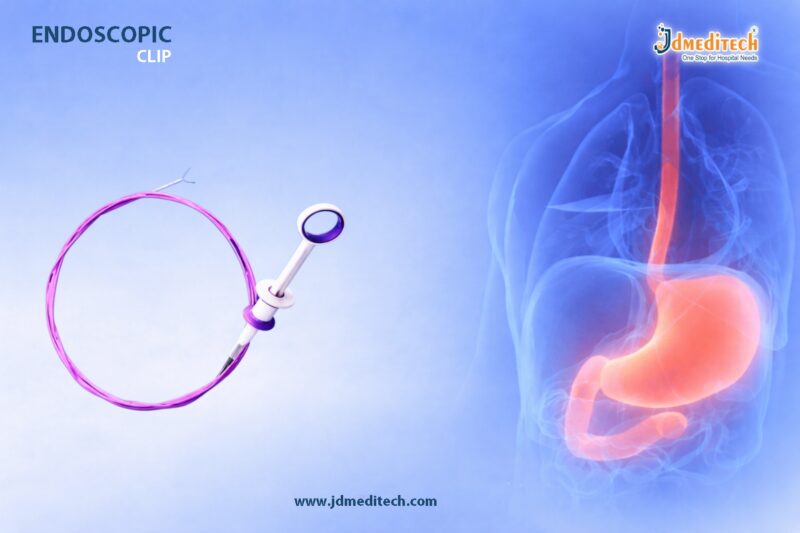
What is an Endoscopic Clip?

An endoscopic clip is a small, mechanical device delivered through an endoscope to grasp, approximate, or compress tissue within the gastrointestinal tract. It is widely used to control bleeding, close mucosal defects, and support healing after endoscopic procedures.

These clips are typically made from biocompatible materials and are designed for precision deployment in challenging anatomical locations.

Key Features of Endoscopic Clip
1. Precise Deployment Mechanism

Endoscopic clips provide controlled and accurate placement, ensuring effective tissue capture even in difficult-to-reach areas.

2. Strong Closure Strength

They deliver reliable compression, which is essential for achieving durable hemostasis and secure tissue approximation.

3. Rotatable Design

Most advanced endoscopic clips feature a 360-degree rotatable system, allowing better positioning and enhanced procedural efficiency.

4. Reopenable Functionality

Some clips can be reopened and repositioned before final deployment, improving accuracy and reducing procedural errors.

5. Biocompatible Material

Manufactured from high-quality materials, these clips ensure patient safety and minimize adverse reactions.

Applications of Endoscopic Clip in GI Procedures
1. Hemostasis

An endoscopic clip is widely used to control bleeding caused by ulcers, lesions, or post-polypectomy complications.

2. Closure of Mucosal Defects

After procedures like Endoscopic Mucosal Resection (EMR) or Endoscopic Submucosal Dissection (ESD), clips help close the defect and promote healing.

3. Management of Perforations

Clips are effective in closing small perforations in the GI tract, reducing the need for surgical intervention.

4. Marking Lesions

Endoscopic clips can be used to mark lesions for future identification during follow-up procedures or surgeries.

5. Securing Feeding Tubes

They assist in stabilizing tubes and other devices within the GI tract.

Advantages of Advanced GI Clipping Solutions
Minimally Invasive Treatment

Endoscopic clips reduce the need for open surgery, leading to faster recovery and less patient discomfort.

Immediate Bleeding Control

They provide rapid hemostasis, which is critical in emergency GI bleeding cases.

Enhanced Procedure Efficiency

With features like rotation and repositioning, clinicians can perform procedures more efficiently.

Reduced Risk of Complications

Effective closure lowers the risk of infection, leakage, and delayed bleeding.

Cost-Effective Solution

By minimizing surgical interventions and hospital stays, endoscopic clips contribute to overall cost savings.

Types of Endoscopic Clips
  • Through-the-Scope (TTS) Clips – Commonly used for routine GI procedures
  • Over-the-Scope Clips (OTSC) – Designed for larger defects and perforations
  • Rotatable Clips – Allow better maneuverability
  • Reopenable Clips – Enable repositioning before deployment
How Endoscopic Clip Works

The endoscopic clip is delivered through the working channel of the endoscope. Once positioned at the target site, the clinician deploys the clip to grasp tissue securely. The clip then remains in place until the tissue heals, after which it may naturally detach and pass through the digestive system.

Why Choose High-Quality Endoscopic Clips?

Selecting a high-quality endoscopic clip ensures:

  • Reliable performance during critical procedures
  • Better patient outcomes
  • Reduced procedural complications
  • Consistent and controlled deployment

For healthcare providers, investing in advanced GI clipping solutions enhances both efficiency and safety.

Conclusion

An endoscopic clip is an indispensable tool in modern gastroenterology. From controlling bleeding to closing defects, it offers a safe, effective, and minimally invasive solution for a wide range of GI conditions. With continuous advancements in design and functionality, endoscopic clips are redefining the standards of endoscopic treatment.


Explore More Product >>

Get Connected:

+91 79909 93062 | +91 63513 72032 | exports@jdmeditech.com

PCN Catheter for Kidney Drainage in Obstructive Uropathy

The PCN catheter kit plays a critical role in modern urology and interventional radiology. It provides a complete solution for safe and effective renal drainage procedures, especially in patients suffering from obstructive uropathy.

When the urinary tract becomes blocked, immediate decompression is essential. Therefore, healthcare professionals rely on a PCN catheter kit to restore urine flow, relieve pressure, and prevent kidney damage.

What is a PCN Catheter Kit?

A PCN catheter kit (Percutaneous Nephrostomy catheter kit) is a sterile, ready-to-use set designed for percutaneous access to the renal pelvis. It allows clinicians to insert a catheter directly into the kidney for drainage.

Moreover, the PCN catheter kit contains all essential components required for a smooth and controlled procedure, reducing preparation time and improving efficiency.

Key Components of a PCN Catheter Kit

A high-quality PCN catheter kit typically includes:

  • Nephrostomy catheter
  • Guidewire for accurate placement
  • Dilators for tract expansion
  • Introducer needle
  • Connecting tube and drainage bag
  • Fixation devices

Because everything comes in one package, the PCN catheter kit ensures procedural convenience and sterility.

Clinical Applications of PCN Catheter Kit

The PCN catheter kit is widely used in various medical conditions. For example:

1. Obstructive Uropathy

It helps relieve blockage caused by stones, tumors, or strictures.

2. Hydronephrosis Management

The PCN catheter kit effectively reduces kidney swelling by draining accumulated urine.

3. Pre-Surgical Drainage

Doctors often use a PCN catheter kit before definitive surgical procedures.

4. Infection Control

It aids in draining infected urine in conditions like pyonephrosis.

Features of a High-Quality PCN Catheter Kit
A reliable PCN catheter kit offers several advanced features:
  • Biocompatible material for patient safety
  • Radiopaque catheter for precise imaging guidance
  • Multiple size options to suit different patient needs
  • Secure locking mechanism to prevent displacement
  • Smooth surface design for easy insertion

These features ensure that the PCN catheter kit delivers consistent and safe performance.

Benefits of Using a PCN Catheter Kit

Healthcare providers prefer a PCN catheter kit because it offers:

Complete Procedural Solution

Everything required is included, reducing setup time.

Enhanced Safety

Sterile packaging minimizes infection risk.

Improved Accuracy

Guidewire and radiopaque markers allow precise placement.

Faster Patient Relief

Immediate drainage reduces pain and complications.

Versatility

Suitable for emergency and planned procedures.

Procedure Overview

Using a PCN catheter kit, clinicians follow these general steps:

  1. Identify the kidney under ultrasound or fluoroscopy
  2. Insert the needle into the renal pelvis
  3. Advance the guidewire
  4. Dilate the tract
  5. Place the nephrostomy catheter
  6. Secure and connect to the drainage system

Because the PCN catheter kit includes all components, the procedure becomes streamlined and efficient.

Why Choose a PCN Catheter Kit from JD Meditech?

At JDMeditech, we provide premium-quality PCN catheter kits designed for reliability and ease of use.

  • Manufactured with strict quality standards
  • Designed for smooth insertion and optimal drainage
  • Available in multiple configurations
  • Trusted by healthcare professionals
Best Practices for Safe Use

To maximize the effectiveness of a PCN catheter kit, follow these tips:

  • Maintain strict aseptic technique
  • Use imaging guidance for accuracy
  • Secure the catheter properly
  • Monitor drainage regularly
  • Replace the catheter as recommended

These steps ensure long-term success and patient safety.

Conclusion

The PCN catheter kit is an essential tool for safe renal drainage procedures. It combines convenience, precision, and reliability in one comprehensive package.

As a result, clinicians can perform procedures efficiently while ensuring optimal patient outcomes. Whether for emergency decompression or planned intervention, the PCN catheter kit remains a cornerstone in urological care.

Explore More Product >>

Get Connected:

+91 79909 93062 | +91 63513 72032 | exports@jdmeditech.com

Gastric Balloon Deflation Device for Safe Endoscopic Removal

The Gastric Balloon Deflation Device plays a critical role in bariatric endoscopy. It enables safe and controlled deflation of intragastric balloons before removal. As weight-loss procedures grow in popularity, healthcare professionals rely on precise tools that enhance safety and efficiency.

In this blog, we explore the features, benefits, applications, and importance of the gastric balloon deflation device in modern endoscopic procedures.

What is a Gastric Balloon Deflation Device?

A gastric balloon deflation device is a specialized endoscopic instrument designed to puncture and deflate intragastric balloons. These balloons are commonly used in non-surgical weight loss treatments.

The device ensures:

  • Controlled puncturing of the balloon
  • Efficient fluid aspiration
  • Safe removal without complications

As a result, it reduces procedural risks and improves patient outcomes.

Why is a Gastric Balloon Deflation Device Important?

The removal of gastric balloons requires precision. Without proper deflation, complications such as mucosal injury or incomplete removal can occur.

Key Importance:
  • Ensures safe balloon removal
  • Prevents leakage and spillage
  • Reduces risk of gastric injury
  • Enhances procedural control

Therefore, a reliable gastric balloon deflation device becomes essential in every bariatric endoscopy setup.

Key Features of Gastric Balloon Deflation Device

Modern gastric balloon deflation devices are engineered for accuracy and ease of use.

1. Sharp Puncture Needle

The device includes a precision needle that allows smooth and controlled balloon puncture.

2. Efficient Aspiration System

It supports rapid fluid evacuation, ensuring complete deflation.

3. Endoscopic Compatibility

The device fits seamlessly with standard endoscopes, making it easy to use.

4. High-Quality Materials

Manufactured using medical-grade materials, it ensures durability and safety.

5. Ergonomic Design

The user-friendly design allows better handling during procedures.

Clinical Applications

The gastric balloon deflation device is widely used in:

  • Bariatric endoscopy procedures
  • Gastric balloon removal
  • Weight loss treatment follow-ups
  • Gastroenterology clinics and hospitals

Because of its versatility, it has become a standard tool in endoscopic practice.

Benefits of Using Gastric Balloon Deflation Device

Using a high-quality gastric balloon deflation device offers multiple advantages.

Improved Patient Safety

Controlled deflation minimizes complications and ensures patient comfort.

Procedure Efficiency

Quick and reliable deflation reduces procedure time.

Enhanced Precision

Accurate puncturing improves overall procedural success.

Reduced Risk

It lowers the chances of balloon rupture-related complications.

How the Gastric Balloon Deflation Device Works

The working mechanism of the gastric balloon deflation device is simple yet effective:

  1. The device is introduced through the endoscope
  2. The needle punctures the balloon
  3. Fluid is aspirated through the system
  4. The deflated balloon is safely removed

This step-by-step approach ensures a smooth and complication-free procedure.

Choosing the Right Gastric Balloon Deflation Device

When selecting a gastric balloon deflation device, consider the following factors:

  • Compatibility with endoscopic systems
  • Needle sharpness and precision
  • Ease of use
  • Sterility and safety standards
  • Manufacturer reliability

Choosing the right device improves both procedural outcomes and clinician confidence.

Maintenance and Safety Considerations

Proper handling of the gastric balloon deflation device is crucial.

  • Always use sterile devices
  • Follow manufacturer guidelines
  • Inspect before use
  • Dispose of single-use devices safely

Adhering to these practices ensures optimal performance and patient safety.

Future Trends in Endoscopic Devices

Advancements in endoscopy continue to improve the design of gastric balloon deflation devices.

Future innovations may include:

  • Enhanced suction control
  • Improved ergonomic designs
  • Integration with advanced imaging systems

These developments will further increase procedural accuracy and safety.

Conclusion

The Gastric Balloon Deflation Device is an essential tool in bariatric and endoscopic procedures. It ensures safe, efficient, and controlled deflation of gastric balloons, reducing risks and improving patient outcomes.

As endoscopic technology evolves, the demand for high-quality gastric balloon deflation devices will continue to grow. Healthcare providers must choose reliable solutions to ensure precision and safety in every procedure.


Explore More Product >>

Get Connected:

+91 79909 93062 | +91 63513 72032 | exports@jdmeditech.com


Double J Stent for Ureteral Patency | Uses, Benefits & Procedure

The Double J Stent plays a critical role in modern urology. It helps maintain ureteral patency, ensuring uninterrupted urine flow from the kidney to the bladder. Doctors widely use this device in cases of obstruction, post-surgical recovery, and stone management.

Because of its unique design and reliable performance, the Double J Stent has become an essential tool in urological procedures. It not only prevents complications but also improves patient outcomes significantly.

What is a Double J Stent?

A Double J Stent is a thin, flexible tube inserted into the ureter. It features a “J” curl at both ends—one in the kidney and the other in the bladder. This design prevents migration and keeps the stent securely in place.

Doctors use the Double J Stent to bypass blockages, reduce pressure, and allow proper urine drainage.

Key Features of Double J Stent

The Double J Stent offers several important features that enhance its clinical performance:

  • Biocompatible material for patient safety
  • Smooth surface for easy insertion and removal
  • Radiopaque design for clear imaging visibility
  • Double curl (J-shape) to prevent displacement
  • Available in multiple sizes and lengths

These features ensure that the Double J Stent performs efficiently in various urological conditions.

Clinical Applications of Double J Stent

Doctors rely on the Double J Stent in a wide range of medical situations:

1. Ureteral Obstruction

The stent helps restore urine flow when stones, tumors, or strictures block the ureter.

2. Post-Surgical Support

After procedures like ureteroscopy or lithotripsy, the Double J Stent prevents swelling-related blockage.

3. Kidney Stone Management

It facilitates drainage and reduces complications during and after stone removal.

4. Ureteral Injuries

The stent supports healing by maintaining ureteral patency.

Procedure: How a Double J Stent is Placed

The placement of a Double J Stent is a minimally invasive procedure:

  1. The doctor inserts a cystoscope through the urethra
  2. A guidewire is advanced into the ureter
  3. The Double J Stent is positioned over the guidewire
  4. Both ends curl into place in the kidney and bladder

The procedure typically takes a short time and does not require major surgery.

Benefits of Double J Stent

The Double J Stent offers multiple benefits for both patients and healthcare providers:

  • Ensures continuous urine flow
  • Reduces pressure in the kidney
  • Prevents complications after surgery
  • Minimizes risk of infection and damage
  • Supports faster recovery

Because of these advantages, the Double J Stent remains a preferred choice in urology.

Possible Side Effects and Considerations

Although the Double J Stent is highly effective, patients may experience minor side effects:

  • Frequent urination
  • Mild discomfort or pain
  • Blood in urine (temporary)
  • Bladder irritation

However, these symptoms are usually manageable and resolve after stent removal.

Care and Maintenance

Proper care ensures optimal performance of the Double J Stent:

  • Stay well hydrated
  • Follow medical advice strictly
  • Attend scheduled follow-ups
  • Avoid delaying stent removal

Timely removal is essential to prevent complications such as encrustation.

Why Choose High-Quality Double J Stent?

A high-quality Double J Stent ensures safety, durability, and consistent performance. Manufacturers focus on precision engineering and strict quality standards to deliver reliable products.

Choosing a trusted supplier like JDMeditech ensures:

  • Superior material quality
  • Accurate sizing options
  • Consistent performance
  • Compliance with medical standards
Conclusion

The Double J Stent is a vital device for maintaining ureteral patency and supporting urological treatments. Its innovative design, ease of use, and clinical reliability make it indispensable in modern healthcare.

By ensuring proper urine flow and reducing complications, the Double J Stent continues to improve patient outcomes and procedural success.


Explore More Product >>

Get Connected:

+91 79909 93062 | +91 63513 72032 | exports@jdmeditech.com

Endoscopy Biopsy Forceps for Accurate GI Tissue Sampling

Endoscopic procedures play a vital role in diagnosing gastrointestinal (GI) conditions. Among the many tools used, endoscopy biopsy forceps stand out as an essential device for collecting tissue samples with precision.

Because accurate diagnosis depends on high-quality specimens, clinicians rely heavily on well-designed biopsy forceps. These instruments allow safe, controlled, and efficient tissue retrieval during procedures like gastroscopy and colonoscopy.

What Are Endoscopy Biopsy Forceps?

Endoscopy biopsy forceps are specialized instruments used during endoscopic procedures to collect tissue samples from the GI tract. Physicians insert these forceps through the working channel of an endoscope.

The device features small jaws at the tip, which open and close to grasp tissue. As a result, clinicians can obtain samples for histopathological examination quickly and safely.

Why Are Endoscopy Biopsy Forceps Important?

Accurate tissue sampling directly impacts diagnosis and treatment planning. Therefore, biopsy forceps play a critical role in modern gastroenterology.

Key Benefits:
  • High Precision : Enables targeted tissue sampling
  • Minimally Invasive : Reduces patient discomfort
  • Quick Procedure : Speeds up diagnostic workflow
  • Reliable Results : Ensures quality specimens for analysis

Moreover, these forceps help detect conditions such as ulcers, infections, polyps, and malignancies at an early stage.

Types of Endoscopy Biopsy Forceps

Different clinical needs require different designs. Consequently, manufacturers offer several types of biopsy forceps:

1. Standard Biopsy Forceps

These are commonly used for routine tissue sampling and general procedures.

2. Alligator Jaw Forceps

They provide a stronger grip and are ideal for fibrotic or tougher tissues.

3. Fenestrated Forceps

These include holes in the jaws, which improve tissue grip and increase sample size.

4. Needle Biopsy Forceps

They feature a central spike that stabilizes tissue before cutting.

5. Hot Biopsy Forceps

These combine electrocautery with biopsy, allowing simultaneous cutting and coagulation.

Key Features of High-Quality Biopsy Forceps

To ensure optimal performance, high-quality endoscopy biopsy forceps include:

  • Sharp Cutting Edges : For clean and precise sampling
  • Ergonomic Handle : For better control and comfort
  • Flexible Shaft : For easy navigation through endoscopes
  • Strong Jaw Alignment : Ensures consistent grip
  • Medical-Grade Materials : Enhances durability and safety

Additionally, single-use options help reduce the risk of cross-contamination.

Applications in Gastrointestinal Procedures

Endoscopy biopsy forceps are widely used in various GI procedures, including:

  • Gastroscopy : Sampling from the esophagus, stomach, and duodenum
  • Colonoscopy : Detecting polyps and colorectal abnormalities
  • ERCP Procedures : Tissue sampling in biliary and pancreatic ducts
  • Upper GI Endoscopy : Diagnosing infections and inflammatory diseases

Because of their versatility, they are indispensable in both diagnostic and therapeutic settings.

How to Choose the Right Biopsy Forceps

Selecting the right forceps improves both efficiency and outcomes. Therefore, consider the following factors:

  • Jaw Type : Based on tissue consistency
  • Cup Size : For adequate sample collection
  • Working Length : Compatible with the endoscope
  • Single-use vs Reusable : Depending on infection control protocols
  • Coating and Flexibility : For smooth insertion

By choosing the appropriate type, clinicians can enhance procedural success.

Safety and Best Practices

To ensure safe usage, healthcare professionals should follow proper guidelines:

  • Always inspect the forceps before use
  • Maintain proper sterilization for reusable devices
  • Avoid excessive force to prevent tissue damage
  • Use compatible endoscopic equipment

Furthermore, training and experience significantly improve handling and outcomes.

Future Trends in Biopsy Forceps Technology

Medical technology continues to evolve. As a result, modern biopsy forceps now feature:

  • Improved jaw designs for larger samples
  • Enhanced flexibility for difficult anatomies
  • Better ergonomics for operator comfort
  • Advanced coatings for smoother performance

These innovations continue to improve diagnostic accuracy and procedural efficiency.

Conclusion

Endoscopy biopsy forceps remain a cornerstone in gastrointestinal diagnostics. Their ability to provide accurate, safe, and efficient tissue sampling makes them indispensable in modern medical practice.

As technology advances, these instruments will continue to evolve, offering even better performance and reliability. Therefore, choosing high-quality biopsy forceps is essential for achieving optimal clinical outcomes.


Explore More Product >>

Get Connected:

+91 79909 93062 | +91 63513 72032 | exports@jdmeditech.com

PTBD Catheter for Biliary Drainage – Safe & Effective Solution

A PTBD catheter (Percutaneous Transhepatic Biliary Drainage catheter) plays a crucial role in managing complex biliary obstructions. It offers a minimally invasive solution to relieve bile duct blockages, especially when endoscopic approaches fail or are not feasible.

In modern interventional radiology, clinicians rely on PTBD catheters to restore bile flow, reduce jaundice, and improve liver function. As a result, this device has become an essential tool in both emergency and elective biliary procedures.

What is a PTBD Catheter?

A PTBD catheter is a flexible medical tube inserted through the skin and liver into the bile ducts. It allows bile to drain either externally into a collection bag or internally into the digestive system.

Doctors typically use this catheter in patients with:

  • Malignant biliary obstruction
  • Benign strictures
  • Post-surgical complications
  • Bile duct injuries

Because of its precision and effectiveness, it is widely preferred in interventional procedures.

Key Features of PTBD Catheter

Modern PTBD catheters are designed for optimal performance and patient safety. Key features include:

  • High-quality biocompatible material for reduced irritation
  • Multiple side holes for efficient bile drainage
  • Radiopaque markers for accurate placement under imaging
  • Flexible yet kink-resistant structure for smooth insertion
  • Secure locking mechanism to prevent displacement

These features ensure consistent drainage and long-term reliability.

How Does a PTBD Catheter Work?

The PTBD procedure involves image-guided catheter placement. First, the clinician inserts a needle into the liver under ultrasound or fluoroscopic guidance. Then, a guidewire helps position the catheter into the obstructed bile duct.

Once placed:

  • Bile drains externally or internally
  • Pressure in the biliary system decreases
  • Liver function gradually improves

Therefore, the procedure provides quick symptomatic relief and prevents complications.

Clinical Applications

PTBD catheters are widely used in various medical scenarios. For instance:

1. Malignant Obstruction

In cancers such as cholangiocarcinoma or pancreatic cancer, the catheter relieves bile duct blockage.

2. Benign Biliary Strictures

It helps manage narrowing caused by inflammation or surgery.

3. Preoperative and Postoperative Care

Doctors use it to stabilize patients before surgery or manage complications afterward.

4. Bile Leak Management

It effectively diverts bile flow, allowing healing of leaks.

Advantages of PTBD Catheter

PTBD catheters offer several benefits over traditional surgical methods:

  • Minimally invasive procedure with faster recovery
  • Immediate relief from jaundice and pain
  • Reduced risk of infection and complications
  • Suitable for high-risk patients
  • Improved quality of life

Moreover, the procedure can be performed under local anesthesia, making it safer for many patients.

Care and Maintenance

Proper care ensures the longevity and effectiveness of the PTBD catheter:

  • Keep the insertion site clean and dry
  • Regularly monitor bile output
  • Flush the catheter as recommended
  • Avoid kinking or pulling the tube
  • Follow scheduled medical check-ups

By maintaining hygiene and monitoring function, patients can prevent complications.

Potential Risks and Considerations

Although PTBD is generally safe, some risks may occur:

  • Infection at the insertion site
  • Bleeding
  • Catheter blockage or displacement
  • Bile leakage

However, with proper technique and care, these risks remain minimal.

Why Choose High-Quality PTBD Catheters?

Choosing a high-quality PTBD catheter ensures better clinical outcomes. Reliable products provide:

  • Smooth insertion and placement
  • Consistent drainage performance
  • Reduced complication rates
Conclusion

The PTBD catheter stands as a lifesaving, minimally invasive solution for complex biliary conditions. It not only restores bile flow but also significantly improves patient comfort and recovery.

As interventional radiology continues to evolve, PTBD catheters remain a cornerstone in biliary drainage procedures, offering precision, safety, and reliability.


Explore More Product >>

Get Connected:

+91 79909 93062 | +91 63513 72032 | exports@jdmeditech.com

Scope Cleaning Brush for Endoscope Maintenance | Safe & Effective Cleaning

Endoscopic procedures demand precision, safety, and strict hygiene standards. Among the essential tools that support these requirements, the scope cleaning brush plays a critical role. It ensures thorough cleaning of endoscopes, preventing contamination and extending the life of delicate instruments.

Healthcare professionals rely on high-quality cleaning accessories to maintain infection control standards. Therefore, choosing the right scope cleaning brush becomes vital for effective endoscope reprocessing.

What is a Scope Cleaning Brush?

A scope cleaning brush is a specialized medical device designed to clean internal channels and external surfaces of endoscopes. It effectively removes biological debris, mucus, and contaminants that accumulate during procedures.

These brushes come in various sizes and designs to suit different types of endoscopes, including gastroscopes, colonoscopes, and bronchoscopes.

Why is Scope Cleaning Important?

Proper cleaning of endoscopes is not optional—it is essential for patient safety. Inadequate cleaning can lead to:

  • Cross-contamination between patients
  • Infection risks
  • Equipment damage
  • Reduced diagnostic accuracy

By using a scope cleaning brush, healthcare providers ensure complete removal of debris before disinfection or sterilization.

Key Features of a High-Quality Scope Cleaning Brush
1. Flexible and Durable Shaft

A flexible shaft allows easy navigation through narrow and curved channels without damaging the scope.

2. Soft yet Effective Bristles

The bristles remove debris efficiently while protecting delicate internal surfaces.

3. Multiple Size Options

Different diameters and lengths ensure compatibility with various endoscope channels.

4. Ergonomic Handle

A comfortable grip improves control and reduces operator fatigue.

5. Single-Use or Reusable Options

Depending on hospital protocols, brushes are available in disposable or reusable formats.

Applications of Scope Cleaning Brush

The scope cleaning brush is widely used in:

  • Gastroenterology (GI endoscopy)
  • Pulmonology (bronchoscopy)
  • Urology procedures
  • ENT endoscopy
  • Surgical endoscopic procedures

It supports thorough cleaning across multiple specialties, making it an indispensable tool in healthcare facilities.

Step-by-Step Endoscope Cleaning Using a Scope Cleaning Brush

To ensure optimal hygiene, follow these steps:

Pre-clean immediately after use
Flush the scope to remove initial debris.
Insert the cleaning brush
Gently pass the brush through all channels.
Brush thoroughly
Move back and forth to dislodge particles.
Rinse with cleaning solution
Use enzymatic detergents for better results.
Inspect the scope
Ensure no residue remains.
Proceed to disinfection or sterilization

Consistent use of a scope cleaning brush ensures compliance with infection control protocols.

Benefits of Using a Scope Cleaning Brush
  • Enhances patient safety
  • Reduces infection risks
  • Improves equipment lifespan
  • Ensures regulatory compliance
  • Maintains optimal performance of endoscopes
How to Choose the Right Scope Cleaning Brush

When selecting a scope cleaning brush, consider:

  • Compatibility with your endoscope model
  • Brush diameter and length
  • Material quality
  • Sterility requirements
  • Manufacturer reliability

Choosing the right brush ensures effective cleaning and long-term cost savings.

Best Practices for Endoscope Maintenance

To maximize results:

  • Use brushes specifically designed for each scope
  • Avoid excessive force during cleaning
  • Replace worn-out brushes regularly
  • Follow hospital infection control guidelines
  • Store brushes in a clean, dry environment
Conclusion

The scope cleaning brush is a small yet powerful tool that plays a vital role in maintaining endoscope hygiene. It ensures effective cleaning, supports infection control, and enhances the longevity of medical equipment.

Healthcare providers must prioritize high-quality cleaning accessories to deliver safe and reliable patient care. By incorporating the right scope cleaning brush into your workflow, you strengthen both clinical outcomes and operational efficiency.


Biliary Balloon Dilator – Precision Tool for Safe Dilation

The Biliary Balloon Dilator is a critical device in modern endoscopic procedures, especially during Endoscopic Retrograde Cholangiopancreatography (ERCP). It enables physicians to perform controlled dilation of strictures in the biliary tract with high precision.

As minimally invasive techniques continue to evolve, the demand for reliable and accurate dilation tools has grown significantly. Therefore, biliary balloon dilators have become essential in improving both procedural efficiency and patient safety.

What is a Biliary Balloon Dilator?

A Biliary Balloon Dilator is a specialized medical device used to widen narrowed bile ducts. It consists of a flexible catheter with an inflatable balloon at its tip, which expands to dilate strictures effectively.

Moreover, these devices are designed to provide:

  • Controlled radial force
  • Uniform expansion
  • Minimal trauma to surrounding tissues

As a result, they are widely used in therapeutic endoscopy.

Key Features of Biliary Balloon Dilators
1. High Precision Balloon Expansion

The balloon expands uniformly, ensuring accurate dilation without causing unnecessary damage.

2. Excellent Trackability

The flexible catheter design allows smooth navigation through complex biliary anatomy.

3. Radiopaque Markers

These markers enhance visibility under fluoroscopy, enabling precise placement.

4. Controlled Inflation System

Physicians can adjust pressure levels for safe and effective dilation.

5. Durable and Biocompatible Material

Manufactured using high-quality materials, these dilators ensure safety and reliability.

Clinical Applications

The Biliary Balloon Dilator plays a vital role in several procedures, including:

  • Dilation of biliary strictures
  • Pre-stent placement preparation
  • Post-stone extraction dilation
  • Management of benign and malignant obstructions

Additionally, it is widely used in both diagnostic and therapeutic ERCP procedures.

Benefits of Using a Biliary Balloon Dilator
Enhanced Procedural Accuracy

The device allows precise control, reducing the risk of complications.

Minimally Invasive Approach

It supports less invasive treatments, leading to faster recovery.

Improved Patient Outcomes

Effective dilation improves bile flow and reduces symptoms.

Time Efficiency

Quick deployment and inflation reduce procedure time.

Why Accuracy Matters in Biliary Dilation

Accuracy is crucial in biliary procedures because even minor errors can lead to complications such as perforation or inadequate dilation.

Therefore, a high-quality Biliary Balloon Dilator ensures:

  • Correct placement
  • Controlled expansion
  • Reduced procedural risks

In addition, it enhances physician confidence during complex cases.

How to Choose the Right Biliary Balloon Dilator

When selecting a biliary balloon dilator, consider the following factors:

  • Balloon diameter and length
  • Compatibility with endoscopic systems
  • Pressure rating
  • Flexibility and trackability
  • Brand reliability and certifications

Choosing the right device ensures optimal performance and patient safety.

Best Practices for Use

To maximize effectiveness:

  • Always confirm positioning using fluoroscopy
  • Inflate gradually to avoid over-expansion
  • Follow recommended pressure guidelines
  • Use sterile and high-quality equipment

These practices help achieve safe and consistent results.

Future Trends in Biliary Dilation Technology

Advancements in medical technology are leading to:

  • More precision-controlled balloons
  • Improved material durability
  • Enhanced navigation capabilities

As innovation continues, biliary balloon dilators will become even more efficient and user-friendly.

Conclusion

The Biliary Balloon Dilator – Engineered for Accuracy stands as a cornerstone in modern endoscopic procedures. Its precision, safety, and reliability make it indispensable for effective biliary dilation.

By enabling controlled and minimally invasive treatment, it significantly improves patient outcomes and procedural success rates.


Explore More Product >>

Get Connected:

+91 79909 93062 | +91 63513 72032 | exports@jdmeditech.com

Proctoscope for Colorectal Examination – Uses & Benefits

A proctoscope for colorectal examination is a vital instrument used to examine the rectum and anal canal. Doctors rely on this tool for accurate diagnosis of colorectal conditions.

In modern healthcare, early detection plays a key role. Therefore, the proctoscope for colorectal examination helps identify issues quickly and improves patient outcomes.

What is a Proctoscope?

A proctoscope for colorectal examination is a rigid, hollow tube equipped with a light source. It allows direct visualization of the rectal lining.

Doctors commonly use this instrument to:

  • Examine rectal abnormalities
  • Identify hemorrhoids
  • Detect tumors
  • Investigate rectal bleeding

Because of its efficiency, the proctoscope for colorectal examination remains essential in clinical practice.

Key Features of Proctoscope for Colorectal Examination

The proctoscope for colorectal examination offers several important features:

  • Bright illumination for clear visibility
  • Smooth edges for patient comfort
  • Strong and durable material
  • Availability in multiple sizes
  • Options for disposable or reusable use

As a result, these features improve both safety and diagnostic accuracy.

Types of Proctoscope for Colorectal Examination

Different types of proctoscope for colorectal examination are available:

1. Disposable Proctoscope

This type reduces infection risk and ensures hygiene.

2. Reusable Proctoscope

Doctors sterilize and reuse this type, making it cost-effective.

3. Illuminated Proctoscope

It includes a built-in light for better visualization.

4. Operative Proctoscope

It allows minor procedures like biopsy and treatment.

Applications of Proctoscope for Colorectal Examination

The proctoscope for colorectal examination helps diagnose multiple conditions, such as:

  • Hemorrhoids
  • Anal fissures
  • Rectal bleeding
  • Polyps
  • Infections
  • Early colorectal cancer

Moreover, doctors can perform minor procedures using the same instrument.

Procedure Using Proctoscope for Colorectal Examination

Doctors follow a simple process when using a proctoscope for colorectal examination:

  1. First, they position the patient comfortably.
  2. Then, they apply lubrication.
  3. Next, they gently insert the proctoscope.
  4. After that, they examine the rectum using light.
  5. Finally, they perform any required procedure.

Therefore, the procedure is quick and minimally invasive.

Advantages of Proctoscope for Colorectal Examination

The proctoscope for colorectal examination provides several advantages:

  • Accurate diagnosis
  • Quick examination
  • Minimally invasive approach
  • Cost-effective solution
  • Early disease detection

In addition, it improves treatment planning significantly.

Safety Considerations

Although the proctoscope for colorectal examination is safe, proper precautions are necessary:

  • Use sterilized instruments
  • Apply adequate lubrication
  • Insert gently
  • Monitor patient comfort

Thus, following these steps ensures safe outcomes.

Why Choose High-Quality Proctoscope for Colorectal Examination?

Choosing a high-quality proctoscope for colorectal examination ensures:

  • Better visualization
  • Higher durability
  • Reduced infection risk
  • Improved patient comfort

Therefore, quality instruments lead to better clinical results.

Conclusion

The proctoscope for colorectal examination plays a crucial role in modern diagnostics. It helps detect conditions early and supports effective treatment.

As healthcare continues to advance, the importance of the proctoscope for colorectal examination will continue to grow.


Explore More Product >>

Get Connected:

+91 79909 93062 | +91 63513 72032 | exports@jdmeditech.com

Catheter Introducer for Safe & Controlled Vascular Access

In modern interventional and diagnostic procedures, achieving safe and reliable vascular access is critical. A catheter introducer plays a key role by providing a controlled entry point into blood vessels. It improves procedural efficiency while minimizing complications.

Healthcare professionals rely on this device to ensure smooth catheter insertion, reduce trauma, and maintain sterility. As a result, it has become an essential component in cardiology, radiology, and critical care.

What is a Catheter Introducer?

A catheter introducer is a medical device designed to facilitate the insertion of catheters into blood vessels. It typically consists of:

  • Introducer sheath
  • Dilator
  • Hemostatic valve
  • Side port for fluid management

Together, these components create a secure and controlled pathway for catheter placement.

Key Features of a Catheter Introducer
1. Smooth Vessel Entry

The dilator ensures gradual vessel expansion. Therefore, it reduces trauma during insertion.

2. Hemostatic Valve System

The valve prevents blood leakage. At the same time, it maintains a clean working environment.

3. Kink-Resistant Sheath

Manufacturers design the sheath for flexibility and strength. As a result, it maintains integrity during complex procedures.

4. Radiopaque Markers

These markers enhance visibility under imaging. Consequently, clinicians can achieve precise placement.

5. Side Port Access

The side port allows fluid infusion or pressure monitoring without interrupting the procedure.

Clinical Applications

A catheter introducer is widely used across various medical specialties:

  • Cardiology : Angiography, angioplasty, and stenting
  • Interventional Radiology : Image-guided vascular procedures
  • Critical Care : Central venous access
  • Nephrology : Dialysis catheter placement
  • Emergency Medicine : Rapid vascular access

Because of its versatility, it supports both routine and complex interventions.

Benefits of Using a Catheter Introducer
Enhanced Safety

The device minimizes vessel trauma and reduces the risk of complications.

Improved Control

Clinicians gain better handling during catheter insertion.

Reduced Blood Loss

The hemostatic valve effectively controls bleeding.

Time Efficiency

Procedures become faster due to smooth catheter navigation.

Versatility

It accommodates a wide range of catheter sizes and procedures.

Why Choose a High-Quality Catheter Introducer?

Not all devices deliver the same performance. A high-quality catheter introducer ensures:

  • Consistent performance
  • Reliable vascular access
  • Enhanced patient safety
  • Compatibility with advanced procedures

Choosing a trusted manufacturer also guarantees adherence to international quality standards.

Best Practices for Safe Use

To achieve optimal results, clinicians should follow these guidelines:

  • Select the appropriate size and type
  • Maintain strict aseptic technique
  • Use imaging guidance when required
  • Monitor for complications such as bleeding or infection
  • Dispose of single-use devices properly

Following these steps improves both safety and outcomes.

Future Trends in Catheter Introducer Design

Medical technology continues to evolve. Modern catheter introducers now feature:

  • Advanced biocompatible materials
  • Improved hemostatic valve designs
  • Enhanced flexibility and trackability
  • Reduced profile for minimally invasive procedures

These innovations aim to further improve patient comfort and procedural success.

Conclusion

A catheter introducer remains a cornerstone in achieving safe and controlled vascular access. It enhances precision, reduces complications, and supports a wide range of procedures.

As healthcare advances, the demand for high-performance introducers will continue to grow. Therefore, selecting the right device is essential for both clinicians and patients.


Explore More Product >>

Get Connected:

+91 79909 93062 | +91 63513 72032 | exports@jdmeditech.com