OTIS Urethrotome – Precision Tool for Effective Urethral Stricture Treatment

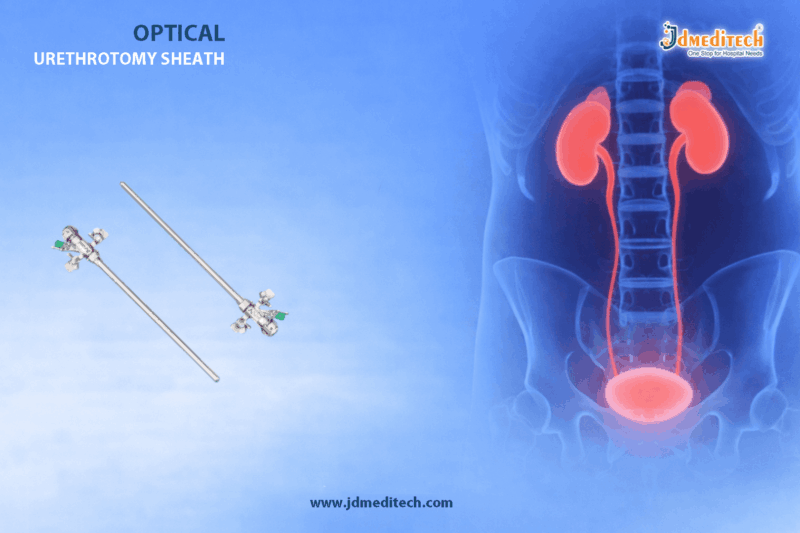
Urethral strictures are a common urological condition that can significantly impact a patient’s quality of life. Effective management requires precision, safety, and reliable instruments. The OTIS Urethrotome stands out as a trusted solution in modern urethral surgery, offering controlled and accurate incision of strictures.

Designed for urologists who demand efficiency and consistency, the OTIS Urethrotome ensures optimal outcomes in urethrotomy procedures. In this article, we explore its design, applications, advantages, and why it remains a preferred instrument in urology.

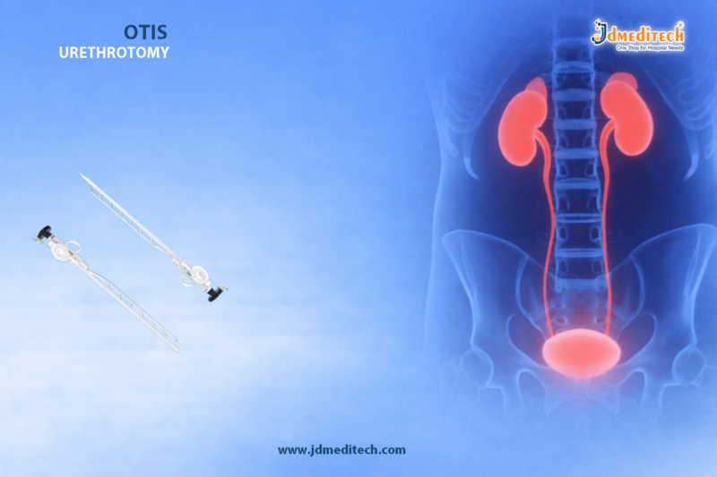
What is an OTIS Urethrotome?

The OTIS Urethrotome is a specialized surgical instrument used to perform internal urethrotomy. It is primarily designed to incise urethral strictures under controlled conditions, allowing restoration of normal urinary flow.

This instrument is widely used in urological surgeries due to its precision mechanism and ease of handling.

Key Features of OTIS Urethrotome
1. Precision Cutting Mechanism

The OTIS Urethrotome is engineered to deliver controlled and accurate incisions, minimizing tissue trauma during urethral stricture treatment.

2. Adjustable Blade System

It features an adjustable blade that allows surgeons to regulate incision depth, ensuring safety and effectiveness.

3. High-Quality Medical-Grade Material

Manufactured using medical-grade stainless steel, the instrument offers durability, corrosion resistance, and long-term reliability.

4. Ergonomic Design

The ergonomic handle provides a firm grip, enabling surgeons to perform procedures with confidence and precision.

5. Reusable and Sterilizable

The OTIS Urethrotome is designed for repeated use and can be easily sterilized, making it cost-effective for healthcare facilities.

Applications in Urethral Surgery

The OTIS Urethrotome plays a crucial role in:

  • Treatment of urethral strictures
  • Internal urethrotomy procedures
  • Restoration of urinary flow
  • Minimally invasive urological interventions

Its precision makes it suitable for both routine and complex urological cases.

Advantages of Using OTIS Urethrotome
1. Minimally Invasive Approach

The instrument supports minimally invasive procedures, reducing recovery time and patient discomfort.

2. Enhanced Surgical Accuracy

With its controlled cutting mechanism, the OTIS Urethrotome allows precise incision, improving surgical outcomes.

3. Reduced Risk of Complications

Accurate incision reduces the chances of unnecessary tissue damage and complications.

4. Cost-Effective Solution

Reusable design ensures long-term value for hospitals and clinics.

5. Trusted by Urologists

The OTIS Urethrotome has gained widespread acceptance among urologists for its reliability and performance.

How OTIS Urethrotome Improves Patient Outcomes

By enabling precise incision of strictures, the OTIS Urethrotome helps:

  • Restore normal urinary function
  • Reduce recurrence rates
  • Improve patient comfort
  • Shorten hospital stays

Its efficiency contributes significantly to better clinical outcomes and patient satisfaction.

Best Practices for Use

To achieve optimal performance with the OTIS Urethrotome, clinicians should:

  • Ensure proper sterilization before use
  • Select appropriate blade size
  • Follow standard urethrotomy protocols
  • Handle with care to maintain precision
Why Choose OTIS Urethrotome from JDMeditech?

At JDMeditech, we focus on delivering high-quality urological instruments that meet global standards. Our OTIS Urethrotome is designed with precision engineering to support safe and effective surgical procedures.

Our Commitment:
  • Premium quality manufacturing
  • Strict quality control
  • Reliable performance
  • Competitive pricing

🌐 Visit: www.jdmeditech.com

Conclusion

The OTIS Urethrotome remains an essential instrument in modern urology, offering precision, safety, and reliability in urethral stricture treatment. Its advanced design and proven effectiveness make it a preferred choice among surgeons worldwide.

Whether for routine procedures or complex cases, investing in a high-quality OTIS Urethrotome ensures better surgical outcomes and improved patient care.


Explore More Product >>

Get Connected:

+91 79909 93062 | +91 63513 72032 | exports@jdmeditech.com


Heparin Sodium Injection – Trusted Anticoagulant for Safe & Effective Blood Thinning

In modern medicine, preventing blood clot formation is critical to ensuring patient safety during surgeries, dialysis, and cardiovascular treatments. Heparin Sodium Injection plays a vital role as a fast-acting anticoagulant that helps maintain smooth blood flow and reduces the risk of life-threatening complications such as thrombosis and embolism.

Widely used across hospitals and clinical setups, this medication has become a trusted anticoagulant solution due to its reliability, rapid action, and proven safety profile.

What is Heparin Sodium Injection?

Heparin Sodium Injection is an anticoagulant medication that works by inhibiting clotting factors in the blood, preventing the formation and growth of clots. It is commonly administered intravenously or subcutaneously under medical supervision.

It is especially effective in situations where immediate anticoagulation is required, making it an essential component in critical care and surgical procedures.

Key Features of Heparin Sodium Injection
  • Rapid Anticoagulant Action : Provides immediate blood-thinning effect
  • Highly Reliable : Trusted by healthcare professionals worldwide
  • Flexible Administration : Can be given IV or subcutaneously
  • Widely Applicable : Suitable for multiple medical conditions
  • Consistent Performance : Ensures predictable anticoagulation outcomes
Clinical Applications
1. Prevention of Blood Clots

Heparin Sodium Injection is widely used to prevent clot formation in patients at high risk, especially during prolonged immobilization or post-surgery.

2. Dialysis Procedures

It is essential in hemodialysis to prevent clotting in the dialysis circuit, ensuring smooth and uninterrupted treatment.

3. Cardiovascular Treatments

Used in conditions such as deep vein thrombosis (DVT), pulmonary embolism (PE), and acute coronary syndromes.

4. Surgical Procedures

Administered during surgeries to maintain proper blood flow and reduce complications caused by clotting.

Benefits of Using Heparin Sodium Injection
  • Immediate Effectiveness : Acts quickly in emergency situations
  • Prevents Serious Complications : Reduces risk of stroke and embolism
  • Improves Patient Outcomes : Ensures safe procedural success
  • Versatile Usage : Applicable in multiple medical specialties
  • Clinically Proven : Backed by decades of medical use
How Heparin Sodium Injection Works

Heparin enhances the activity of antithrombin III, a natural inhibitor of blood clotting factors such as thrombin and factor Xa. By blocking these clotting pathways, it prevents fibrin formation, which is essential for clot development.

This mechanism ensures that blood remains fluid and reduces the chances of dangerous clot formation.

Safety and Precautions

While Heparin Sodium Injection is highly effective, proper usage and monitoring are essential:

  • Administer only under medical supervision
  • Monitor coagulation parameters (e.g., APTT levels)
  • Avoid in patients with active bleeding disorders
  • Use cautiously in patients with hypertension or liver disease
  • Watch for signs of heparin-induced thrombocytopenia (HIT)
Storage and Handling
  • Store in a cool, dry place away from direct sunlight
  • Maintain recommended temperature conditions
  • Ensure sterile handling to prevent contamination
  • Keep out of reach of unauthorized personnel
Why Choose JD Meditech?

At JDMeditech, we are committed to delivering high-quality medical solutions that meet international standards. Our Heparin Sodium Injection ensures:

  • Reliable and consistent performance
  • Strict quality control and compliance
  • Safe packaging and easy handling
  • Trusted by healthcare professionals

🌐 Visit: www.jdmeditech.com

Conclusion

Heparin Sodium Injection remains a cornerstone in anticoagulant therapy, offering fast, reliable, and effective prevention of blood clot formation. Its wide range of applications in dialysis, surgery, and cardiovascular care makes it an indispensable tool in modern healthcare.

For healthcare providers seeking a trusted anticoagulant solution, Heparin Sodium Injection continues to deliver proven results with safety and precision.


Explore More Product >>

Get Connected:

+91 79909 93062 | +91 63513 72032 | exports@jdmeditech.com


Gastrology Cytology Brush for Accurate GI Cell Sampling

In modern gastroenterology, early and precise diagnosis plays a critical role in improving patient outcomes. One of the most essential tools used during endoscopic procedures is the gastrology cytology brush, designed specifically for accurate GI cell sampling.

This minimally invasive device enables clinicians to collect high-quality cellular samples from the gastrointestinal (GI) tract, aiding in the detection of infections, inflammation, and malignancies.

At JDMeditech, we provide high-quality cytology brushes that ensure precision, safety, and consistent performance in every procedure.

What is a Gastrology Cytology Brush?

A gastrology cytology brush is a specialized medical device used during endoscopy to collect cells from the lining of the gastrointestinal tract.

It consists of:

  • A flexible catheter
  • Soft, atraumatic bristles
  • Ergonomic handle for controlled movement

The brush is inserted through the endoscope channel and gently brushed against the target tissue to obtain cell samples for laboratory analysis.

Importance of Accurate GI Cell Sampling

Accurate GI cell sampling is essential for diagnosing various gastrointestinal conditions, including:

  • Gastrointestinal cancers (esophageal, gastric, colorectal)
  • Infections and inflammatory diseases
  • Barrett’s esophagus
  • Precancerous lesions

The gastrology cytology brush ensures:

  • High cellular yield
  • Minimal tissue trauma
  • Reliable diagnostic results

Early detection using effective sampling tools can significantly improve treatment success rates.

Key Features of Gastrology Cytology Brush
1. High-Quality Bristles for Effective Sampling

The brush is designed with soft yet durable bristles that maximize cell collection without damaging tissue.

2. Flexible and Kink-Resistant Catheter

Ensures smooth navigation through the endoscope and easy access to targeted areas.

3. Atraumatic Tip Design

Reduces patient discomfort and enhances procedural safety.

4. Ergonomic Control Handle

Allows precise manipulation and controlled brushing during procedures.

5. Sterile and Single-Use Design

Minimizes the risk of cross-contamination and ensures patient safety.

Applications in Gastroenterology

The gastrology cytology brush is widely used in:

  • Endoscopic procedures (EGD, ERCP)
  • Biliary duct sampling
  • Esophageal and gastric lesion evaluation
  • Pancreatic duct investigations

Its versatility makes it an indispensable tool in both diagnostic and screening procedures.

Advantages of Using Gastrology Cytology Brush
1. Minimally Invasive Sampling

Reduces the need for surgical biopsies in many cases.

2. Quick and Efficient Procedure

Enables rapid sample collection during routine endoscopy.

3. High Diagnostic Accuracy

Provides sufficient cellular material for precise analysis.

4. Enhanced Patient Comfort

Designed to minimize irritation and trauma.

5. Cost-Effective Solution

Reduces overall diagnostic costs by enabling early detection.

Why Choose JD Meditech Cytology Brushes?

At JDMeditech, we focus on delivering advanced medical devices that meet international standards. Our gastrology cytology brush offers:

  • Superior material quality
  • Precision-engineered design
  • Consistent performance
  • Strict sterilization standards
  • Compatibility with major endoscopes

We are committed to supporting healthcare professionals with reliable tools for better patient care.

Best Practices for Using Cytology Brush

To ensure optimal results:

  • Use gentle brushing techniques to avoid tissue damage
  • Rotate the brush evenly across the lesion
  • Ensure proper handling and disposal after use
  • Follow sterilization and safety protocols

Proper technique significantly improves sample quality and diagnostic accuracy.

Future of GI Cytology Sampling

With advancements in gastroenterology, cytology tools are evolving rapidly. Innovations include:

  • Enhanced brush designs for higher cell yield
  • Integration with imaging technologies
  • Improved catheter flexibility and control

The gastrology cytology brush will continue to play a key role in early detection and precision diagnostics.

Conclusion

The gastrology cytology brush is a vital instrument for accurate GI cell sampling, enabling early diagnosis and effective treatment of gastrointestinal diseases.

Its precision, safety, and efficiency make it an essential tool in modern endoscopic practice.

For high-quality and reliable cytology brushes, trust JDMeditech—your partner in advanced medical solutions.


Explore More Product >>

Get Connected:

+91 79909 93062 | +91 63513 72032 | exports@jdmeditech.com


Advanced Visual Obturator for Minimally Invasive Surgery

In modern minimally invasive surgery, precision and safety are critical. One device that significantly enhances both is the Visual Obturator. Designed to provide direct visualization during trocar insertion, this advanced surgical tool reduces complications and improves procedural accuracy.

The Visual Obturator is widely used in laparoscopic and endoscopic procedures, offering surgeons a controlled and clear entry into body cavities. In this blog, we explore its features, benefits, applications, and why it has become an essential tool in surgical practice.

What is a Visual Obturator?

A Visual Obturator is a specialized surgical instrument used alongside trocars to enable real-time visualization during entry into the abdominal or thoracic cavity. Unlike traditional blind insertion techniques, it allows surgeons to see tissue layers as they advance, ensuring safer access.

Why Visual Obturator is Essential in Modern Surgery

Traditional trocar insertion carries risks such as:

  • Organ injury
  • Vascular damage
  • Incorrect placement

The Visual Obturator for precision entry addresses these challenges by providing direct visual control, significantly reducing complications.

Key Features of Visual Obturator
1. Direct Visualization Technology

The device integrates with endoscopic systems, allowing surgeons to monitor entry in real time.

2. Ergonomic Design

Its user-friendly design ensures:

  • Comfortable grip
  • Better maneuverability
  • Enhanced control during insertion
3. High-Quality Medical-Grade Materials

Manufactured from durable, sterilizable materials ensuring:

  • Longevity
  • Resistance to corrosion
  • Compliance with medical standards
4. Compatibility with Trocar Systems

The Visual Obturator is compatible with various trocar sizes, making it versatile across procedures.

5. Smooth Penetration Tip

Designed for minimal tissue trauma and precise entry.

Advantages of Using a Visual Obturator
 Enhanced Safety

Direct visualization minimizes the risk of internal injuries.

 Improved Accuracy

Surgeons can precisely control the insertion path.

 Reduced Complications

Lower chances of bleeding, organ damage, and misplacement.

 Faster Procedures

Better control leads to quicker and more efficient surgeries.

Better Patient Outcomes

Less trauma results in faster recovery and reduced hospital stays.

Applications of Visual Obturator

The Visual Obturator for precision entry is widely used in:

  • Laparoscopic surgeries
  • Gynecological procedures
  • General surgeries
  • Urological interventions
  • Thoracoscopic procedures

Its versatility makes it a must-have tool in operating rooms.

How Visual Obturator Improves Surgical Workflow

By eliminating blind insertion, the Visual Obturator:

  • Enhances surgeon confidence
  • Improves procedural efficiency
  • Reduces intraoperative risks
  • Supports better clinical outcomes
Comparison: Visual Obturator vs Traditional Obturator
Feature Visual Obturator Traditional Obturator
Visualization Direct Blind
Safety High Moderate
Accuracy High Limited
Risk of Injury Low Higher
Control Excellent Limited
Best Practices for Using Visual Obturator
  • Ensure proper sterilization before use
  • Use compatible trocar systems
  • Maintain steady insertion under visualization
  • Follow standard surgical protocols
Why Choose JDMeditech Visual Obturator

At JDMeditech, we focus on delivering precision-engineered medical devices designed for safety and performance.

Our Advantages:
  • Superior build quality
  • Precision engineering
  • Reliable performance
  • Compliance with global medical standards
Conclusion

The Visual Obturator for precision entry represents a major advancement in minimally invasive surgery. By offering direct visual control, it significantly enhances safety, accuracy, and surgical efficiency.

For healthcare professionals seeking reliable and advanced surgical tools, the Visual Obturator is an essential addition to modern operating setups.


Explore More Product >>

Get Connected:

+91 79909 93062 | +91 63513 72032 | exports@jdmeditech.com


Permanent Dialysis Catheter Kit for Long-Term Use

For patients with chronic kidney disease (CKD) or end-stage renal disease (ESRD), reliable long-term vascular access is critical for effective hemodialysis. Among the available options, permanent dialysis catheter kits provide a dependable and efficient solution, especially when arteriovenous fistulas or grafts are not feasible.

Modern permanent dialysis access catheter kits are designed to ensure patient safety, long-term functionality, and ease of insertion, making dialysis procedures smoother for both clinicians and patients.

What is a Permanent Dialysis Catheter?

A permanent dialysis catheter (also known as a tunneled dialysis catheter) is a flexible, biocompatible tube inserted into a central vein—typically the internal jugular vein—to facilitate long-term hemodialysis.

Unlike temporary catheters, these are:

  • Tunneled under the skin
  • Equipped with a cuff for infection prevention
  • Designed for extended use (weeks to months or longer)
What is Included in a Permanent Dialysis Catheter Kit?

A high-quality permanent dialysis catheter kit includes all essential components required for safe and efficient catheter placement:

Key Components:
  • Dialysis catheter (dual lumen or triple lumen)
  • Introducer needle
  • Guidewire
  • Dilators
  • Peel-away sheath
  • Tunneler
  • Scalpel
  • Syringes and accessories

These kits are pre-sterilized and ready-to-use, reducing procedural time and infection risks.

Benefits of Permanent Dialysis Catheter Kits
1. Long-Term Reliability

Designed for extended use, permanent dialysis catheter kits ensure consistent blood flow rates required for effective dialysis.

2. Reduced Infection Risk

The Dacron cuff promotes tissue ingrowth, acting as a barrier against infection and catheter displacement.

3. Easy Placement Procedure

With a complete set of tools in one kit, clinicians can perform smooth and efficient catheter insertion.

4. Patient Comfort

Soft, flexible materials improve patient comfort during long-term use.

5. High Biocompatibility

Made from medical-grade materials such as polyurethane or silicone, minimizing adverse reactions.

Applications of Permanent Dialysis Access

Permanent dialysis catheter kits are widely used in:

  • Long-term hemodialysis treatment
  • Patients awaiting AV fistula maturation
  • Emergency backup access
  • Patients unsuitable for surgical access creation
Key Features to Look for in a Quality Catheter Kit

When choosing a trusted permanent dialysis catheter kit, consider:

  •  Optimal lumen size for high flow rates
  •  Kink-resistant and durable catheter design
  •  Radiopaque material for imaging visibility
  •  Soft tip to reduce vessel trauma
  •  Complete sterile kit packaging
  •  Secure locking connectors
Procedure Overview (Simplified)
  1. Site Preparation – Sterilization and local anesthesia
  2. Venous Access – Needle insertion into the target vein
  3. Guidewire Placement – Ensures accurate catheter positioning
  4. Tunneling – Subcutaneous pathway creation
  5. Catheter Insertion – Through peel-away sheath
  6. Securing the Catheter – Suturing and dressing

Note: The procedure should always be performed by trained medical professionals under sterile conditions.

Why Choose Trusted Catheter Kits?

Using high-quality permanent dialysis catheter kits ensures:

  • Improved procedural success rates
  • Reduced complications
  • Enhanced patient outcomes
  • Time-efficient clinical workflow

Reliable manufacturers focus on precision engineering, strict quality control, and compliance with global medical standards.

Conclusion

In modern nephrology care, permanent dialysis catheter kits for long-term use play a vital role in ensuring reliable and safe vascular access. Their ease of use, infection control features, and patient-friendly design make them an essential choice for healthcare providers.

Choosing a trusted, high-quality catheter kit not only improves clinical outcomes but also enhances the overall dialysis experience for patients.


Explore More Product >>

Get Connected:

+91 79909 93062 | +91 63513 72032 | exports@jdmeditech.com


PTFE Guide Wire Solutions for Cardiovascular Excellence

In modern cardiovascular interventions, precision and control are critical for successful outcomes. Among the essential tools used by interventional cardiologists, the PTFE guide wire stands out as a key component for navigating complex vascular pathways with ease and accuracy.

Designed with advanced coating technology, PTFE guide wire solutions offer smooth navigation, reduced friction, and enhanced procedural efficiency. This blog explores how PTFE guide wires contribute to cardiovascular excellence and why they are indispensable in interventional procedures.

What is a PTFE Guide Wire?

A PTFE guide wire (Polytetrafluoroethylene-coated guide wire) is a specialized medical device used in cardiovascular procedures to guide catheters, balloons, and stents through blood vessels.

The PTFE coating provides:

  • Ultra-low friction surface
  • Enhanced trackability
  • Improved maneuverability in tortuous anatomy

This makes PTFE guide wires ideal for both diagnostic and interventional cardiology procedures.

Why PTFE Guide Wire Solutions are Essential in Cardiology
1. Superior Lubricity for Smooth Navigation

The PTFE coating ensures minimal resistance, allowing the guide wire to glide effortlessly through vessels. This is particularly beneficial in:

  • Narrow arteries
  • Calcified lesions
  • Complex vascular structures
2. Enhanced Control and Precision

PTFE guide wires offer excellent torque control, enabling physicians to:

  • Navigate difficult pathways
  • Reach target lesions accurately
  • Improve procedural success rates
3. Reduced Risk of Vessel Trauma

The smooth surface minimizes friction against vessel walls, reducing:

  • Endothelial damage
  • Risk of complications
  • Patient discomfort
4. Versatility Across Procedures

PTFE guide wires are widely used in:

  • Coronary angiography
  • Percutaneous coronary intervention (PCI)
  • Peripheral vascular procedures
  • Endovascular treatments
Key Features of High-Quality PTFE Guide Wires

When selecting a PTFE guide wire solution, the following features are crucial:

  • Hydrophobic PTFE Coating for consistent performance
  • High Torque Response for precise control
  • Flexible Tip Design to navigate complex anatomy
  • Kink Resistance for durability
  • Radiopaque Markers for enhanced visibility under imaging
Applications in Cardiovascular Procedures
1. Coronary Interventions

PTFE guide wires are essential for accessing coronary arteries and delivering devices during angioplasty and stenting.

2. Peripheral Vascular Procedures

Used for navigating peripheral arteries in cases of:

  • Peripheral artery disease (PAD)
  • Chronic total occlusions (CTO)
3. Diagnostic Angiography

Facilitates smooth catheter placement for accurate imaging and diagnosis.

Benefits of Using PTFE Guide Wire Solutions
  • Improved Procedural Efficiency
  • Shorter Procedure Time
  • Higher Success Rate in Complex Cases
  • Enhanced Patient Safety
  • Reduced Operator Fatigue
How to Choose the Right PTFE Guide Wire

Selecting the appropriate guide wire depends on:

  • Procedure Type (diagnostic vs interventional)
  • Vessel Anatomy (straight vs tortuous)
  • Lesion Complexity
  • Required Flexibility and Support

Consulting with a trusted medical device provider ensures optimal product selection.

Why Choose JDMeditech PTFE Guide Wire Solutions

At JDMeditech, we deliver advanced PTFE guide wire solutions designed for superior performance in cardiovascular procedures.

Our Advantages:
  • Premium medical-grade materials
  • Precision-engineered design
  • Consistent quality and reliability
  • Compatibility with standard catheter systems
  • Competitive pricing for global markets
Future of PTFE Guide Wire Technology

With continuous innovation, PTFE guide wires are evolving to include:

  • Hybrid coatings for enhanced performance
  • Improved tip flexibility
  • Advanced imaging compatibility

These advancements will further enhance cardiovascular excellence and patient outcomes.

Conclusion

The PTFE guide wire is a cornerstone of modern cardiovascular interventions. Its ability to provide smooth navigation, precise control, and reduced risk makes it indispensable in both routine and complex procedures.

By choosing high-quality PTFE guide wire solutions, healthcare providers can achieve better clinical outcomes, improve efficiency, and ensure patient safety.


Explore More Product >>

Get Connected:

+91 79909 93062 | +91 63513 72032 | exports@jdmeditech.com


Optical Urethrotomy Sheath for Urethral Stricture Surgery

Urethral stricture is a common urological condition that can significantly impact a patient’s quality of life, causing difficulty in urination, urinary retention, and recurrent infections. Modern minimally invasive techniques have revolutionized its treatment, and one of the most essential tools used in this procedure is the Optical Urethrotomy Sheath.

The Optical Urethrotomy Sheath for urethral stricture surgery is specifically designed to facilitate direct vision internal urethrotomy (DVIU), ensuring precision, safety, and improved surgical outcomes. In this blog, we will explore its features, benefits, applications, and why it is a crucial instrument in urological procedures.

What is an Optical Urethrotomy Sheath?

An Optical Urethrotomy Sheath is a specialized endoscopic instrument used during urethrotomy procedures to treat urethral strictures. It acts as a guiding channel for optical systems and surgical instruments, allowing surgeons to perform incisions under direct visualization.

This sheath is typically used along with:

  • Endoscopes
  • Cold knife or laser fibers
  • Irrigation systems

Its primary function is to provide a stable and clear pathway for safe and precise surgical intervention.

Importance in Urethral Stricture Surgery

The Optical Urethrotomy Sheath for urethral stricture surgery plays a critical role in ensuring procedural success. It allows surgeons to:

  • Visualize the stricture directly
  • Perform controlled incisions
  • Minimize trauma to surrounding tissues
  • Reduce complications and recovery time

This makes it an indispensable tool in Direct Vision Internal Urethrotomy (DVIU) procedures.

Key Features of Optical Urethrotomy Sheath
1. High-Quality Medical-Grade Material

Manufactured using premium stainless steel or medical-grade materials, ensuring durability and corrosion resistance.

2. Precision Engineering

Designed for smooth insertion and accurate alignment during surgery.

3. Optimal Visualization

Compatible with high-definition endoscopes, providing clear visibility of the urethral passage.

4. Ergonomic Design

Ensures ease of handling for surgeons, reducing fatigue during procedures.

5. Efficient Irrigation Channel

Allows continuous fluid flow to maintain a clear surgical field.

6. Compatibility

Works seamlessly with:

  • Optical systems
  • Cold knife urethrotomes
  • Laser fibers
Benefits of Using Optical Urethrotomy Sheath

Using an Optical Urethrotomy Sheath for urethral stricture surgery offers several clinical advantages:

✔ Enhanced Surgical Accuracy

Direct visualization allows precise incision of strictures.

✔ Minimally Invasive Approach

Reduces the need for open surgery, leading to faster recovery.

✔ Reduced Risk of Complications

Lower chances of bleeding, infection, and tissue damage.

✔ Improved Patient Outcomes

Better success rates and reduced recurrence when used correctly.

✔ Time-Efficient Procedure

Streamlined design helps in quicker surgical execution.

Applications in Urology

The Optical Urethrotomy Sheath is widely used in:

  • Treatment of anterior urethral strictures
  • Direct Vision Internal Urethrotomy (DVIU)
  • Laser urethrotomy procedures
  • Diagnostic urethroscopy
  • Minimally invasive urological interventions
How It Works

During urethral stricture surgery:

  1. The sheath is inserted into the urethra.
  2. An optical system is introduced for visualization.
  3. A cutting instrument (cold knife or laser) is passed through the sheath.
  4. The surgeon precisely incises the stricture under direct vision.
  5. Irrigation ensures a clear operative field throughout the procedure.

This controlled approach ensures safety and effectiveness.

Why Choose a High-Quality Optical Urethrotomy Sheath?

Choosing a premium-quality Optical Urethrotomy Sheath for urethral stricture surgery is essential for achieving optimal results. A high-quality sheath ensures:

  • Smooth insertion without trauma
  • Superior visualization
  • Long-lasting performance
  • Compatibility with advanced surgical systems

For healthcare providers, investing in reliable instruments directly impacts surgical efficiency and patient satisfaction.

Maintenance and Sterilization

Proper care is crucial for maintaining performance and hygiene:

  • Clean immediately after use
  • Use approved sterilization methods (autoclaving)
  • Inspect for wear and damage
  • Store in a dry, sterile environment
Conclusion

The Optical Urethrotomy Sheath for urethral stricture surgery is a vital instrument that enables precise, safe, and minimally invasive treatment of urethral strictures. With its advanced design and compatibility with modern surgical systems, it plays a crucial role in improving patient outcomes and surgical efficiency.

For healthcare professionals seeking reliability and precision, investing in a high-quality urethrotomy sheath is a smart and essential choice.


Explore More Product >>

Get Connected:

+91 79909 93062 | +91 63513 72032 | exports@jdmeditech.com


Bicarbonate Cartridge (BiCart) for Hemodialysis – Compact & Efficient Dialysis Solution

Hemodialysis is a life-saving procedure for patients with kidney failure, where maintaining the correct dialysate composition is crucial for effective treatment. One of the most important components in this process is the Bicarbonate Cartridge (BiCart), a modern solution designed to deliver accurate bicarbonate concentration safely and efficiently.

The Bicarbonate Cartridge (BiCart) for Hemodialysis offers a compact, hygienic, and machine-compatible alternative to traditional bicarbonate mixing systems. It simplifies workflow, minimizes contamination risks, and ensures consistent dialysis performance.

What is a Bicarbonate Cartridge (BiCart)?

A Bicarbonate Cartridge (BiCart) is a pre-filled, single-use container that holds dry sodium bicarbonate powder. It is designed to connect directly to compatible hemodialysis machines, where it dissolves automatically with purified water to form bicarbonate concentrate.

This ready-to-use system eliminates the need for manual preparation, ensuring precision and reducing operational errors.

Why Bicarbonate is Essential in Hemodialysis

Bicarbonate plays a vital role in dialysis by:

  • Correcting metabolic acidosis in patients
  • Maintaining acid-base balance
  • Supporting overall physiological stability
  • Enhancing patient comfort during treatment

The Bicarbonate Cartridge (BiCart) for Hemodialysis ensures that this critical component is delivered consistently and accurately.

Key Features of Bicarbonate Cartridge (BiCart)
1. Compact and Space-Saving Design

The BiCart is designed to occupy minimal space, making it ideal for dialysis centers with limited storage.

2. Pre-Filled and Ready-to-Use

Each cartridge comes pre-loaded with medical-grade bicarbonate powder, eliminating manual mixing and saving time.

3. High Compatibility with Dialysis Machines

BiCart systems are engineered to work seamlessly with most modern hemodialysis machines, ensuring smooth integration.

4. Hygienic and Single-Use System

Being disposable, the cartridge significantly reduces the risk of contamination and bacterial growth.

5. Consistent Concentration Delivery

The automated mixing process ensures accurate bicarbonate levels in every dialysis session.

6. Reduced Operational Errors

By removing manual handling, BiCart minimizes human errors and improves treatment reliability.

Advantages of Using BiCart in Hemodialysis
Improved Infection Control

Traditional bicarbonate tanks can harbor bacteria if not cleaned properly. BiCart eliminates this risk with its single-use design.

Time Efficiency

Dialysis staff can save valuable preparation time, allowing them to focus more on patient care.

Enhanced Patient Safety

Accurate bicarbonate concentration ensures stable treatment conditions, reducing complications.

Lower Maintenance Requirements

No need for cleaning tanks or mixing containers, which reduces workload and maintenance costs.

Consistent Dialysis Quality

Each cartridge provides uniform performance, ensuring standardized dialysis sessions.

Applications of Bicarbonate Cartridge (BiCart)

The Bicarbonate Cartridge (BiCart) for Hemodialysis is widely used in:

  • Dialysis centers
  • Hospitals and nephrology units
  • Home hemodialysis setups
  • Intensive care units (ICUs)

Its versatility makes it a preferred choice across various healthcare environments.

How BiCart Works
  1. The cartridge is connected to a compatible dialysis machine
  2. Purified water flows into the cartridge
  3. The bicarbonate powder dissolves automatically
  4. The machine mixes it with acid concentrate
  5. The final dialysate is delivered to the patient

This automated process ensures precision and eliminates manual intervention.

Comparison: BiCart vs Traditional Bicarbonate Mixing
Feature BiCart System Traditional System
Preparation Ready-to-use Manual mixing required
Hygiene High (single-use) Risk of contamination
Accuracy Consistent Variable
Maintenance Minimal High
Time Required Low High
Why Choose JD Meditech Bicarbonate Cartridge?

At JDMeditech, we focus on delivering high-quality dialysis solutions that meet international standards. Our Bicarbonate Cartridge (BiCart) for Hemodialysis is designed for:

  • Reliable performance
  • Superior safety standards
  • Easy machine compatibility
  • Consistent clinical outcomes

We ensure that every product supports healthcare professionals in delivering efficient and safe dialysis treatment.

Best Practices for Using BiCart
  • Always check compatibility with your dialysis machine
  • Store cartridges in a clean, dry environment
  • Use within the recommended shelf life
  • Dispose of used cartridges safely as per medical waste guidelines
Conclusion

The Bicarbonate Cartridge (BiCart) for Hemodialysis represents a significant advancement in dialysis technology. Its compact design, hygienic single-use system, and machine compatibility make it an ideal choice for modern dialysis centers.

By improving efficiency, safety, and consistency, BiCart systems help healthcare providers deliver better patient outcomes with minimal operational complexity.


Explore More Product >>

+91 79909 93062 | +91 63513 72032 | exports@jdmeditech.com


Short Term Hemodialysis Catheter for Temporary Dialysis Access | Complete Guide

A Short Term Hemodialysis Catheter for Temporary Dialysis Access is a critical medical device used to provide immediate vascular access for patients requiring urgent hemodialysis. These catheters are designed for short-duration use, ensuring safe, efficient, and rapid blood flow during dialysis procedures.

In emergency or acute care settings, when permanent access like AV fistula or graft is not available, a short term hemodialysis catheter becomes a lifesaving solution.

What is a Short Term Hemodialysis Catheter?

A Short Term Hemodialysis Catheter for Temporary Dialysis Access is a flexible, sterile tube inserted into a large central vein—commonly the internal jugular, femoral, or subclavian vein—to facilitate dialysis treatment.

These catheters are specifically designed for:

  • Immediate use
  • Short duration (typically up to 2–3 weeks)
  • High flow rates for efficient dialysis
Why is Temporary Dialysis Access Needed?

Temporary access is essential in situations such as:

  • Acute kidney injury (AKI)
  • Emergency dialysis requirements
  • Waiting period for AV fistula maturation
  • Failure of permanent vascular access
  • Critical care or ICU dialysis support

A Short Term Hemodialysis Catheter for Temporary Dialysis Access ensures that treatment can begin without delay.

Key Features of Short Term Hemodialysis Catheters
1. Biocompatible Material

Made from medical-grade polyurethane or silicone to reduce thrombosis and infection risk.

2. High Flow Lumen Design

Dual lumen or triple lumen configurations allow efficient blood flow rates for optimal dialysis performance.

3. Kink-Resistant Structure

Ensures uninterrupted blood flow and durability during use.

4. Radiopaque Markings

Facilitates accurate placement under imaging guidance.

5. Smooth Tip Design

Minimizes vessel trauma during insertion.

6. Pre-attached Extensions & Clamps

Improves ease of handling and reduces contamination risk.

Types of Short Term Hemodialysis Catheters
  • Non-tunneled Catheters – Most commonly used for short-term access
  • Dual Lumen Catheters – Standard for hemodialysis
  • Triple Lumen Catheters – Allows medication administration along with dialysis
Insertion Sites for Temporary Dialysis Catheter

The effectiveness of a Short Term Hemodialysis Catheter for Temporary Dialysis Access depends on correct placement:

  • Internal Jugular Vein (Preferred) – Lower infection risk
  • Femoral Vein – Easy access in emergencies
  • Subclavian Vein – Less preferred due to stenosis risk
Advantages of Short Term Hemodialysis Catheters
  • Immediate dialysis access
  • Quick and minimally invasive placement
  • Suitable for emergency situations
  • No surgical procedure required
  • Cost-effective for short-term use
Risks and Considerations

While highly effective, these catheters may have some risks:

  • Infection (Catheter-related bloodstream infections)
  • Thrombosis
  • Catheter dysfunction
  • Vessel injury
  • Air embolism (rare)

Proper sterile techniques and monitoring significantly reduce these risks.

Best Practices for Safe Use

To ensure optimal performance of a Short Term Hemodialysis Catheter for Temporary Dialysis Access:

  • Maintain strict aseptic technique
  • Regularly flush catheter lumens
  • Monitor for signs of infection
  • Ensure proper fixation to prevent displacement
  • Replace with permanent access as soon as possible
Short Term vs Long Term Dialysis Catheters
Feature Short Term Catheter Long Term Catheter
Duration Up to 2–3 weeks Months to years
Placement Non-tunneled Tunneled
Infection Risk Higher Lower
Use Case Emergency dialysis Chronic dialysis
Conclusion

A Short Term Hemodialysis Catheter for Temporary Dialysis Access plays a vital role in emergency and acute dialysis care. Its ability to provide immediate vascular access makes it indispensable in critical situations.

However, due to associated risks, it is recommended only for short-term use, with a transition to permanent access planned as early as possible.

For healthcare providers, choosing a high-quality catheter ensures patient safety, efficient dialysis, and better clinical outcomes.


Explore More Product >>

Get Connected:

+91 79909 93062 | +91 63513 72032 | exports@jdmeditech.com


Manual Lithotripters in Gastroenterology – Safe & Effective Stone Removal

In modern gastroenterology, the management of biliary and pancreatic duct stones has evolved significantly. Among the essential tools used in endoscopic procedures, manual lithotripters in gastroenterology play a crucial role in safely and effectively fragmenting large or difficult stones.

These devices are widely used during Endoscopic Retrograde Cholangiopancreatography (ERCP) procedures, enabling physicians to crush stones that cannot be removed using standard extraction techniques. Their precision, reliability, and cost-effectiveness make them indispensable in both advanced hospitals and routine clinical settings.

What is a Manual Lithotripter?

A manual lithotripter is a mechanical medical device designed to crush large stones located in the bile duct or pancreatic duct. It works by applying controlled mechanical force to fragment stones into smaller pieces, making them easier to extract.

Unlike powered or electrohydraulic lithotripsy systems, manual lithotripters are:

  • Simple to operate
  • Highly reliable
  • Cost-efficient
Importance of Manual Lithotripters in Gastroenterology

The role of manual lithotripters in gastroenterology has become increasingly significant due to the rising incidence of gallstones and bile duct stones.

Why They Are Essential:
  • Enable removal of large or impacted stones
  • Reduce the need for surgical intervention
  • Improve success rates of ERCP procedures
  • Offer a minimally invasive solution

Manual lithotripters are particularly valuable in situations where standard baskets fail to retrieve stones.

Key Features of Manual Lithotripters

Modern manual lithotripters are engineered for performance, safety, and ease of use.

1. High Mechanical Strength

Designed to exert strong crushing force for effective stone fragmentation.

2. Ergonomic Handle Design

Provides better control and reduces operator fatigue during procedures.

3. Durable Construction

Made from medical-grade stainless steel and high-quality polymers for long-term use.

4. Compatibility with ERCP Accessories

Works seamlessly with stone retrieval baskets and guidewires.

5. Smooth Operation Mechanism

Ensures controlled and precise crushing without sudden force application.

Clinical Applications

Manual lithotripters are widely used in various gastroenterological procedures:

1. Bile Duct Stone Removal

Crushing large common bile duct (CBD) stones during ERCP.

2. Pancreatic Duct Stone Management

Used in selected cases for pancreatic stone fragmentation.

3. Difficult Stone Cases

Ideal for impacted or hard stones that cannot be removed by baskets alone.

4. Rescue Procedures

Used when standard stone retrieval techniques fail.

Advantages of Manual Lithotripters
✔ Cost-Effective Solution

Compared to advanced lithotripsy systems, manual lithotripters are more affordable.

✔ High Success Rate

Effective in breaking large stones, improving procedural outcomes.

✔ Minimally Invasive

Eliminates the need for open or laparoscopic surgery in many cases.

✔ Easy Handling

Simple mechanism ensures ease of use even in complex procedures.

✔ Reliable Performance

No dependency on electricity or complex equipment.

Manual vs. Advanced Lithotripsy Techniques
Feature Manual Lithotripter Advanced Lithotripsy (Laser/EHL)
Cost Low High
Complexity Simple Advanced
Equipment Needed Minimal Specialized setup
Accessibility Widely available Limited to advanced centers
Effectiveness High for most stones Ideal for very hard stones

Manual lithotripters remain the first-line solution in many clinical scenarios due to their efficiency and practicality.

Safety Considerations

While manual lithotripters are generally safe, proper technique is essential:

  • Ensure correct basket positioning before crushing
  • Avoid excessive force to prevent duct injury
  • Use fluoroscopic guidance during ERCP
  • Always follow standard procedural protocols
Future of Manual Lithotripters in Gastroenterology

Despite advancements in laser and electrohydraulic lithotripsy, manual lithotripters in gastroenterology continue to hold strong clinical relevance.

Future innovations may include:

  • Improved ergonomic designs
  • Enhanced durability materials
  • Better integration with endoscopic systems

However, their simplicity, affordability, and effectiveness ensure their continued use worldwide.

Conclusion

The role of manual lithotripters in modern gastroenterology is both significant and indispensable. These devices provide a safe, efficient, and cost-effective solution for managing difficult biliary and pancreatic stones.

As healthcare systems strive for minimally invasive and accessible treatments, manual lithotripters remain a trusted tool for gastroenterologists performing ERCP procedures.


Explore More Product >>

Get Connected:

+91 79909 93062 | +91 63513 72032 | exports@jdmeditech.com