Cardiac Resynchronization Therapy – CRT Treatment Guide

Heart failure affects millions of people worldwide, significantly reducing quality of life and increasing mortality risk. Fortunately, Cardiac Resynchronization Therapy has emerged as a revolutionary solution. This advanced treatment improves heart function, reduces symptoms, and helps patients live longer and healthier lives.

In this detailed guide, we will explore how Cardiac Resynchronization Therapy works, its benefits, procedure, and why it is considered a breakthrough in modern cardiology.

What is Cardiac Resynchronization Therapy?

Cardiac Resynchronization Therapy (CRT) is a specialized treatment designed to improve the coordination of heart contractions. In many heart failure patients, the heart’s ventricles do not beat in sync, which reduces pumping efficiency.

CRT uses a small implantable device, similar to a pacemaker, to restore synchronized heart rhythms. As a result, Cardiac Resynchronization Therapy enhances cardiac performance and reduces complications.

Why is Cardiac Resynchronization Therapy Needed?

Heart failure often leads to electrical conduction delays in the heart. This causes inefficient pumping and worsens symptoms like fatigue and shortness of breath.

Key reasons to consider Cardiac Resynchronization Therapy:
  • Improves irregular heart rhythms
  • Enhances blood circulation
  • Reduces hospitalization rates
  • Improves survival outcomes
  • Boosts overall quality of life

Therefore, Cardiac Resynchronization Therapy plays a crucial role in managing advanced heart failure.

How Does Cardiac Resynchronization Therapy Work?

Cardiac Resynchronization Therapy works by delivering electrical impulses to both ventricles, ensuring they contract simultaneously.

The system includes:
  • A pulse generator (battery-powered device)
  • Leads (wires) placed in the heart chambers
  • A programming system to control timing

Because of this synchronized action, Cardiac Resynchronization Therapy restores efficient heart pumping and reduces strain on the heart.

Types of Cardiac Resynchronization Therapy Devices

There are two main types of Cardiac Resynchronization Therapy devices:

1. CRT-P (Pacemaker)

This device helps regulate heart rhythm without defibrillation.

2. CRT-D (Defibrillator)

This advanced device includes a defibrillator that can prevent sudden cardiac arrest.

Choosing the right Cardiac Resynchronization Therapy device depends on the patient’s condition and risk factors.

Procedure of Cardiac Resynchronization Therapy

The Cardiac Resynchronization Therapy procedure is minimally invasive and usually completed within a few hours.

Step-by-step process:
  1. Local anesthesia is administered
  2. Leads are inserted through veins into the heart
  3. The device is implanted under the skin near the chest
  4. The system is tested and programmed

After implantation, Cardiac Resynchronization Therapy begins improving heart function almost immediately.

Benefits of Cardiac Resynchronization Therapy

Patients receiving Cardiac Resynchronization Therapy often experience significant improvements.

Major benefits include:
  • Improved heart efficiency
  • Reduced symptoms of heart failure
  • Better exercise tolerance
  • Lower risk of hospitalization
  • Enhanced survival rates

Because of these advantages, Cardiac Resynchronization Therapy is widely recommended by cardiologists.

Risks and Considerations

Although Cardiac Resynchronization Therapy is generally safe, some risks may occur.

Possible risks:
  • Infection at the implant site
  • Lead displacement
  • Device malfunction
  • Bleeding or swelling

However, with proper medical care, complications related to Cardiac Resynchronization Therapy remain minimal.

Who is an Ideal Candidate for Cardiac Resynchronization Therapy?

Not every patient with heart failure qualifies for Cardiac Resynchronization Therapy.

Ideal candidates include:
  • Patients with moderate to severe heart failure
  • Individuals with delayed ventricular conduction
  • Patients not responding well to medication
  • Those with reduced ejection fraction

A cardiologist evaluates whether Cardiac Resynchronization Therapy is suitable based on diagnostic tests.

Recovery and Lifestyle After CRT

Recovery after Cardiac Resynchronization Therapy is usually quick. Most patients resume normal activities within a few weeks.

Post-procedure care tips:
  • Avoid heavy lifting initially
  • Attend regular follow-ups
  • Monitor device function
  • Maintain a heart-healthy lifestyle

By following these guidelines, patients can maximize the benefits of Cardiac Resynchronization Therapy.

Future of Cardiac Resynchronization Therapy

Advancements in technology continue to improve Cardiac Resynchronization Therapy devices. Innovations like wireless pacing and AI-driven monitoring are shaping the future of heart failure treatment.

As research progresses, Cardiac Resynchronization Therapy will become even more effective and accessible.

Conclusion

Cardiac Resynchronization Therapy has transformed the management of heart failure. By restoring synchronized heart function, it significantly improves patient outcomes and quality of life.

If you or your patients are dealing with advanced heart failure, Cardiac Resynchronization Therapy offers a reliable and life-enhancing solution.


Explore More Product >>

+91 79909 93062 | +91 63513 72032 | exports@jdmeditech.com
Laparoscopic Choledocholithotomy – Bile Duct Stone Removal

Choledocholithotomy is a highly effective surgical procedure used to remove stones from the common bile duct. These stones, also known as choledocholithiasis, can cause severe pain, infection, and life-threatening complications if left untreated.

Today, advancements in medical technology have made choledocholithotomy a minimally invasive and safer solution. As a result, patients experience faster recovery, reduced hospital stays, and fewer complications.

What is Choledocholithotomy?

Choledocholithotomy refers to the surgical removal of stones from the bile duct. Surgeons perform this procedure when stones block bile flow and lead to symptoms such as jaundice, abdominal pain, and infection.

In many cases, doctors prefer minimally invasive approaches such as laparoscopic or endoscopic techniques. These methods improve patient outcomes and reduce surgical risks.

Why is Choledocholithotomy Needed?

Doctors recommend choledocholithotomy when bile duct stones cannot pass naturally or be removed through less invasive methods like ERCP.

Common Indications Include:
  • Obstructive jaundice
  • Severe abdominal pain
  • Cholangitis (bile duct infection)
  • Pancreatitis caused by bile duct stones
  • Failed ERCP stone removal

Without timely choledocholithotomy, these conditions can worsen quickly and lead to serious complications.

Types of Choledocholithotomy Procedures
1. Open Choledocholithotomy

Surgeons make a larger incision to access the bile duct directly. Although effective, this method is less common today due to longer recovery times.

2. Laparoscopic Choledocholithotomy

This minimally invasive approach uses small incisions and specialized instruments. It offers quicker recovery, minimal pain, and reduced scarring.

3. Endoscopic Approach (ERCP-Assisted)

In some cases, surgeons combine choledocholithotomy with ERCP techniques to remove stones without external incisions.

Step-by-Step Procedure

Understanding the steps of choledocholithotomy helps patients feel more confident before surgery.

  1. Anesthesia Administration – The patient receives general anesthesia
  2. Access to Bile Duct – Surgeons use laparoscopic or open techniques
  3. Stone Identification – Imaging tools locate the stones
  4. Stone Removal – Specialized instruments extract the stones
  5. Drain Placement (if needed) – A T-tube may be inserted
  6. Closure – The incision is closed carefully

Each step in choledocholithotomy focuses on safety and complete stone removal.

Advantages of Minimally Invasive Choledocholithotomy

Modern choledocholithotomy techniques offer several benefits:

  • Smaller incisions and less scarring
  • Reduced postoperative pain
  • Faster recovery time
  • Shorter hospital stay
  • Lower risk of infection

Because of these advantages, laparoscopic choledocholithotomy has become the preferred choice in many hospitals.

Risks and Complications

Although choledocholithotomy is generally safe, some risks may occur:

  • Infection
  • Bleeding
  • Bile leakage
  • Injury to surrounding structures
  • Recurrence of stones

However, experienced surgeons significantly reduce these risks during choledocholithotomy.

Recovery After Choledocholithotomy

Recovery depends on the type of procedure performed.

  • Laparoscopic surgery : 1–2 weeks recovery
  • Open surgery : 3–6 weeks recovery

Patients usually resume normal activities quickly after choledocholithotomy, especially with minimally invasive techniques.

Postoperative Care Tips:
  • Follow dietary recommendations
  • Avoid heavy lifting initially
  • Take prescribed medications
  • Attend follow-up visits

Proper care ensures a smooth recovery after choledocholithotomy.

Role of Advanced Medical Devices

High-quality surgical instruments play a critical role in successful choledocholithotomy procedures. Precision tools ensure accurate stone removal and improve patient safety.

At JDMeditech, we provide advanced medical devices designed for minimally invasive gastrointestinal and surgical procedures, ensuring reliability and superior performance.

Conclusion

Choledocholithotomy remains a trusted and effective solution for bile duct stone removal. With the rise of minimally invasive techniques, the procedure has become safer, faster, and more patient-friendly.

Early diagnosis and timely choledocholithotomy can prevent serious complications and restore normal bile flow effectively.


Explore More Product >>

Get Connected:

+91 79909 93062 | +91 63513 72032 | exports@jdmeditech.com
Distal Pancreatectomy – Surgical Removal of the Pancreas Tail

Distal pancreatectomy is a specialized surgical procedure used to remove the tail of the pancreas. Surgeons perform distal pancreatectomy mainly to treat tumors, cysts, or chronic pancreatic conditions affecting the distal portion of the gland.

Moreover, distal pancreatectomy plays a crucial role in modern gastroenterological surgery. It helps patients achieve better outcomes when less invasive treatments are not effective.

What Is Distal Pancreatectomy?

Distal pancreatectomy refers to the surgical removal of the body and tail of the pancreas. In many cases, surgeons also remove the spleen during distal pancreatectomy, depending on the disease condition.

In addition, distal pancreatectomy can be performed using:

  • Open surgery
  • Laparoscopic techniques
  • Robotic-assisted procedures

Therefore, the choice of technique depends on the patient’s condition and the surgeon’s expertise.

Why Is Distal Pancreatectomy Performed?

Doctors recommend distal pancreatectomy for several medical conditions. Most importantly, distal pancreatectomy helps in removing diseased pancreatic tissue effectively.

Common Indications Include:
  • Pancreatic tumors (benign or malignant)
  • Pancreatic cysts
  • Neuroendocrine tumors
  • Chronic pancreatitis
  • Trauma to the pancreas

Furthermore, early intervention with distal pancreatectomy can significantly improve survival rates in cancer patients.

Procedure of Distal Pancreatectomy

Distal pancreatectomy involves a series of carefully planned surgical steps.

Step-by-Step Process:
  1. The patient receives general anesthesia
  2. The surgeon makes an incision or inserts laparoscopic instruments
  3. The pancreas tail is identified and isolated
  4. Blood vessels supplying the pancreas are controlled
  5. The diseased portion is removed
  6. The surgical site is closed securely

In minimally invasive distal pancreatectomy, surgeons use small incisions. As a result, patients experience faster recovery and less postoperative pain.

Types of Distal Pancreatectomy

Distal pancreatectomy can be classified into different types based on surgical approach.

1. Open Distal Pancreatectomy

Surgeons perform this method through a large incision. It is suitable for complex cases.

2. Laparoscopic Distal Pancreatectomy

This minimally invasive approach uses small incisions and a camera. Therefore, it reduces recovery time.

3. Robotic Distal Pancreatectomy

This advanced technique offers greater precision and flexibility. Consequently, it improves surgical outcomes.

Benefits of Distal Pancreatectomy

Distal pancreatectomy offers several advantages for patients.

  • Effective removal of tumors
  • Minimally invasive options available
  • Faster recovery in laparoscopic cases
  • Reduced postoperative pain
  • Improved quality of life

Additionally, distal pancreatectomy allows precise removal of diseased tissue while preserving healthy structures.

Risks and Complications

Although distal pancreatectomy is generally safe, some risks may occur.

Possible Complications Include:
  • Pancreatic fistula
  • Infection
  • Bleeding
  • Delayed gastric emptying
  • Diabetes (in rare cases)

However, with modern techniques and experienced surgeons, the risk of complications is significantly reduced.

Recovery After Distal Pancreatectomy

Recovery after distal pancreatectomy varies depending on the surgical method.

Postoperative Care Includes:
  • Hospital stay of 5–10 days
  • Pain management
  • Gradual return to normal diet
  • Regular follow-up visits

Furthermore, patients undergoing laparoscopic distal pancreatectomy often recover faster compared to open surgery.

Distal Pancreatectomy vs Other Pancreatic Surgeries

Distal pancreatectomy differs from other pancreatic procedures.

  • Whipple Procedure : Removes the head of the pancreas
  • Total Pancreatectomy : Removes the entire pancreas

In contrast, distal pancreatectomy focuses only on the body and tail, making it less extensive in many cases.

Role of Advanced Surgical Instruments

Modern surgical instruments play a vital role in distal pancreatectomy procedures.

At JDMeditech, we provide high-quality medical devices designed for precision and safety in advanced surgeries. Our instruments support surgeons in achieving optimal outcomes in procedures like distal pancreatectomy.

Conclusion

Distal pancreatectomy is a highly effective surgical solution for treating diseases affecting the pancreas tail. With advancements in minimally invasive techniques, distal pancreatectomy has become safer and more efficient.

Therefore, early diagnosis and timely distal pancreatectomy can significantly improve patient outcomes and quality of life.


Explore More Product >>

+91 79909 93062 | +91 63513 72032 | exports@jdmeditech.com
 

Cardiac Sympathectomy for Severe Heart Arrhythmias

Heart arrhythmias can disrupt normal cardiac function and, in severe cases, become life-threatening. While medications and catheter-based treatments often help, some patients continue to experience uncontrolled irregular heart rhythms. In such cases, Cardiac Sympathectomy offers an effective surgical solution.

This advanced procedure targets the sympathetic nerves responsible for triggering abnormal heart rhythms. As a result, it helps reduce arrhythmia episodes and improves patient outcomes.

What is Cardiac Sympathectomy?

Cardiac Sympathectomy is a minimally invasive surgical procedure that interrupts specific sympathetic nerves supplying the heart. These nerves play a key role in regulating heart rate and rhythm.

By reducing excessive nerve stimulation, the procedure helps stabilize the heart’s electrical activity. Therefore, it is especially beneficial for patients with refractory or life-threatening arrhythmias.

Why is Cardiac Sympathectomy Performed?

Doctors recommend Cardiac Sympathectomy when other treatments fail to control arrhythmias effectively. It is particularly useful in conditions such as:

  • Ventricular tachycardia (VT)
  • Long QT syndrome
  • Catecholaminergic polymorphic ventricular tachycardia (CPVT)
  • Recurrent arrhythmias despite medication
  • Patients not responding to implantable cardioverter-defibrillator (ICD) therapy

Moreover, this procedure reduces the frequency of sudden cardiac events, making it a critical intervention in high-risk patients.

How Does Cardiac Sympathectomy Work?

The procedure focuses on removing or disabling portions of the sympathetic nerve chain. Typically, surgeons perform it using video-assisted thoracoscopic surgery (VATS).

Step-by-Step Procedure:
  1. The patient receives general anesthesia
  2. Small incisions are made in the chest
  3. A thoracoscope (camera) is inserted
  4. The surgeon identifies the sympathetic nerves
  5. Selected nerve segments are removed or interrupted
  6. Incisions are closed after ensuring stability

Because of its minimally invasive nature, recovery is usually faster compared to open surgery.

Benefits of Cardiac Sympathectomy

Cardiac Sympathectomy provides several important benefits:

  • Reduces frequency of arrhythmia episodes
  • Lowers risk of sudden cardiac death
  • Improves effectiveness of ICD therapy
  • Minimally invasive with quicker recovery
  • Enhances quality of life

Additionally, it offers long-term relief for patients with drug-resistant arrhythmias.

Risks and Considerations

Although Cardiac Sympathectomy is generally safe, some risks may occur:

  • Infection or bleeding
  • Pneumothorax (collapsed lung)
  • Temporary nerve-related effects
  • Compensatory sweating

However, experienced surgeons significantly reduce these risks, ensuring better outcomes.

Recovery After Cardiac Sympathectomy

Recovery is typically smooth and quick. Most patients:

  • Stay in the hospital for 1–3 days
  • Resume normal activities within 1–2 weeks
  • Experience reduced arrhythmia symptoms over time

Follow-up care remains essential to monitor heart rhythm and overall progress.

Who is a Suitable Candidate?

Not all patients require Cardiac Sympathectomy. Doctors carefully evaluate candidates based on:

  • Severity of arrhythmia
  • Response to medications
  • Risk of sudden cardiac arrest
  • Overall health condition

Therefore, a cardiologist and cardiac surgeon work together to determine suitability.

Conclusion

Cardiac Sympathectomy stands as a life-saving surgical option for patients suffering from severe and resistant heart arrhythmias. When conventional treatments fail, this procedure provides a reliable way to control abnormal heart rhythms and reduce life-threatening risks.

With advancements in minimally invasive techniques, Cardiac Sympathectomy continues to improve patient outcomes and offers renewed hope for individuals with complex cardiac conditions.


Explore More Product >>

Get Connected:

+91 79909 93062 | +91 63513 72032 | exports@jdmeditech.com
Spider Basket for Stone Retrieval in Urology

The Spider Basket plays a crucial role in modern urology. It is widely used for stone retrieval during minimally invasive procedures, especially in ureteroscopy. Because of its unique design, the Spider Basket ensures secure stone capture, precise control, and reduced trauma to surrounding tissues.

As urological procedures continue to evolve, surgeons rely on tools like the Spider Basket to improve efficiency and patient outcomes. Therefore, understanding its features and benefits is essential for healthcare professionals.

What is a Spider Basket?

A Spider Basket is a specialized urological device designed to capture and retrieve stones from the urinary tract. It consists of flexible wires arranged in a basket-like structure, allowing it to open and close smoothly inside the ureter or kidney.

Moreover, the Spider Basket is commonly used in:

  • Ureteroscopy procedures
  • Kidney stone removal
  • Minimally invasive endourological surgeries

Because of its flexible and expandable design, the Spider Basket can easily adapt to different stone sizes and shapes.

Key Features of Spider Basket

The Spider Basket offers several advanced features that enhance its performance:

1. Flexible Multi-Wire Design

The basket wires expand like a spider web, ensuring maximum stone capture efficiency.

2. Strong Grip on Stones

It provides a firm hold, which reduces the risk of stone slippage during retrieval.

3. Smooth Opening and Closing Mechanism

Surgeons can control the basket precisely, improving procedural accuracy.

4. Minimally Invasive Operation

Its design reduces trauma to the urinary tract, leading to faster patient recovery.

5. Compatibility with Endoscopic Instruments

The Spider Basket works seamlessly with ureteroscopes and other endourology tools.

Applications of Spider Basket in Urology
The Spider Basket is widely used in various urological procedures:
Ureteral Stone Retrieval
It helps in capturing and removing stones from the ureter safely.
Kidney Stone Extraction

During flexible ureteroscopy, the Spider Basket retrieves stones from the renal pelvis.

Fragment Removal After Lithotripsy

After laser lithotripsy, the Spider Basket collects broken stone fragments efficiently.

Diagnostic and Therapeutic Procedures

It assists in both diagnosis and treatment within the urinary system.

Advantages of Using Spider Basket

Using a Spider Basket provides multiple benefits:

  • Ensures high precision stone retrieval
  • Reduces risk of tissue damage
  • Improves procedure success rate
  • Enhances surgeon control and visibility
  • Supports faster recovery for patients

Additionally, the Spider Basket minimizes complications, making it a preferred choice in modern urology.

Procedure: How Spider Basket is Used

During a urological procedure:

  1. The surgeon inserts a ureteroscope into the urinary tract.
  2. The Spider Basket is advanced through the working channel.
  3. The basket opens around the stone.
  4. The surgeon captures the stone securely.
  5. The basket is carefully withdrawn along with the stone.

As a result, the procedure becomes efficient and minimally invasive.

Why Choose Spider Basket from JDMeditech?

At JDMeditech, we offer high-quality Spider Basket solutions designed for precision and reliability.

Our Key Highlights:
  • Premium-grade materials
  • Excellent flexibility and durability
  • Ergonomic design for easy handling
  • Strict quality control standards
Maintenance and Handling Tips

To ensure optimal performance of the Spider Basket:

  • Always sterilize before use
  • Handle gently to avoid wire damage
  • Store in a dry, clean environment
  • Follow manufacturer guidelines strictly

Proper handling increases both device lifespan and safety.

Conclusion

The Spider Basket is an advanced stone retrieval tool for urological procedures, offering precision, safety, and efficiency. With its innovative design and reliable performance, it has become an essential instrument in modern endourology.

As minimally invasive techniques continue to grow, the Spider Basket remains a trusted solution for effective stone management.


Explore More Product >>

+91 79909 93062 | +91 63513 72032 | exports@jdmeditech.com
Lateral Internal Sphincterotomy – Best Surgery for Chronic Anal Fissures

Lateral Internal Sphincterotomy is a widely accepted and highly effective surgical procedure used to treat chronic anal fissures. When conservative treatments fail, this procedure provides long-term relief by reducing anal sphincter pressure and promoting healing.

In this blog, we will explore everything about Lateral Internal Sphincterotomy, including its procedure, benefits, indications, and recovery.

What is Lateral Internal Sphincterotomy?

Lateral Internal Sphincterotomy is a minor surgical procedure in which a small portion of the internal anal sphincter muscle is carefully divided. As a result, muscle tension decreases, blood flow improves, and the fissure heals naturally.

This procedure is considered the gold standard treatment for chronic anal fissures due to its high success rate and low recurrence.

Understanding Chronic Anal Fissures

Chronic anal fissures are small tears in the lining of the anal canal that persist for more than 6 weeks. They often cause:

  • Severe pain during bowel movements
  • Bleeding
  • Burning sensation
  • Spasm of the anal sphincter

When these symptoms persist, Lateral Internal Sphincterotomy becomes the most reliable solution.

Indications for Lateral Internal Sphincterotomy

Doctors recommend Lateral Internal Sphincterotomy in the following cases:

  • Chronic anal fissures not responding to medication
  • Recurrent fissures
  • Severe sphincter spasm
  • Persistent pain affecting quality of life

Therefore, this procedure is ideal when non-surgical treatments fail.

Procedure of Lateral Internal Sphincterotomy

The Lateral Internal Sphincterotomy procedure is simple and typically performed under local or general anesthesia.

Step-by-Step Overview
  1. The patient is positioned comfortably
  2. A small incision is made near the anal region
  3. The internal sphincter muscle is partially divided
  4. The incision is either left open or closed with sutures

The entire Lateral Internal Sphincterotomy procedure usually takes 15–30 minutes.

Benefits of Lateral Internal Sphincterotomy

Lateral Internal Sphincterotomy offers several advantages:

  • High success rate (over 90%)
  • Rapid pain relief
  • Minimal recurrence
  • Quick recovery time
  • Improved blood flow for healing

Moreover, it significantly improves patient comfort and quality of life.

Risks and Complications

Although Lateral Internal Sphincterotomy is safe, some risks may include:

  • Minor bleeding
  • Infection
  • Temporary incontinence (rare)
  • Delayed wound healing

However, these complications are uncommon when performed by experienced surgeons.

Recovery After Lateral Internal Sphincterotomy

Recovery after Lateral Internal Sphincterotomy is generally smooth.

Post-Surgery Care
  • Maintain proper hygiene
  • Take sitz baths regularly
  • Follow a high-fiber diet
  • Stay hydrated
  • Avoid straining during bowel movements

Most patients return to normal activities within a few days.

Why Lateral Internal Sphincterotomy is the Gold Standard

Lateral Internal Sphincterotomy remains the preferred treatment because:

  • It directly addresses the root cause (muscle spasm)
  • It ensures long-term healing
  • It reduces recurrence significantly

Therefore, it is trusted worldwide by surgeons and patients alike.

Medical Equipment Used in the Procedure

For performing Lateral Internal Sphincterotomy, surgeons rely on precision instruments such as:

  • Surgical blades
  • Forceps
  • Retractors
  • Suturing materials

High-quality surgical instruments, like those offered by JDMeditech, ensure safety, accuracy, and optimal surgical outcomes.

Conclusion

Lateral Internal Sphincterotomy is a safe, effective, and trusted surgical solution for chronic anal fissures. With its high success rate and quick recovery, it offers long-lasting relief for patients who do not respond to conservative treatments.

If you are dealing with persistent anal fissures, consulting a specialist for Lateral Internal Sphincterotomy can be a life-changing decision.

Forcep Basket for Stone Retrieval – Versatile Urology Tool

The Forcep Basket for Stone Retrieval is an essential device in modern urology. It helps surgeons remove stones safely and efficiently from the urinary tract. As minimally invasive procedures continue to grow, the Forcep Basket for Stone Retrieval has become a preferred choice.

Moreover, this device improves precision and reduces complications. As a result, patients experience faster recovery and less discomfort. Therefore, many urologists rely on the Forcep Basket for Stone Retrieval for effective stone management.

What is a Forcep Basket for Stone Retrieval?

A Forcep Basket for Stone Retrieval is a specialized instrument used to capture and extract urinary stones. It combines a flexible basket with a forcep-like control mechanism.

In addition, the Forcep Basket for Stone Retrieval allows smooth navigation through the urinary tract. Because of this, it works effectively in ureteroscopy and cystoscopy procedures.

Key Features of Forcep Basket for Stone Retrieval

The Forcep Basket for Stone Retrieval offers several important features that enhance performance:

Flexible Construction

It uses durable materials like nitinol or stainless steel. Therefore, it ensures both strength and flexibility.

Secure Stone Capture

The basket design allows the Forcep Basket for Stone Retrieval to hold stones firmly during removal.

Smooth Navigation

Its ergonomic structure ensures easy insertion and precise control.

Multiple Size Options

Different sizes of Forcep Basket for Stone Retrieval are available for various stone types.

Minimally Invasive Use

It supports less invasive procedures, which improves patient safety.

Applications in Urology

The Forcep Basket for Stone Retrieval is widely used in different procedures.

Ureteroscopy

During ureteroscopy, the Forcep Basket for Stone Retrieval removes stones from the ureter with precision.

Kidney Stone Removal

It helps extract stones from the kidney without damaging surrounding tissue.

Bladder Stone Extraction

Surgeons use the Forcep Basket for Stone Retrieval to safely remove bladder stones.

Post-Lithotripsy Procedures

After stone fragmentation, it collects remaining fragments efficiently.

Advantages of Forcep Basket for Stone Retrieval

The Forcep Basket for Stone Retrieval provides multiple advantages:

  • Improves surgical accuracy
  • Reduces tissue trauma
  • Enhances stone removal efficiency
  • Supports minimally invasive techniques
  • Promotes faster patient recovery

Furthermore, the Forcep Basket for Stone Retrieval increases overall procedural success.

How Does Forcep Basket for Stone Retrieval Work?

The Forcep Basket for Stone Retrieval follows a simple process:

  1. The device enters through an endoscope
  2. The basket opens near the stone
  3. The Forcep Basket for Stone Retrieval captures the stone
  4. The basket closes securely
  5. The surgeon removes the device carefully

Thus, the process ensures safe and controlled extraction.

Why Choose a High-Quality Forcep Basket for Stone Retrieval?

Choosing a reliable Forcep Basket for Stone Retrieval improves outcomes. High-quality devices provide better control and durability.

At JDMeditech, we design each Forcep Basket for Stone Retrieval with precision. Therefore, our products meet global quality standards and clinical needs.

Maintenance and Handling Tips

Proper handling of the Forcep Basket for Stone Retrieval ensures long-term performance:

  • Clean and sterilize before use
  • Inspect the device regularly
  • Avoid excessive force
  • Store in a dry environment

By following these steps, the Forcep Basket for Stone Retrieval remains effective.

Future Trends

The Forcep Basket for Stone Retrieval continues to evolve. New designs improve flexibility and safety.

For example, manufacturers now develop disposable versions. In addition, advanced materials enhance durability. Therefore, the Forcep Basket for Stone Retrieval will remain a key tool in urology.

Conclusion

The Forcep Basket for Stone Retrieval is a versatile and reliable device for stone removal. It supports safe, efficient, and minimally invasive procedures.

As technology advances, the Forcep Basket for Stone Retrieval will continue to improve patient care and surgical outcomes.


Explore More Product >>

Get Connected:

+91 79909 93062 | +91 63513 72032 | exports@jdmeditech.com
Hemorrhoidectomy – A Surgical Solution for Hemorrhoids

Hemorrhoidectomy is a highly effective surgical solution for hemorrhoids that provides long-term relief from severe or recurring symptoms. When non-surgical treatments fail, hemorrhoidectomy becomes the most reliable option for patients suffering from painful, bleeding, or prolapsed hemorrhoids.

In this blog, we will explore hemorrhoidectomy, its procedure, benefits, recovery, and why it remains a trusted surgical solution for hemorrhoids.

What is Hemorrhoidectomy?

Hemorrhoidectomy is a surgical procedure used to remove hemorrhoids, which are swollen veins located in the lower rectum or anus. This surgical solution for hemorrhoids is typically recommended for advanced cases where conservative treatments do not work.

Hemorrhoidectomy directly removes the affected tissue, ensuring permanent relief and reducing the chances of recurrence.

Types of Hemorrhoids Treated by Hemorrhoidectomy

Hemorrhoidectomy is used to treat different types of hemorrhoids, including:

  • Internal Hemorrhoids : Located inside the rectum and may cause bleeding
  • External Hemorrhoids : Found under the skin around the anus and often painful
  • Prolapsed Hemorrhoids : Hemorrhoids that protrude outside the anus
  • Thrombosed Hemorrhoids : Hemorrhoids with blood clots causing severe pain

In all these cases, hemorrhoidectomy serves as a definitive surgical solution for hemorrhoids.

When is Hemorrhoidectomy Needed?

Doctors recommend hemorrhoidectomy when:

  • Hemorrhoids cause persistent pain and bleeding
  • Non-surgical treatments fail to provide relief
  • Hemorrhoids become prolapsed or thrombosed
  • There is recurrent infection or irritation
  • Daily activities are affected due to discomfort

In such conditions, hemorrhoidectomy offers a permanent surgical solution for hemorrhoids.

Hemorrhoidectomy Procedure

The hemorrhoidectomy procedure is performed under local, spinal, or general anesthesia. It involves the following steps:

  1. Preparation : The patient is positioned appropriately, and anesthesia is administered
  2. Excision : The surgeon carefully removes the swollen hemorrhoidal tissue
  3. Closure : The wound may be left open (open hemorrhoidectomy) or closed with sutures (closed hemorrhoidectomy)
  4. Recovery Monitoring : The patient is monitored before discharge

Modern hemorrhoidectomy techniques ensure precision, safety, and minimal complications.

Benefits of Hemorrhoidectomy

Hemorrhoidectomy offers several advantages as a surgical solution for hemorrhoids:

  • Permanent Relief : Removes hemorrhoids completely
  • Low Recurrence Rate : Long-lasting results
  • Improved Comfort :  Eliminates pain, itching, and irritation
  • Effective for Severe Cases : Ideal for advanced hemorrhoids
  • Better Quality of Life : Restores normal daily activities
Recovery After Hemorrhoidectomy

Recovery from hemorrhoidectomy usually takes a few weeks. Patients may experience mild discomfort, but proper care ensures smooth healing.

Post-Surgery Care Tips:
  • Maintain proper hygiene
  • Follow a high-fiber diet
  • Stay hydrated
  • Avoid straining during bowel movements
  • Take prescribed medications

Most patients recover fully and experience significant improvement in symptoms after hemorrhoidectomy.

Risks and Complications

Like any surgery, hemorrhoidectomy may have some risks, including:

  • Pain or discomfort
  • Bleeding
  • Infection
  • Temporary difficulty in bowel movements

However, with proper medical care, hemorrhoidectomy remains a safe surgical solution for hemorrhoids.

Hemorrhoidectomy vs Non-Surgical Treatments

While non-surgical treatments like medications, dietary changes, or rubber band ligation can help in early stages, they may not provide permanent relief.

Hemorrhoidectomy stands out as the most effective surgical solution for hemorrhoids, especially in severe or recurrent cases.

Conclusion

Hemorrhoidectomy is a proven surgical solution for hemorrhoids that ensures long-term relief and improved quality of life. For patients dealing with severe symptoms, this procedure offers a reliable and permanent treatment option.

If you are experiencing persistent hemorrhoid issues, consulting a specialist for hemorrhoidectomy can be the right step toward recovery.


Explore More Product >>

+91 79909 93062 | +91 63513 72032 | exports@jdmeditech.com
Tumor Removal Surgery – Safe & Effective Treatment for Abnormal Growths

Tumor removal surgery plays a critical role in modern healthcare. It offers a reliable way to eliminate abnormal growths from the body. These growths, commonly known as tumors, can be benign or malignant. Therefore, early detection and timely tumor removal surgery are essential for better outcomes.

In many cases, tumor removal surgery not only relieves symptoms but also prevents the spread of cancer. As medical technology advances, surgeons now perform tumor removal surgery with greater precision and minimal invasiveness.

What is Tumor Removal Surgery?

Tumor removal surgery refers to a medical procedure used to remove abnormal tissue growth from the body. Doctors recommend tumor removal surgery when a tumor causes pain, disrupts organ function, or shows signs of malignancy.

Surgeons aim to remove the entire tumor while preserving as much healthy tissue as possible. In addition, tumor removal surgery often works alongside other treatments such as chemotherapy or radiation therapy.

Types of Tumors Treated with Tumor Removal Surgery

Tumor removal surgery treats a wide range of abnormal growths. These include:

  • Benign tumors : Non-cancerous growths that may still cause complications
  • Malignant tumors : Cancerous growths that require urgent tumor removal surgery
  • Precancerous tumors : Abnormal cells that may develop into cancer
  • Solid tumors : Found in organs like the liver, lungs, brain, or kidneys

Because each case differs, doctors choose tumor removal surgery based on tumor size, location, and type.

Common Techniques Used in Tumor Removal Surgery

Modern tumor removal surgery includes several advanced techniques. These methods improve safety and recovery.

1. Open Surgery

Surgeons make a larger incision to access and remove the tumor. This method suits complex or large tumors.

2. Minimally Invasive Surgery

This tumor removal surgery uses small incisions and specialized tools. It reduces pain and speeds up recovery.

3. Laparoscopic Surgery

Doctors use a camera and thin instruments to perform tumor removal surgery with high precision.

4. Robotic-Assisted Surgery

Robotic systems enhance control and accuracy during tumor removal surgery.

Benefits of Tumor Removal Surgery

Tumor removal surgery offers several important benefits:

  • Eliminates abnormal growths effectively
  • Prevents cancer progression
  • Relieves pain and discomfort
  • Improves organ function
  • Enhances quality of life

Moreover, early tumor removal surgery increases survival rates significantly.

Risks and Considerations

Although tumor removal surgery is generally safe, some risks may occur:

  • Infection
  • Bleeding
  • Damage to surrounding tissues
  • Recurrence of tumor

However, experienced surgeons minimize these risks through advanced techniques and careful planning.

Recovery After Tumor Removal Surgery

Recovery after tumor removal surgery depends on the procedure type and patient condition. Most patients experience faster healing with minimally invasive techniques.

Post-Surgery Care Includes:
  • Regular follow-up visits
  • Medication management
  • Proper wound care
  • Balanced diet and rest

Patients should follow medical advice closely to ensure smooth recovery after tumor removal surgery.

When is Tumor Removal Surgery Necessary?

Doctors recommend tumor removal surgery when:

  • The tumor grows rapidly
  • It causes pain or discomfort
  • It affects organ function
  • There is a risk of cancer

In such cases, timely tumor removal surgery becomes essential for long-term health.

Future of Tumor Removal Surgery

The future of tumor removal surgery looks promising. Innovations such as robotic surgery, AI-assisted diagnostics, and image-guided procedures continue to improve outcomes.

As a result, tumor removal surgery becomes safer, faster, and more effective for patients worldwide.

Conclusion

Tumor removal surgery remains one of the most effective solutions for treating abnormal growths. It not only removes harmful tumors but also protects overall health. With advancements in medical technology, tumor removal surgery continues to evolve and deliver better results.

If detected early, tumor removal surgery can be life-saving. Therefore, regular check-ups and timely medical intervention are crucial.


Explore More Product >>

Get Connected:

+91 79909 93062 | +91 63513 72032 | exports@jdmeditech.com
SS Stone Retrieval Basket for Urological Stone Extraction

The SS Stone Retrieval Basket is a vital instrument in modern urology, designed to safely and effectively remove stones from the urinary tract. As minimally invasive procedures continue to advance, urologists rely heavily on precision tools like the SS Stone Retrieval Basket to ensure successful outcomes.

Moreover, this device plays a crucial role in procedures such as ureteroscopy and cystoscopy. It allows surgeons to capture and extract stones without causing trauma to surrounding tissues. Therefore, the demand for a durable and reliable SS Stone Retrieval Basket continues to grow across healthcare facilities.

What is an SS Stone Retrieval Basket?

An SS Stone Retrieval Basket is a specialized medical device made from high-quality stainless steel (SS). It features flexible wires arranged in a basket-like structure that can open and close to trap stones inside the urinary tract.

In addition, the stainless steel construction ensures excellent durability, corrosion resistance, and long-term usability. Because of these features, the SS Stone Retrieval Basket remains a preferred choice among urologists worldwide.

Key Features of SS Stone Retrieval Basket
1. Durable Stainless Steel Construction

The SS Stone Retrieval Basket is made from premium-grade stainless steel, ensuring strength and resistance to wear and tear. As a result, it offers long-lasting performance even after repeated use.

2. Flexible and Controlled Operation

The basket wires are designed to provide flexibility while maintaining control. Consequently, surgeons can easily navigate through narrow anatomical pathways.

3. Secure Stone Grip

The device ensures a firm grip on stones, minimizing the risk of slippage during extraction. This feature enhances procedural safety and efficiency.

4. Smooth Opening and Closing Mechanism

The SS Stone Retrieval Basket allows precise opening and closing, which helps in capturing stones of various sizes effectively.

5. Compatibility with Endoscopic Procedures

It is widely used with ureteroscopes and cystoscopes, making it an essential tool in minimally invasive urological procedures.

Applications in Urology

The SS Stone Retrieval Basket is used in several urological procedures, including:

  • Ureteroscopy – For removing ureteral stones
  • Cystoscopy – For bladder stone extraction
  • Kidney Stone Management – For retrieving fragments after lithotripsy
  • Post-Lithotripsy Procedures – For clearing residual stones

Furthermore, it significantly reduces the need for open surgery, making it a preferred option for both patients and surgeons.

Advantages of Using SS Stone Retrieval Basket
1. Minimally Invasive Approach

The SS Stone Retrieval Basket supports minimally invasive techniques, reducing patient discomfort and recovery time.

2. High Precision and Control

Surgeons can precisely capture stones without damaging surrounding tissues.

3. Cost-Effective Solution

Due to its durability, the SS Stone Retrieval Basket offers long-term cost benefits for healthcare providers.

4. Reduced Complications

Because of its efficient design, it minimizes complications such as tissue injury or incomplete stone removal.

Why Choose SS Stone Retrieval Basket from JDMeditech?

At JDMeditech, we provide high-quality SS Stone Retrieval Basket solutions designed to meet international standards.

Our Key Benefits:
  • Premium-grade stainless steel construction
  • Precision-engineered design
  • Strict quality control processes
  • Reliable performance in clinical settings
  • Competitive pricing for global buyers
How to Use SS Stone Retrieval Basket Safely

To ensure optimal performance of the SS Stone Retrieval Basket, follow these guidelines:

  • Always use under trained medical supervision
  • Ensure proper sterilization before use
  • Carefully navigate through the urinary tract
  • Avoid excessive force during stone capture
  • Inspect the device after each procedure
Maintenance and Sterilization

Proper maintenance extends the lifespan of the SS Stone Retrieval Basket. Therefore:

  • Clean immediately after use
  • Use approved sterilization methods (e.g., autoclaving)
  • Store in a dry and sterile environment
  • Regularly inspect for damage or wear
Future of Stone Retrieval Technology

With continuous innovation in urology, devices like the SS Stone Retrieval Basket are becoming more advanced. For instance, improvements in flexibility, design, and material strength are enhancing procedural success rates.

Additionally, integration with advanced endoscopic systems will further improve precision and patient outcomes.

Conclusion

The SS Stone Retrieval Basket stands out as a durable, reliable, and essential tool for urological stone extraction. Its stainless steel construction, precision design, and compatibility with minimally invasive procedures make it indispensable in modern urology.

If you are looking for a high-quality SS Stone Retrieval Basket, JDMeditech offers trusted solutions that combine durability with performance.


Explore More Product >>

Get Connected:

+91 79909 93062 | +91 63513 72032 | exports@jdmeditech.com
Liver Transplant Surgery – Lifesaving Liver Treatment

Liver transplantation surgery is one of the most advanced and lifesaving procedures in modern gastroenterology. It replaces a damaged or failing liver with a healthy donor liver. As a result, patients suffering from severe liver diseases get a second chance at life.

Moreover, liver transplantation surgery has significantly improved survival rates over the years. With better surgical techniques and post-operative care, patients now experience faster recovery and long-term success.

What is Liver Transplantation Surgery?

Liver transplantation surgery is a medical procedure in which surgeons remove a diseased liver and replace it with a healthy liver from a donor. Typically, the donor may be deceased or living.

In addition, this procedure becomes necessary when the liver loses its ability to function properly. Since the liver performs essential tasks like detoxification, metabolism, and bile production, its failure can be life-threatening.

When is Liver Transplantation Surgery Needed?

Doctors recommend liver transplantation surgery when other treatments fail. Common conditions include:

  • Chronic liver disease (cirrhosis)
  • Liver cancer (hepatocellular carcinoma)
  • Acute liver failure
  • Alcohol-related liver damage
  • Hepatitis B and C infections
  • Genetic liver disorders

Therefore, early evaluation plays a crucial role in determining eligibility for liver transplantation surgery.

Types of Liver Transplantation Surgery
1. Deceased Donor Liver Transplant

In this type, surgeons use a liver from a brain-dead donor. It remains the most common form of liver transplantation surgery.

2. Living Donor Liver Transplant

Here, a healthy person donates a portion of their liver. Interestingly, the liver regenerates in both the donor and recipient after liver transplantation surgery.

3. Split Liver Transplant

In some cases, surgeons divide a donor liver into two parts for two recipients. This approach increases the availability of organs.

Procedure of Liver Transplantation Surgery

The process of liver transplantation surgery involves several steps:

1. Pre-Transplant Evaluation

Doctors assess the patient’s condition, perform blood tests, and check compatibility.

2. Donor Matching

A suitable donor liver is identified based on blood type, size, and urgency.

3. Surgical Procedure
  • Surgeons remove the diseased liver
  • They implant the healthy donor liver
  • Blood vessels and bile ducts are carefully connected
4. Post-Surgery Care

After liver transplantation surgery, patients stay in the ICU for monitoring. Gradually, they move to recovery wards.

Benefits of Liver Transplantation Surgery

Liver transplantation surgery offers multiple benefits:

  • Restores normal liver function
  • Improves quality of life
  • Increases life expectancy
  • Eliminates symptoms like jaundice and fatigue
  • Provides a long-term solution for liver failure

Hence, it remains a highly effective treatment option.

Risks and Complications

Although liver transplantation surgery is safe, some risks exist:

  • Organ rejection
  • Infection
  • Bleeding
  • Blood clots
  • Side effects of immunosuppressants

However, with proper medical care, doctors manage these complications effectively.

Recovery After Liver Transplantation Surgery

Recovery after liver transplantation surgery requires patience and discipline. Patients must:

  • Take prescribed medications regularly
  • Attend follow-up visits
  • Maintain a healthy diet
  • Avoid alcohol and smoking

Typically, most patients resume normal activities within a few months.

Role of Advanced Medical Technology

Modern advancements have improved the success of liver transplantation surgery. Innovations include:

  • Minimally invasive surgical techniques
  • Advanced imaging systems
  • Improved organ preservation methods
  • Enhanced ICU care

As a result, survival rates continue to rise globally.

Conclusion

In conclusion, liver transplantation surgery stands as a remarkable breakthrough in gastroenterology. It not only saves lives but also restores hope for patients with severe liver diseases.

If diagnosed early and managed properly, liver transplantation surgery can offer long-term survival and an improved quality of life. Therefore, consulting a specialist at the right time is essential.


Explore More Product >>

Get Connected:

+91 79909 93062 | +91 63513 72032 | exports@jdmeditech.com
Nitinol Stone Retrieval Basket for Safe Stone Removal

The Nitinol Stone Retrieval Basket plays a vital role in modern urology. It enables surgeons to remove stones from the urinary tract with high precision. As minimally invasive procedures become more common, this advanced device ensures safer and more efficient outcomes.

Moreover, the Nitinol Stone Retrieval Basket offers flexibility, durability, and superior control. Because of these advantages, it has become an essential tool in endourological procedures.

What is a Nitinol Stone Retrieval Basket?

A Nitinol Stone Retrieval Basket is a specialized medical device used to capture and extract stones from the kidney, ureter, or bladder. It is commonly used during ureteroscopy and other minimally invasive procedures.

Nitinol, a nickel-titanium alloy, gives the basket unique properties. It provides excellent flexibility and shape memory, which allows the basket to adapt easily within the urinary tract.

Key Features of Nitinol Stone Retrieval Basket
1. Superior Flexibility

The Nitinol Stone Retrieval Basket easily navigates complex anatomical pathways. As a result, it reduces the risk of tissue damage.

2. Shape Memory Technology

Nitinol allows the basket to return to its original shape. Therefore, it ensures consistent performance during stone retrieval.

3. High Strength and Durability

Despite its flexibility, the basket maintains excellent strength. Consequently, it securely captures stones without breaking.

4. Minimally Invasive Design

The slim and flexible design supports minimally invasive procedures. Hence, patients experience faster recovery and less discomfort.

5. Precise Stone Capture

The basket structure ensures a firm grip on stones. This improves procedural success and reduces the need for repeat interventions.

Applications in Urology

The Nitinol Stone Retrieval Basket is widely used in various urological procedures:

  • Ureteroscopy (URS) for ureteral stones
  • Flexible ureteroscopy for kidney stones
  • Stone extraction after lithotripsy
  • Bladder stone removal procedures

Because of its versatility, it remains a preferred choice among urologists.

Advantages of Using Nitinol Stone Retrieval Basket
Enhanced Patient Safety

The flexible design minimizes trauma to the urinary tract. Therefore, complications are reduced significantly.

Improved Procedural Efficiency

The Nitinol Stone Retrieval Basket allows quick and effective stone capture. As a result, procedure time decreases.

Better Clinical Outcomes

Precise stone removal reduces residual fragments. Hence, it improves overall treatment success rates.

Cost-Effective Solution

Fewer complications and repeat procedures lower overall healthcare costs.

How the Procedure Works

During the procedure, a ureteroscope is inserted into the urinary tract. Then, the Nitinol Stone Retrieval Basket is passed through the working channel.

Once the stone is located, the basket opens and captures it securely. After that, the surgeon carefully withdraws the basket along with the stone. This process ensures safe and efficient removal.

Why Choose JDMeditech Nitinol Stone Retrieval Basket?

At JDMeditech, we focus on delivering high-quality medical devices. Our Nitinol Stone Retrieval Basket is designed for precision, safety, and durability.

Our Key Benefits:
  • Premium-grade Nitinol material
  • Ergonomic and user-friendly design
  • High flexibility with excellent control
  • Reliable performance in complex procedures


Maintenance and Handling Tips

To ensure optimal performance of the Nitinol Stone Retrieval Basket:

  • Handle the device carefully during insertion and removal
  • Avoid excessive force to maintain structural integrity
  • Follow sterilization protocols strictly
  • Inspect the basket before use

Proper handling increases longevity and ensures patient safety.

Conclusion

The Nitinol Stone Retrieval Basket has transformed the way urologists manage stone removal. Its flexibility, strength, and precision make it an indispensable tool in minimally invasive procedures.

As healthcare continues to advance, devices like the Nitinol Stone Retrieval Basket will play an even greater role. Choosing a reliable supplier like JDMeditech ensures quality, performance, and trust in every procedure.


Explore More Product >>

Get Connected:

+91 79909 93062 | +91 63513 72032 | exports@jdmeditech.com
Stone Cone Basket – Prevent Stone Migration in Urology

The Stone Cone Basket is an essential device in modern urology. It helps prevent stone migration during procedures like ureteroscopy and lithotripsy. When treating kidney or ureteral stones, migration can complicate the process. Therefore, using a Stone Cone Basket improves both safety and efficiency.

Moreover, urologists rely on this tool to ensure better clinical outcomes. As minimally invasive procedures grow in popularity, the importance of the Stone Cone Basket continues to increase.

What is a Stone Cone Basket?

A Stone Cone Basket is a specialized urological device designed to block the upward movement of stones during fragmentation. It is typically inserted into the ureter before lithotripsy begins.

In addition, the Stone Cone Basket forms a helical or cone-shaped barrier. This structure prevents fragments from migrating toward the kidney.

Why Stone Migration is a Problem

Stone migration is a common challenge in urology. When stones move upward during treatment, it can:

  • Increase procedure time
  • Require additional interventions
  • Reduce treatment success rate
  • Cause patient discomfort

However, by using a Stone Cone Basket, surgeons can effectively prevent these issues. As a result, the procedure becomes more controlled and predictable.

Key Features of Stone Cone Basket

The Stone Cone Basket offers several advanced features that enhance performance:

  • Flexible Design : Allows smooth navigation through the ureter
  • Nitinol Construction : Ensures durability and shape memory
  • Helical Coil Structure : Prevents stone migration effectively
  • Radiopaque Markers : Improve visibility under imaging
  • Single-use Sterility : Maintains hygiene and reduces infection risk

Because of these features, the Stone Cone Basket is widely preferred in urological procedures.

How Stone Cone Basket Works

The working principle of a Stone Cone Basket is simple yet highly effective.

First, the device is inserted past the stone. Then, it is deployed to form a cone-shaped barrier. After that, lithotripsy is performed to break the stone into smaller fragments.

Consequently, the Stone Cone Basket traps the fragments and prevents them from moving upward. This ensures complete and efficient stone removal.

Applications in Urology

The Stone Cone Basket is commonly used in various procedures, including:

  • Ureteroscopy (URS)
  • Laser lithotripsy
  • Pneumatic lithotripsy
  • Kidney and ureteral stone management

Furthermore, it plays a crucial role in minimally invasive urological surgeries.

Benefits of Using Stone Cone Basket

Using a Stone Cone Basket provides multiple benefits:

  • Prevents stone migration
  • Improves stone-free rates
  • Reduces need for repeat procedures
  • Saves procedural time
  • Enhances patient safety

Therefore, it is considered a must-have tool in modern urology.

Stone Cone Basket vs Traditional Methods

Traditional methods often fail to control stone migration effectively. In contrast, the Stone Cone Basket provides a reliable mechanical barrier.

Additionally, it reduces dependence on complex techniques. This makes the procedure simpler and more efficient for surgeons.

Choosing the Right Stone Cone Basket

When selecting a Stone Cone Basket, consider the following:

  • Size compatibility with ureter
  • Material quality (preferably nitinol)
  • Flexibility and deployment mechanism
  • Sterility and packaging

Choosing a high-quality Stone Cone Basket ensures better performance and safety.

Why Choose JDMeditech Stone Cone Basket?

At JDMeditech, we provide premium-quality Stone Cone Basket solutions designed for precision and reliability.

Our products offer:

  • High-grade nitinol construction
  • Excellent flexibility and control
  • Strict quality standards
  • Competitive pricing
Conclusion

The Stone Cone Basket is a vital innovation in urology. It effectively prevents stone migration, ensuring smoother and safer procedures.

As a result, it enhances surgical efficiency and patient outcomes. For healthcare professionals, investing in a reliable Stone Cone Basket is a smart decision.


Explore More Product >>

Get Connected:

+91 79909 93062 | +91 63513 72032 | exports@jdmeditech.com
Colon Polypectomy Surgery – Minimally Invasive Cancer Prevention

Colon polypectomy surgery plays a crucial role in preventing colorectal cancer. It is a minimally invasive procedure that removes abnormal growths, known as polyps, from the colon lining. These polyps can become cancerous over time. Therefore, early removal through colon polypectomy surgery significantly reduces cancer risk.

Moreover, doctors commonly perform this procedure during a colonoscopy. As a result, patients benefit from early detection and immediate treatment in one session. Because of its effectiveness and safety, colon polypectomy surgery has become a standard preventive measure in modern gastroenterology.

What is Colon Polypectomy Surgery?

Colon polypectomy surgery is a medical procedure used to remove polyps from the inner wall of the colon. These polyps vary in size and shape. While some remain harmless, others can develop into colorectal cancer if left untreated.

During colon polypectomy surgery, a gastroenterologist inserts a colonoscope into the colon. Then, specialized tools remove the polyp safely. This approach avoids open surgery and ensures faster recovery.

Types of Colon Polyps

Understanding polyp types helps determine the need for colon polypectomy surgery:

  • Adenomatous Polyps : These are precancerous and require removal
  • Hyperplastic Polyps : Usually benign but monitored carefully
  • Inflammatory Polyps : Often linked to inflammatory bowel disease
  • Sessile Serrated Polyps : Higher cancer risk if untreated

Because some polyps carry cancer potential, doctors strongly recommend colon polypectomy surgery for timely removal.

Why is Colon Polypectomy Surgery Important?

Colon polypectomy surgery offers multiple benefits. Most importantly, it prevents cancer before it develops. In addition, it supports early diagnosis and improves long-term outcomes.

Key Benefits:
  • Prevents colorectal cancer
  • Removes precancerous growths early
  • Reduces need for major surgery
  • Provides quick recovery
  • Enhances digestive health

Therefore, regular screening combined with colon polypectomy surgery ensures proactive healthcare management.

Procedure of Colon Polypectomy Surgery

The procedure is simple, safe, and minimally invasive. Doctors usually perform it during a colonoscopy.

Step-by-Step Process:
  1. Preparation : The patient undergoes bowel cleansing
  2. Sedation : Mild sedation ensures comfort
  3. Colonoscope Insertion : A flexible tube examines the colon
  4. Polyp Removal : Tools like snares or forceps remove polyps
  5. Inspection : The doctor checks for additional abnormalities

Because the procedure is minimally invasive, colon polypectomy surgery does not require large incisions.

Techniques Used in Colon Polypectomy Surgery

Doctors use different techniques depending on polyp size and type:

  • Snare Polypectomy : Uses a wire loop to cut polyps
  • Hot Biopsy Forceps : Applies heat to remove small polyps
  • Endoscopic Mucosal Resection (EMR) : Removes larger polyps
  • Endoscopic Submucosal Dissection (ESD) : For complex lesions

Each technique ensures precise removal during colon polypectomy surgery.

Recovery After Colon Polypectomy Surgery

Recovery is usually quick and smooth. Most patients return home the same day.

Post-Procedure Care:
  • Mild bloating or discomfort may occur
  • Avoid heavy meals for a short period
  • Follow doctor’s dietary advice
  • Monitor for unusual symptoms

Because of its minimally invasive nature, colon polypectomy surgery allows faster healing compared to traditional surgery.

Risks and Complications

Although generally safe, colon polypectomy surgery carries minor risks:

  • Bleeding at removal site
  • Perforation (rare)
  • Infection (very rare)

However, experienced professionals significantly minimize these risks.

Who Should Consider Colon Polypectomy Surgery?

Doctors recommend colon polypectomy surgery for:

  • Patients with detected colon polyps
  • Individuals over 45 undergoing screening
  • People with family history of colorectal cancer
  • Patients with gastrointestinal symptoms

Early intervention through colon polypectomy surgery improves outcomes and reduces complications.

Role of Advanced Endoscopic Devices

Modern endoscopic tools improve the safety and efficiency of colon polypectomy surgery. High-quality snares, biopsy forceps, and electrosurgical units ensure precise removal.

Conclusion

Colon polypectomy surgery is a powerful preventive procedure in modern medicine. It removes potentially dangerous polyps before they turn cancerous. Moreover, its minimally invasive approach ensures safety, comfort, and quick recovery.

Therefore, regular screening combined with timely colon polypectomy surgery can save lives. Investing in early detection is the key to long-term health and cancer prevention.


Explore More Product >>

Get Connected:

+91 79909 93062 | +91 63513 72032 | exports@jdmeditech.com
Patent Ductus Arteriosus Closure – Minimally Invasive Heart Repair

Patent Ductus Arteriosus Closure is a modern and minimally invasive procedure used to treat a common congenital heart defect. In this condition, a blood vessel called the ductus arteriosus fails to close after birth. As a result, abnormal blood flow occurs between the aorta and pulmonary artery.

Fortunately, Patent Ductus Arteriosus Closure helps restore normal circulation effectively. Moreover, it reduces complications and improves long-term heart health. Today, cardiologists widely prefer this procedure because it offers faster recovery and minimal discomfort.

What is Patent Ductus Arteriosus (PDA)?

Patent Ductus Arteriosus is a condition in which the ductus arteriosus remains open after birth. Normally, this vessel closes within a few days of life. However, when it stays open, it causes excess blood flow to the lungs.

As a result, the heart and lungs experience increased workload. Therefore, timely Patent Ductus Arteriosus Closure becomes essential to prevent complications such as heart failure or pulmonary hypertension.

Why is Patent Ductus Arteriosus Closure Needed?

Doctors recommend Patent Ductus Arteriosus Closure when the condition leads to noticeable symptoms or complications.

Common Reasons Include:
  • Abnormal heart murmur
  • Rapid breathing or shortness of breath
  • Poor weight gain in infants
  • Frequent respiratory infections
  • Risk of heart enlargement

In addition, early Patent Ductus Arteriosus Closure helps avoid severe cardiovascular issues later in life.

Types of Patent Ductus Arteriosus Closure Procedures

There are two main methods for performing Patent Ductus Arteriosus Closure. However, minimally invasive techniques are now the preferred choice.

1. Transcatheter Closure (Minimally Invasive)

This is the most common method of Patent Ductus Arteriosus Closure.

  • A catheter is inserted through a blood vessel, usually in the groin
  • A closure device is guided to the heart
  • The device seals the open ductus arteriosus
Advantages:
  • No major surgery required
  • Faster recovery
  • Minimal pain
  • Short hospital stay
2. Surgical Closure

In some cases, especially in premature infants, surgery may be required.

  • A small incision is made in the chest
  • The ductus arteriosus is tied or clipped

Although effective, surgical Patent Ductus Arteriosus Closure involves longer recovery compared to catheter-based procedures.

Procedure of Patent Ductus Arteriosus Closure

The process of Patent Ductus Arteriosus Closure is straightforward and safe.

Step-by-Step Overview:
  1. The patient receives local or general anesthesia
  2. A catheter is inserted into a blood vessel
  3. The catheter is guided to the heart using imaging techniques
  4. A closure device is deployed to seal the duct
  5. Blood flow returns to normal

Typically, the procedure takes about 1 to 2 hours.

Benefits of Patent Ductus Arteriosus Closure

Patent Ductus Arteriosus Closure offers multiple advantages for patients of all ages.

  • Improves heart efficiency
  • Prevents long-term complications
  • Reduces symptoms quickly
  • Minimally invasive with small or no scars
  • Short recovery time

Therefore, it has become a preferred solution in modern cardiology.

Risks and Complications

Although Patent Ductus Arteriosus Closure is generally safe, some risks may occur.

  • Bleeding at the catheter insertion site
  • Device displacement (rare)
  • Infection
  • Allergic reaction to contrast dye

However, with experienced cardiologists and advanced equipment, complications remain minimal.

Recovery After Patent Ductus Arteriosus Closure

Recovery after Patent Ductus Arteriosus Closure is usually quick and smooth.

  • Most patients return home within 24 hours
  • Normal activities can resume within a few days
  • Follow-up visits ensure proper healing

In addition, patients often experience immediate improvement in symptoms.

Who Needs Patent Ductus Arteriosus Closure?

Not all cases require immediate treatment. However, doctors recommend Patent Ductus Arteriosus Closure for:

  • Infants with significant PDA
  • Children with symptoms
  • Adults with untreated PDA
  • Patients at risk of complications

Early diagnosis and treatment improve outcomes significantly.

Conclusion

Patent Ductus Arteriosus Closure is a highly effective and minimally invasive solution for treating congenital heart defects. It not only restores normal blood flow but also prevents serious complications.

With modern techniques like transcatheter closure, patients benefit from faster recovery and improved quality of life. Therefore, timely Patent Ductus Arteriosus Closure plays a crucial role in ensuring long-term heart health.


Explore More Product >>

Get Connected:

+91 79909 93062 | +91 63513 72032 | exports@jdmeditech.com
Paw Basket Retrieval – Efficient Stone Removal in Urology

PAW BASKEY-urology products jdmeditech

Urological procedures have evolved significantly with the introduction of advanced tools designed for precision and patient safety. Among these innovations, the Paw Basket stone retrieval device stands out as a reliable solution for removing urinary stones effectively.

In modern endourology, surgeons require instruments that offer both control and efficiency. Therefore, the Paw Basket stone retrieval system plays a crucial role in improving outcomes during procedures like ureteroscopy and cystoscopy.

What is a Paw Basket?

A Paw Basket stone retrieval device is a specialized urological instrument used to capture and remove stones from the urinary tract. It typically features flexible wires arranged in a basket-like structure, which allows it to securely grasp stones.

Moreover, the design of the Paw Basket stone retrieval system ensures minimal trauma to surrounding tissues. As a result, it becomes an essential tool for urologists performing minimally invasive procedures.

Key Features of Paw Basket Stone Retrieval Devices

The Paw Basket stone retrieval device offers several advanced features that enhance its performance:

  • Flexible Wire Design : Ensures smooth navigation through the urinary tract
  • Strong Gripping Mechanism : Allows secure stone capture
  • Atraumatic Tip : Minimizes mucosal injury
  • High Durability : Suitable for repeated clinical use
  • Precise Control Handle : Provides better maneuverability

Additionally, these features collectively improve procedural efficiency and patient safety.

Applications in Urological Procedures

The Paw Basket stone retrieval device is widely used in various urological interventions. Some of the key applications include:

1. Ureteroscopy (URS)

During ureteroscopy, the Paw Basket stone retrieval tool helps in capturing and extracting stones from the ureter.

2. Kidney Stone Removal

It is also used in procedures involving renal stones, ensuring safe removal without fragmentation.

3. Cystoscopy

In bladder stone management, the Paw Basket stone retrieval device assists in retrieving smaller calculi effectively.

4. Post-Lithotripsy Fragment Removal

After lithotripsy, this device helps in clearing residual stone fragments.

Benefits of Using Paw Basket Stone Retrieval

Using the Paw Basket stone retrieval device offers multiple advantages:

  • Minimally Invasive Approach : Reduces patient discomfort
  • Improved Success Rate : Ensures complete stone removal
  • Reduced Procedure Time : Enhances surgical efficiency
  • Lower Risk of Complications : Minimizes tissue damage
  • Better Visualization : Works effectively with endoscopic systems

Furthermore, these benefits make it a preferred choice among urologists worldwide.

How Paw Basket Enhances Surgical Precision

Precision is critical in urology. The Paw Basket stone retrieval device provides excellent control during procedures.

Because of its ergonomic handle and flexible structure, surgeons can easily navigate complex anatomical pathways. Consequently, this leads to improved accuracy and reduced risk of stone migration.

Materials and Design Considerations

The Paw Basket stone retrieval device is usually made from high-quality materials such as stainless steel or nitinol. These materials provide flexibility and strength.

In addition, the basket configuration is designed to adapt to different stone sizes. Therefore, it ensures efficient capture without slipping.

Why Choose JDMeditech Paw Basket?

At JDMeditech, we focus on delivering high-performance urological instruments. Our Paw Basket stone retrieval devices are designed to meet international quality standards.

Key Advantages:
  • Premium-grade materials
  • Precision-engineered design
  • Consistent performance
  • Cost-effective solutions
  • Trusted by healthcare professional
Best Practices for Using Paw Basket Stone Retrieval

To achieve optimal results, follow these best practices:

  • Always ensure proper visualization before capturing the stone
  • Use gentle movements to avoid tissue injury
  • Select the appropriate basket size
  • Maintain sterility throughout the procedure

Additionally, proper training and handling improve overall effectiveness.

Future Trends in Stone Retrieval Technology

The future of Paw Basket stone retrieval devices looks promising. Innovations are focusing on improved flexibility, enhanced grip strength, and better compatibility with advanced endoscopic systems.

As technology advances, these devices will continue to improve patient outcomes and surgical efficiency.

Conclusion

The Paw Basket stone retrieval device has become an indispensable tool in modern urology. Its ability to provide safe, efficient, and minimally invasive stone removal makes it highly valuable in clinical practice.

By choosing high-quality solutions like those offered by JDMeditech, healthcare providers can ensure better procedural success and patient satisfaction.


Explore More Product >>

Get Connected:

+91 79909 93062 | +91 63513 72032 | exports@jdmeditech.com
Esophageal Dilation Surgery for Swallowing Disorders

Esophageal Dilation Surgery is a proven treatment for swallowing disorders, also known as dysphagia. When the esophagus becomes narrow, patients often struggle to swallow food and liquids. Therefore, Esophageal Dilation Surgery helps restore normal swallowing by widening the narrowed passage.

Moreover, this procedure improves patient comfort and overall quality of life. Because of its effectiveness, doctors widely recommend Esophageal Dilation Surgery for long-term relief.

What is Esophageal Dilation Surgery?

Esophageal Dilation Surgery is a minimally invasive procedure that expands a narrowed esophagus. In most cases, doctors use endoscopic techniques to perform Esophageal Dilation Surgery safely and accurately.

During the procedure, specialists insert a dilator or balloon into the esophagus. Then, they gradually widen the affected area. As a result, Esophageal Dilation Surgery allows food to pass smoothly.

Causes of Swallowing Disorders

Several conditions can lead to esophageal narrowing. Therefore, identifying the cause is important before performing Esophageal Dilation Surgery.

Common causes include:

  • Acid reflux (GERD) leading to strictures
  • Esophageal rings and webs
  • Scar tissue formation
  • Radiation damage
  • Eosinophilic esophagitis
  • Tumors or abnormal growth

Because these conditions restrict the esophagus, Esophageal Dilation Surgery becomes necessary in many cases.

When is Esophageal Dilation Surgery Required?

Doctors recommend Esophageal Dilation Surgery when symptoms persist despite medication. In addition, early treatment prevents complications.

Typical symptoms include:

  • Difficulty swallowing
  • Food getting stuck in the throat
  • Chest discomfort during meals
  • Weight loss
  • Frequent regurgitation

If these symptoms worsen, Esophageal Dilation Surgery provides effective relief.

Techniques Used in Esophageal Dilation Surgery

Doctors use different methods in Esophageal Dilation Surgery depending on the condition.

Balloon Dilation

First, a balloon is inserted into the narrowed area. Then, it is inflated to widen the esophagus evenly.

Bougie Dilators

In this method, doctors pass flexible tubes of increasing size. As a result, Esophageal Dilation Surgery gradually expands the esophagus.

Guidewire-Assisted Dilation

Here, a guidewire ensures precise placement of dilators. Therefore, Esophageal Dilation Surgery becomes safer and more controlled.

Procedure Steps

Understanding the process helps patients feel more confident about Esophageal Dilation Surgery.

  1. First, the patient receives sedation
  2. Then, an endoscope is inserted
  3. The doctor identifies the narrowed section
  4. A dilator or balloon is positioned
  5. The esophagus is gently widened
  6. Finally, the instruments are removed

Overall, Esophageal Dilation Surgery usually takes less than one hour.

Benefits of Esophageal Dilation Surgery

Esophageal Dilation Surgery offers several important benefits. For example:

  • Immediate improvement in swallowing
  • Minimally invasive approach
  • Quick recovery time
  • Better nutrition and hydration
  • Reduced discomfort

Therefore, Esophageal Dilation Surgery is a preferred treatment option for dysphagia.

Risks of Esophageal Dilation Surgery

Although Esophageal Dilation Surgery is safe, some risks may occur. However, complications remain rare.

Possible risks include:

  • Mild throat pain
  • Minor bleeding
  • Esophageal perforation (very rare)
  • Recurrence of narrowing

Even so, experienced doctors perform Esophageal Dilation Surgery with high success rates.

Recovery After Esophageal Dilation Surgery

Recovery after Esophageal Dilation Surgery is usually fast. In most cases, patients return home the same day.

After the procedure:

  • Patients start with soft foods
  • They avoid spicy or hot meals
  • Doctors may prescribe medications
  • Follow-up visits ensure proper healing

As a result, Esophageal Dilation Surgery leads to quick improvement.

Long-Term Results

Esophageal Dilation Surgery provides long-lasting relief in many patients. However, some patients may require repeat procedures.

With proper care, Esophageal Dilation Surgery helps maintain normal swallowing function. Therefore, regular monitoring ensures better outcomes.

Role of Medical Devices

High-quality instruments play a key role in Esophageal Dilation Surgery. Advanced dilators and guidewires improve precision and safety.

At JDMeditech, we provide reliable medical devices that support Esophageal Dilation Surgery and enhance clinical performance.

Conclusion

Esophageal Dilation Surgery is a safe and effective solution for swallowing disorders. By widening the esophagus, it restores normal function and improves patient comfort.

If swallowing problems continue, Esophageal Dilation Surgery offers a dependable path to relief and better health.


Explore More Product >>

Get Connected:

+91 79909 93062 | +91 63513 72032 | exports@jdmeditech.com
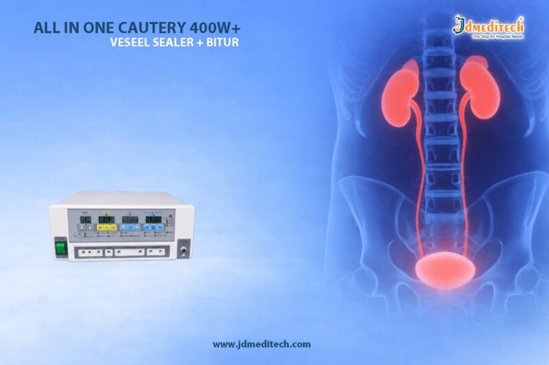
Gastrojejunostomy – Surgical Bypass for Gastric Obstruction Explained

Gastrojejunostomy surgical bypass for gastric obstruction plays a vital role in modern gastrointestinal surgery. Doctors use this procedure to create a direct connection between the stomach and the jejunum, effectively bypassing a blocked or narrowed part of the digestive tract.

In many cases, gastric obstruction results from conditions such as tumors, peptic ulcers, or severe inflammation. Therefore, gastrojejunostomy provides an effective solution that restores digestive flow and improves patient comfort.

What is Gastrojejunostomy?

Gastrojejunostomy is a surgical procedure in which a surgeon connects the stomach directly to the jejunum, which is the middle section of the small intestine. As a result, food bypasses the obstructed part of the stomach or duodenum.

This gastrojejunostomy surgical bypass for gastric obstruction is commonly performed when normal gastric emptying becomes impossible.

Why is Gastrojejunostomy Performed?

Doctors recommend gastrojejunostomy in several clinical situations. Most importantly, it helps when other treatments fail or when obstruction becomes severe.

Common Indications
  • Gastric outlet obstruction
  • Stomach or pancreatic cancer
  • Chronic peptic ulcer disease
  • Duodenal strictures
  • Congenital abnormalities

Moreover, surgeons often choose this gastrojejunostomy surgical bypass for gastric obstruction to provide palliative relief in advanced cancer cases.

Types of Gastrojejunostomy

Different surgical techniques exist depending on the patient’s condition and surgeon’s expertise.

1. Open Gastrojejunostomy

Surgeons perform this procedure through a larger abdominal incision. Although it allows direct access, it usually requires longer recovery.

2. Laparoscopic Gastrojejunostomy

This minimally invasive approach uses small incisions and a camera. As a result, patients experience less pain and faster healing.

3. Endoscopic Gastrojejunostomy

In selected cases, doctors use endoscopic techniques to create the bypass without major surgery. Therefore, it reduces hospital stay significantly.

Step-by-Step Procedure

Understanding the procedure helps patients feel more confident and prepared.

  1. First, the patient receives general anesthesia.
  2. Next, the surgeon accesses the stomach and jejunum.
  3. Then, a connection (anastomosis) is created between them.
  4. After that, the surgeon ensures proper blood flow and alignment.
  5. Finally, the incision is closed carefully.

This structured approach ensures that the gastrojejunostomy surgical bypass for gastric obstruction works effectively.

Benefits of Gastrojejunostomy

This procedure offers multiple clinical advantages.

  • Restores normal food passage
  • Relieves symptoms like vomiting and nausea
  • Improves nutritional intake
  • Enhances quality of life
  • Provides palliative care in cancer patients

Additionally, laparoscopic methods reduce complications and speed up recovery.

Risks and Complications

Although gastrojejunostomy is generally safe, certain risks may occur.

  • Infection
  • Bleeding
  • Anastomotic leakage
  • Delayed gastric emptying
  • Dumping syndrome

However, experienced surgeons minimize these risks through proper technique and care.

Recovery and Aftercare

Recovery depends on the surgical method used.

  • Patients usually stay in the hospital for a few days
  • Doctors gradually reintroduce liquids and soft foods
  • Regular follow-ups ensure proper healing
  • Patients must follow dietary guidelines strictly

Furthermore, early mobilization and proper nutrition support faster recovery after gastrojejunostomy surgical bypass for gastric obstruction.

Role of Medical Devices in Gastrojejunostomy

High-quality surgical instruments play a crucial role in successful outcomes.

At JDMeditech, we provide precision-engineered medical devices designed for gastrointestinal procedures. These include:

  • Surgical staplers
  • Anastomosis devices
  • Endoscopic accessories
  • Guidewires and dilation tools

Reliable instruments improve surgical accuracy and patient safety.

Conclusion

Gastrojejunostomy surgical bypass for gastric obstruction remains a highly effective solution for managing complex gastrointestinal conditions. Not only does it restore digestive function, but it also significantly improves patient quality of life.

As surgical techniques continue to advance, minimally invasive approaches are making this procedure safer and more efficient. Therefore, healthcare providers increasingly rely on this method for both curative and palliative treatment.


Explore More Product >>

Get Connected:

+91 79909 93062 | +91 63513 72032 | exports@jdmeditech.com
Esophagectomy Procedure – Complete Guide to Surgery, Purpose & Recovery

The esophagectomy procedure is a major surgery used to remove part or all of the esophagus. Doctors mainly perform this procedure to treat esophageal cancer. However, it is also helpful for certain severe non-cancerous conditions.

Although it is a complex operation, modern techniques have made the esophagectomy procedure safer and more effective. As a result, many patients experience better recovery and improved quality of life.

What is an Esophagectomy Procedure?

The esophagectomy procedure involves removing the damaged section of the esophagus. After that, surgeons rebuild the food pathway using the stomach or, in some cases, a part of the intestine.

Because of this reconstruction, patients can swallow food again after recovery. Therefore, the esophagectomy procedure plays a critical role in restoring normal digestive function.

Why is the Esophagectomy Procedure Performed?

Doctors recommend the esophagectomy procedure for several serious conditions. Most importantly, it is a key treatment for cancer.

Common reasons include:
  • Esophageal cancer
  • Barrett’s esophagus with severe dysplasia
  • Advanced achalasia
  • Severe esophageal strictures
  • Chemical or caustic injury

In many cases, early treatment with the esophagectomy procedure can be life-saving.

Types of Esophagectomy Procedure

There are different approaches to the esophagectomy procedure. The choice depends on the patient’s condition and the stage of the disease.

1. Transhiatal Esophagectomy

This method involves incisions in the abdomen and neck. It avoids opening the chest, which often leads to faster recovery.

2. Transthoracic Esophagectomy

In this approach, surgeons open the chest. This provides better visibility, especially for complex tumors.

3. Minimally Invasive Esophagectomy

This modern technique uses small incisions and advanced instruments. As a result, patients experience less pain and quicker healing.

Today, many hospitals prefer minimally invasive esophagectomy procedures because of better outcomes.

Step-by-Step Esophagectomy Procedure

The esophagectomy procedure is performed in several stages. Each step is carefully planned to ensure patient safety.

  1. General Anesthesia
    The patient is fully sedated.
  2. Surgical Access
    The surgeon makes incisions in the required areas.
  3. Removal of Diseased Tissue
    The damaged part of the esophagus is removed.
  4. Reconstruction
    The stomach is reshaped and connected to the remaining esophagus.
  5. Closure and Monitoring
    The surgical area is closed, and the patient is moved to intensive care.

Overall, the esophagectomy procedure requires precision and expertise at every step.

Recovery After Esophagectomy Procedure

Recovery after the esophagectomy procedure takes time and proper care. However, structured rehabilitation helps patients heal faster.

Recovery includes:
  • Close monitoring in ICU
  • Gradual shift from liquid to solid diet
  • Nutritional support if needed
  • Breathing and physical exercises

Most patients recover within a few weeks. Additionally, regular follow-ups improve long-term results after the esophagectomy procedure.

Risks and Complications

Like any major surgery, the esophagectomy procedure carries some risks. However, careful medical care reduces these risks significantly.

Possible complications:
  • Infection
  • Leakage at the surgical connection
  • Breathing problems
  • Difficulty swallowing
  • Acid reflux

Even though complications may occur, modern advancements have made the esophagectomy procedure much safer.

Advancements in Esophagectomy Procedure

In recent years, the esophagectomy procedure has improved greatly. New technologies have enhanced both safety and outcomes.

Key advancements include:
  • Robotic-assisted surgery
  • High-definition imaging
  • Improved anesthesia techniques
  • Enhanced Recovery After Surgery (ERAS) protocols

Because of these innovations, patients now experience faster recovery and better results after the esophagectomy procedure.

Conclusion

The esophagectomy procedure is a vital surgical treatment for serious esophageal conditions. While it is complex, modern techniques have made it more effective and safer.

Most importantly, early diagnosis and timely treatment can greatly improve outcomes. With proper care, patients undergoing the esophagectomy procedure can achieve a better quality of life.


Explore More Product >>

Get Connected:

+91 79909 93062 | +91 63513 72032 | exports@jdmeditech.com
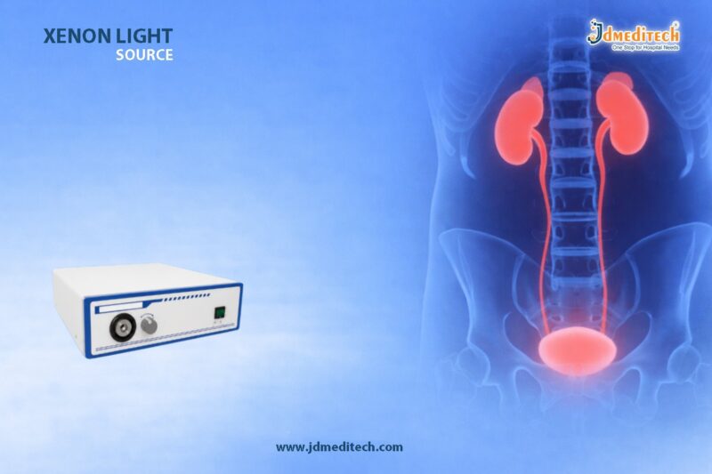
Amplatz Renal Dilator for Minimally Invasive Access

The Amplatz Renal Dilator plays a vital role in modern urology. Surgeons use it in minimally invasive procedures such as Percutaneous Nephrolithotomy (PCNL) to create safe access to the kidney.

Today, doctors prefer minimally invasive techniques because they reduce complications and speed up recovery. Therefore, the Amplatz Renal Dilator helps improve surgical precision and patient outcomes.

What is an Amplatz Renal Dilator?

The Amplatz Renal Dilator is a specialized medical instrument that expands the nephrostomy tract during kidney procedures.

Surgeons pass the Amplatz Renal Dilator over a guidewire in a controlled sequence. This method allows them to widen the tract gradually. Its rigid structure provides stability and ensures precise dilation.

Why Do Surgeons Use Amplatz Renal Dilator?

Surgeons use the Amplatz Renal Dilator to create direct and safe access to the kidney. They commonly use it in:

  • Percutaneous Nephrolithotomy (PCNL)
  • Kidney stone removal procedures
  • Nephrostomy tract dilation
  • Endourological surgeries

In addition, it helps protect surrounding tissues and allows smooth instrument insertion.

Key Features of Amplatz Renal Dilator
1. Step-by-Step Dilation

The Amplatz Renal Dilator comes in multiple sizes. Surgeons increase the size gradually, which improves safety and control.

2. Strong and Durable Material

Manufacturers use high-quality medical-grade materials to produce the Amplatz Renal Dilator. This ensures durability and consistent performance.

3. Smooth Surface Design

The smooth surface allows easy insertion. It also reduces friction and minimizes tissue damage.

4. Guidewire Compatibility

The Amplatz Renal Dilator works seamlessly with standard guidewires. This compatibility supports efficient workflow during procedures.

5. Easy Handling

The ergonomic design gives surgeons better grip and control. As a result, they perform procedures with greater accuracy.

How Does Amplatz Renal Dilator Work?

Surgeons follow a clear and controlled process when using the Amplatz Renal Dilator:

  1. First, they insert a guidewire into the kidney.
  2. Then, they introduce a small-sized Amplatz Renal Dilator.
  3. Next, they use larger dilators step by step.
  4. Finally, they achieve the required tract size.

This gradual method ensures safe and effective dilation.

Benefits of Amplatz Renal Dilator

The Amplatz Renal Dilator offers several important advantages:

  • Provides minimally invasive kidney access
  • Reduces tissue trauma
  • Improves surgical precision
  • Speeds up patient recovery
  • Lowers the risk of complications

Additionally, it helps surgeons complete procedures more efficiently.

Clinical Applications

Doctors use the Amplatz Renal Dilator in many urological procedures, including:

  • PCNL for kidney stone removal
  • Renal access creation
  • Advanced endourological procedures

It performs well in both routine and complex cases.

Why Surgeons Prefer Amplatz Renal Dilator

Surgeons prefer the Amplatz Renal Dilator because it:

  • Delivers consistent dilation
  • Provides strong stability
  • Allows smooth instrument access
  • Reduces procedural risks

Because of these advantages, it has become a trusted choice in urology.

Maintenance and Sterilization

Proper maintenance keeps the Amplatz Renal Dilator in optimal condition:

  • Clean it immediately after use
  • Use approved cleaning solutions
  • Follow standard sterilization protocols
  • Inspect it before every procedure

These steps help maintain safety and extend the instrument’s lifespan.

Conclusion

The Amplatz Renal Dilator remains a key instrument in minimally invasive kidney procedures. It allows surgeons to create precise and controlled access while protecting surrounding tissues.

As medical technology advances, surgeons will continue to rely on the Amplatz Renal Dilator for safe and effective outcomes. High-quality instruments always lead to better patient care.


Explore More Product >>

Get Connected:

+91 79909 93062 | +91 63513 72032 | exports@jdmeditech.com
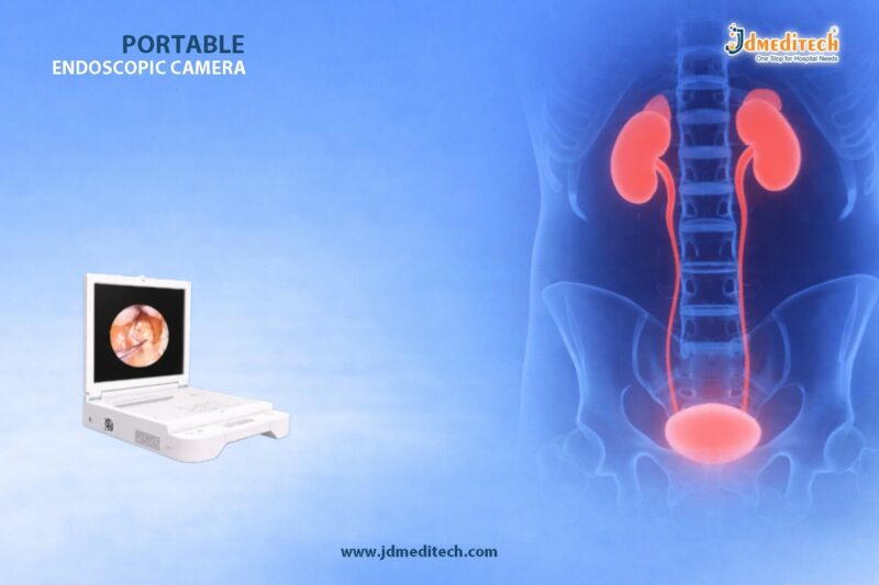
ASD/VSD Closure Surgery: Expert Heart Care Guide

ASD/VSD closure surgery is an important treatment for congenital heart defects. It helps correct abnormal openings in the heart wall and improves blood flow. Doctors often recommend ASD/VSD closure surgery when a heart defect affects normal heart function, causes symptoms, or increases the risk of future complications.

In many patients, early diagnosis leads to better treatment planning. As a result, ASD/VSD closure surgery plays a major role in long-term heart care. This procedure can reduce symptoms, protect the lungs, and support healthier circulation. Moreover, modern cardiac teams now perform ASD/VSD closure surgery with advanced imaging, precision devices, and safer surgical techniques.

This expert guide explains what ASD/VSD closure surgery is, why it is needed, how it is performed, and what patients can expect during recovery.

What Is ASD/VSD Closure Surgery?

ASD/VSD closure surgery is a heart procedure used to repair septal defects. A septal defect is a hole in the wall that separates the chambers of the heart. These openings allow blood to move in the wrong direction. Therefore, the heart may work harder than normal.

There are two common types of septal defects:

Atrial Septal Defect (ASD)

An ASD is a hole between the upper chambers of the heart, called the atria.

Ventricular Septal Defect (VSD)

A VSD is a hole between the lower chambers of the heart, called the ventricles.

Because both conditions can affect circulation, ASD/VSD closure surgery helps restore more normal blood flow. In addition, it lowers the chance of heart enlargement, lung pressure problems, and rhythm issues.

Why Is ASD/VSD Closure Surgery Needed?

Not every small heart defect needs immediate intervention. However, larger defects or symptomatic cases often require ASD/VSD closure surgery. Doctors decide this after evaluating heart size, blood flow changes, symptoms, and overall patient health.

Common reasons for treatment include:
  • Shortness of breath
  • Tiring easily during daily activity
  • Poor weight gain in children
  • Recurrent chest infections
  • Enlargement of heart chambers
  • Increased pressure in the lungs
  • Risk of irregular heartbeat
  • Risk of stroke in some ASD cases

When left untreated, some defects may lead to long-term damage. For that reason, ASD/VSD closure surgery is often recommended at the right stage to prevent future complications.

Types of ASD/VSD Closure Surgery

Doctors may choose different approaches depending on the location and size of the defect. Although both aim to close the opening, the treatment method can vary.

1. Catheter-Based Closure

This method is less invasive. The doctor inserts a catheter through a blood vessel, usually in the groin, and guides it to the heart. Then a closure device seals the defect.

Benefits include:

  • Smaller access site
  • Shorter hospital stay
  • Faster recovery
  • Less postoperative discomfort

For suitable patients, catheter-based ASD/VSD closure surgery offers an effective and efficient option.

2. Open Heart Surgical Closure

Some defects are too large, too complex, or not suitable for device closure. In those cases, surgeons perform open repair. They close the defect using stitches or a surgical patch.

This method is often chosen when:

  • The defect has a complex shape
  • There are multiple openings
  • There are associated heart abnormalities
  • Device closure is not appropriate

Even in complex situations, ASD/VSD closure surgery gives strong long-term results when performed by experienced heart specialists.

How ASD/VSD Closure Surgery Is Performed

Although the exact steps depend on the technique used, the overall treatment process follows a careful plan. Cardiac teams first evaluate the defect with tests such as echocardiography, ECG, chest imaging, and blood work. After that, the patient is prepared for the procedure.

Typical steps include:
  1. The patient receives anesthesia or sedation.
  2. The care team monitors heart rhythm and vital signs.
  3. The surgeon or cardiologist reaches the defect by catheter or surgical access.
  4. The opening is closed with a device, stitches, or a patch.
  5. Blood flow is checked to confirm successful repair.
  6. The patient moves to recovery for observation.

Because precision is essential, imaging guidance improves the safety of ASD/VSD closure surgery. Furthermore, skilled heart teams use specialized instruments and closure systems to achieve accurate results.

Benefits of ASD/VSD Closure Surgery

The main goal of ASD/VSD closure surgery is to improve heart function and reduce strain on the cardiovascular system. Once the abnormal opening is repaired, circulation becomes more efficient.

Major benefits include:
  • Better oxygen-rich blood flow
  • Reduced workload on the heart
  • Lower risk of lung pressure problems
  • Improved physical activity tolerance
  • Better growth and development in children
  • Reduced risk of future cardiac complications

In many patients, ASD/VSD closure surgery leads to a clear improvement in quality of life. Patients often breathe more comfortably, feel less tired, and recover normal function over time.

Risks and Safety Considerations

Like any cardiac procedure, ASD/VSD closure surgery carries some risks. However, the procedure is generally safe when performed in an appropriate hospital setting.

Possible risks may include:
  • Bleeding
  • Infection
  • Arrhythmia
  • Reaction to anesthesia
  • Device movement in rare cases
  • Residual leak in uncommon situations

Even so, most patients do well after ASD/VSD closure surgery. Careful planning, modern technology, and proper follow-up greatly reduce the overall risk.

Recovery After ASD/VSD Closure Surgery

Recovery varies from one patient to another. It also depends on whether the procedure was catheter-based or open surgery. In general, catheter procedures allow earlier discharge, while open repair needs a longer recovery period.

Recovery expectations:
  • Catheter closure: shorter hospital stay
  • Surgical closure: more recovery time
  • Follow-up visits: essential in both cases
  • Activity: resumed gradually as advised by the doctor

After ASD/VSD closure surgery, patients must follow medical advice closely. Doctors may prescribe medicines, recommend follow-up imaging, and provide activity guidelines. As recovery progresses, most patients return to normal routines safely.

Role of Technology in ASD/VSD Closure Surgery

Modern cardiology continues to improve ASD/VSD closure surgery. Today, heart specialists use advanced imaging, refined closure devices, and better procedural planning. As a result, treatment is more accurate and patient-friendly than before.

Important advancements include:

  • High-quality imaging guidance
  • Improved occluder devices
  • Better surgical patch materials
  • Safer anesthesia support
  • Enhanced postoperative monitoring

Because of these innovations, ASD/VSD closure surgery now offers better comfort, reliable closure, and stronger long-term results.

Importance of Quality Cardiac Instruments

Successful ASD/VSD closure surgery depends not only on the surgeon’s skill but also on the quality of the instruments and procedural support. Precision tools help improve control, visibility, and treatment safety during heart procedures.

At JDMeditech, we understand the value of reliable medical equipment in advanced heart care. High-quality cardiac instruments support better clinical performance and smoother procedural outcomes.

Conclusion

ASD/VSD closure surgery is a proven treatment for congenital heart defects that affect normal circulation. It helps close abnormal openings in the heart, improves blood flow, and protects long-term heart health. Whether performed by catheter-based technique or open repair, ASD/VSD closure surgery remains an essential part of modern heart care.

With early diagnosis, skilled specialists, and the right technology, ASD/VSD closure surgery can deliver safe treatment and meaningful recovery. For patients and care providers alike, it remains a highly effective solution in congenital cardiac management.


Explore More Product >>

Get Connected:

+91 79909 93062 | +91 63513 72032 | exports@jdmeditech.com
 

Amplatz Dilator Set for Percutaneous Tract Dilation

The Amplatz Dilator Set for percutaneous tract dilation plays a critical role in modern urology. It allows surgeons to create a safe and controlled access tract during minimally invasive procedures. Most importantly, it ensures precision while reducing tissue trauma.

Today, urologists widely use the Amplatz Dilator Set for percutaneous tract dilation in procedures such as Percutaneous Nephrolithotomy (PCNL). As a result, it has become an essential tool in advanced endourology.

What is an Amplatz Dilator Set?

An Amplatz Dilator Set for percutaneous tract dilation is a series of graduated dilators designed to enlarge a percutaneous access tract. Typically, surgeons insert these dilators sequentially over a guidewire to achieve the desired tract size.

Moreover, the set often includes:

  • Multiple dilators of increasing diameters
  • A central obturator for support
  • Optional working sheath

Because of this design, the Amplatz Dilator Set for percutaneous tract dilation ensures smooth and controlled dilation.

Key Features of Amplatz Dilator Set

The Amplatz Dilator Set for percutaneous tract dilation offers several advanced features that improve procedural outcomes:

1. Graduated Dilator Design

Each dilator increases in size gradually. Therefore, it minimizes trauma and enhances patient safety.

2. High-Quality Medical Material

Manufacturers use durable, biocompatible materials. As a result, the device ensures strength and reliability during procedures.

3. Smooth Surface Finish

The polished surface reduces friction. Consequently, insertion becomes easier and more controlled.

4. Compatibility with Guidewires

The system works seamlessly with standard guidewires. This compatibility improves procedural efficiency.

5. Sterile and Ready-to-Use Options

Many sets come pre-sterilized. Hence, they reduce preparation time in the operating room.

Applications in Medical Procedures

The Amplatz Dilator Set for percutaneous tract dilation is widely used across various urological interventions:

  • Percutaneous Nephrolithotomy (PCNL) – for kidney stone removal
  • Nephrostomy procedures – to establish drainage pathways
  • Renal access creation – for diagnostic and therapeutic procedures

Among these, PCNL remains the most common application. In this procedure, surgeons rely heavily on the Amplatz Dilator Set for percutaneous tract dilation for safe tract formation.

Step-by-Step Working Principle

Understanding how the Amplatz Dilator Set for percutaneous tract dilation works helps highlight its importance:

  1. First, the surgeon inserts a guidewire into the targeted renal area
  2. Then, the initial small dilator is advanced over the guidewire
  3. Subsequently, larger dilators are introduced sequentially
  4. Finally, the tract reaches the desired diameter for instrument access

Because of this stepwise approach, the Amplatz Dilator Set for percutaneous tract dilation ensures controlled and safe dilation.

Advantages of Using Amplatz Dilator Set

The Amplatz Dilator Set for percutaneous tract dilation offers numerous clinical advantages:

  • Ensures precise tract dilation
  • Minimizes tissue trauma
  • Reduces procedural complications
  • Improves surgical efficiency
  • Supports minimally invasive techniques

Furthermore, it allows surgeons to maintain better control throughout the procedure.

Why Choose JDMeditech Amplatz Dilator Set?

At JDMeditech, we focus on quality, precision, and performance. Our Amplatz Dilator Set for percutaneous tract dilation meets international medical standards and delivers reliable results.

Key Benefits:
  • Precision-engineered design
  • High-quality medical-grade materials
  • Smooth and atraumatic dilation
  • Wide range of sizes available
  • Strict quality control and sterilization

Therefore, healthcare professionals trust JDMeditech for dependable urology solutions.

Safety and Handling Tips

To achieve optimal results with the Amplatz Dilator Set for percutaneous tract dilation, follow these guidelines:

  • Always use under trained medical supervision
  • Ensure proper sterilization before use
  • Select the correct dilator size sequence
  • Handle with care to maintain sterility

By following these steps, practitioners can maximize safety and efficiency.

Conclusion

The Amplatz Dilator Set for percutaneous tract dilation remains a cornerstone in modern urological procedures. It provides a safe, efficient, and precise method for tract creation, especially in PCNL.

In conclusion, choosing a high-quality solution like JDMeditech’s Amplatz Dilator Set for percutaneous tract dilation ensures better outcomes and improved patient safety.


Explore More Product >>

Get Connected:

+91 79909 93062 | +91 63513 72032 | exports@jdmeditech.com
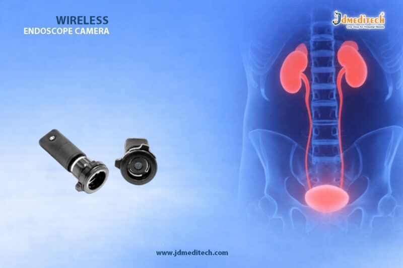
Maze Procedure for Atrial Fibrillation – Surgical Treatment Guide

Atrial fibrillation (AFib) is one of the most common cardiac rhythm disorders worldwide. It disrupts the heart’s normal electrical signals and increases the risk of stroke, heart failure, and other complications. While medications and catheter-based treatments help many patients, some cases require advanced surgical intervention.

The Maze Procedure for Atrial Fibrillation is a highly effective surgical solution. It restores normal heart rhythm by creating controlled scar tissue that blocks abnormal electrical signals. Therefore, it plays a crucial role in the surgical management of atrial fibrillation.

What is Atrial Fibrillation?

Atrial fibrillation is an irregular and often rapid heart rhythm. It originates in the atria, the upper chambers of the heart.

Common Causes
  • High blood pressure
  • Coronary artery disease
  • Heart valve disorders
  • Obesity and lifestyle factors
Symptoms
  • Palpitations
  • Shortness of breath
  • Fatigue
  • Dizziness

However, some patients may not experience noticeable symptoms, which makes early diagnosis important.

What is the Maze Procedure for Atrial Fibrillation?

The Maze Procedure for Atrial Fibrillation is a surgical technique designed to eliminate abnormal electrical pathways in the heart.

Surgeons create a “maze” of scar tissue in the atria using:

  • Radiofrequency energy
  • Cryotherapy (freezing)
  • Surgical incisions

As a result, electrical signals can only travel through a controlled pathway, restoring normal sinus rhythm.

Types of Maze Procedures
1. Cox Maze III Procedure

This is the traditional “cut-and-sew” technique. Although it is highly effective, it is more invasive.

2. Cox Maze IV Procedure

This modern version uses energy sources like radiofrequency or cryoablation instead of incisions. Therefore, it reduces surgical complexity.

3. Minimally Invasive Maze Procedure

Surgeons perform this approach using small incisions and specialized instruments. As a result, recovery is faster and complications are fewer.

Indications for Maze Procedure

Doctors recommend the Maze Procedure for Atrial Fibrillation in the following cases:

  • Persistent or long-standing AFib
  • Failure of medications or catheter ablation
  • Patients undergoing other cardiac surgeries (e.g., valve repair or CABG)
  • High risk of stroke

Thus, it becomes a reliable option when other treatments do not provide lasting results.

Step-by-Step Surgical Process
1. Preoperative Evaluation

Doctors assess heart function using ECG, echocardiography, and imaging.

2. Anesthesia and Access

The patient receives general anesthesia. Surgeons access the heart through open surgery or minimally invasive techniques.

3. Creating the Maze Pattern

Surgeons create scar lines using radiofrequency or cryotherapy to block abnormal signals.

4. Restoring Normal Rhythm

The controlled pathways allow electrical impulses to travel properly.

5. Closure and Monitoring

Finally, surgeons close the incision and monitor the patient closely.

Benefits of Maze Procedure

The Maze Procedure for Atrial Fibrillation offers several advantages:

  • High success rate in restoring normal rhythm
  • Reduced risk of stroke
  • Improved heart function
  • Long-term solution compared to medications
  • Can be combined with other cardiac surgeries
Risks and Complications

Although effective, the procedure carries some risks:

  • Bleeding
  • Infection
  • Stroke
  • Need for pacemaker in some cases

However, with modern techniques, complication rates are significantly reduced.

Recovery After Maze Procedure

Recovery depends on the surgical approach.

Typical Recovery Timeline
  • Hospital stay: 5–7 days
  • Full recovery: 4–8 weeks
Postoperative Care
  • Medications to control rhythm
  • Regular follow-up visits
  • Lifestyle modifications

Therefore, proper aftercare ensures better long-term outcomes.

Maze Procedure vs Catheter Ablation
Feature Maze Procedure Catheter Ablation
Invasiveness Surgical Minimally invasive
Success Rate Higher in complex AFib Moderate
Recovery Time Longer Shorter
Best For Severe AFib cases Early-stage AFib
Latest Advances in Maze Procedure

Modern innovations have improved the Maze Procedure for Atrial Fibrillation:

  • Minimally invasive robotic-assisted surgery
  • Advanced energy sources for precision
  • Hybrid procedures combining surgery and catheter techniques

As a result, outcomes continue to improve with reduced recovery time.

Why Choose Surgical Management for AFib?

Surgical intervention provides a durable solution when other treatments fail. Moreover, the Maze Procedure for Atrial Fibrillation offers long-term rhythm control and improved quality of life.

Conclusion

The Maze Procedure for Atrial Fibrillation remains a gold standard in the surgical management of complex AFib cases. It effectively restores normal heart rhythm by controlling abnormal electrical signals.

If medications and catheter-based treatments do not work, this surgical approach offers a reliable and long-lasting solution. Therefore, it plays a vital role in modern cardiac care.


Explore More Product >>

Get Connected:

+91 79909 93062 | +91 63513 72032 | exports@jdmeditech.com
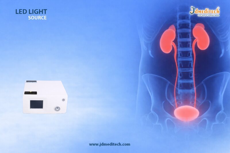
Heart Transplantation Surgery – Procedure, Benefits & Recovery

Heart transplantation surgery is one of the most advanced and life-saving procedures in modern cardiology. It is primarily performed for patients suffering from end-stage heart failure when all other treatments have failed.

Over the years, heart transplantation surgery has evolved significantly, offering improved survival rates and better quality of life. Moreover, with advancements in surgical techniques and post-operative care, outcomes continue to improve globally.

What is Heart Transplantation Surgery?

Heart transplantation surgery is a procedure in which a diseased or failing heart is replaced with a healthy donor heart. Typically, the donor heart comes from a brain-dead individual whose organs are still functioning.

This procedure is considered only when:

  • Medications are no longer effective
  • Medical devices fail to support heart function
  • The patient has severe, irreversible heart damage

Therefore, heart transplantation surgery becomes the last and most effective option for survival.

Who Needs Heart Transplantation Surgery?All Posts

Not every heart patient qualifies for heart transplantation surgery. However, it is recommended in severe cases such as:

1. End-Stage Heart Failure

When the heart cannot pump enough blood to meet the body’s needs.

2. Cardiomyopathy

A condition where the heart muscle becomes weak or enlarged.

3. Severe Coronary Artery Disease

Blocked arteries leading to irreversible heart damage.

4. Congenital Heart Defects

Complex heart abnormalities present from birth.

5. Failed Previous Cardiac Surgeries

When earlier procedures no longer provide relief.

In such cases, heart transplantation surgery significantly improves survival chances.

Pre-Transplant Evaluation Process

Before undergoing heart transplantation surgery, patients must go through a detailed evaluation. This ensures that they are suitable candidates.

Key Evaluations Include:
  • Blood tests and imaging
  • Cardiac function assessment
  • Psychological evaluation
  • Infection screening

Additionally, patients are placed on a waiting list based on urgency, compatibility, and availability of donor organs.

Heart Transplantation Surgery Procedure

The heart transplantation surgery procedure is complex and requires a highly skilled cardiac team.

Step-by-Step Process:
  1. Anesthesia Administration
    The patient is placed under general anesthesia.
  2. Heart Removal
    The diseased heart is carefully removed.
  3. Donor Heart Implantation
    The healthy donor heart is connected to major blood vessels.
  4. Restarting the Heart
    The new heart is stimulated to start beating.
  5. Monitoring and Stabilization
    The patient is closely monitored in the ICU.

As a result, heart transplantation surgery restores normal blood circulation and cardiac function.

Benefits of Heart Transplantation Surgery

There are several advantages of heart transplantation surgery, including:

  • Improved Survival Rate
  • Better Quality of Life
  • Increased Physical Activity
  • Relief from Severe Symptoms

Furthermore, many patients return to near-normal daily activities after recovery.

Risks and Complications

Although heart transplantation surgery is highly effective, it also carries certain risks:

1. Organ Rejection

The body may attack the new heart.

2. Infection

Due to immunosuppressive medications.

3. Blood Clots

Which can lead to stroke or heart attack.

4. Side Effects of Medications

Including kidney damage and high blood pressure.

However, with proper care and monitoring, these risks can be managed effectively.

Post-Transplant Care and Recovery

Recovery after heart transplantation surgery is crucial for long-term success.

Important Care Measures:
  • Lifelong immunosuppressive medications
  • Regular follow-up visits
  • Healthy diet and lifestyle
  • Cardiac rehabilitation

In addition, patients must strictly follow medical advice to prevent complications.

Success Rate of Heart Transplantation Surgery

The success rate of heart transplantation surgery has improved significantly due to modern advancements.

  • 1-year survival rate: 85–90%
  • 5-year survival rate: 70–75%

Therefore, heart transplantation surgery remains one of the most reliable solutions for advanced heart failure.

Latest Advancements in Heart Transplantation

Modern innovations are transforming heart transplantation surgery, such as:

  • Artificial heart support devices
  • Improved organ preservation techniques
  • Minimally invasive monitoring
  • Advanced immunosuppressive therapies

These advancements continue to enhance patient outcomes and reduce complications.

Conclusion

In conclusion, heart transplantation surgery is a life-saving solution for patients with severe heart disease. While the procedure is complex, it offers a second chance at life for many individuals.

With continuous medical advancements and proper post-operative care, heart transplantation surgery provides excellent long-term results and improved quality of life.


Explore More Product >>

Get Connected:

+91 79909 93062 | +91 63513 72032 | exports@jdmeditech.com
S Curved Dilator Set for Smooth Urethral Dilation

The S Curved Dilator Set for Smooth Urethral Dilation is an essential tool in modern urology. It is specifically designed to provide safe, gradual, and controlled urethral dilation, especially in patients with urethral strictures. Because of its unique S-shaped curvature, this instrument allows better navigation through the natural anatomy of the urethra.

Moreover, urologists prefer the S Curved Dilator Set for Smooth Urethral Dilation because it minimizes trauma while ensuring procedural efficiency. As a result, it has become a reliable solution in both diagnostic and therapeutic urological procedures.

What is an S Curved Dilator Set?

An S Curved Dilator Set for Smooth Urethral Dilation is a collection of progressively sized dilators designed with a gentle S-shaped curve. This design matches the anatomical pathway of the urethra, allowing smoother insertion.

In addition, each dilator is carefully calibrated to ensure gradual expansion, which is critical for patient safety. Therefore, the set plays a vital role in treating urethral narrowing without causing unnecessary discomfort.

Key Features of S Curved Dilator Set for Smooth Urethral Dilation
1. Anatomical S-Curved Design

First and foremost, the S Curved Dilator Set for Smooth Urethral Dilation features a unique curvature that follows the natural urethral path. Consequently, it reduces resistance during insertion.

2. Smooth Surface Finish

Furthermore, the dilators have a polished surface. This ensures minimal friction, thereby reducing the risk of tissue injury.

3. Graduated Sizes

Each S Curved Dilator Set for Smooth Urethral Dilation includes multiple sizes. Hence, clinicians can perform step-by-step dilation safely.

4. High-Quality Material

Typically, the set is made from medical-grade stainless steel or high-quality polymer, ensuring durability and easy sterilization.

5. Ergonomic Handling

In addition, the handle design allows better grip and control. As a result, precision during procedures improves significantly.

Clinical Applications

The S Curved Dilator Set for Smooth Urethral Dilation is widely used in:

  • Treatment of urethral strictures
  • Pre-operative urethral preparation
  • Post-surgical urethral management
  • Diagnostic urological procedures
  • Gradual urethral calibration

Because of its versatility, it is a must-have instrument in urology departments.

Advantages of Using S Curved Dilator Set for Smooth Urethral Dilation
Enhanced Patient Comfort

Due to its anatomical design, the S Curved Dilator Set for Smooth Urethral Dilation ensures less discomfort during insertion.

Reduced Risk of Trauma

Moreover, the smooth finish and gradual sizing significantly lower the chances of urethral injury.

Improved Procedural Accuracy

Since the device offers better control, urologists can perform dilation more precisely.

Time-Efficient Procedures

Additionally, the ease of insertion helps reduce procedure time, thereby improving workflow in clinical settings.

Step-by-Step Procedure Overview
  1. First, proper sterilization of the S Curved Dilator Set for Smooth Urethral Dilationis ensured.
  2. Next, lubrication is applied to minimize friction.
  3. Then, the smallest dilator is gently inserted into the urethra.
  4. Gradually, larger sizes are introduced to achieve the desired dilation.
  5. Finally, the procedure is completed while monitoring patient comfort.
Maintenance and Sterilization

To maintain performance, the S Curved Dilator Set for Smooth Urethral Dilation must be properly cleaned and sterilized after each use.

  • Use autoclaving or approved sterilization methods
  • Inspect for any damage before reuse
  • Store in a clean, dry environment
Why Choose JDMeditech?

At JDMeditech, we provide high-quality S Curved Dilator Set for Smooth Urethral Dilation designed to meet international medical standards.

  • Precision-engineered instruments
  • Reliable performance
  • Strict quality control
  • Competitive pricing
Conclusion

The S Curved Dilator Set for Smooth Urethral Dilation stands out as a specialized, reliable, and patient-friendly solution for urethral dilation procedures. Because of its anatomical design, smooth finish, and graduated sizing, it ensures both clinical efficiency and patient safety.

Therefore, for urologists aiming to deliver precise and comfortable treatment, this instrument is an indispensable addition to their surgical toolkit.


Explore More Product >>

+91 79909 93062 | +91 63513 72032 | exports@jdmeditech.com