Hydrocelectomy Surgery – Advanced Treatment for Hydrocele Removal

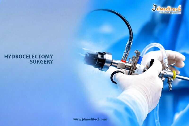
Hydrocelectomy surgery is a specialized urologic procedure performed to treat a hydrocele, a condition in which fluid accumulates around the testicle inside the scrotum. Although hydroceles are usually painless, they can gradually enlarge and cause discomfort, heaviness, swelling, and difficulty performing daily activities.

In many cases, small hydroceles may resolve naturally. However, persistent or large hydroceles often require surgical treatment for permanent relief. Therefore, hydrocelectomy is considered one of the most effective and reliable procedures for managing this condition.

Moreover, modern surgical advancements have significantly improved the safety, precision, and recovery outcomes associated with hydrocelectomy surgery.

What Is Hydrocelectomy Surgery?
Understanding the Procedure

Hydrocelectomy is a surgical procedure designed to remove or repair the hydrocele sac surrounding the testicle. Essentially, the surgery eliminates excess fluid buildup while also preventing recurrence.

The procedure may involve:

  • Draining accumulated fluid
  • Removing the hydrocele sac
  • Reshaping or everting the tissue lining
  • Securing surrounding tissues to reduce fluid reaccumulation

In most cases, hydrocelectomy is performed as a day-care or short-stay procedure under anesthesia.

What Is a Hydrocele?
Understanding the Condition

A hydrocele develops when fluid collects within the thin sac surrounding the testicle. Although the condition is common in newborns, it can also occur in adolescents and adult males.

In adults, hydroceles may develop due to:

  • Injury or trauma
  • Infection
  • Inflammation
  • Previous surgery
  • Unknown or idiopathic causes

While hydroceles are generally non-cancerous, they should still be evaluated by a qualified urologist. This is important because other scrotal conditions may produce similar symptoms.

Symptoms of Hydrocele
Common Signs and Symptoms

Patients with hydrocele may experience several noticeable symptoms. For example, common signs include:

  • Scrotal swelling
  • Feeling of heaviness in the scrotum
  • Mild discomfort
  • Enlarged appearance of one testicle
  • Difficulty walking or sitting comfortably
  • Cosmetic concerns

As the hydrocele enlarges, symptoms may become more noticeable and interfere with routine activities.

When Is Hydrocelectomy Surgery Needed?
Indications for Surgical Treatment

Doctors may recommend hydrocelectomy surgery when conservative management no longer provides relief. Specifically, surgery may be advised when:

  • The hydrocele becomes large
  • Symptoms worsen over time
  • Movement or walking becomes difficult
  • Cosmetic appearance is significantly affected
  • Infection or complications occur
  • The hydrocele persists for a prolonged period

Consequently, timely surgical treatment can improve comfort and prevent further complications.

Preoperative Evaluation Before Hydrocelectomy
Diagnostic Tests and Assessment

Before surgery, the urologist performs a complete evaluation to confirm the diagnosis. Typically, preoperative assessment may include:

  • Physical examination
  • Scrotal ultrasound
  • Blood investigations
  • Urine analysis
  • Review of medical history

Additionally, these tests help rule out hernias, tumors, infections, or other scrotal abnormalities.

How Hydrocelectomy Surgery Is Performed
Step-by-Step Surgical Procedure

Hydrocelectomy surgery is usually performed under general or spinal anesthesia. During the procedure, the surgeon carefully follows several important steps:

  1. First, a small incision is made in the scrotum or groin region.
  2. Next, the hydrocele sac is exposed carefully.
  3. The accumulated fluid is then drained.
  4. After that, the sac is removed, folded, or repaired.
  5. Bleeding is controlled using surgical techniques.
  6. Finally, the incision is closed with sutures.

Generally, the procedure takes approximately 30 to 60 minutes.

Advanced Surgical Techniques in Hydrocelectomy
Modern Approaches for Better Outcomes

Modern hydrocelectomy techniques focus on improving patient safety and comfort. As a result, surgeons now use methods that provide:

  • Minimal tissue damage
  • Reduced bleeding
  • Smaller incisions
  • Improved cosmetic appearance
  • Faster recovery
  • Lower recurrence rates

Furthermore, advanced surgical instruments and refined techniques contribute to better long-term outcomes.

Recovery After Hydrocelectomy Surgery
Postoperative Care and Healing

Most patients recover smoothly after hydrocelectomy surgery. Nevertheless, proper postoperative care remains essential for optimal healing.

Doctors commonly recommend:

  • Wearing scrotal support garments
  • Avoiding strenuous physical activity
  • Keeping the surgical area clean and dry
  • Taking prescribed medications regularly
  • Applying cold packs if advised

Although mild swelling and discomfort are common initially, these symptoms usually improve within a few days.

In addition, most patients can resume normal activities within 1 to 2 weeks.

Possible Risks and Complications
Understanding Surgical Safety

Hydrocelectomy is generally considered a safe surgical procedure. However, like any operation, it may involve certain risks, including:

  • Infection
  • Bleeding
  • Temporary swelling
  • Bruising
  • Mild postoperative pain
  • Fluid recurrence
  • Rare injury to nearby structures

Fortunately, choosing an experienced urologic surgeon significantly reduces the risk of complications.

Benefits of Hydrocelectomy Surgery
Advantages of Surgical Hydrocele Treatment

Hydrocelectomy offers several important benefits for patients suffering from persistent hydrocele. For instance, the procedure provides:

  • Permanent fluid removal
  • Improved scrotal appearance
  • Relief from discomfort and heaviness
  • Better mobility
  • Reduced recurrence risk
  • Enhanced quality of life

Therefore, many patients experience substantial long-term improvement following surgery.

Who Is a Suitable Candidate for Hydrocelectomy?
Patient Eligibility Factors

Patients may benefit from hydrocelectomy surgery if they experience:

  • Persistent hydrocele
  • Large scrotal swelling
  • Ongoing discomfort
  • Recurrent hydrocele after aspiration
  • Functional difficulties
  • Cosmetic concerns

Ultimately, a urologist determines the most suitable treatment plan after evaluating the patient’s overall condition and symptoms.

Preventing Hydrocele Complications
Importance of Early Medical Attention

Early diagnosis and treatment can help prevent several complications. In particular, prompt medical care may reduce the risk of:

  • Progressive swelling
  • Chronic discomfort
  • Infection
  • Pressure-related symptoms
  • Delayed diagnosis of underlying conditions

Therefore, patients should seek medical attention whenever persistent scrotal swelling develops.

Conclusion

Hydrocelectomy surgery is a precise and highly effective urologic procedure used for the treatment of hydrocele. By removing excess fluid and repairing the hydrocele sac, the surgery provides lasting relief from swelling, discomfort, and mobility problems.

Additionally, modern surgical advancements have improved safety, reduced complications, and shortened recovery times. As a result, hydrocelectomy remains a preferred treatment option for persistent or symptomatic hydroceles.

Patients experiencing scrotal swelling or discomfort should consult an experienced urologist for accurate diagnosis and timely treatment. Early intervention not only improves comfort but also enhances overall quality of life.


Explore More Product >>

Get Connected:
+91 79909 93062 | +91 63513 72032 | exports@jdmeditech.com

Radical Orchiectomy Surgery – Complete Testis and Spermatic Cord Removal

Radical orchiectomy surgery is a specialized urologic procedure performed to remove the entire testis along with the spermatic cord through an inguinal incision. Surgeons commonly recommend this procedure for patients diagnosed with suspected or confirmed testicular cancer. In certain cases, it may also help manage severe testicular trauma, chronic infection, or hormone-sensitive conditions.

This surgery plays a critical role in both diagnosis and treatment. By completely removing the affected testicle and spermatic cord, doctors can prevent the spread of disease and obtain tissue for accurate pathological evaluation.

Modern surgical techniques have significantly improved the safety, precision, and recovery outcomes associated with radical orchiectomy surgery.

What Is Radical Orchiectomy Surgery?
Understanding the Procedure

Radical orchiectomy is a surgical operation that involves the removal of:

  • The affected testicle
  • The spermatic cord
  • Associated surrounding tissues when necessary

Unlike a simple orchiectomy, which removes only the testicle through the scrotum, radical orchiectomy uses an inguinal approach to minimize the risk of cancer spread.

The surgery is considered the standard primary treatment for most testicular tumors.

Why Is Radical Orchiectomy Surgery Performed?
Common Medical Indications

Doctors may recommend radical orchiectomy surgery for several conditions, including:

  • Testicular cancer
  • Suspicious testicular masses
  • Severe testicular injury
  • Chronic infection not responding to treatment
  • Hormone-sensitive cancers
  • Testicular torsion with irreversible damage

Early surgical intervention often improves treatment success and long-term survival outcomes.

Symptoms That May Require Surgical Evaluation
Warning Signs Patients Should Not Ignore

Patients experiencing the following symptoms should seek immediate medical attention:

  • Lump or swelling in the testicle
  • Persistent testicular pain
  • Feeling of heaviness in the scrotum
  • Sudden enlargement of one testicle
  • Fluid accumulation in the scrotum
  • Lower abdominal or groin discomfort

Many testicular tumors are painless during the early stages, making regular self-examination important.

Preoperative Evaluation Before Radical Orchiectomy
Important Diagnostic Tests

Before surgery, doctors typically perform:

  • Physical examination
  • Scrotal ultrasound
  • Blood tumor marker tests
  • CT scan or MRI imaging
  • Routine blood investigations
  • Chest imaging when cancer spread is suspected

These tests help determine the stage of disease and guide treatment planning.

How Radical Orchiectomy Surgery Is Performed
Step-by-Step Surgical Procedure

The surgery is usually performed under general anesthesia and follows these steps:

  1. A small incision is made in the groin area.
  2. The surgeon carefully isolates the spermatic cord.
  3. The entire testicle and spermatic cord are removed.
  4. Blood vessels are secured to prevent bleeding.
  5. The incision is closed using sutures.

The operation generally takes between 45 minutes and 90 minutes.

Minimally Invasive and Advanced Surgical Techniques
Modern Innovations in Urologic Surgery

Advanced surgical practices may include:

  • Microsurgical precision techniques
  • Improved hemostatic devices
  • Enhanced anesthesia protocols
  • Better cosmetic closure methods
  • Faster recovery pathways

These innovations help reduce pain, minimize complications, and shorten hospital stays.

Recovery After Radical Orchiectomy Surgery
Postoperative Care and Healing

Most patients recover well after surgery. Common postoperative recommendations include:

  • Rest for several days
  • Avoid heavy lifting
  • Wear scrotal support garments
  • Keep the incision clean and dry
  • Take prescribed medications as directed

Patients usually return to normal daily activities within 2 to 4 weeks.

Possible Risks and Complications
Understanding Surgical Safety

Although radical orchiectomy is generally safe, potential risks may include:

  • Bleeding
  • Infection
  • Scrotal swelling
  • Pain or discomfort
  • Blood clot formation
  • Fertility concerns
  • Hormonal changes

Choosing an experienced urologic surgeon significantly reduces complication risks.

Fertility and Hormonal Considerations
Long-Term Reproductive Health

Many patients continue to maintain normal hormone levels and fertility after removal of one testicle. However, doctors may discuss:

  • Sperm banking before surgery
  • Testosterone monitoring
  • Fertility preservation options
  • Follow-up hormonal evaluation

These considerations are especially important for younger patients.

Follow-Up Care After Surgery
Importance of Regular Monitoring

Postoperative follow-up may include:

  • Physical examinations
  • Tumor marker testing
  • CT scans or ultrasounds
  • Oncology consultations when needed

Regular monitoring helps detect recurrence early and supports long-term health management.

Benefits of Radical Orchiectomy Surgery
Advantages of Early Surgical Treatment

Key benefits include:

  • Complete removal of diseased tissue
  • Accurate cancer diagnosis
  • Prevention of disease spread
  • Improved survival outcomes
  • Relief from pain and discomfort
  • Effective long-term management

Timely treatment often provides the best clinical outcomes.

Who Is a Suitable Candidate?
Patient Eligibility Factors

Patients may qualify for radical orchiectomy surgery if they have:

  • Suspicious testicular masses
  • Confirmed testicular cancer
  • Severe irreversible testicular damage
  • Persistent symptoms affecting quality of life

A urologist evaluates each patient individually before recommending surgery.

Conclusion

Radical orchiectomy surgery is a highly effective and advanced surgical procedure used for the complete removal of the testis and spermatic cord. It remains the gold standard treatment for suspected or confirmed testicular cancer and other severe testicular conditions.

With modern surgical advancements, patients can expect improved safety, faster recovery, and excellent treatment outcomes. Early diagnosis and timely surgical intervention play a crucial role in preserving long-term health and preventing disease progression.

If you experience any unusual testicular symptoms, consult an experienced urologist for proper evaluation and treatment guidance.


Explore More Product >>

Get Connected:
+91 79909 93062 | +91 63513 72032 | exports@jdmeditech.com

Orchiectomy Surgery – Advanced Urologic Treatment for Cancer and Hormonal Disorders

Orchiectomy Surgery is a commonly performed urologic procedure that involves the removal of one or both testicles for therapeutic or preventive medical reasons. Doctors often recommend this surgery for conditions such as testicular cancer, advanced prostate cancer, severe trauma, infection, or hormone-related disorders.

Today, modern surgical techniques allow surgeons to perform orchiectomy with greater precision, improved safety, and faster recovery. In addition, minimally invasive approaches and advanced postoperative care help reduce complications and improve patient comfort.

As a result, orchiectomy surgery continues to play a major role in urologic oncology, hormonal management, and specialized urologic treatment.

What Is Orchiectomy Surgery?
Understanding the Procedure

Orchiectomy Surgery is a surgical procedure in which a surgeon removes one or both testicles through an incision in the groin or scrotal area.

The testicles produce sperm and testosterone, which is the primary male hormone. Therefore, removing the testicles can help control hormone-sensitive diseases and eliminate diseased tissue.

Depending on the medical condition, surgeons may perform:

  • Removal of one testicle (unilateral orchiectomy)
  • Removal of both testicles (bilateral orchiectomy)
  • Partial orchiectomy in selected cases

Furthermore, doctors may combine the procedure with additional cancer treatments or reconstructive options when necessary.

Why Doctors Perform Orchiectomy Surgery
Conditions That May Require Treatment

Doctors recommend orchiectomy surgery for several urologic and oncologic conditions.

Common medical indications include:

  • Testicular cancer
  • Advanced prostate cancer
  • Severe testicular trauma
  • Chronic infection
  • Testicular torsion with irreversible damage
  • Hormone-sensitive cancers
  • Gender-affirming surgical treatment
  • Undescended testicle complications

If left untreated, some of these conditions may lead to serious health complications. Therefore, early surgical intervention often improves treatment outcomes and patient safety.

Types of Orchiectomy Surgery
Different Surgical Approaches

Surgeons perform different types of orchiectomy procedures depending on the patient’s diagnosis and treatment goals.

Simple Orchiectomy

Doctors remove one or both testicles through a scrotal incision, usually for hormonal management.

Radical Inguinal Orchiectomy

Surgeons remove the testicle through an incision in the groin, mainly for testicular cancer treatment.

Bilateral Orchiectomy

This procedure removes both testicles to reduce testosterone production significantly.

Partial Orchiectomy

In selected cases, surgeons remove only the diseased portion of the testicle while preserving healthy tissue.

Consequently, the surgical technique depends on the underlying medical condition and patient requirements.

Understanding the Role of the Testicles
Importance in Male Reproductive Health

The testicles perform important reproductive and hormonal functions in the male body.

Their primary roles include:

  • Production of testosterone
  • Sperm production
  • Regulation of male reproductive health
  • Support of sexual function
  • Maintenance of muscle and bone health

However, certain diseases may affect normal testicular function and require surgical treatment.

As a result, doctors carefully evaluate each patient before recommending orchiectomy surgery.

Symptoms That May Indicate the Need for Orchiectomy
Common Warning Signs

Patients with serious testicular or hormonal conditions may develop symptoms that require medical evaluation.

Common symptoms include:

  • Testicular lump or swelling
  • Persistent testicular pain
  • Scrotal heaviness
  • Enlarged lymph nodes
  • Hormone-related symptoms
  • Testicular injury
  • Blood in semen
  • Lower abdominal discomfort

In some cases, testicular cancer may not cause pain initially. Therefore, early diagnosis remains extremely important.

Diagnostic Evaluation Before Orchiectomy Surgery
Important Preoperative Assessments

Before surgery, doctors perform several evaluations to confirm the diagnosis and plan treatment.

Medical History and Physical Examination

First, the urologist reviews symptoms, medical history, and physical findings.

Ultrasound Examination

Scrotal ultrasound helps evaluate masses, swelling, or structural abnormalities.

Blood Tests

Doctors may check tumor markers, hormone levels, and general health indicators.

CT Scan or MRI

Imaging studies help determine whether cancer has spread beyond the testicle.

Biopsy Evaluation

In some cases, pathology evaluation confirms the diagnosis after surgical removal.

Consequently, detailed assessment improves surgical planning and long-term treatment outcomes.

How Orchiectomy Surgery Is Performed
Step-by-Step Surgical Procedure

Doctors usually perform orchiectomy under general or regional anesthesia.

Administration of Anesthesia

First, the anesthesia team ensures patient comfort before surgery begins.

Surgical Incision

Next, the surgeon creates an incision in the groin or scrotal region depending on the procedure type.

Isolation of the Testicle

The surgeon carefully isolates the affected testicle and associated structures.

Removal of the Testicle

Afterward, the surgeon removes the diseased testicle safely.

Control of Blood Vessels

The physician secures blood vessels carefully to prevent bleeding.

Closure of the Incision

Finally, the surgeon closes the incision using sutures or surgical staples.

As a result, the procedure effectively removes diseased tissue and supports further treatment when necessary.

Benefits of Orchiectomy Surgery
Advantages of Surgical Treatment

Orchiectomy offers several important therapeutic benefits depending on the underlying condition.

Major advantages include:

  • Effective cancer treatment
  • Hormonal control for prostate cancer
  • Removal of diseased tissue
  • Reduced cancer progression risk
  • Relief from severe testicular pain
  • Improved long-term disease management
  • Minimally invasive surgical options in selected cases

Moreover, early surgical treatment often improves survival and recovery outcomes.

Recovery After Orchiectomy Surgery
Postoperative Healing and Care

Most patients recover smoothly after orchiectomy surgery. However, proper postoperative care remains essential.

Pain Management

Doctors prescribe medications to reduce discomfort after surgery.

Wound Care

Patients should keep the surgical area clean and dry during healing.

Activity Restrictions

Doctors usually recommend avoiding heavy lifting and strenuous activity temporarily.

Scrotal Support

Supportive garments may help reduce swelling and improve comfort.

Follow-Up Visits

Regular follow-up appointments help monitor healing and pathology results.

Consequently, most patients return to normal daily activities within a few weeks.

Risks and Possible Complications
Understanding Potential Surgical Risks

Although orchiectomy surgery is generally safe, some complications may occasionally occur.

Possible risks include:

  • Bleeding
  • Infection
  • Swelling or bruising
  • Pain or discomfort
  • Scar formation
  • Hormonal changes
  • Fertility concerns
  • Emotional or psychological effects

Nevertheless, experienced surgeons and proper postoperative care significantly reduce complication risks.

Orchiectomy Surgery for Prostate Cancer
Hormonal Management in Advanced Disease

Doctors commonly perform bilateral orchiectomy to reduce testosterone levels in patients with advanced prostate cancer.

Since testosterone can stimulate prostate cancer growth, lowering hormone levels helps slow disease progression.

Compared to long-term hormone injections, orchiectomy provides permanent hormonal suppression with immediate effectiveness.

Therefore, the procedure remains an important treatment option in selected prostate cancer patients.

Orchiectomy Surgery for Testicular Cancer
Effective Oncologic Treatment

Radical inguinal orchiectomy serves as the primary treatment for most testicular cancers.

The procedure helps doctors:

  • Remove the cancerous testicle
  • Confirm the diagnosis
  • Determine cancer type and stage
  • Guide additional treatment planning

Furthermore, early treatment significantly improves long-term survival rates in testicular cancer patients.

Orchiectomy vs Hormone Therapy
Comparing Two Treatment Approaches

Doctors use both orchiectomy and medical hormone therapy to manage hormone-sensitive conditions.

Feature Orchiectomy Surgery Hormone Therapy
Testosterone Reduction Immediate Gradual
Procedure Type Surgical Medication-based
Long-Term Maintenance Minimal Ongoing treatment
Cost Over Time Lower long-term Higher long-term
Reversibility Permanent Often reversible

As a result, treatment choice depends on patient preference, medical condition, and long-term management goals.

Tips for Faster Recovery After Surgery
Helpful Recovery Recommendations

Patients can support healing and reduce complications by following these recommendations:

  • Take medications as prescribed
  • Maintain proper wound hygiene
  • Avoid strenuous activities temporarily
  • Wear supportive garments
  • Attend follow-up appointments regularly
  • Report unusual swelling or fever immediately

In addition, emotional support and counseling may help some patients adjust after surgery.

Long-Term Outlook After Orchiectomy Surgery
Life After Treatment

Many patients recover well and return to normal activities after orchiectomy surgery. Furthermore, regular follow-up care helps doctors monitor long-term health and treatment success.

Patients often experience improvements in:

  • Disease control
  • Pain relief
  • Hormonal management
  • Overall health stability
  • Cancer treatment outcomes

However, some patients may require hormone replacement therapy or additional cancer treatments depending on the condition.

Conclusion
Advanced Urologic Care for Hormonal and Oncologic Conditions

Orchiectomy Surgery remains a highly effective and reliable treatment for several urologic, hormonal, and oncologic conditions. By removing diseased or hormone-producing testicular tissue, the procedure helps doctors manage cancers, hormonal disorders, trauma, and other serious medical problems successfully.

Most importantly, modern surgical techniques allow surgeons to perform orchiectomy safely with improved precision, reduced complications, and faster recovery. Therefore, with early diagnosis, expert surgical care, and proper follow-up, patients can achieve better treatment outcomes and improved quality of life.


Explore More Product >>

Get Connected:
+91 79909 93062 | +91 63513 72032 | exports@jdmeditech.com 

Meatal Dilatation Surgery – Minimally Invasive Treatment for Meatal Stenosis

Meatal Dilatation Surgery is a minimally invasive urologic procedure used to widen the urethral opening and restore smooth urinary flow. Doctors commonly perform this procedure to treat meatal stenosis, a condition in which the external opening of the urethra becomes narrow and restricts urine passage.

Today, meatal dilatation offers a safe and effective solution for patients experiencing urinary discomfort, weak urine stream, or difficulty urinating. In addition, modern urologic techniques allow physicians to perform the procedure with greater precision, reduced discomfort, and faster recovery.

As a result, patients often experience immediate improvement in urinary function and overall comfort after treatment.

What Is Meatal Dilatation Surgery?
Understanding the Procedure

Meatal Dilatation Surgery is a minimally invasive procedure that gently enlarges the external urethral opening, also known as the urinary meatus.

During the procedure, the urologist uses specialized dilators or surgical instruments to stretch the narrowed opening carefully. Consequently, urine can pass more freely through the urethra.

Unlike major reconstructive surgeries, meatal dilatation usually requires minimal tissue disruption. Therefore, most patients recover quickly and resume normal activities within a short period.

Furthermore, doctors may perform the procedure under local or general anesthesia depending on the patient’s age and condition.

Why Doctors Perform Meatal Dilatation Surgery
Conditions That May Require Treatment

Doctors recommend meatal dilatation when narrowing of the urethral opening interferes with normal urination.

Common conditions include:

  • Meatal stenosis
  • Weak urinary stream
  • Painful urination
  • Difficulty starting urination
  • Urinary spraying
  • Incomplete bladder emptying
  • Recurrent urinary tract infections
  • Post-circumcision urethral narrowing

If left untreated, severe narrowing may lead to bladder problems and urinary discomfort. Therefore, early treatment helps prevent complications and improves urinary health.

Understanding Meatal Stenosis
Causes of Urethral Opening Narrowing

Meatal stenosis occurs when the opening at the tip of the urethra becomes abnormally narrow.

Several factors may contribute to this condition, including:

  • Inflammation or irritation
  • Recurrent infections
  • Scar tissue formation
  • Previous catheter use
  • Trauma to the urethral opening
  • Post-circumcision irritation in children

As the opening narrows, urine flow becomes restricted. Consequently, patients may strain while urinating or develop an abnormal urine stream.

Moreover, untreated meatal stenosis may eventually affect bladder function and urinary comfort.

Symptoms That May Indicate the Need for Meatal Dilatation
Common Warning Signs

Patients with meatal narrowing often develop symptoms that gradually worsen over time.

Common symptoms include:

  • Thin or weak urine stream
  • Difficulty urinating
  • Pain or burning during urination
  • Urinary spraying
  • Increased urination frequency
  • Incomplete bladder emptying
  • Straining while urinating
  • Recurrent urinary infections

In children, parents may notice changes in the urine stream or prolonged urination time. Therefore, medical evaluation should not be delayed.

Diagnostic Evaluation Before Meatal Dilatation Surgery
Important Preoperative Assessments

Before recommending treatment, doctors perform several evaluations to confirm the diagnosis and determine the severity of narrowing.

Medical History and Physical Examination

First, the urologist reviews urinary symptoms, medical history, and prior infections or procedures.

Examination of the Urethral Opening

The physician carefully examines the urinary meatus to assess narrowing.

Urine Analysis

Doctors use urine tests to detect infection or inflammation.

Uroflowmetry

This test measures urinary flow strength and identifies obstruction severity.

Ultrasound Examination

In some cases, ultrasound helps evaluate bladder emptying and urinary retention.

Consequently, detailed assessment helps doctors plan the most appropriate treatment approach.

How Meatal Dilatation Surgery Is Performed
Step-by-Step Surgical Procedure

Doctors usually perform meatal dilatation as a short outpatient procedure.

Administration of Anesthesia

First, the physician administers local or general anesthesia for patient comfort.

Gentle Instrument Insertion

Next, the doctor carefully inserts specialized dilators into the narrowed urethral opening.

Controlled Dilatation

The physician gradually widens the narrowed meatus using progressively larger instruments.

Assessment of Urinary Opening

Afterward, the doctor evaluates the urethral opening to ensure adequate expansion.

Completion of the Procedure

Finally, the physician removes the instruments carefully after achieving proper urinary passage size.

As a result, patients usually experience improved urinary flow immediately after treatment.

Benefits of Meatal Dilatation Surgery
Advantages of Minimally Invasive Treatment

Meatal dilatation offers several important benefits for patients with meatal stenosis.

Major advantages include:

  • Minimally invasive treatment
  • Improved urinary flow
  • Reduced urinary discomfort
  • Short procedure duration
  • Faster recovery
  • Minimal tissue trauma
  • Outpatient treatment in many cases
  • Quick return to daily activities

Moreover, the procedure helps prevent long-term urinary complications associated with urethral narrowing.

Recovery After Meatal Dilatation Surgery
Postoperative Healing and Care

Most patients recover quickly after meatal dilatation surgery. However, proper postoperative care remains important for smooth healing.

Mild Temporary Discomfort

Some patients experience slight burning during urination for a short period.

Hydration

Doctors encourage patients to drink plenty of water to support urinary tract healing.

Hygiene Care

Maintaining proper hygiene helps reduce infection risk.

Activity Restrictions

Patients should avoid strenuous physical activities temporarily after the procedure.

Follow-Up Visits

Regular follow-up appointments help monitor healing and urinary improvement.

Consequently, most patients resume normal routines within a few days.

Risks and Possible Complications
Understanding Potential Risks

Although meatal dilatation is generally safe, some complications may occasionally occur.

Possible risks include:

  • Mild bleeding
  • Temporary urinary discomfort
  • Urinary tract infection
  • Recurrence of narrowing
  • Local irritation

Nevertheless, experienced urologists and proper postoperative care significantly reduce these risks.

Furthermore, early treatment often improves long-term outcomes.

Who Is an Ideal Candidate for Meatal Dilatation?
Patients Who May Benefit Most

Doctors commonly recommend meatal dilatation for:

  • Children with meatal stenosis
  • Adults with urethral opening narrowing
  • Patients with weak urinary stream
  • Individuals experiencing painful urination
  • Patients with recurrent urinary infections related to narrowing

Therefore, early diagnosis and proper evaluation remain essential for successful treatment.

Meatal Dilatation vs Meatotomy
Comparing Two Urologic Treatments

Doctors use both meatal dilatation and meatotomy to treat meatal stenosis. However, the procedures differ in technique and severity management.

Feature Meatal Dilatation Meatotomy
Procedure Type Minimally invasive stretching Minor surgical incision
Tissue Cutting No Yes
Recovery Time Faster Slightly longer
Best for Mild Narrowing Yes Moderate to severe cases
Outpatient Procedure Common Common

As a result, doctors select the procedure based on the severity of narrowing and patient condition.

Tips for Faster Recovery After Surgery
Helpful Recovery Recommendations

Patients can support healing and reduce complications by following these recommendations:

  • Drink adequate fluids daily
  • Maintain proper hygiene
  • Follow medication instructions carefully
  • Avoid excessive physical strain temporarily
  • Attend follow-up appointments
  • Report worsening symptoms immediately

In addition, early postoperative care helps maintain long-term urinary health.

Long-Term Outlook After Meatal Dilatation Surgery
Life After Treatment

Most patients experience significant improvement in urinary flow and comfort after meatal dilatation surgery.

Patients commonly notice improvements in:

  • Urinary stream strength
  • Ease of urination
  • Reduced urinary discomfort
  • Better bladder emptying
  • Overall quality of life

However, some patients may require repeat treatment if narrowing returns over time.

Nevertheless, regular monitoring helps maintain successful long-term results.

Conclusion
Advanced Minimally Invasive Care for Meatal Stenosis

Meatal Dilatation Surgery remains a safe, effective, and minimally invasive treatment for restoring normal urinary flow in patients with meatal stenosis. By gently widening the narrowed urethral opening, the procedure improves urinary comfort, reduces obstruction, and supports better bladder function.

Most importantly, modern urologic techniques allow doctors to perform the procedure with greater precision, minimal discomfort, and faster recovery. Therefore, with proper diagnosis, expert care, and timely treatment, patients can achieve long-term relief and improved urinary health.


Explore More Product >>

Get Connected:
+91 79909 93062 | +91 63513 72032 | exports@jdmeditech.com 

Stripped Guide Wire – High-Flexibility Medical Access Wire for Smooth Instrument Guidance

A Stripped Guide Wire is an essential medical device widely used in urology, endourology, interventional radiology, and minimally invasive surgical procedures. Designed for smooth navigation through narrow anatomical pathways, this highly flexible guide wire helps physicians safely guide catheters, dilators, stents, and other surgical instruments during diagnostic and therapeutic procedures.

Today, stripped guide wires play a major role in modern minimally invasive interventions because they provide excellent maneuverability, controlled access, and improved procedural precision. Moreover, their flexible construction allows easier navigation through tortuous anatomy while minimizing tissue trauma.

As a result, stripped guide wires have become indispensable tools in advanced surgical and endoscopic procedures requiring safe and controlled instrument guidance.

What Is a Stripped Guide Wire?
Understanding the Medical Device

A Stripped Guide Wire is a thin, flexible wire designed to establish and maintain access within the body during minimally invasive procedures.

The term “stripped” generally refers to a guide wire with a specially processed or exposed flexible section that enhances maneuverability and navigation through complex anatomical structures.

During medical procedures, the guide wire is first inserted into the target pathway. Subsequently, other instruments such as catheters, sheaths, stents, or dilators are advanced over the wire with greater precision and control.

Therefore, the guide wire acts as a stable pathway for safe instrument placement.

Why Stripped Guide Wires Are Used
Importance in Minimally Invasive Procedures

Doctors use stripped guide wires to facilitate safe access during various diagnostic and therapeutic interventions.

Common purposes include:

  • Smooth catheter placement
  • Instrument guidance during endourology procedures
  • Ureteral access support
  • Stent placement assistance
  • Navigation through narrow anatomical pathways
  • Reduced tissue trauma during instrument advancement
  • Improved procedural accuracy

Furthermore, the flexible design helps physicians maneuver through curved or obstructed passages more effectively.

Key Features of Stripped Guide Wires
Advanced Design Characteristics

Modern stripped guide wires are engineered with several important features that improve procedural performance and patient safety.

High Flexibility

The wire bends smoothly through complex anatomical structures without excessive resistance.

Smooth Surface Finish

A smooth outer surface minimizes friction during instrument advancement.

Excellent Torque Control

Physicians can guide the wire accurately even in difficult pathways.

Kink Resistance

The guide wire maintains structural stability during manipulation.

Hydrophilic or PTFE Coating

Some guide wires include specialized coatings to improve lubricity and navigation.

Radiopaque Visibility

Enhanced visibility under fluoroscopy allows accurate positioning during procedures.

As a result, these features contribute to safer and more efficient surgical access.

Common Medical Applications of Stripped Guide Wires
Procedures That Use Guide Wires

Stripped guide wires are widely used across multiple medical specialties.

Urology and Endourology

Guide wires assist during:

  • Ureteroscopy
  • PCNL procedures
  • Ureteral stent placement
  • Cystoscopy
  • Urethral dilatation
  • Stone retrieval procedures
Interventional Radiology

They help guide catheters during vascular and nonvascular interventions.

Gastroenterology

Guide wires support endoscopic access procedures within the digestive tract.

Cardiology

Specialized guide wires are used during angioplasty and catheter-based cardiac procedures.

Consequently, guide wires remain essential components of minimally invasive medicine.

How a Stripped Guide Wire Works
Step-by-Step Instrument Guidance Process

Guide wires help establish a safe pathway for instrument insertion during medical procedures.

Initial Wire Placement

First, the physician carefully inserts the guide wire into the target anatomical pathway.

Navigation Through Anatomy

Next, the flexible tip navigates through narrow or curved structures safely.

Confirmation of Position

Imaging guidance such as fluoroscopy may confirm accurate placement.

Advancement of Instruments

Afterward, catheters or surgical instruments are passed over the guide wire.

Maintenance of Access

The guide wire maintains continuous access throughout the procedure.

Completion of the Procedure

Finally, the guide wire is removed after successful instrument placement or treatment completion.

As a result, the procedure becomes safer, smoother, and more controlled.

Types of Stripped Guide Wires
Different Guide Wire Variations

Several types of stripped guide wires are available depending on procedural requirements.

Straight Tip Guide Wires

These wires are commonly used for routine access procedures.

Angled Tip Guide Wires

Angled tips improve navigation through tortuous anatomy.

Hydrophilic Guide Wires

These wires feature lubricious coatings for smoother advancement.

Stiff Guide Wires

Stiffer wires provide enhanced support for larger instruments.

Hybrid Guide Wires

Hybrid designs combine flexibility and support in a single device.

Therefore, physicians select the guide wire according to procedural complexity and anatomical needs.

Benefits of Using Stripped Guide Wires
Advantages in Surgical and Endoscopic Procedures

Stripped guide wires offer numerous clinical and procedural advantages.

Major benefits include:

  • Smooth instrument guidance
  • Improved procedural precision
  • Enhanced flexibility
  • Reduced tissue trauma
  • Better navigation through narrow passages
  • Increased procedural safety
  • Faster instrument placement
  • Reliable anatomical access
  • Improved surgical efficiency

Moreover, modern guide wire technology helps reduce complications during minimally invasive procedures.

Materials Used in Stripped Guide Wires
Advanced Medical-Grade Construction

Manufacturers use specialized materials to improve durability, flexibility, and patient safety.

Common materials include:

  • Stainless steel
  • Nitinol alloys
  • PTFE coatings
  • Hydrophilic polymers
  • Radiopaque materials

As a result, these materials help optimize wire performance during complex interventions.

Safety Considerations During Guide Wire Use
Important Procedural Precautions

Although stripped guide wires are highly safe when used properly, careful handling remains essential.

Key safety considerations include:

  • Avoiding excessive force
  • Confirming correct wire positioning
  • Maintaining sterile technique
  • Monitoring fluoroscopic guidance
  • Preventing wire kinking or looping
  • Using appropriate wire type for the procedure

Therefore, experienced medical professionals should always handle guide wires during interventions.

Choosing the Right Stripped Guide Wire
Factors That Influence Selection

Doctors choose guide wires based on several procedural and anatomical factors.

Important considerations include:

  • Procedure type
  • Anatomical complexity
  • Required flexibility
  • Support requirements
  • Coating preference
  • Wire diameter and length
  • Compatibility with surgical instruments

Consequently, selecting the correct guide wire improves procedural success and patient safety.

Role of Stripped Guide Wires in Endourology
Importance in Modern Urologic Procedures

In endourology, stripped guide wires are especially valuable because they help maintain precise urinary tract access during delicate procedures.

They are frequently used for:

  • Kidney stone management
  • Ureteral stenting
  • Flexible ureteroscopy
  • Percutaneous nephrolithotomy (PCNL)
  • Urethral procedures

Furthermore, their flexibility and smooth navigation help reduce trauma within the urinary tract.

Maintenance and Sterility Considerations
Ensuring Safe Device Handling

Proper handling and sterility are essential for safe guide wire usage.

Healthcare professionals should:

  • Use sterile packaging
  • Inspect wires before use
  • Avoid damaged or kinked wires
  • Follow manufacturer recommendations
  • Dispose of single-use devices appropriately

As a result, strict procedural protocols help maintain patient safety and device performance.

Future Advancements in Guide Wire Technology
Innovations in Minimally Invasive Access Devices

Modern medical technology continues to improve guide wire performance and procedural outcomes.

Future developments may include:

  • Enhanced hydrophilic coatings
  • Improved torque control
  • Advanced shape-memory materials
  • Better radiopacity
  • Reduced friction technology
  • Smart navigation systems

Therefore, guide wire innovation continues to support safer and more precise minimally invasive procedures.

Conclusion
Advanced Access Support for Modern Minimally Invasive Procedures

The Stripped Guide Wire remains an essential medical device for achieving smooth, controlled, and precise instrument guidance during minimally invasive procedures. With its high flexibility, superior maneuverability, and reliable access support, the guide wire plays a critical role in modern urology, endourology, interventional radiology, and other advanced medical specialties.

Most importantly, modern stripped guide wires help improve procedural efficiency while reducing tissue trauma and enhancing patient safety. Therefore, with continued advancements in guide wire technology, minimally invasive interventions will continue to become safer, faster, and more effective.


Explore More Product >>

Get Connected:
+91 79909 93062 | +91 63513 72032 | exports@jdmeditech.com 

Multiband Ligator Set – Advanced Endoscopic Variceal Ligation Device

The Multiband Ligator Set helps gastroenterologists manage esophageal varices and gastrointestinal bleeding through advanced endoscopic treatment. Doctors use this device during endoscopic variceal ligation (EVL) procedures to place elastic bands around enlarged veins and stop dangerous bleeding.

Today, many specialists prefer multiband ligation because it offers a minimally invasive, fast, and highly effective treatment option. In addition, modern multiband systems allow physicians to deploy several bands during a single procedure, which improves efficiency and reduces treatment time.

As a result, the Multiband Ligator Set plays an important role in therapeutic endoscopy and advanced gastrointestinal care.

What Is a Multiband Ligator Set?
Understanding the Device

A Multiband Ligator Set is an endoscopic accessory that supports variceal ligation procedures inside the esophagus. Doctors attach the device to the tip of an endoscope before starting the procedure.

The system contains multiple preloaded elastic bands that help physicians ligate enlarged veins quickly and accurately. During treatment, the doctor suctions the targeted varix into the ligator cap and then releases an elastic band around it. Consequently, blood flow decreases, and the treated varix gradually shrinks.

Unlike older single-band systems, the multiband design allows doctors to place multiple bands without removing the endoscope repeatedly. Therefore, the procedure becomes faster and more convenient.

Why Doctors Use Multiband Ligator Sets
Importance in Gastrointestinal Endoscopy

Doctors use multiband ligator sets mainly to treat and prevent bleeding from esophageal varices caused by portal hypertension.

Common clinical uses include:

  • Esophageal variceal ligation
  • Prevention of recurrent variceal bleeding
  • Emergency gastrointestinal bleeding control
  • Management of portal hypertension complications
  • Therapeutic upper GI endoscopy procedures

Furthermore, the device helps physicians reduce bleeding risks and improve patient safety during endoscopic treatment.

Understanding Esophageal Varices
Causes and Clinical Risks

Esophageal varices develop when increased pressure inside the portal venous system enlarges veins in the lower esophagus.

Several conditions can trigger this problem, including:

  • Liver cirrhosis
  • Chronic liver disease
  • Portal hypertension
  • Advanced liver fibrosis
  • Hepatic venous obstruction

As pressure rises, the veins become swollen and fragile. Consequently, they can rupture and cause severe internal bleeding.

Therefore, early diagnosis and timely endoscopic treatment remain extremely important.

Key Features of the Multiband Ligator Set
Advanced Design Characteristics

Manufacturers design modern multiband ligator systems with features that improve procedural control and treatment accuracy.

Multiple Preloaded Bands

The device allows doctors to deploy several bands during one procedure without reloading.

Transparent Ligation Cap

The clear cap improves endoscopic visibility during treatment.

Smooth Band Release Mechanism

The trigger system enables controlled and accurate band deployment.

Flexible Endoscope Compatibility

Most systems fit standard therapeutic endoscopes easily.

Durable Medical-Grade Construction

High-quality materials support reliable performance during complex procedures.

As a result, physicians can perform safer and more efficient variceal ligation procedures.

How the Multiband Ligator Set Works
Step-by-Step Procedure

Doctors use the multiband ligator set during upper GI endoscopy procedures.

Device Preparation

First, the physician attaches the ligator system securely to the endoscope tip.

Endoscope Insertion

Next, the doctor advances the endoscope carefully into the esophagus.

Identification of Varices

The physician locates enlarged or bleeding varices that require treatment.

Suction of the Target Vein

Afterward, the doctor suctions the selected varix into the ligator cap.

Elastic Band Deployment

The physician then releases an elastic band around the varix to block blood flow.

Additional Band Placement

If necessary, the doctor deploys additional bands during the same session.

Procedure Completion

Finally, the physician removes the endoscope after completing treatment successfully.

Consequently, the procedure reduces the risk of variceal rupture and bleeding.

Benefits of Using a Multiband Ligator Set
Advantages in Endoscopic Treatment

The Multiband Ligator Set offers several important clinical benefits.

Major advantages include:

  • Minimally invasive treatment
  • Effective bleeding control
  • Faster procedure completion
  • Multiple band deployment capability
  • Reduced need for repeated endoscope insertion
  • Improved procedural efficiency
  • Better treatment precision
  • Reduced patient recovery time
  • Lower risk of severe bleeding complications

Moreover, physicians achieve excellent treatment outcomes with modern multiband ligation systems.

Common Procedures That Use Multiband Ligator Sets
Gastroenterology Applications

Doctors commonly use multiband ligator sets during several therapeutic endoscopic procedures.

These procedures include:

  • Endoscopic variceal ligation
  • Emergency GI bleeding management
  • Preventive variceal therapy
  • Portal hypertension treatment
  • Follow-up variceal eradication procedures

Therefore, the device remains an essential tool in modern gastroenterology.

Materials Used in Multiband Ligator Sets
High-Quality Medical Construction

Manufacturers use durable and biocompatible materials to ensure device safety and procedural reliability.

Common materials include:

  • Medical-grade polymers
  • Elastic rubber bands
  • Flexible catheter tubing
  • Transparent endoscopic caps
  • Stainless steel components

As a result, the device performs consistently during advanced endoscopic procedures.

Safety Considerations During Endoscopic Ligation
Important Procedural Precautions

Doctors must follow proper technique to maintain safety during variceal ligation procedures.

Key safety precautions include:

  • Maintaining clear endoscopic visualization
  • Applying controlled suction carefully
  • Positioning bands accurately
  • Monitoring for active bleeding
  • Following sterile procedural protocols
  • Avoiding excessive tissue capture

In addition, trained endoscopy specialists should always perform the procedure.

Recovery After Multiband Variceal Ligation
Post-Procedure Care and Monitoring

Most patients recover smoothly after endoscopic variceal ligation. However, physicians still recommend careful follow-up care.

Mild Throat Irritation

Some patients develop temporary throat discomfort after endoscopy.

Dietary Recommendations

Doctors often advise soft foods and adequate hydration for a short period.

Monitoring for Symptoms

Patients should contact their doctor immediately if they experience severe chest pain or vomiting blood.

Follow-Up Endoscopy

Physicians may schedule additional endoscopic sessions to monitor and treat remaining varices.

Consequently, regular follow-up care supports long-term treatment success.

Multiband Ligator Set vs Injection Sclerotherapy
Comparing Two Endoscopic Treatments

Doctors use both multiband ligation and injection sclerotherapy to manage esophageal varices. However, multiband ligation offers several important advantages.

Feature Multiband Ligation Injection Sclerotherapy
Treatment Method Elastic band placement Chemical injection
Procedure Efficiency Higher Moderate
Risk of Ulcer Formation Lower Higher
Bleeding Control Excellent Effective
Recovery Speed Faster Moderate
Repeat Sessions Sometimes needed Sometimes needed

As a result, many specialists now prefer multiband ligation for variceal management.

Choosing the Right Multiband Ligator Set
Important Selection Factors

Doctors and healthcare facilities consider several factors before selecting a ligator system.

Important considerations include:

  • Number of available bands
  • Endoscope compatibility
  • Ease of deployment
  • Visualization quality
  • Device flexibility
  • Clinical application requirements
  • Physician preference

Therefore, selecting the appropriate system improves procedural efficiency and treatment outcomes.

Future Advancements in Multiband Ligation Technology
Innovations in Endoscopic Devices

Medical technology continues to improve endoscopic ligation systems and gastrointestinal treatment methods.

Future advancements may include:

  • Enhanced deployment mechanisms
  • Improved visualization systems
  • Better ergonomic controls
  • Advanced biocompatible materials
  • Increased endoscope compatibility
  • More efficient multi-band systems

Consequently, innovation continues to improve patient safety and procedural precision in therapeutic endoscopy.

Conclusion
Advanced Endoscopic Support for Variceal Treatment

The Multiband Ligator Set remains a highly effective and reliable solution for endoscopic variceal ligation and gastrointestinal bleeding management. With its ability to deploy multiple bands quickly and accurately, the device improves procedural efficiency, enhances treatment precision, and supports better patient outcomes.

Most importantly, this advanced endoscopic technology helps doctors manage potentially life-threatening variceal bleeding through minimally invasive treatment. Therefore, with expert endoscopic care and modern ligation systems, patients can receive safer, faster, and more effective gastrointestinal treatment.


Explore More Product >>

Get Connected:
+91 79909 93062 | +91 63513 72032 | exports@jdmeditech.com 

Optical Internal Urethrotomy Surgery – Minimally Invasive Treatment for Urethral Strictures

Optical Internal Urethrotomy Surgery is a minimally invasive endoscopic procedure used to treat urethral strictures and restore normal urinary flow. Doctors commonly perform this surgery when scar tissue narrows the urethra and obstructs the passage of urine.

Today, optical internal urethrotomy has become a widely used urologic treatment because it offers precision, reduced surgical trauma, and faster recovery compared to open reconstructive procedures. In addition, modern endoscopic technology allows surgeons to visualize the urethra clearly and perform accurate incision of the narrowed segment.

As a result, many patients experience significant improvement in urinary symptoms and overall quality of life after treatment.

What Is Optical Internal Urethrotomy Surgery?
Understanding the Procedure

Optical Internal Urethrotomy Surgery, also known as OIU or Direct Vision Internal Urethrotomy (DVIU), is an endoscopic procedure that treats urethral strictures by making a controlled incision in the narrowed urethral segment.

During the procedure, the surgeon inserts a specialized endoscopic instrument called a urethrotome into the urethra. The instrument contains a camera and cutting mechanism that allows direct visualization and precise incision of scar tissue.

Consequently, the urethral passage widens, allowing urine to flow more freely.

Unlike open urethral reconstruction, this procedure does not require large surgical incisions. Therefore, patients usually recover faster with less postoperative discomfort.

Why Doctors Perform Optical Internal Urethrotomy Surgery
Conditions That May Require Treatment

Doctors recommend optical internal urethrotomy for patients experiencing urethral narrowing and urinary obstruction.

Common medical indications include:

  • Urethral stricture disease
  • Weak urinary stream
  • Difficulty urinating
  • Incomplete bladder emptying
  • Urinary retention
  • Recurrent urinary tract infections
  • Scar tissue inside the urethra
  • Post-traumatic urethral narrowing
  • Post-surgical urethral strictures

If left untreated, urethral strictures may lead to bladder dysfunction, recurrent infections, and kidney-related complications. Therefore, early treatment helps preserve urinary health and improve patient comfort.

Understanding Urethral Strictures
Causes of Urethral Narrowing

A urethral stricture develops when scar tissue forms inside the urethra and restricts urine flow.

Several factors may contribute to this condition, including:

  • Urinary tract infections
  • Previous catheterization
  • Pelvic trauma
  • Prior urologic surgery
  • Inflammatory conditions
  • Radiation therapy
  • Sexually transmitted infections

As scar tissue increases, the urethral opening becomes progressively narrower. Consequently, patients often experience worsening urinary symptoms over time.

Furthermore, severe strictures may eventually cause complete urinary blockage if not treated appropriately.

Symptoms That May Indicate the Need for Optical Internal Urethrotomy
Common Warning Signs

Patients with urethral strictures commonly develop symptoms that interfere with daily urinary function.

Common symptoms include:

  • Weak urine stream
  • Straining during urination
  • Painful urination
  • Frequent urination
  • Spraying urine stream
  • Urinary urgency
  • Incomplete bladder emptying
  • Urinary retention
  • Recurrent urinary tract infections
  • Blood in urine

In many cases, these symptoms gradually worsen. Therefore, early medical evaluation remains extremely important.

Diagnostic Evaluation Before Optical Internal Urethrotomy Surgery
Important Preoperative Assessments

Before recommending surgery, doctors perform several tests to confirm the diagnosis and evaluate the severity of the stricture.

Medical History and Physical Examination

First, the urologist reviews symptoms, previous procedures, infections, and urinary difficulties.

Urine Analysis

Doctors perform urine tests to detect infection or inflammation.

Uroflowmetry

This test measures urinary flow strength and identifies obstruction severity.

Ultrasound Examination

Ultrasound helps evaluate bladder emptying and urinary retention.

Urethroscopy

The physician may directly examine the urethra using a small endoscopic camera.

Retrograde Urethrogram

Special imaging studies help determine the location and length of the stricture.

Consequently, detailed evaluation helps doctors select the most suitable treatment approach.

How Optical Internal Urethrotomy Surgery Is Performed
Step-by-Step Surgical Procedure

Doctors usually perform optical internal urethrotomy under spinal or general anesthesia.

Administration of Anesthesia

First, the anesthesia team ensures patient comfort before the procedure begins.

Insertion of the Urethrotome

Next, the surgeon inserts the urethrotome gently into the urethra.

Visualization of the Stricture

The endoscopic camera allows the surgeon to identify the narrowed segment clearly.

Controlled Incision of Scar Tissue

Afterward, the surgeon carefully incises the scar tissue to widen the urethral passage.

Restoration of Urinary Flow

The widened urethra allows urine to pass more freely after treatment.

Catheter Placement

The surgeon may place a temporary urinary catheter to support healing.

Completion of the Procedure

Finally, the surgeon removes the instruments after confirming adequate urethral expansion.

As a result, many patients notice immediate improvement in urinary flow.

Benefits of Optical Internal Urethrotomy Surgery
Advantages of Minimally Invasive Endoscopic Treatment

Optical internal urethrotomy offers several important benefits compared to open surgery.

Major advantages include:

  • Minimally invasive treatment
  • Improved urinary flow
  • Reduced surgical trauma
  • Faster recovery
  • Shorter hospital stay
  • Minimal external scarring
  • Reduced postoperative discomfort
  • Quick return to normal activities
  • Precise endoscopic visualization

Moreover, the procedure provides effective symptom relief in selected patients with short urethral strictures.

Recovery After Optical Internal Urethrotomy Surgery
Postoperative Healing and Care

Most patients recover smoothly after optical internal urethrotomy surgery. However, proper postoperative care remains essential.

Temporary Urinary Discomfort

Patients may experience mild burning during urination for a short period.

Catheter Care

If a catheter is placed, proper hygiene and handling remain important.

Hydration

Doctors encourage patients to drink sufficient water to support healing.

Activity Restrictions

Patients should avoid heavy lifting and strenuous activities temporarily.

Follow-Up Visits

Regular follow-up appointments help doctors monitor healing and detect recurrence early.

Consequently, many patients resume normal activities within a short recovery period.

Risks and Possible Complications
Understanding Potential Surgical Risks

Although optical internal urethrotomy is generally safe, some complications may occasionally occur.

Possible risks include:

  • Bleeding
  • Urinary tract infection
  • Temporary urinary discomfort
  • Recurrence of urethral stricture
  • Difficulty urinating temporarily
  • Urethral injury

Nevertheless, experienced urologists and modern endoscopic techniques significantly reduce complication risks.

Furthermore, proper postoperative care helps improve long-term outcomes.

Who Is an Ideal Candidate for Optical Internal Urethrotomy?
Patients Who May Benefit Most

Doctors commonly recommend optical internal urethrotomy for:

  • Patients with short urethral strictures
  • Individuals with recurrent urinary obstruction
  • Patients seeking minimally invasive treatment
  • Men with weak urinary stream
  • Patients with limited scar tissue involvement

However, patients with long or complex strictures may require urethroplasty instead.

Therefore, careful evaluation remains essential before selecting treatment.

Optical Internal Urethrotomy vs Urethroplasty
Comparing Two Urethral Stricture Treatments

Doctors use both optical internal urethrotomy and urethroplasty to treat urethral strictures. However, the procedures differ in complexity and long-term outcomes.

Feature Optical Internal Urethrotomy Urethroplasty
Procedure Type Endoscopic Open reconstructive surgery
Invasiveness Minimally invasive More invasive
Recovery Time Faster Longer
Hospital Stay Shorter Longer
Best for Short Strictures Yes Yes
Long-Term Success Moderate Higher

As a result, doctors choose the treatment based on stricture severity, recurrence risk, and patient condition.

Tips for Faster Recovery After Surgery
Helpful Recovery Recommendations

Patients can support healing and reduce complications by following these recommendations:

  • Drink plenty of fluids
  • Follow medication instructions carefully
  • Maintain catheter hygiene
  • Avoid strenuous activities temporarily
  • Attend scheduled follow-up visits
  • Report worsening symptoms immediately

In addition, healthy urinary habits may help reduce future recurrence risk.

Long-Term Outlook After Optical Internal Urethrotomy Surgery
Life After Treatment

Many patients experience significant improvement in urinary flow and bladder emptying after optical internal urethrotomy.

Patients commonly notice improvements in:

  • Urinary stream strength
  • Ease of urination
  • Reduced urinary retention
  • Better bladder control
  • Overall quality of life

However, some strictures may recur over time and require repeat treatment or reconstructive surgery.

Nevertheless, regular follow-up care helps doctors monitor urinary health effectively.

Conclusion
Advanced Endoscopic Care for Urethral Strictures

Optical Internal Urethrotomy Surgery remains a safe, effective, and minimally invasive treatment for urethral strictures and urinary obstruction. By using precision endoscopic technology to incise scar tissue and widen the urethra, the procedure restores urinary flow while minimizing surgical trauma.

Most importantly, modern endourology techniques allow surgeons to perform optical internal urethrotomy with improved accuracy, faster recovery, and enhanced patient comfort. Therefore, with early diagnosis, expert surgical care, and proper follow-up, patients can achieve long-term symptom relief and improved urinary health.


Explore More Product >>

Get Connected:
+91 79909 93062 | +91 63513 72032 | exports@jdmeditech.com 

Urethroplasty Surgery – Advanced Reconstructive Treatment for Urethral Strictures

Urethroplasty Surgery is an advanced reconstructive urologic procedure used to repair urethral strictures and restore normal urinary flow. Doctors commonly recommend this surgery when scar tissue narrows the urethra and causes persistent urinary obstruction, discomfort, or recurrent infections.

Today, urethroplasty is considered one of the most effective long-term treatments for urethral stricture disease because it provides durable results and lower recurrence rates compared to temporary treatments. In addition, modern reconstructive techniques allow surgeons to repair complex urethral narrowing with greater precision and improved patient outcomes.

As a result, many patients experience significant improvement in urinary function, bladder emptying, and overall quality of life after surgery.

What Is Urethroplasty Surgery?
Understanding the Procedure

Urethroplasty Surgery is a reconstructive surgical procedure that repairs or rebuilds narrowed sections of the urethra caused by scar tissue.

During the procedure, the surgeon removes the damaged portion of the urethra or reconstructs the narrowed segment using healthy tissue. Consequently, the urinary passage becomes wider and allows urine to flow normally again.

Unlike temporary treatments such as urethral dilatation or internal urethrotomy, urethroplasty focuses on definitive long-term repair. Therefore, doctors often recommend it for recurrent or complex urethral strictures.

Furthermore, surgeons may use tissue grafts from the inside of the mouth or nearby tissues to support reconstruction when necessary.

Why Doctors Perform Urethroplasty Surgery
Conditions That May Require Treatment

Doctors recommend urethroplasty surgery for patients with significant or recurrent urethral narrowing.

Common medical indications include:

  • Urethral stricture disease
  • Recurrent urethral strictures
  • Weak urinary stream
  • Difficulty urinating
  • Incomplete bladder emptying
  • Urinary retention
  • Recurrent urinary tract infections
  • Post-traumatic urethral injury
  • Failed previous urethral procedures

If left untreated, severe urethral strictures may affect bladder function and kidney health. Therefore, timely surgical repair helps prevent long-term complications and improves urinary function.

Understanding Urethral Stricture Disease
Causes of Urethral Narrowing

A urethral stricture develops when scar tissue forms inside the urethra and restricts urine flow.

Several factors may contribute to this condition, including:

  • Pelvic trauma
  • Previous catheterization
  • Urinary tract infections
  • Prior urologic surgery
  • Inflammatory diseases
  • Radiation therapy
  • Sexually transmitted infections

As scar tissue gradually increases, the urethral channel becomes narrower. Consequently, patients may develop worsening urinary symptoms and recurrent infections.

Moreover, severe strictures may eventually cause complete urinary blockage if not treated appropriately.

Symptoms That May Indicate the Need for Urethroplasty Surgery
Common Warning Signs

Patients with urethral strictures often experience symptoms that interfere with daily urinary function.

Common symptoms include:

  • Weak urine stream
  • Straining during urination
  • Frequent urination
  • Painful urination
  • Spraying urine stream
  • Incomplete bladder emptying
  • Urinary urgency
  • Recurrent urinary tract infections
  • Urinary retention
  • Blood in urine

In many cases, symptoms gradually worsen over time. Therefore, early evaluation by a urologist remains extremely important.

Diagnostic Evaluation Before Urethroplasty Surgery
Important Preoperative Assessments

Before surgery, doctors perform several evaluations to confirm the diagnosis and determine the exact location and severity of the stricture.

Medical History and Physical Examination

First, the urologist reviews symptoms, prior surgeries, trauma history, and urinary difficulties.

Urine Analysis

Doctors perform urine tests to detect infection or inflammation.

Uroflowmetry

This test measures urinary flow strength and obstruction severity.

Ultrasound Examination

Ultrasound helps evaluate bladder emptying and urinary retention.

Urethroscopy

The physician may directly examine the urethra using an endoscopic camera.

Retrograde Urethrogram

Special imaging studies identify the length and location of the stricture accurately.

Consequently, detailed evaluation helps surgeons plan the most appropriate reconstructive approach.

Types of Urethroplasty Surgery
Different Reconstructive Techniques

Surgeons perform different urethroplasty techniques depending on the stricture location and severity.

Excision and Primary Anastomosis Urethroplasty

The surgeon removes the scarred segment and reconnects the healthy urethral ends directly.

Substitution Urethroplasty

The surgeon uses tissue grafts or flaps to reconstruct the narrowed urethra.

Buccal Mucosal Graft Urethroplasty

The surgeon uses tissue from the inside of the cheek to rebuild the urethra.

Staged Urethroplasty

Complex strictures may require reconstruction in multiple surgical stages.

Therefore, surgeons choose the technique based on individual patient anatomy and disease severity.

How Urethroplasty Surgery Is Performed
Step-by-Step Surgical Procedure

Doctors usually perform urethroplasty under general anesthesia.

Administration of Anesthesia

First, the anesthesia team ensures patient comfort throughout the procedure.

Surgical Exposure

Next, the surgeon creates an incision to access the affected urethral segment.

Identification of the Stricture

The surgeon carefully identifies the scarred or narrowed section.

Removal or Reconstruction

Afterward, the surgeon removes scar tissue or reconstructs the urethra using healthy tissue or grafts.

Restoration of Urinary Continuity

The surgeon reconnects or rebuilds the urethra to restore normal urine flow.

Catheter Placement

A urinary catheter is placed temporarily to support healing.

Closure of the Incision

Finally, the surgeon closes the surgical area carefully.

As a result, the repaired urethra allows improved urinary function after recovery.

Benefits of Urethroplasty Surgery
Advantages of Definitive Urethral Reconstruction

Urethroplasty offers several important long-term benefits for patients with urethral strictures.

Major advantages include:

  • Definitive stricture repair
  • Improved urinary flow
  • Lower recurrence rates
  • Better bladder emptying
  • Reduced urinary infections
  • Long-term symptom relief
  • Improved quality of life
  • Durable surgical outcomes

Moreover, reconstructive surgery often provides better long-term success compared to temporary treatments.

Recovery After Urethroplasty Surgery
Postoperative Healing and Care

Recovery after urethroplasty surgery requires proper care and follow-up monitoring.

Catheter Management

Patients usually keep a urinary catheter in place for several weeks during healing.

Pain Management

Doctors prescribe medications to control discomfort after surgery.

Activity Restrictions

Patients should avoid heavy lifting and strenuous activities temporarily.

Hydration

Adequate water intake supports urinary tract healing.

Follow-Up Imaging

Doctors may perform imaging tests before catheter removal to confirm proper healing.

Consequently, most patients gradually return to normal daily activities after recovery.

Risks and Possible Complications
Understanding Potential Surgical Risks

Although urethroplasty surgery is highly effective, some complications may occasionally occur.

Possible risks include:

  • Bleeding
  • Infection
  • Urinary discomfort
  • Stricture recurrence
  • Urinary leakage
  • Erectile dysfunction in rare cases
  • Wound healing problems

Nevertheless, experienced reconstructive urologists significantly reduce complication risks through advanced surgical techniques.

Furthermore, proper postoperative care helps improve long-term outcomes.

Who Is an Ideal Candidate for Urethroplasty Surgery?
Patients Who May Benefit Most

Doctors commonly recommend urethroplasty for:

  • Patients with recurrent urethral strictures
  • Individuals with long strictures
  • Patients with failed prior treatments
  • Men with severe urinary obstruction
  • Patients seeking long-term repair

However, patient evaluation remains essential before selecting the appropriate surgical approach.

Therefore, detailed diagnostic assessment helps determine candidacy accurately.

Urethroplasty vs Optical Internal Urethrotomy
Comparing Two Urethral Stricture Treatments

Doctors use both urethroplasty and optical internal urethrotomy to treat urethral strictures. However, the procedures differ significantly in long-term effectiveness.

Feature Urethroplasty Surgery Optical Internal Urethrotomy
Procedure Type Reconstructive surgery Endoscopic incision
Long-Term Success Higher Moderate
Recurrence Rate Lower Higher
Recovery Time Longer Faster
Best for Complex Strictures Yes Limited

As a result, surgeons often recommend urethroplasty for complex or recurrent strictures.

Tips for Faster Recovery After Surgery
Helpful Recovery Recommendations

Patients can support healing and reduce complications by following these recommendations:

  • Follow catheter care instructions carefully
  • Drink adequate fluids daily
  • Avoid strenuous activities temporarily
  • Attend follow-up appointments regularly
  • Take medications as prescribed
  • Report unusual symptoms immediately

In addition, healthy urinary habits may support long-term recovery and reduce recurrence risk.

Long-Term Outlook After Urethroplasty Surgery
Life After Treatment

Many patients experience major improvement in urinary function after urethroplasty surgery. Furthermore, successful reconstruction often provides long-lasting symptom relief.

Patients commonly notice improvements in:

  • Urinary stream strength
  • Bladder emptying
  • Daily comfort
  • Reduced infections
  • Overall quality of life

However, regular follow-up care remains important for monitoring long-term outcomes.

Nevertheless, most patients achieve durable urinary improvement after successful reconstruction.

Conclusion
Advanced Reconstructive Solution for Urethral Strictures

Urethroplasty Surgery remains one of the most effective and reliable treatments for urethral stricture disease. By reconstructing or repairing narrowed sections of the urethra, the procedure restores normal urinary flow and provides long-term symptom relief.

Most importantly, modern reconstructive urologic techniques allow surgeons to achieve excellent functional outcomes with improved precision and reduced recurrence rates. Therefore, with early diagnosis, expert surgical care, and proper follow-up, patients can achieve lasting urinary health and improved quality of life.


Explore More Product >>

Get Connected:
+91 79909 93062 | +91 63513 72032 | exports@jdmeditech.com 

Meatotomy Surgery – Safe and Effective Treatment for Meatal Stenosis

Meatotomy Surgery is a minor urologic procedure that helps restore normal urinary flow by widening the urethral opening. Doctors commonly perform this surgery to treat meatal stenosis, a condition in which the opening at the tip of the urethra becomes abnormally narrow.

Today, meatotomy surgery offers a safe, effective, and minimally invasive solution for patients experiencing urinary difficulty, weak urine stream, or painful urination. In addition, modern surgical techniques allow urologists to perform the procedure with greater precision, minimal discomfort, and faster recovery.

As a result, many patients experience immediate improvement in urinary flow and overall urinary comfort after treatment.

What Is Meatotomy Surgery?
Understanding the Procedure

Meatotomy Surgery is a minor surgical procedure that enlarges the external urethral opening, also known as the urinary meatus.

During the procedure, the surgeon makes a small controlled incision to widen the narrowed opening. Consequently, urine can pass more freely through the urethra.

Unlike major reconstructive surgeries, meatotomy requires minimal tissue disruption and usually takes a short time to complete. Therefore, most patients recover quickly and return to normal activities within a few days.

Furthermore, doctors commonly perform the procedure on both children and adults depending on the underlying condition.

Why Doctors Perform Meatotomy Surgery
Conditions That May Require Treatment

Doctors recommend meatotomy surgery when narrowing of the urethral opening interferes with normal urination.

Common medical indications include:

  • Meatal stenosis
  • Weak urinary stream
  • Painful urination
  • Difficulty starting urination
  • Urinary spraying
  • Recurrent urinary tract infections
  • Incomplete bladder emptying
  • Urethral opening scarring

If left untreated, severe narrowing may affect bladder function and cause ongoing urinary discomfort. Therefore, early surgical treatment helps prevent complications and improves urinary health.

Understanding Meatal Stenosis
Causes of Urethral Opening Narrowing

Meatal stenosis occurs when the urinary opening at the tip of the penis becomes narrow due to irritation, inflammation, or scar tissue formation.

Several factors may contribute to this condition, including:

  • Chronic irritation
  • Recurrent infections
  • Previous catheter use
  • Scar tissue formation
  • Trauma to the urethral opening
  • Post-circumcision inflammation

As the opening becomes narrower, urine flow decreases. Consequently, patients may strain during urination or develop an abnormal spraying urine stream.

Moreover, untreated narrowing may eventually increase bladder pressure and urinary discomfort.

Symptoms That May Indicate the Need for Meatotomy Surgery
Common Warning Signs

Patients with meatal stenosis often develop urinary symptoms that gradually worsen over time.

Common symptoms include:

  • Thin or weak urine stream
  • Spraying urine stream
  • Difficulty urinating
  • Pain or burning during urination
  • Frequent urination
  • Straining while urinating
  • Incomplete bladder emptying
  • Recurrent urinary infections

In children, parents may notice prolonged urination time or changes in urine direction. Therefore, timely medical evaluation remains extremely important.

Diagnostic Evaluation Before Meatotomy Surgery
Important Preoperative Assessments

Before recommending surgery, doctors perform several evaluations to confirm the diagnosis and determine the severity of narrowing.

Medical History and Physical Examination

First, the urologist reviews urinary symptoms, medical history, and previous infections or procedures.

Examination of the Urethral Opening

The physician carefully examines the urinary meatus to assess narrowing severity.

Urine Analysis

Doctors use urine tests to detect infection or inflammation.

Uroflowmetry

This test measures urinary flow strength and identifies urinary obstruction.

Ultrasound Examination

In selected cases, ultrasound helps evaluate bladder emptying and urinary retention.

Consequently, proper evaluation helps doctors select the most appropriate treatment plan.

How Meatotomy Surgery Is Performed
Step-by-Step Surgical Procedure

Doctors usually perform meatotomy surgery as a short outpatient procedure under local or general anesthesia.

Administration of Anesthesia

First, the doctor administers anesthesia to ensure patient comfort during the procedure.

Identification of the Narrowed Opening

Next, the surgeon carefully identifies the narrowed urethral opening.

Small Surgical Incision

The physician then makes a controlled incision to widen the urinary opening.

Hemostasis and Tissue Adjustment

Afterward, the surgeon controls minor bleeding and ensures proper urinary passage size.

Completion of the Procedure

Finally, the surgeon completes the procedure after confirming adequate urine flow.

As a result, patients usually notice immediate improvement in urination.

Benefits of Meatotomy Surgery
Advantages of Minimally Invasive Treatment

Meatotomy surgery provides several important benefits for patients with meatal stenosis.

Major advantages include:

  • Improved urinary flow
  • Minimally invasive treatment
  • Short procedure duration
  • Faster recovery
  • Reduced urinary discomfort
  • Better bladder emptying
  • Outpatient treatment in many cases
  • Minimal surgical trauma
  • Quick return to daily activities

Moreover, early treatment helps prevent long-term urinary complications.

Recovery After Meatotomy Surgery
Postoperative Healing and Care

Most patients recover quickly after meatotomy surgery. However, proper postoperative care remains important for smooth healing.

Mild Temporary Discomfort

Some patients experience slight burning or irritation during urination initially.

Hydration

Doctors encourage adequate water intake to support urinary tract healing.

Hygiene Care

Proper hygiene helps reduce infection risk and promotes recovery.

Activity Restrictions

Patients should avoid strenuous activities temporarily after surgery.

Follow-Up Appointments

Regular follow-up visits help doctors monitor healing and urinary improvement.

Consequently, many patients return to normal activities within a few days.

Risks and Possible Complications
Understanding Potential Surgical Risks

Although meatotomy surgery is generally safe, certain complications may occasionally occur.

Possible risks include:

  • Mild bleeding
  • Temporary discomfort
  • Infection
  • Scar tissue recurrence
  • Local irritation

Nevertheless, experienced urologists and proper postoperative care significantly reduce complication risks.

Furthermore, most patients achieve excellent long-term outcomes after treatment.

Who Is an Ideal Candidate for Meatotomy Surgery?
Patients Who May Benefit Most

Doctors commonly recommend meatotomy surgery for:

  • Children with meatal stenosis
  • Adults with narrowed urethral opening
  • Patients with weak urine stream
  • Individuals experiencing urinary spraying
  • Patients with recurrent urinary infections related to narrowing

Therefore, early diagnosis and timely surgical management remain essential for successful treatment.

Meatotomy Surgery vs Meatal Dilatation
Comparing Two Urologic Treatments

Doctors use both meatotomy and meatal dilatation to treat meatal stenosis. However, the procedures differ in technique and long-term effectiveness.

Feature Meatotomy Surgery Meatal Dilatation
Procedure Type Minor surgical incision Urethral stretching
Long-Term Effectiveness Higher Moderate
Tissue Cutting Yes No
Recovery Time Short Very short
Best for Severe Narrowing Yes Mild cases

As a result, doctors choose the treatment based on narrowing severity and patient condition.

Tips for Faster Recovery After Surgery
Helpful Recovery Recommendations

Patients can support healing and reduce complications by following these recommendations:

  • Drink plenty of fluids
  • Maintain proper hygiene
  • Follow medication instructions carefully
  • Avoid excessive physical activity temporarily
  • Attend scheduled follow-up visits
  • Report worsening symptoms immediately

In addition, proper postoperative care helps maintain long-term urinary health.

Long-Term Outlook After Meatotomy Surgery
Life After Treatment

Most patients experience significant improvement in urinary flow and comfort after meatotomy surgery.

Patients commonly notice improvements in:

  • Urinary stream strength
  • Ease of urination
  • Reduced urinary discomfort
  • Better bladder emptying
  • Improved quality of life

However, a small number of patients may develop recurrent narrowing over time.

Nevertheless, regular follow-up care helps doctors monitor urinary function effectively.

Conclusion
Effective Surgical Solution for Meatal Stenosis

Meatotomy Surgery remains a safe, simple, and highly effective treatment for restoring normal urinary flow in patients with meatal stenosis. By widening the narrowed urethral opening, the procedure improves urination, reduces discomfort, and supports better bladder function.

Most importantly, modern urologic techniques allow doctors to perform meatotomy with excellent precision, minimal discomfort, and rapid recovery. Therefore, with early diagnosis, expert surgical care, and proper follow-up, patients can achieve long-term symptom relief and improved urinary health.


Explore More Product >>

Get Connected:
+91 79909 93062 | +91 63513 72032 | exports@jdmeditech.com 

Urethral Dilatation Surgery – Minimally Invasive Treatment for Urethral Stricture

Urethral Dilatation Surgery is a modern minimally invasive urologic procedure used to widen narrowed portions of the urethra and restore smooth urinary flow. In most cases, doctors perform this procedure to treat urethral strictures, which develop when scar tissue causes narrowing inside the urinary passage.

Today, urethral dilatation has become a widely preferred treatment because it provides effective symptom relief without requiring major surgery. Moreover, advanced endourology techniques now allow surgeons to perform controlled urethral expansion with greater precision, improved safety, and faster recovery.

As a result, patients suffering from urinary blockage, weak urine flow, or painful urination often experience significant improvement after treatment. Furthermore, early intervention may help prevent long-term bladder and urinary complications.

What Is Urethral Dilatation Surgery?
Understanding the Procedure

Urethral Dilatation Surgery is a minimally invasive procedure performed to stretch and widen a narrowed urethra using specialized medical instruments.

During the procedure, the urologist carefully inserts dilators or balloon devices into the urethra to expand the restricted segment gradually. Consequently, the urinary passage becomes wider, allowing urine to flow more freely.

Unlike open reconstructive surgery, urethral dilatation usually requires minimal tissue trauma. Therefore, recovery is generally quicker and more comfortable for patients.

In addition, many procedures are performed using endoscopic guidance, which improves visualization and procedural accuracy.

Why Urethral Dilatation Surgery Is Performed
Conditions That May Require Treatment

Doctors commonly recommend urethral dilatation when narrowing inside the urethra interferes with normal urinary function.

Common conditions treated include:

  • Urethral stricture disease
  • Weak urinary stream
  • Difficulty urinating
  • Incomplete bladder emptying
  • Urinary retention
  • Recurrent urinary tract infections
  • Post-surgical urethral narrowing
  • Scar tissue inside the urethra
  • Urethral obstruction after trauma

If left untreated, urethral narrowing may worsen over time. Consequently, patients may develop bladder dysfunction, urinary infections, or kidney-related complications.

Therefore, timely treatment plays an important role in maintaining urinary health.

Understanding Urethral Stricture Disease
Causes of Urethral Narrowing

A urethral stricture occurs when scar tissue develops inside the urethra and restricts the normal passage of urine.

Several factors may contribute to this condition, including:

  • Previous catheter placement
  • Urinary tract infections
  • Pelvic trauma
  • Prior urologic surgery
  • Radiation therapy
  • Inflammatory conditions
  • Sexually transmitted infections

As scar tissue gradually increases, the urethral opening becomes narrower. As a result, patients often experience increasing difficulty while urinating.

Furthermore, severe narrowing may eventually lead to complete urinary blockage if not treated properly.

Symptoms That May Indicate the Need for Urethral Dilatation
Common Warning Signs

Patients with urethral strictures often develop urinary symptoms that become progressively more bothersome.

Common symptoms include:

  • Weak urine stream
  • Straining during urination
  • Burning sensation while urinating
  • Frequent urination
  • Spraying urine stream
  • Incomplete bladder emptying
  • Sudden urinary urgency
  • Urinary retention
  • Blood in urine
  • Recurrent urinary infections

In many cases, these symptoms gradually affect daily comfort and quality of life. Therefore, medical evaluation should not be delayed.

Diagnostic Evaluation Before Urethral Dilatation Surgery
Important Preoperative Investigations

Before recommending treatment, doctors perform several diagnostic tests to confirm the location and severity of urethral narrowing.

Medical History and Physical Examination

First, the urologist reviews urinary symptoms, medical history, and any prior surgeries or infections.

Urine Analysis

Next, urine tests help identify infection, inflammation, or blood in the urinary tract.

Uroflowmetry

This test measures urinary flow rate and helps assess obstruction severity.

Ultrasound Examination

Ultrasound imaging evaluates bladder emptying and detects urinary retention.

Urethroscopy

In many cases, a small camera is inserted into the urethra to directly visualize the narrowing.

Retrograde Urethrogram

Additionally, special imaging studies may be performed to determine the exact length and location of the stricture.

Consequently, detailed evaluation helps doctors choose the most suitable treatment approach.

How Urethral Dilatation Surgery Is Performed
Step-by-Step Surgical Procedure

Urethral dilatation is usually performed under local, spinal, or general anesthesia depending on the complexity of the condition.

Administration of Anesthesia

First, anesthesia or numbing medication is administered to ensure patient comfort.

Insertion of Dilators

Next, the surgeon gently inserts specialized dilators into the urethra.

Gradual Expansion of the Narrowed Area

The narrowed segment is then widened gradually using progressively larger instruments.

Balloon Dilatation Technique

In some cases, a balloon catheter is inflated carefully to stretch the scar tissue.

Endoscopic Guidance

Meanwhile, endoscopic visualization may be used to improve procedural precision and safety.

Catheter Placement

Afterward, a temporary urinary catheter may be inserted to support healing and urine drainage.

Completion of the Procedure

Finally, the instruments are removed carefully once adequate urethral expansion is achieved.

As a result, urinary flow often improves immediately after treatment.

Types of Urethral Dilatation Procedures
Different Treatment Techniques

Depending on the patient’s condition, doctors may choose different urethral dilatation methods.

Sequential Urethral Dilatation

This method uses progressively larger dilators to widen the urethra gradually.

Balloon Urethral Dilatation

Here, a small balloon device is expanded inside the narrowed segment to stretch scar tissue.

Endoscopic Urethral Dilatation

This approach uses direct camera visualization to improve treatment accuracy.

Therefore, the chosen technique depends on stricture size, severity, and patient needs.

Benefits of Urethral Dilatation Surgery
Advantages of Minimally Invasive Treatment

Urethral dilatation offers several important benefits compared to more extensive surgical procedures.

Major advantages include:

  • Minimally invasive treatment
  • Improved urinary flow
  • Reduced urinary obstruction
  • Faster recovery
  • Shorter hospital stay
  • Minimal tissue damage
  • Reduced postoperative discomfort
  • Outpatient treatment in many cases
  • Quicker return to normal activities

Moreover, the procedure may be repeated if urethral narrowing returns in the future.

Recovery After Urethral Dilatation Surgery
Postoperative Healing and Care

Recovery after urethral dilatation is usually smooth and relatively quick. However, proper postoperative care remains essential.

Temporary Urinary Discomfort

Patients may experience mild burning or irritation during urination for a short period.

Hydration

Additionally, drinking plenty of water helps flush the urinary tract and supports healing.

Catheter Care

If a catheter is placed, proper hygiene and care are extremely important.

Activity Restrictions

Patients should also avoid strenuous activities and heavy lifting during early recovery.

Follow-Up Visits

Regular follow-up appointments help monitor healing and detect any recurrence early.

Consequently, most patients return to normal daily activities within a short time.

Risks and Possible Complications
Understanding Potential Risks

Although urethral dilatation is generally considered safe, certain complications may occasionally occur.

Possible risks include:

  • Bleeding
  • Urinary tract infection
  • Temporary urinary discomfort
  • Urethral injury
  • Recurrence of narrowing
  • Difficulty urinating temporarily

Nevertheless, experienced urologists and modern endoscopic techniques significantly reduce these risks.

Furthermore, following postoperative instructions carefully may help prevent complications.

Who Is an Ideal Candidate for Urethral Dilatation?
Patients Who May Benefit Most

Urethral dilatation may be recommended for:

  • Patients with short urethral strictures
  • Individuals with mild-to-moderate narrowing
  • Patients experiencing recurrent urinary blockage
  • Men with weak urinary stream
  • Individuals seeking minimally invasive treatment

However, patients with long or complex strictures may require reconstructive surgery instead.

Therefore, careful medical evaluation is necessary before treatment selection.

Urethral Dilatation vs Urethroplasty
Comparing Two Urethral Stricture Treatments

Both urethral dilatation and urethroplasty are effective treatments for urethral strictures. However, the procedures differ significantly.

Feature Urethral Dilatation Urethroplasty
Procedure Type Minimally invasive Reconstructive surgery
Recovery Time Faster Longer
Hospital Stay Shorter Longer
Best for Short Strictures Yes Yes
Repeat Procedures Sometimes needed Less common
Long-Term Success Moderate Higher

As a result, treatment choice depends on stricture severity, recurrence risk, and patient health.

Tips for Faster Recovery After Surgery
Helpful Recovery Recommendations

Patients can support recovery and reduce complications by following these important recommendations:

  • Drink plenty of water daily
  • Take medications as prescribed
  • Avoid heavy lifting temporarily
  • Maintain proper catheter hygiene
  • Attend follow-up appointments regularly
  • Report worsening symptoms immediately

In addition, maintaining healthy urinary habits may help reduce future stricture recurrence.

Long-Term Outlook After Urethral Dilatation
Life After Treatment

Many patients experience major improvement in urinary flow and bladder emptying after urethral dilatation. Furthermore, early treatment often prevents worsening urinary complications.

Patients commonly notice improvements in:

  • Urinary stream strength
  • Bladder control
  • Daily comfort
  • Reduced urinary retention
  • Overall quality of life

However, some patients may require repeat procedures if scar tissue redevelops over time.

Nevertheless, regular follow-up care helps monitor long-term urinary health effectively.

Conclusion
Advanced Minimally Invasive Care for Urethral Narrowing

Urethral Dilatation Surgery remains a safe, effective, and minimally invasive solution for treating urethral strictures and urinary obstruction. By carefully widening narrowed sections of the urethra, the procedure helps restore normal urine flow and improve urinary comfort.

Most importantly, modern endourology techniques now allow controlled urethral expansion with minimal trauma, improved precision, and faster recovery. Therefore, with early diagnosis, expert urologic care, and proper follow-up, patients can achieve long-term symptom relief and better urinary health.


Explore More Product >>

Get Connected:
+91 79909 93062 | +91 63513 72032 | exports@jdmeditech.com