Chronic Kidney Disease And Dialysis Procedure

What Is Chronic Kidney Disease?
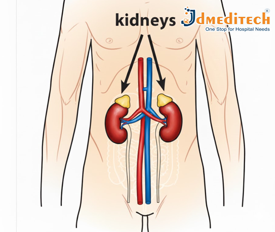
Chronic Kidney Disease (CKD) is a long-term health condition in which the kidneys slowly lose their ability to function properly over time. The kidneys are responsible for filtering waste products, toxins, and excess fluid from the blood. When they become damaged, these harmful substances start to build up in the body, leading to serious health problems.
CKD develops gradually, often over months or years, and may not show noticeable symptoms in its early stages. As the disease progresses, kidney function continues to decline, eventually leading to End-Stage Renal Disease (ESRD)—a stage where the kidneys can no longer support the body’s needs. At this point, patients require dialysis or a kidney transplant to survive.

What are the symptoms of CKD?
Chronic Kidney Disease (CKD) is a progressive condition, and its symptoms often appear gradually as kidney function deteriorates. In the early stages, CKD may not show any noticeable symptoms because the kidneys can still compensate for minor damage. However, as the disease progresses, the buildup of toxins, excess fluid, and electrolyte imbalance leads to a variety of physical and metabolic symptoms.
Patients may begin experiencing persistent fatigue, weakness, and lack of energy due to anemia and toxin accumulation. Swelling in the feet, ankles, hands, and face is common as the kidneys lose their ability to remove extra fluid. Many individuals notice changes in urination, such as foamy urine, increased nighttime urination, or reduced urine output.
Other symptoms include nausea, vomiting, loss of appetite, and a metallic taste, which result from rising waste levels in the bloodstream. High blood pressure, muscle cramps, itchy or dry skin, and difficulty concentrating also indicate declining kidney function. In advanced stages, CKD can lead to breathlessness, severe fluid retention, chest discomfort, and almost no urine production, signaling the onset of kidney failure.
Recognizing these symptoms early and seeking medical evaluation can significantly slow disease progression and improve long-term outcomes.

Symptoms of advanced CKD

Common Symptoms of Advanced CKD (Stage 4–5)
1. Severe Fatigue and Weakness
Due to anemia and toxin buildup, patients feel extremely tired and weak and lack energy.
2. Swelling (Edema)
Significant swelling appears in the legs, ankles, feet, hands, and sometimes around the face due to fluid retention.
3. Very Little or No Urine Output
The kidneys fail to produce normal amounts of urine, even though fluid is building up inside the body.
4. Shortness of Breath
Fluid buildup in the lungs causes difficulty breathing, even while resting.
5. Nausea and Frequent Vomiting
Excess waste in the bloodstream leads to digestive discomfort, nausea, and loss of appetite.
6. Extreme Itching (Uremic Pruritus)
High levels of waste products cause persistent and sometimes severe itching.

What Is Hemodialysis?
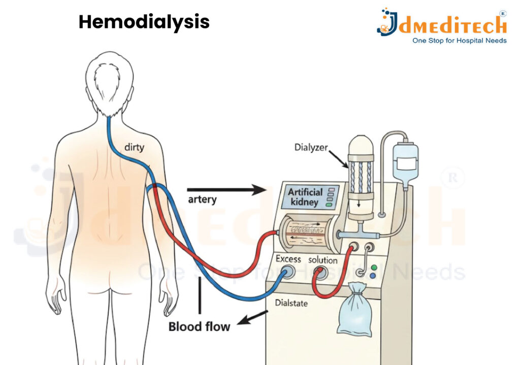
Hemodialysis is a medical procedure used to remove waste products, toxins, and excess fluid from the blood when the kidneys are no longer able to perform these functions effectively. It acts as an artificial kidney for people with advanced Chronic Kidney Disease (CKD) or kidney failure.
During hemodialysis, blood is taken out of the body, cleaned using a machine called a dialyzer, and then returned to the body. This process helps maintain fluid balance, remove harmful substances, and keep electrolytes like potassium and sodium at healthy levels.

How Does Hemodialysis Work?
1. A vascular access (fistula, graft, or catheter) is created to allow blood to flow in and out of the body.
2. Blood travels through tubing into the dialysis machine.
3. Inside the dialyzer, the blood passes through a special filter that removes waste and excess water.
4. Clean blood is then returned to the body through the same access.
5. This cycle continues for the duration of the session.
What Is Peritoneal Dialysis?

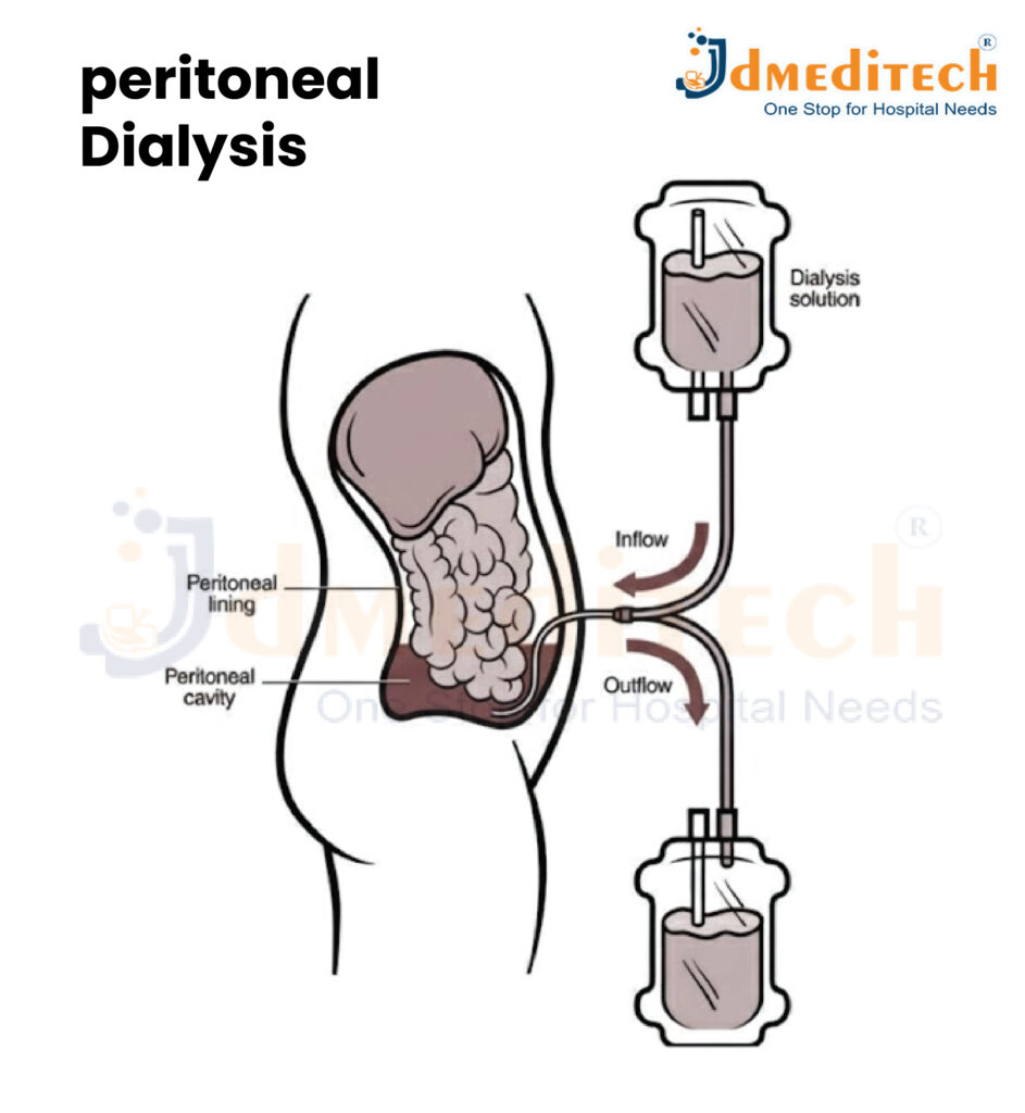
Peritoneal dialysis (PD) is a type of dialysis that uses the lining of your abdomen—called the peritoneal membrane—to filter waste and excess fluid from the blood. Instead of using a machine like hemodialysis, this method relies on the body’s natural membrane as a filter.

A sterile dialysis fluid is introduced into the abdominal cavity through a soft tube called a peritoneal dialysis catheter. Waste products pass from the blood into the fluid. After a few hours, the used fluid is drained out and replaced with fresh solution.
Peritoneal dialysis is often done at home, giving patients more flexibility and independence.

Types of Peritoneal Dialysis
1. Continuous Ambulatory Peritoneal Dialysis (CAPD)
• Manual process
• No machine required
• Usually 3–4 exchanges per day
• The patient can walk and do daily activities while fluid is inside the abdomen
2. Automated Peritoneal Dialysis (APD)
• Performed using a machine called a cycler
• Usually done at night while the patient sleeps
• More convenient for many people

What Happens During Peritoneal Dialysis?
Peritoneal dialysis (PD) cleans the blood by using the natural lining of the abdomen—the peritoneal membrane—as a filter. The process happens inside the body and does not require blood to leave the bloodstream. Instead, a special cleansing fluid removes waste and extra fluid.

Conclusion

Chronic kidney disease is a progressive condition that requires timely diagnosis and continuous management. When the kidneys fail, dialysis becomes essential to sustain life. Understanding different types of dialysis and how they work helps patients and families make informed healthcare decisions.
With proper treatment, lifestyle changes, and regular follow-up, CKD patients can lead stable and meaningful lives while preparing for the possibility of a kidney transplant.

Hemodialysis SETUP

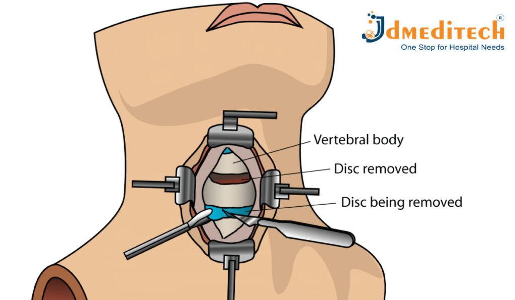
ACDF (Anterior Cervical Discectomy And Fusion) Surgery

ACDF (Anterior Cervical Discectomy and Fusion) is a widely performed spine surgery used to treat nerve compression in the neck caused by conditions like herniated discs, degenerative disc disease, or spinal stenosis. It helps relieve neck pain, arm pain, numbness, and weakness and restores normal movement.
What is ACDF?
Anterior Cervical Discectomy and Fusion (ACDF) is a surgical procedure used to treat conditions in the neck—particularly those where a disk in the cervical spine is herniated or degenerative, causing pressure on nerves or the spinal cord.
During ACDF:
• The surgeon approaches the spine from the front (through the throat/neck area) and removes the diseased or herniated disk (and often bone spurs), thereby relieving pressure on nerves or the spinal cord.
• Then, a “fusion” is done: adjacent vertebrae are stabilized using a bone graft or spacer (sometimes a cage), and plates/screws are often fixed to hold them in place while the bone fuses over time.
How Is ACDF Surgery Performed?
ACDF is usually performed under general anesthesia and takes about 1–2 hours, depending on the number of levels treated. Incision on the front of the neck
A small cut is made to access the cervical spine.

1. Disc removal
The damaged or herniated disc is carefully removed.
2. Nerve decompression
Bone spurs or tissue pressing on nerves are cleared.
3. Placement of bone graft or cage
An implant is inserted to maintain disc height.
4. Fusion with plate and screws
Metal hardware stabilizes the spine and helps fusion.
5. Closure
The incision is stitched and covered.

Benefits of ACDF Surgery
• Relieves neck and arm pain
• Restores nerve function
• Improves mobility and strength
• Stabilizes the spine
• Corrects compression of spinal cord or nerves
• Prevents worsening neurological symptoms

What Happens During ACDF Surgery?

ACDF (Anterior Cervical Discectomy and Fusion) is a structured spine procedure done to relieve pressure on the spinal cord or nerves in the neck. The surgery typically takes 1–2 hours, depending on the number of levels involved. Here is a step-by-step explanation of what happens:
1. Anesthesia and Positioning
• The patient is given general anesthesia, ensuring they remain fully asleep and pain-free.
• They are positioned on their back with the neck slightly extended to allow the surgeon clear access to the cervical spine.
2. Small Incision on the Front of the Neck
• The surgeon makes a small incision (3–4 cm) on one side of the front of the neck, usually near a natural skin crease.
• This approach avoids major muscles and allows a gentle, safe path to the spine.
3. Accessing the Cervical Spine
• Soft tissues like the esophagus, trachea, and neck muscles are carefully moved aside—not cut.
• Specialized instruments are used to expose the problematic disc between the cervical vertebrae.

4. Removing the Damaged/Herniated Disc (Discectomy)

• The surgeon removes the entire damaged or herniated disc causing pressure on nerves or the spinal cord.
• Any bone spurs (osteophytes) or fragments pressing on nerves are also removed.
• This step creates space and relieves compression.
5. Preparing the Disc Space
• After removing the disc, the empty disc space is cleaned and prepared.
• This helps promote proper fusion and alignment.
6. Inserting a Bone Graft or Cage
• A bone graft or interbody cage is placed into the disc space.
• This material helps the two vertebrae grow together (fuse) over time.
• The graft is usually:
o Patient’s own bone (rare)
o Donor bone
o Synthetic material
7. Stabilizing With a Plate and Screws (Fusion)
• A small titanium plate is fixed to the front of the vertebrae using screws.
• This keeps the spine stable and holds the graft securely until fusion occurs naturally.
8. Closing the Incision
• Once stability is confirmed:
o Muscles and soft tissues are placed back gently.
o The skin is closed with sutures.
• A small bandage is applied.
9. Recovery in the Hospital
• After surgery, the patient is moved to the recovery room.
• Most patients:
o Are awake within minutes
o Can walk the same day
o Stay 1 day in the hospital
• A neck collar may be recommended to support healing.
10. Healing & Fusion Process
• Although recovery begins immediately, the fusion process takes 3–6 months.
• Follow-up X-rays monitor how well the bones are fusing.

Conclusion

ACDF is a well-established, commonly performed spinal surgery that—for the right patient—can greatly improve quality of life by relieving nerve compression, pain, and weakness and restoring function. At the same time, as with any major surgery, it carries risks and requires careful patient selection, meticulous surgical technique, and disciplined postoperative care.

Explore More Product >>

Get Connected:
+91 79909 93062 | +91 63513 72032 | exports@jdmeditech.com

Laparoscopic Surgery

What Is Laparoscopic Surgery?
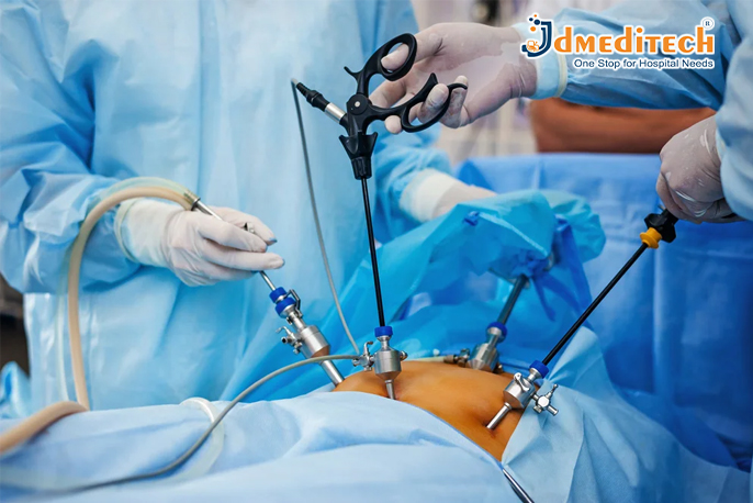
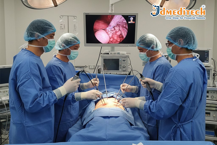
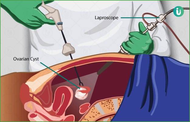
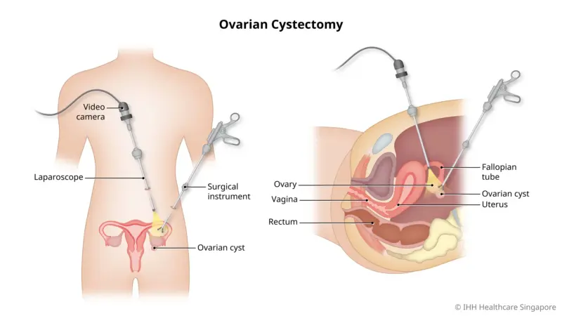
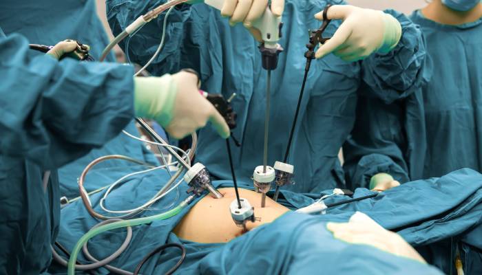
Laparoscopic surgery involves the use of a laparoscope, a thin tube with a high-resolution camera and light source. This device allows surgeons to view internal organs on a digital monitor in real time. Through additional small incisions, other instruments are inserted to perform the surgical procedure with precision.
This technique is commonly used for abdominal and pelvic surgeries, making it a preferred option for both surgeons and patients.

How Is Laparoscopic Surgery Performed?
1. Anesthesia is administered to ensure the patient feels no pain.
2. Small incisions are created in the abdomen.
3. Carbon dioxide gas is used to gently inflate the abdominal cavity for better visibility.
4. A laparoscope is inserted to project a clear image on a monitor.
5. Specialized surgical instruments are inserted through additional ports.
6. After completing the procedure, instruments are removed, CO₂ is released, and incisions are closed with sutures.

Common Procedures Performed Through Laparoscopy
Laparoscopic surgery can be used to diagnose and treat many conditions. Some widely performed laparoscopic procedures include:
• Laparoscopic Cholecystectomy (Gallbladder removal)
• Laparoscopic Appendectomy
• Laparoscopic Hernia Repair
• Laparoscopic Hysterectomy
• Laparoscopic Ovarian Cyst Removal
• Laparoscopic Bariatric Surgery
• Laparoscopic Diagnostic Procedures

Benefits of Laparoscopic Surgery
Laparoscopic procedures offer significant advantages over open surgery:
✔ Smaller Incisions
Only 0.5 cm to 1 cm incisions are made, resulting in minimal tissue damage.
✔ Less Pain & Discomfort
Patients experience less postoperative pain due to reduced trauma.
✔ Faster Recovery Time
Most patients can return to normal activities much sooner.
✔ Minimal Blood Loss
Smaller incisions help reduce bleeding during surgery.
✔ Lower Infection Risk
Smaller wounds reduce the chances of infection.
✔ Better Cosmetic Results
Tiny scars heal quickly and are barely visible.

Conclusion
Laparoscopic surgery has revolutionized the medical world with its minimally invasive nature, faster recovery, and excellent surgical outcomes. As technology continues to advance, more procedures are being performed laparoscopically, offering patients safer and more effective treatment options.

Explore More Product >>

Get Connected:
+91 79909 93062 | +91 63513 72032 | exports@jdmeditech.com

Hysteroscopy Surgery

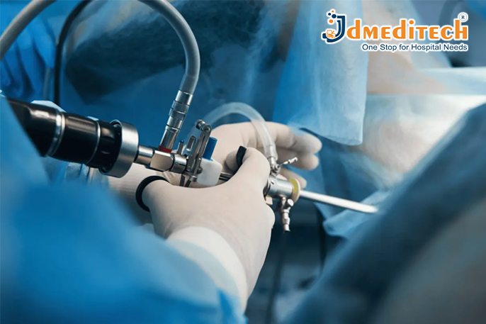
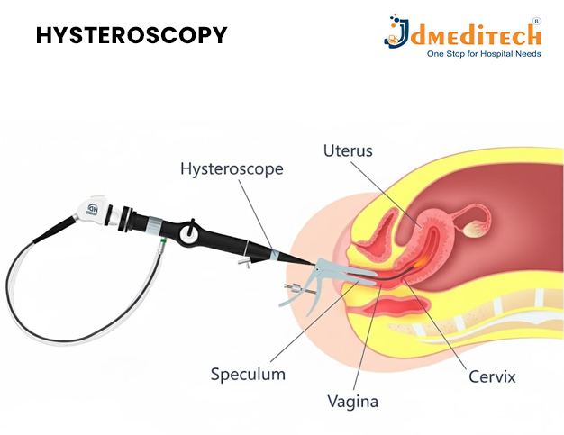
Hysteroscopy surgery allows a gynaecologist to examine and treat conditions inside the uterus. The doctor uses a thin, lighted instrument called a hysteroscope to view the uterine cavity.

During the procedure, the doctor guides the hysteroscope through the vagina and cervix. Therefore, hysteroscopy usually does not require an abdominal incision. Doctors may perform it for diagnostic purposes or to treat a uterine condition.

What Is Hysteroscopy?

Hysteroscopy is a minimally invasive procedure that provides a clear view of the cervix and uterine cavity. The hysteroscope includes a small camera that displays images on a monitor.

As a result, the doctor can examine the uterine lining and identify abnormalities. These may include polyps, fibroids, adhesions, scar tissue and structural changes.

Doctors may also recommend hysteroscopy to investigate abnormal uterine bleeding, infertility or repeated miscarriages. However, the reason for the procedure will depend on the patient’s symptoms and medical history.

Types of Hysteroscopy Surgery

There are two main types of hysteroscopy: diagnostic hysteroscopy and operative hysteroscopy. The doctor chooses the appropriate type according to the purpose of the procedure.

What Is Diagnostic Hysteroscopy?

Diagnostic hysteroscopy helps a doctor examine the inside of the uterus. It can help identify the cause of abnormal bleeding, infertility or repeated pregnancy loss.

First, the doctor guides a thin hysteroscope through the vagina and cervix. The camera then displays the uterine cavity on a monitor.

Consequently, the doctor can inspect the uterine lining without making an abdominal incision. The doctor may also collect a small tissue sample when necessary.

What Is Operative Hysteroscopy?

Operative hysteroscopy allows the doctor to treat certain conditions inside the uterus. The doctor may perform it after an ultrasound or diagnostic hysteroscopy identifies a problem.

During treatment, the surgeon passes small instruments through an operating channel in the hysteroscope. These instruments can cut, grasp or remove abnormal tissue.

Moreover, the surgeon may diagnose and treat the problem during the same appointment. However, some patients may require a separate procedure.

Diagnostic and operative hysteroscopy procedure

When Is Operative Hysteroscopy Used?

A gynaecologist may recommend operative hysteroscopy for several uterine conditions. The following are some common reasons for the procedure.

1. Uterine Polyps

Uterine polyps are soft tissue growths inside the uterus. They may contribute to heavy bleeding, irregular periods or fertility problems. Therefore, the surgeon may use hysteroscopy to remove them.

2. Submucosal Fibroids

Submucosal fibroids grow towards or inside the uterine cavity. They can cause heavy periods, pelvic discomfort or reproductive problems.

In suitable cases, the surgeon can remove these fibroids through operative hysteroscopy. However, the treatment choice depends on their size and location.

3. Uterine Septum

A uterine septum is a wall of tissue that divides the uterine cavity. This condition may affect fertility or increase the risk of miscarriage.

When appropriate, the surgeon can cut the septum during hysteroscopy. Afterwards, the doctor may recommend follow-up care or imaging.

4. Uterine Adhesions

Uterine adhesions are bands of scar tissue inside the uterus. Doctors may also refer to severe uterine adhesions as Asherman syndrome.

These adhesions can contribute to light or absent periods, pelvic pain and fertility problems. Therefore, the surgeon may use hysteroscopy to locate and divide them.

5. Retained Pregnancy Tissue

Sometimes, tissue may remain inside the uterus after a miscarriage, delivery or pregnancy termination. This tissue can contribute to persistent bleeding, pain or infection.

In selected cases, hysteroscopy allows the doctor to see and remove the remaining tissue. The patient’s condition will determine the safest treatment method.

6. Removal of an Embedded or Difficult IUD

An intrauterine device may move, become embedded or lose its visible removal threads. Consequently, routine removal may become difficult.

Hysteroscopy helps the doctor locate the device directly. The doctor can then remove it with specialised instruments when clinically appropriate.

7. Abnormal Uterine Bleeding

Heavy, irregular or prolonged bleeding can have several causes. For this reason, doctors first evaluate the patient before recommending treatment.

If a structural problem causes the bleeding, operative hysteroscopy may help treat it. Examples include polyps and certain fibroids.

8. Endometrial Abnormalities

Hysteroscopy helps the doctor inspect unusual areas of the uterine lining. In addition, the doctor can collect tissue for laboratory examination.

A biopsy can provide more information about the abnormal tissue. Nevertheless, the doctor must review the laboratory result before confirming a diagnosis.

9. Evaluation of the Tubal Openings

The doctor can inspect the openings of the fallopian tubes during hysteroscopy. In selected cases, a specialist may also perform tubal cannulation near the uterine opening.

However, hysteroscopy cannot treat every type of fallopian tube blockage. Additional tests or fertility treatments may be necessary.

How to Prepare for Hysteroscopy Surgery

Before hysteroscopy surgery, the doctor reviews the patient’s symptoms and medical history. The doctor may also perform a pregnancy test, blood test, ultrasound or pelvic examination.

In addition, patients should tell the doctor about their medicines, allergies and existing health conditions. This information helps the medical team plan the procedure safely.

The doctor may ask the patient to avoid food and drinks before hysteroscopy with sedation or anaesthesia. Therefore, patients should carefully follow the instructions provided by their hospital.

What Happens During Hysteroscopy?

First, the medical team positions the patient and prepares the treatment area. The doctor then passes the hysteroscope through the vagina and cervix.

Next, fluid may gently expand the uterine cavity. This step helps the doctor obtain a clearer view of the uterine lining.

During diagnostic hysteroscopy, the doctor examines the cavity and may collect a biopsy. During operative hysteroscopy, the surgeon uses miniature instruments to perform the required treatment.

Finally, the doctor removes the hysteroscope and monitors the patient during recovery. The total procedure time varies according to the treatment.

Benefits of Hysteroscopy Surgery

Hysteroscopy offers several potential benefits for suitable patients:

  • Direct examination of the uterine cavity
  • No abdominal surgical incision
  • Accurate identification of uterine abnormalities
  • Diagnosis and treatment during one procedure in some cases
  • Targeted removal of abnormal tissue
  • Shorter recovery than many open surgical procedures
  • Same-day discharge for many patients

However, individual results may vary. The condition, treatment type and patient’s overall health can affect the outcome.

Recovery After Hysteroscopy

Many patients return home on the same day. However, patients who receive sedation or general anaesthesia may need someone to accompany them.

Mild cramping and light vaginal bleeding can occur after the procedure. These symptoms often improve within a few days.

During recovery, patients should follow their doctor’s advice about bathing, exercise, sexual activity and tampon use. They should also take only the medicines their doctor recommends.

Possible Risks of Hysteroscopy

Hysteroscopy is generally safe, but every medical procedure has possible risks. These may include:

  • Bleeding
  • Infection
  • Injury to the cervix
  • Perforation of the uterus
  • Reaction to anaesthesia
  • Complications related to the fluid used during the procedure
  • Development of scar tissue

The doctor should explain the expected benefits and individual risks before the procedure.

When Should You Contact a Doctor?

Patients should contact their doctor if they experience severe or worsening symptoms after hysteroscopy. Important warning signs include:

  • Heavy vaginal bleeding
  • Severe abdominal or pelvic pain
  • Fever or chills
  • Foul-smelling vaginal discharge
  • Dizziness or fainting
  • Persistent vomiting

Conclusion

Hysteroscopy surgery helps doctors diagnose and treat several conditions inside the uterus. Furthermore, it provides a direct view of the uterine cavity without an abdominal incision.

The procedure may help manage polyps, fibroids, adhesions and other uterine abnormalities. Nevertheless, its suitability depends on each patient’s symptoms and medical condition.

Anyone experiencing abnormal bleeding, fertility problems or persistent pelvic symptoms should consult a qualified gynaecologist. The doctor can recommend suitable tests and treatment options.

This article provides general educational information. It does not replace professional medical advice, diagnosis or treatment.

Explore More Products >>

Get Connected

+91 79909 93062 | +91 63513 72032 | exports@jdmeditech.com

Dilation and Curettage (D&C)

What is dilation and curettage (D&C)?
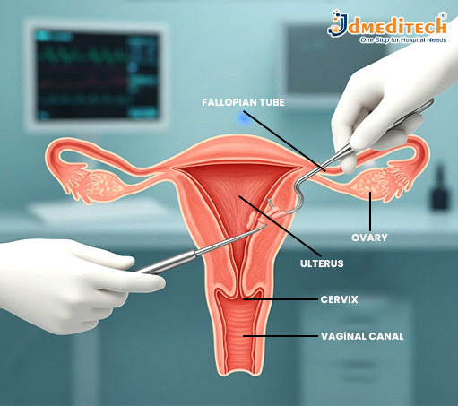
Dilation and curettage, sometimes known as D&C, is a gynecological procedure that involves the gentle opening of the cervix—the lower part of the uterus—and the removal of tissue from inside the uterus using a specialized instrument called a curette.
Depending on the circumstances, D&C can also be performed under suction—that is, vacuum—rather than sharp curettage.

Why is D&C used?
D&C may be both a diagnostic and therapeutic procedure. Indications for a D&C include:
• Investigating and diagnosing abnormal uterine bleeding, postmenopausal bleeding, and unexplained changes in menstrual cycles.
• Removing abnormal endometrial tissue, such as polyps, overgrown uterine lining, or suspected precancerous/cancerous changes.
• Clearing the uterus following a miscarriage or termination of pregnancy to ensure that no fetal or placental tissue remains.
• Removal of retained fragments of placenta after delivery or abortion, which may cause hemorrhage or infection.
Due to its flexibility and effectiveness, D&C remains one of the most common procedures when uterine sampling or tissue removal is required.

What to Expect—Procedure & Recovery Before the Procedure
•Your physician will discuss with you your medical history and advise any preoperative preparations, such as fasting or the use of any medication, as may be required.
• You will be asked to empty your bladder before the procedure. Depending on the indication and clinical circumstance, local or general anesthesia might be administered.

During the Procedure
• The speculum is inserted gently to visualize the cervix. Then, the cervix is slowly dilated using graduated dilators.
• Once dilation is sufficient, a metal or plastic curette or suction device is introduced to remove lining or tissue from the uterus.
• The tissue removed may be sent to the laboratory for analysis—looking for polyps, abnormal cells, or other pathology—if the D&C was performed for diagnostic purposes.
• The procedure itself is relatively quick; many D&Cs take about 10–20 minutes.
After the Procedure: Recovery & Home Care
•After D&C, many people experience mild cramping (like menstrual cramps) and light bleeding or spotting for a few days.
• Use sanitary pads, not tampons, to decrease your infection risk. Do not engage in vaginal intercourse, nor insert a tampon into the vagina, until your doctor lets you know that this will be safe, which is generally in about a week, though timing may vary.
• Most individuals return to normal activities within a few days.
•Many surgeons will schedule a follow-up appointment within 1–2 weeks, especially if the tissue was sent for pathology, to review the results and make sure the healing is normal.

Why Choose JD Meditech for Gynecology Instruments? ISO-certified manufacturer
• International-standard surgical instruments
• Rust-free, medical-grade stainless steel
• Ergonomic, surgeon-friendly design
• Export quality products with reliable performance
Our various products enjoy immense trust in hospitals, clinics, and healthcare facilities across the world.
Conclusion
D&C is a safe and effective diagnostic and treatment method for a variety of uterine problems. Availing themselves of the most advanced gynecology instruments offered by JD Meditech, healthcare professionals can conduct D&C with precision and confidence, ensuring better outcomes and patient care.

Explore More Product >>

Get Connected:
+91 79909 93062 | +91 63513 72032 | exports@jdmeditech.com

Manufacturing of Medical Devices in India

India’s medical device manufacturing industry is growing rapidly. As a result, the country is becoming an important source of reliable and affordable healthcare technology.

Government initiatives, skilled professionals and modern facilities support this growth. Moreover, rising healthcare demand creates new opportunities for manufacturers and exporters.

Therefore, many healthcare providers now consider India a preferred destination for cost-effective medical devices.

Introduction to Medical Device Manufacturing in India

India manufactures a wide range of medical devices for domestic and international markets. These products include surgical instruments, endoscopy equipment and hospital supplies.

In addition, Indian manufacturers continue to improve their production systems. They also focus on product quality, safety and regulatory compliance.

Companies such as JDMeditech contribute to this sector. The company supplies medical devices for several clinical specialties.

Advanced Manufacturing Capabilities

Indian medical device manufacturers use modern equipment and controlled production processes. Consequently, they can produce accurate and consistent products.

Many manufacturing facilities include:

  • Precision engineering systems
  • CNC and automated machinery
  • Quality inspection and testing laboratories
  • Sterilization facilities
  • Controlled cleanroom environments
  • Product assembly and packaging areas

Furthermore, manufacturers follow documented quality procedures throughout production. These procedures support product consistency and traceability.

Major Medical Devices Manufactured in India

India produces medical devices for many healthcare specialties. The following categories represent some important areas of manufacturing.

1. Urology Instruments

Urology instruments support diagnosis and minimally invasive treatment. Manufacturers offer various reusable instruments, devices and accessories.

  • TURP resectoscopes
  • Rigid and flexible ureteroscopes
  • URS forceps and stone baskets
  • RIRS scopes and accessories
  • Laser surgery instruments

2. Laparoscopy and Endoscopy Equipment

Laparoscopy and endoscopy devices help doctors perform minimally invasive procedures. Therefore, manufacturers focus on precision, visibility and ease of use.

  • Laparoscopic hand instruments
  • Telescopes and camera systems
  • Insufflators and light sources
  • Trocars and cannulas
  • Endoscopy accessories

3. Gynaecology and Obstetrics Devices

Indian manufacturers also supply instruments for gynaecological examinations and procedures. For example, this category may include:

  • Hysteroscopes
  • D&C instruments
  • Vacuum delivery systems
  • Gynaecology forceps and scissors

4. ENT and General Surgery Devices

Surgeons require dependable instruments for accurate handling and tissue management. Hence, this category includes several specialised devices.

  • Sinuscopes
  • Rigid endoscopes
  • Surgical forceps
  • Surgical scissors
  • General surgery instruments

5. Hospital Equipment

Hospital equipment supports patient care in operating rooms, intensive care units and emergency departments. Common products include:

  • Operating tables
  • LED operation theatre lights
  • ICU equipment
  • Emergency care devices
  • Patient-monitoring equipment

JDMeditech supplies products across several medical categories. In addition, the company serves customers in India and international markets.

Quality Standards and Regulatory Compliance

Quality plays an essential role in medical device manufacturing. Therefore, manufacturers must follow the regulations that apply to their products and target markets.

Manufacturers may implement quality management systems for design, production and inspection. In addition, they may perform product testing and maintain technical documentation.

However, requirements can vary by product, risk classification and destination country. Exporters must confirm all applicable rules before supplying a device.

Medical Device Exports from India

India exports medical devices and surgical equipment to many international markets. Competitive pricing and expanding production capabilities support this growth.

Moreover, manufacturers increasingly focus on global quality expectations. They also improve product documentation, labelling and packaging.

Exporters commonly supply:

  • Surgical instruments
  • Urology devices
  • Endoscopy equipment
  • Diagnostic products
  • Hospital supplies
  • Disposable medical products

Support for Medical Device Exports

Government departments and regulatory authorities continue to support the medical device sector. For example, their initiatives may improve manufacturing practices and export procedures.

Important areas of development include:

  • Good manufacturing practices
  • Export labelling requirements
  • Clinical evaluation guidance
  • Adverse-event reporting
  • Licensing and certification processes
  • Access to manufacturer information

As a result, Indian manufacturers can improve their access to international markets. However, each company must meet the regulations of the importing country.

Medical device manufacturing and exports from India

The Future of Medical Device Manufacturing in India

India’s medical device sector has strong potential for continued growth. Investment in research, technology and automation will support future development.

Additionally, manufacturers are exploring advanced healthcare solutions. These developments include:

  • Robotic surgical systems
  • Artificial intelligence in diagnostics
  • Minimally invasive surgical instruments
  • Laser-based medical technology
  • Advanced imaging systems
  • Digital healthcare solutions

Meanwhile, domestic manufacturing can help reduce dependence on imported devices. It can also improve access to affordable healthcare technology.

Why Choose an Indian Medical Device Manufacturer?

Indian manufacturers combine engineering expertise with competitive production costs. Therefore, they can support healthcare providers in diverse markets.

Key advantages may include:

  • A wide range of medical products
  • Competitive pricing
  • Skilled engineering professionals
  • Modern manufacturing facilities
  • Customised product solutions
  • International export support

Furthermore, reliable manufacturers provide product information and regulatory documents. They may also offer assistance with packaging and logistics.

Conclusion

Medical device manufacturing in India continues to develop. Modern technology, skilled professionals and government initiatives support this progress.

As a result, India is strengthening its position in the global healthcare market. The country can offer reliable and affordable medical devices to healthcare providers worldwide.

JDMeditech contributes to this mission by supplying urology, endoscopy and surgical products. Moreover, the company supports healthcare professionals in India and overseas markets.

Explore More Products >>

Get Connected

+91 79909 93062 | +91 63513 72032 | exports@jdmeditech.com

Vaginal Hysterectomy Surgery

A vaginal hysterectomy is one of the most preferred minimally invasive procedures in modern gynecology for the removal of the uterus. At JDMeditech, we support healthcare professionals with advanced surgical instruments and reliable medical devices that ensure safety, precision, and high-quality outcomes during gynecological procedures.
Vaginal hysterectomy offers faster recovery, minimal pain, and no visible scars, making it a popular choice for many women. Here is a complete guide to help patients and healthcare providers understand this procedure better.

What Is a Vaginal Hysterectomy?
A vaginal hysterectomy involves removing the uterus (womb) through the vaginal canal. In some cases, the cervix, fallopian tubes, or ovaries may also be removed depending on the patient’s condition.
It is commonly recommended for women who do not plan future pregnancies and are suffering from chronic reproductive health issues that cannot be treated with medication or minimally invasive procedures.

When Is Vaginal Hysterectomy Recommended?
Doctors may suggest a vaginal hysterectomy for conditions such as
• Uterine fibroids
• Heavy or irregular menstrual bleeding
• Pelvic organ prolapse
• Endometriosis
• Chronic pelvic pain
• Adenomyosis
• Abnormal uterine bleeding
• Recurrent uterine infections

Advantages of Vaginal Hysterectomy
Vaginal hysterectomy provides several benefits for patients, including:
✔ No Visible Scars
The entire procedure is performed through the vagina, leaving no external marks.
✔ Faster Recovery
Most patients recover quicker compared to abdominal surgery.
✔ Less Postoperative Pain
Minimal tissue damage results in reduced pain and discomfort.
✔ Shorter Hospital Stay
Patients are usually discharged within 1–2 days.
✔ Lower Risk of Complications
Reduced chances of bleeding or infection.

Conclusion
Vaginal hysterectomy is a safe, efficient, and minimally invasive surgical technique that offers numerous benefits for women suffering from chronic gynecological issues. With the right technology and surgical instruments—such as those provided by JDMeditech—healthcare professionals can ensure optimal outcomes with enhanced safety and precision.

Explore More Product >>

Get Connected:
+91 79909 93062 | +91 63513 72032 | exports@jdmeditech.com

Labiaplasty Surgery

Labiaplasty is a surgical procedure that involves trimming or reshaping the labia minora to achieve a more balanced and comfortable vaginal structure. The goal of this surgery is not only aesthetic enhancement but also improved functionality in daily activities.
• Discomfort while walking, exercising, or wearing tight clothing
• Pain during intercourse
• Irritation or hygiene issues
• Asymmetry in the size or shape of the labia
• Desire for a more proportionate vaginal appearance

Who Can Benefit from Labiaplasty?
Labiaplasty is suitable for women experiencing:
• Enlarged or elongated labia due to genetics, childbirth, aging, or hormonal changes
• Functional discomfort during sexual intercourse or physical activities
• Emotional or psychological dissatisfaction with the appearance of their intimate area
A consultation with a qualified gynecologist or cosmetic surgeon helps determine whether the procedure is the right choice based on health history and expectations.

Benefits of Labiaplasty
• Enhanced comfort during activities like cycling, walking, or exercise
• Reduction of discomfort during sexual intercourse
• Improved intimate hygiene
• Boosted self-confidence and body image
• Correction of congenital or postpartum asymmetry

Recovery Tips
• Use cold compresses for swelling
• Avoid tight clothing during initial healing
• Maintain proper hygiene as instructed by the doctor
• Avoid sexual intercourse and strenuous exercise for 4–6 weeks
• Take prescribed medications for pain and inflammation

Risks & Considerations
• Swelling or bruising
• Temporary numbness
• Infection
• Scarring
• Asymmetry (rare and correctable)

Conclusion
Labiaplasty surgery offers a transformative solution for women seeking physical comfort, confidence, and improved intimate wellness. With the right guidance and expert care, patients can achieve natural-looking results and an enhanced quality of life.

Explore More Product >>

Get Connected:
+91 79909 93062 | +91 63513 72032 | exports@jdmeditech.com

Angioplasty Surgery

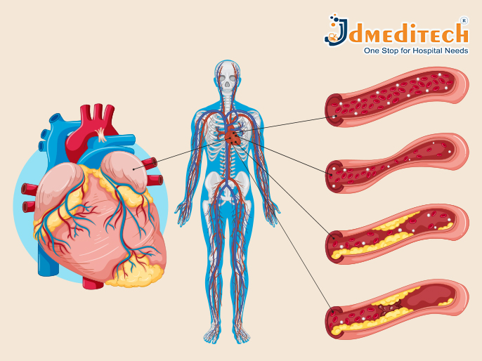
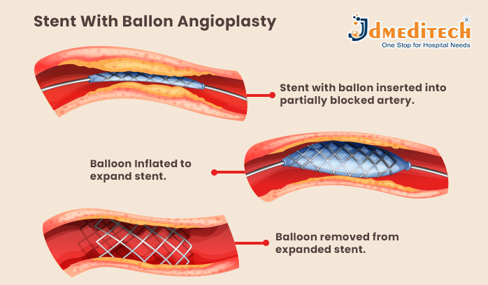
Angioplasty is a procedure in which a cardiologist inserts a small balloon inside a blocked artery. When the balloon is inflated, it pushes the plaque against the artery walls, widening the blood vessel. In many cases, a stent (a small metal mesh tube) is placed to keep the artery open permanently.

When Is Angioplasty Recommended?
Doctors may suggest angioplasty for patients experiencing:
• Coronary Artery Disease (CAD)
Plaque buildup narrows the arteries and reduces blood supply to the heart.
• Heart Attack
Angioplasty can quickly restore blood flow and reduce damage to the heart muscle.
• Severe Chest Pain (Angina)
Especially when pain does not improve with medication.
• Blockages Detected in Tests
Such as stress tests, CT scans, or angiograms.

How Angioplasty Is Performed
1. Preparation
• Local anesthesia is applied.
• A catheter is inserted through the groin or wrist.
• Using X-ray guidance, the catheter is moved toward the blocked artery.
2. Balloon Inflation
• A tiny balloon at the tip of the catheter is inflated.
• This compresses plaque and widens the artery.
3. Stent Placement
• A metal stent is placed in most cases.
• The stent stays in the artery to prevent re-narrowing.
4. Completion
• The catheter is removed.
• The entire procedure usually takes 30–60 minutes.

Benefits of Angioplasty
• Quickly restores blood flow
• Reduces chest pain
• Minimizes heart damage during a heart attack
• Shorter recovery time compared to open-heart surgery
• High success rate with stent placement

Risks and Complications
Although angioplasty is generally safe, some risks may include:
• Bleeding at the catheter insertion site
• Re-narrowing of the artery (restenosis)
• Blood clots in the stent
• Irregular heartbeats
• Rare chances of heart attack or stroke

Recovery After Angioplasty
Most patients recover quickly and may return home within a day. Recovery tips include:
• Avoid heavy lifting for a week
• Take prescribed blood-thinning medications
• Follow a heart-healthy diet
• Regular exercise as advised by the doctor
• Scheduled follow-ups to monitor progress
Lifestyle changes play a key role in preventing future blockages.

Conclusion
Angioplasty surgery is a safe, effective, and life-saving procedure for opening blocked arteries and improving heart function. With modern medical technology and expert cardiac care, patients can enjoy faster recovery and a healthier future. Understanding the procedure helps individuals make confident decisions about their heart health.

Explore More Product >>

Get Connected:
+91 79909 93062 | +91 63513 72032 | exports@jdmeditech.com

C-Section Surgery (Cesarean Delivery)

A Cesarean delivery, commonly known as a C-section, is a surgical procedure used to deliver a baby safely when vaginal birth is not possible or poses risks to the mother or child. Thanks to advanced medical equipment and modern surgical standards, C-sections today are highly safe and widely practiced around the world.

Why a C-Section Is Performed
Doctors may recommend a C-section for several medical reasons:

1. Complications During Labor
• Labor not progressing as expected
• Signs of fetal distress
• Issues with the umbilical cord
2. Maternal Health Concerns
• High blood pressure or pre-eclampsia
• Certain active infections
• Placenta previa (placenta covering or blocking the birth canal)
3. Baby’s Position
• Breech position (baby coming feet first)
• Transverse position (lying sideways)
4. Multiple Pregnancy
• Twins, triplets, or higher-order multiples
• Complications affecting one or more babies
5. Previous C-Sections
• Some women may require a repeat C-section depending on uterine healing and pregnancy conditions.

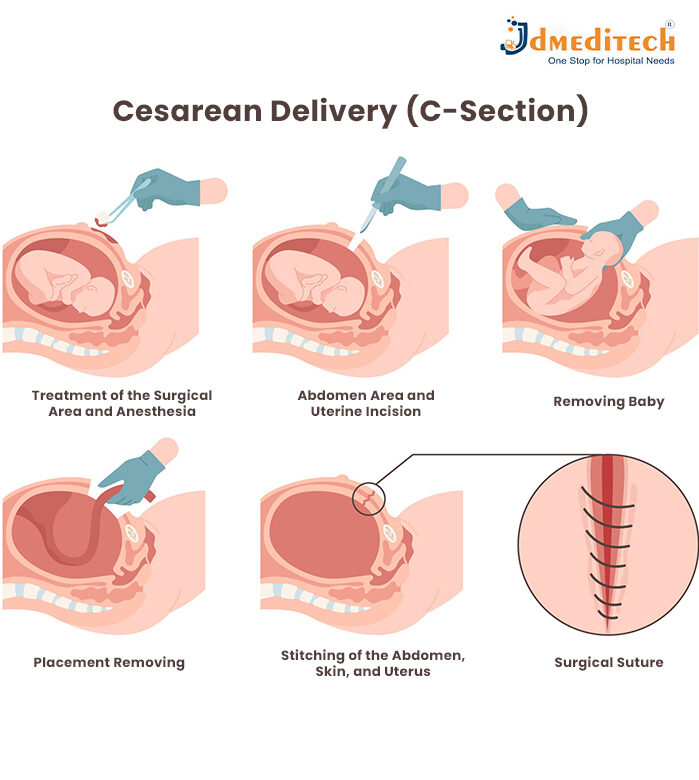
How C-Section Surgery Is Performed

1. Preparation
• The mother receives spinal or epidural anesthesia.
• Vital signs are monitored, and the abdominal area is sterilized.
2. Making the Incision
• A horizontal (bikini line) incision is made on the lower abdomen.
• A second incision is created in the uterus to reach the baby.
3. Delivery of the Baby
• The baby is gently lifted out of the uterus.
• The umbilical cord is cut, and a pediatric team assesses the newborn.
4. Closing the Incisions
• The uterus and abdominal layers are carefully sutured.
• The entire operation typically takes about 40–60 minutes.

Recovery After a C-Section

Recovery from a C-section takes longer than a vaginal birth because it involves major abdominal surgery. Typical recovery steps include:
• Staying in the hospital for 2–4 days
• Pain management and adequate rest
• Avoiding lifting heavy objects
• Gentle walking to improve circulation
• Scheduling follow-up visits to monitor incision healing
Most mothers recover fully within 4–6 weeks.

Benefits of a C-Section
• A safer option in emergency situations
• Protects the health of both mother and baby in high-risk pregnancies
• Planned C-Sections allow scheduling convenience for families

Risks and Considerations
As with any surgical procedure, a C-section carries some risks, such as:
• Infection or excessive bleeding
• Blood clots
• Longer healing period
• Increased risk of breathing problems in babies delivered before 39 weeks (when not medically necessary)
Healthcare professionals evaluate each case carefully before recommending cesarean delivery.

Conclusion
A C-section is a highly effective and reliable method of childbirth when normal delivery isn’t possible or safe. With advanced surgical techniques, professional medical care, and high-quality equipment—such as those supported by JD Meditech—mothers and babies can expect safe outcomes and a smooth recovery.
Understanding the procedure empowers parents, reduces anxiety, and prepares families for a confident birthing experience.

Explore More Product >>

Get Connected:
+91 79909 93062 | +91 63513 72032 | exports@jdmeditech.com

Instruments Used in TURBT (Transurethral Resection of Bladder Tumor) Surgery

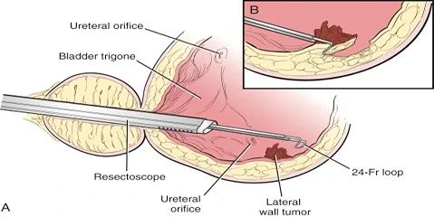
During TURBT (Transurethral Resection of Bladder Tumor), a specialized set of urological instruments is used to access and remove bladder tumors via the urethra—without external incisions.

Instrument Names Used in TURBT Surgery:
Resectoscope
Working Element (active part of resectoscope)
Cutting Loop Electrode (monopolar or bipolar)
Light Cable and Camera Head
Irrigation Tubing Set
Saline or Glycine Irrigation Fluid
Ellik Evacuator or Toomey Syringe
Cautery Electrode / Coagulation Electrode
Bladder Distension Sheath
Biopsy Forceps (if tissue sampling is needed)
Electrosurgical Generator Unit (ESU)
e.g., Bipolar Saline Electrosurgical Unit – 400W
Suction Tubing and Collection Canister
Operating Table with Leg Supports (Lithotomy Position)

These instruments work together to perform the resection and removal of bladder tumors through the urethra, without open surgery.

TURBT (Transurethral Resection of Bladder Tumor): Procedure, Benefits & Recovery

TURBT (Transurethral Resection of Bladder Tumor) is typically needed when a person is suspected to have or has been diagnosed with a bladder tumor, especially at early stages. It plays a crucial role in both diagnosis and initial treatment.

We need TURBT (Transurethral Resection of Bladder Tumor) in the following situations:
1. Diagnosis of Bladder Cancer
When a person has symptoms like:
Blood in urine (hematuria)
Frequent or painful urination
Imaging or cystoscopy shows a suspicious mass or lesion in the bladder.
TURBT helps confirm if the tumor is cancerous, and determines the type, grade, and depth of invasion.

2. Initial Treatment of Early-Stage Bladder Cancer
Especially for non-muscle invasive bladder cancer (NMIBC):
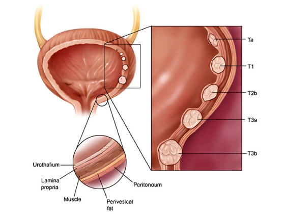
Ta (confined to bladder lining)
T1 (into connective tissue)
CIS (flat carcinoma in situ)
TURBT removes the tumor completely, often followed by intravesical therapy (e.g. BCG).

3. Restaging or Repeat TURBT
If the initial tumor is:
High-grade
T1 stage
A second TURBT (within 2–6 weeks) is often done to ensure complete removal and accurate staging.

4. Relief of Urinary Symptoms in Advanced Cancer
When the tumor causes:
Bleeding
Blockage of urine flow
TURBT may be done for palliative purposes, not cure.

Procedure of TURBT (Transurethral Resection of Bladder Tumor)
1. Preoperative Preparation
Admission: Usually done as a day-care or short hospital stay.
Anesthesia: Spinal or general anesthesia is administered.
Positioning: Patient lies on their back with legs supported in stirrups (lithotomy position).

2. Insertion of the Resectoscope
A resectoscope (a thin tube with a camera and surgical loop) is gently inserted through the urethra into the bladder.
No external incision is made.

3. Visual Inspection of the Bladder
The surgeon examines the inside of the bladder for:
Tumor(s) — their size, location, and number
Bladder wall condition
Any suspicious areas for biopsy

4. Tumor Resection
The tumor is shaved off in layers using the resectoscope’s electric loop.
If necessary, tissue from the base of the tumor (including muscle layer) is also taken to check how deep the tumor has grown.

5. Control of Bleeding (Hemostasis)
The surgical area is cauterized to stop bleeding.
Irrigation fluid is used to flush out blood clots and tissue pieces from the bladder.

6. Tissue Collection for Biopsy
Tumor and bladder wall tissue samples are collected and sent for pathological analysis to determine:
Cancer presence
Type, grade, and stage

7. Catheter Placement
A Foley catheter is inserted into the bladder to:
Drain urine
Prevent blockage from clots
Allow bladder to heal
Bladder irrigation may be used to prevent clot formation.

Postoperative Care
Hospital Stay: Usually 1–2 days depending on recovery.
Catheter Removal: Within 24–72 hours typically.
Pathology Report: Usually ready within 7–10 days to guide further treatment.

Duration of Procedure
Typically takes 30–90 minutes depending on the number and size of tumors.

Instruments Used in Bladder Augmentation Surgery: Step-by-Step Guide

During Bladder Augmentation Surgery, various surgical instruments are used to perform the procedure efficiently and safely. Here’s a list of common instruments:

step-by-step list of instruments

1. Preparation Phase:
Surgical Scalpels: For making the initial incision in the abdominal area.
Needle Holder: To securely hold needles for suturing.

2. Accessing the Bladder:
Surgical Scissors: To dissect and open up layers of tissue to reach the bladder.
Bladder Retractors: Used to hold the bladder open for better access.

3. Harvesting Intestinal Tissue:
Intestinal Forceps: To clamp and hold the intestinal segment to be used for augmentation.
Bowel Clamp: To temporarily stop the blood flow to the portion of the intestine being removed.
Scissors or Electrocautery: To cut the section of the intestine.

4. Preparing and Attaching Intestinal Tissue to the Bladder:
Needle Holder and Suturing Instruments: For stitching the harvested segment of intestine to the bladder.
Surgical Scissors: For trimming any excess tissue.
Electrocautery (Diathermy): For coagulating small blood vessels to minimize bleeding.

5. Closing the Incision:
Sutures (Absorbable or Non-absorbable): To close the abdominal and bladder incisions.
Staplers: Occasionally used for closing the larger incisions in the bladder or abdominal wall.

6. Post-Operative Care:
Catheters: A catheter is inserted into the bladder for draining urine during recovery.
Drainage Tubes: May be used to prevent fluid buildup after surgery.

7. Final Inspection and Closure:
Sterile Drapes and Covers: To ensure a sterile environment during surgery.
Sutures or Staplers: To close the abdominal incision.

These instruments work together to ensure that the bladder augmentation procedure is successful and safe.

Bladder Augmentation Surgery: Procedure, Benefits & Recovery Guide

Bladder Augmentation is needed when the bladder is too small, stiff, or unable to store urine properly, causing serious problems.

Bladder Augmentation is needed when:
Bladder cannot hold enough urine (small bladder capacity).
Bladder is too stiff or high-pressure, risking kidney damage.
Severe urinary incontinence that doesn’t improve with medication or catheter use.
Neurogenic bladder (due to spinal cord injury, spina bifida, etc.).
Congenital bladder problems like bladder exstrophy or other birth defects.
Chronic bladder inflammation or damage from radiation, infection, or long-term catheter use.
Urine backing up to kidneys (vesicoureteral reflux) due to poor bladder function.

Procedure of Bladder Augmentation (Augmentation Cystoplasty)

1. Anesthesia
The patient is placed under general anesthesia (completely unconscious).

2. Opening the Abdomen
A surgical incision is made in the lower abdomen to access the bladder.

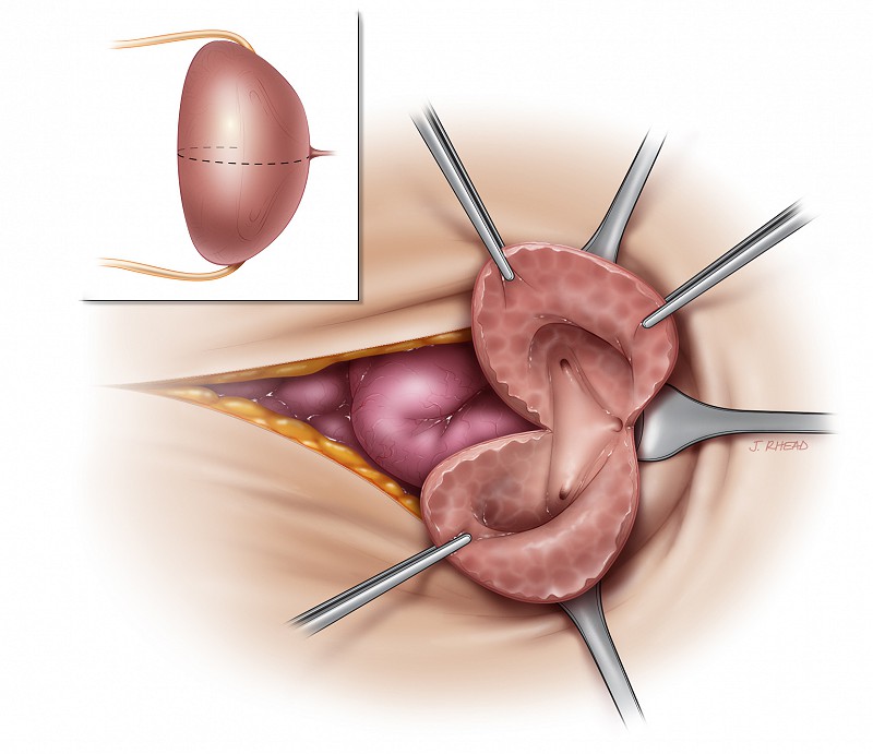
3. Opening the Bladder
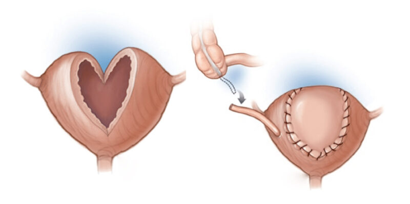
The surgeon cuts open the bladder at the top to prepare it for expansion.

4. Removing a Section of Intestine
A small piece of intestine (usually ileum) is removed.
The intestine is then reconnected so digestion continues normally.

5. Attaching Intestine to Bladder
The intestinal segment is reshaped and sewn to the bladder to increase its size and reduce pressure.

6. Closing the Incision
A catheter is placed into the bladder to drain urine.
The abdominal incision is closed with stitches.

7. Post-Surgery Recovery
Hospital stay: 7–10 days.
Catheter use: 2–3 weeks.
Patient may need to learn self-catheterization for bladder emptying.

Benefits
Increased bladder capacity – can hold more urine comfortably.
Reduced bladder pressure – protects the kidneys from damage.
Improved continence – helps control urine leakage.
Better quality of life – less urgency, fewer infections, and more independence.

Instruments Used in Cystectomy Surgery: A Complete Guide

Here is a list of specific instruments used in cystectomy surgery (bladder removal surgery):

Basic Surgical Instruments:
Scalpel – For making precise incisions in the skin and tissues.
Mayo scissors – For cutting soft tissues during dissection.
Metzenbaum scissors – For finer dissection of delicate tissues.
Needle holder – Used to hold needles while suturing.
Kelly clamp – For clamping blood vessels and tissues to control bleeding.
Mosquito clamp – A smaller version of the Kelly clamp, used for smaller blood vessels.

Instruments for Bladder and Pelvic Surgery:
Balfour retractor – A self-retaining retractor to hold the abdominal cavity open for better access.
DeBakey forceps – For grasping tissues delicately, used to hold and manipulate tissues.
Allis forceps – Used to grasp tissues, especially in deeper regions.
Surgical suction – Used to remove blood and fluids, maintaining a clear surgical field.
Electrocautery (Bovie) – An electrical tool used to cut tissue and coagulate blood vessels to minimize bleeding.
Bladder dissector – To carefully separate the bladder from surrounding tissues.

Laparoscopic or Robotic-Assisted Instruments (if minimally invasive):
Trocars – Tubes inserted into the abdomen to create access points for laparoscopic instruments.
Laparoscope – A small camera inserted through a trocar to view the internal surgical site.
Graspers – Instruments used for holding and manipulating tissues during laparoscopic surgery.
Laparoscopic scissors – Special scissors for cutting tissues during minimally invasive surgery.
Endoscopic stapler – Used for stapling tissues together, often in laparoscopic procedures.

Urinary Diversion Instruments:
Bowel clamps – Used to isolate portions of the bowel for creating urinary diversions, such as an ileal conduit.
Catheters – Used for draining urine during the procedure and post-surgery.
Stents – Tubes used to keep the urinary system open after surgery.
Staplers – Used in creating an ileal conduit or a neobladder.
Stoma measuring device – To measure and create a proper site for the urinary diversion.

Closure Instruments:
Sutures – To close incisions after the procedure.
Staples – Used to close larger incisions or areas that require quicker closure.

These instruments are used to carry out different stages of the cystectomy procedure, including bladder removal, tissue dissection, and urinary diversion creation.

Cystectomy Surgery: Procedure, Types, Recovery & Risks Explained

A cystectomy is a surgical procedure to remove all or part of the urinary bladder. It is most commonly performed to treat bladder cancer, but it may also be done for other conditions like chronic bladder inflammation, severe trauma, or congenital defects.

Types of Cystectomy:
Partial Cystectomy – Only a part of the bladder is removed (usually if cancer is small and localized).
Radical Cystectomy – The entire bladder is removed.
Bladder Cancer (most common reason)
Especially if it is muscle-invasive or high-grade and does not respond to treatments like chemotherapy or BCG.
Severe Bladder Damage
From trauma, radiation therapy, or chronic inflammation.
Painful Bladder Conditions
Such as interstitial cystitis that does not respond to other treatments.
Birth Defects
Rarely, if the bladder is malformed and non-functional.
Neurological Bladder Dysfunction
In very severe cases where the bladder cannot empty or store urine properly, and other treatments fail.

Step-by-Step Cystectomy Procedure
1. Pre-operative Preparation
Medical tests: Blood work, urine tests, imaging (CT/MRI), EKG.
Bowel prep: Cleansing of the intestines if a urinary diversion will involve the bowel.
Fasting: No food or drink for several hours before surgery.
Consent: Patient is informed and signs surgical consent.

2. Anesthesia
General anesthesia is administered — the patient is fully unconscious and pain-free.

3. Positioning and Sterile Prep
The patient is positioned on the operating table.
The abdomen is cleaned and sterilized to prevent infection.

4. Surgical Incision
Open surgery: A vertical incision is made in the lower abdomen.
Minimally invasive (laparoscopic/robotic): Several small incisions are made for camera and tools.

5. Removal of the Bladder and Nearby Organs
The bladder is carefully detached from surrounding tissues.
In men: Bladder + prostate + seminal vesicles are removed

In women: Bladder + uterus + part of vagina + ovaries (sometimes) are removed.

6. Lymph Node Dissection
Pelvic lymph nodes are removed and sent for testing to check for cancer spread.

7. Urinary Diversion Construction

Since the bladder is gone, a new pathway for urine is created:
Ileal conduit: A piece of small intestine is used to connect ureters to a stoma (urine collects in a bag).
Neobladder: A new bladder made from intestine, connected to the urethra (you can urinate normally).
Continent reservoir: A pouch is created inside the body and emptied using a catheter.

8. Closure
All internal structures are checked for leaks.
Drains and catheters are placed.
The incision is closed with sutures or staples.

9. Recovery in Hospital
ICU or recovery room monitoring.
Pain control, IV fluids, and gradual reintroduction of diet.
Patient learns to manage urine diversion (if needed).
Hospital stay: usually 5–10 days.

Instruments in Shock Wave Lithotripsy (SWL) for Kidney Stones

The main instrument used in SWL is the lithotripter — a specialized medical device designed to generate and focus shock waves to break kidney or ureteral stones.

Step-by-Step Instruments Used in Shock Wave Lithotripsy (SWL)

1. Pre-Procedure Stage
Patient Evaluation – Ultrasound Machine | X-ray/CT Scanner – To locate the stone and assess size/location
Vital Monitoring – BP Monitor | ECG Monitor – To monitor heart rate, oxygen, blood pressure
Sedation (if required) – IV line, syringes, anesthetic setup – For patient comfort and pain control

2. Positioning and Preparation
Positioning Patient – Adjustable SWL Table – To properly align the patient with the machine
Shock Transmission Medium – Water cushion, gel pad, or water bath – Helps conduct shock waves to the body

3. Lithotripsy Procedure
Stone Localization – Fluoroscopy or Ultrasound Guidance – Real-time targeting of the stone
Shock Wave Delivery – Lithotripter Machine – Generates and focuses shock waves on stone

Types of Lithotripters:
Electrohydraulic Lithotripter
Electromagnetic Lithotripter
Piezoelectric Lithotripter

4. Post-Procedure Monitoring
Vital Sign Monitoring – ECG, Pulse Oximeter, BP Monitor – To check patient stability after treatment
Urine Output Observation – Urine collection system, Urinometer – To detect stone fragments and urine flow
Follow-Up Imaging – Ultrasound or X-ray – To confirm stone clearance

Shock Wave Lithotripsy (SWL): Guide to Procedure & Recovery

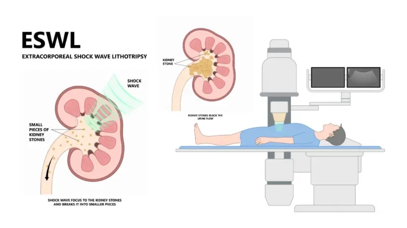
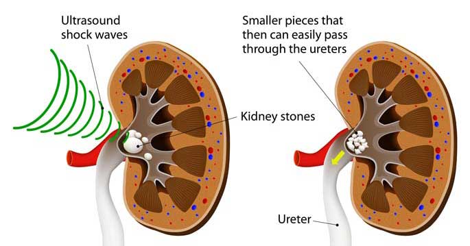
Shock Wave Lithotripsy (SWL) is a non-invasive medical procedure used to break kidney or ureteral stones into smaller pieces using high-energy sound waves (shock waves). These small fragments can then pass naturally through the urinary tract

You may need SWL if

You have kidney stones or ureter stones between 4 mm to 20 mm.
The stone is visible on X-ray or ultrasound.
The stone is in the kidney or upper ureter.
You have pain, bleeding, or infection due to stones.
You want to avoid surgery.
Other treatments (like medications) did not work.

Here is a step-by-step explanation of how Shock Wave Lithotripsy (SWL) is performed

1. Pre-Procedure Preparation
Medical Evaluation – Imaging tests (X-ray, CT scan, or ultrasound) are done to locate the stone and determine its size and position.
Fasting – You may be asked to avoid food and drink for 6–8 hours before the procedure.
Medication Review – Blood thinners or certain medications may be stopped temporarily.
Anesthesia – Light sedation or general anesthesia may be used to reduce pain or movement during the procedure.

2. During the Procedure

Positioning – You lie on a water-filled cushion or a special table, depending on the machine used.
Imaging Guidance – The doctor uses X-ray or ultrasound to target the stone accurately.
Shock Wave Delivery – A lithotripter machine generates high-energy sound waves focused on the stone.
Stone Fragmentation – The shock waves pass through the skin and break the stone into small fragments.
Duration – The procedure typically lasts 30 to 60 minutes.

3. Post-Procedure Care
Observation – You may be monitored for a few hours and discharged the same day.
Symptoms – Mild pain, blood in urine, or bruising at the skin site is common.
Hydration – You are advised to drink plenty of fluids to flush out stone fragments.
Medications – Pain relievers and sometimes antibiotics are prescribed.
Follow-up – Imaging is done after a few weeks to ensure the stone is cleared completely.

You may pass stone fragments over several days to weeks.
Some patients may need repeat SWL sessions if stones are not completely broken.
If SWL fails, other options like URS (ureteroscopy) or PCNL may be considered.

Key Instruments in Ureteroscopy (URS) for Effective Treatment

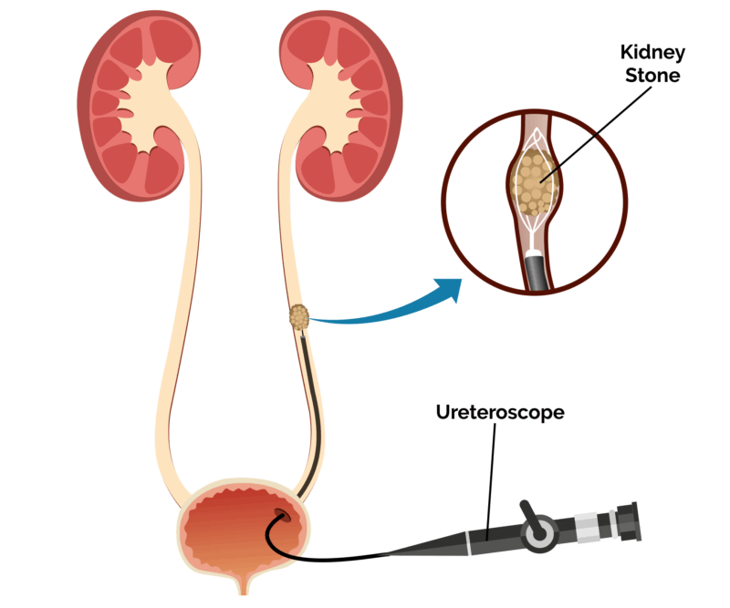
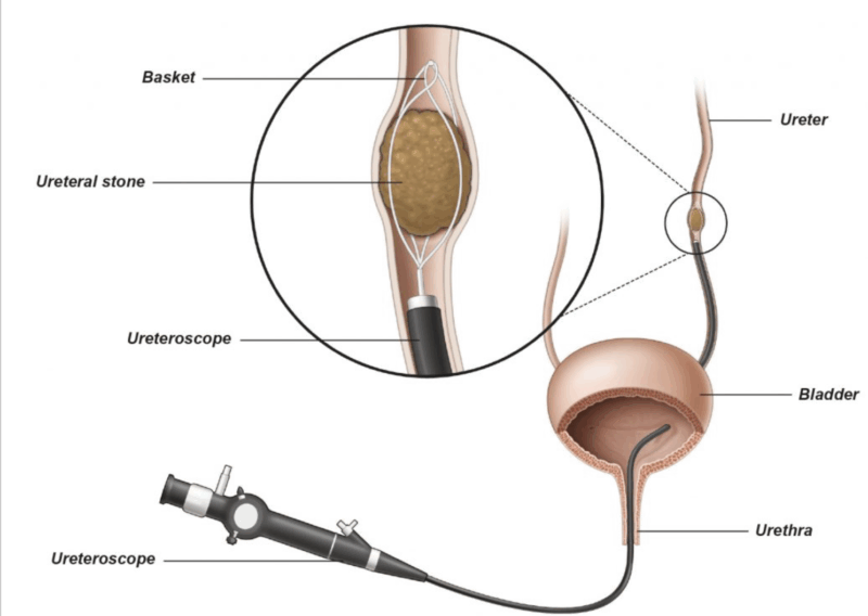
Ureteroscopy (URS) is a minimally invasive urological procedure used to diagnose and treat conditions of the ureter and kidney, such as stones, tumors, or strictures. It involves the insertion of a ureteroscope (a thin, flexible or rigid scope) through the urethra and bladder into the ureter.

Here is a list of instrument names used in Ureteroscopy (URS):

1. Ureteroscope
Types:
Rigid ureteroscope – for lower ureter.
Flexible ureteroscope – for upper ureter and kidney.
Semi-rigid ureteroscope – for mid-ureter and some upper ureter access.
Allows visualization and access to the ureter and renal pelvis.

2. Guidewires
Hydrophilic Guidewire – for easy ureteral navigation.
Zebra Wire / Amplatz Wire – for support and safety.
PTFE Guide Wire – To safely access and guide instruments into the ureter.
Fluoroscopy – confirms stent position.

3. Access Sheaths
Ureteral Access Sheath (UAS) – facilitates repeated passage of instruments, reduces intrarenal pressure, and protects the ureter.
URS Forceps – To grasp and retrieve stones or tissue samples.
Bugbee Electrode – For cauterization and tumor ablation during URS.

4. Irrigation System
Maintains clear vision by flushing the field.
Manual syringe or pressurized bag system.

5. Stone Retrieval Devices
Stone retrieval baskets (Nitinol / Stainless) – To capture and remove stone fragments from the ureter/kidney.
Grasping Forceps – to retrieve stones or tissue.

6. Lithotripsy Devices (for stone fragmentation)
Holmium:YAG Laser – most commonly used.
Ultrasonic lithotripter
Pneumatic lithotripter

7. Dilators and Catheters
Ureteral Dilators or metal dilators – to widen the ureteral orifice.
Balloon Dilators – for precise dilation.

8. Stents and Tubes
Double-J (DJ) Stents – placed post-procedure to prevent obstruction and promote healing.
Ureteral Catheters – for dye injection or drainage.

9. Contrast Media and Monitoring Tools
Ureteroscope withdrawal – under vision to avoid trauma.
C-arm Fluoroscopy Unit – real-time X-ray guidance.
LED Light Source – Provides bright illumination for endoscopic visualization.
Fiber Optic Cable – Transmits light from the LED source to the ureteroscope.

Ureteroscopy (URS) Procedure: Steps, Benefits, and Recovery

Ureteroscopy (URS) allows a urologist to examine and treat problems inside the ureter and kidney. During the procedure, the doctor passes a thin instrument through the urinary tract. Therefore, URS does not require an external surgical cut.

Doctors commonly recommend ureteroscopy for urinary stones. However, they may also perform it to investigate bleeding, narrowing or other urinary tract problems.

When Is Ureteroscopy Needed?

A urologist may recommend ureteroscopy when imaging tests identify a problem in the ureter or kidney. In addition, the procedure can help when symptoms continue without a clear cause.

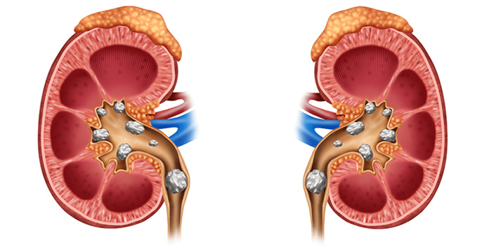
Kidney or Ureteral Stones

Stones can cause pain, bleeding or urinary blockage. Therefore, the doctor may use URS to locate and treat them.

Ureteral Obstruction

Stones, scar tissue or narrowing can block urine flow. In this situation, ureteroscopy helps the doctor find and treat the obstruction.

Suspected Urinary Tract Tumour

A doctor may recommend URS after detecting an unusual growth. During the procedure, the doctor can inspect the area and collect a tissue sample.

Blood in the Urine

Unexplained blood in the urine requires medical evaluation. As a result, the urologist may perform ureteroscopy to examine the upper urinary tract.

Ureteral Stricture

A ureteral stricture narrows the ureter and restricts urine flow. Therefore, the doctor may use URS to examine or treat the narrowed area.

Recurring Urinary Infections

Stones or urinary blockages may contribute to repeated infections. Consequently, the urologist may recommend URS to identify an underlying cause.

Follow-Up After Previous Treatment

Sometimes, doctors perform ureteroscopy after stone treatment or another urinary procedure. This follow-up allows them to check the result and address any remaining problem.

Preparation for Ureteroscopy

Before the procedure, the medical team reviews the patient’s health and medications. The doctor may also request blood tests, urine tests or imaging scans.

In addition, the patient may need to stop eating and drinking for a specific period. However, patients should follow only the instructions provided by their medical team.

Patients should tell the doctor about blood-thinning medicines, allergies and existing medical conditions. Moreover, they should report symptoms of a urinary infection before the procedure.

Ureteroscopy Procedure: Step by Step

1. Administration of Anaesthesia

First, the anaesthesia team provides suitable anaesthesia. General anaesthesia commonly keeps the patient asleep and comfortable during the procedure.

2. Patient Positioning

Next, the medical team positions the patient on the procedure table. The team also cleans and prepares the treatment area.

3. Examination of the Bladder

Then, the urologist passes a cystoscope through the urethra and into the bladder. This instrument helps the doctor locate the opening of the ureter.

4. Insertion of the Ureteroscope

After that, the doctor guides the ureteroscope into the ureter. When necessary, the doctor advances it towards the kidney.

5. Examination and Diagnosis

The ureteroscope provides a clear view of the urinary tract. Therefore, the doctor can identify stones, tumours, strictures or other abnormalities.

6. Treatment During URS

If the doctor finds a stone, specialised instruments can remove it. Alternatively, laser energy can break the stone into smaller fragments.

The doctor may also collect a tissue sample from a suspicious area. In addition, specialised tools can help treat certain strictures or obstructions.

7. Ureteral Stent Placement

Sometimes, the doctor places a ureteral stent to support urine flow. The stent can also help the ureter heal after treatment.

However, not every patient requires a stent. The doctor decides according to the procedure and the patient’s condition.

8. Completion of the Procedure

Finally, the urologist removes the ureteroscope and other instruments. The medical team then moves the patient to a recovery area.

How Long Does Ureteroscopy Take?

The duration depends on the location and complexity of the problem. Many procedures take approximately 30 to 90 minutes. However, complex cases may require more time.

Many patients return home on the same day. Nevertheless, the doctor may recommend observation or an overnight stay in some cases.

Benefits of Ureteroscopy

Ureteroscopy offers several potential benefits for suitable patients:

  • No external surgical incision
  • Direct examination of the ureter and kidney
  • Diagnosis and treatment during one procedure
  • Effective treatment for many urinary stones
  • Access to stones in different urinary tract locations
  • Collection of tissue samples when necessary
  • A relatively short recovery period for many patients

However, treatment results can vary. Stone size, location, anatomy and overall health may affect the outcome.

Recovery After Ureteroscopy

After URS, patients may experience mild burning during urination. They may also notice urinary urgency, discomfort or a small amount of blood in the urine.

Furthermore, a ureteral stent may cause bladder irritation or flank discomfort. These symptoms often improve after the doctor removes the stent.

During recovery, patients should follow their doctor’s instructions carefully. They should also take prescribed medicines and attend follow-up appointments.

Although many people resume light activities within a few days, recovery time varies. Therefore, patients should ask their doctor when they can return to work, exercise or driving.

Possible Risks of Ureteroscopy

Ureteroscopy is a common procedure, but every medical procedure has potential risks. These may include:

  • Urinary tract infection
  • Bleeding
  • Pain or discomfort
  • Temporary difficulty urinating
  • Injury to the ureter
  • Ureteral narrowing
  • Need for an additional procedure
  • Anaesthesia-related complications

Patients should discuss their individual risks with a qualified urologist before treatment.

When Should You Contact Your Doctor?

After the procedure, contact the treating doctor if symptoms become severe or unusual. In particular, seek medical advice for:

  • Fever or chills
  • Severe or increasing pain
  • Heavy bleeding or blood clots
  • Inability to urinate
  • Persistent nausea or vomiting
  • Signs of infection

Conclusion

Ureteroscopy helps urologists diagnose and treat conditions inside the ureter and kidney. Most commonly, doctors use the procedure to manage kidney and ureteral stones.

Moreover, URS provides direct access to the urinary tract without an external incision. Recovery is often relatively quick, although results and recovery times vary.

Patients should consult a qualified urologist for personalised advice. The doctor can explain the expected benefits, possible risks and available treatment options.

This content provides general educational information and does not replace professional medical advice, diagnosis or treatment.

Instruments Used in Percutaneous Nephrolithotomy(PCNL) Surgery

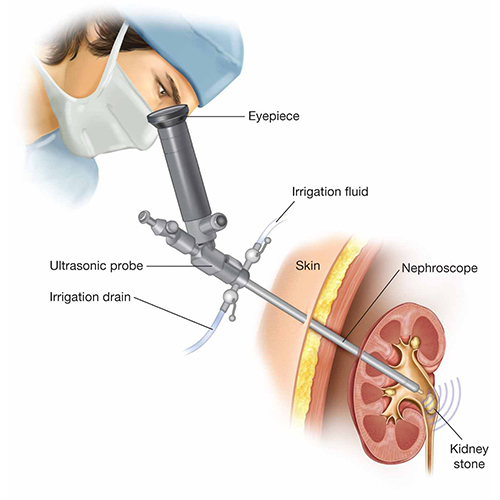
Percutaneous Nephrolithotomy (PCNL) surgery — a procedure used to remove large or complex kidney stones — several specialized surgical instruments and equipment are used.

Key Equipment Used in PCNL Surgery:

Fluoroscopy or Ultrasound Machine
For real-time imaging to guide the surgeon during the procedure.

Nephrostomy Needle (e.g., Chiba needle)
Used to puncture the kidney through the skin and access the collecting system.

Guidewire (e.g., Zebra wire, J-tip wire)
A flexible wire placed through the needle to maintain access to the kidney.

Dilators (serial or balloon dilators / Fascial Dilators / Metal or Plastic Serial Dilators)
Used to widen the tract from the skin to the kidney, allowing for instrument passage.

Amplatz Sheath (Working Sheath)
A hollow tube that provides a working channel to the kidney for instruments.

Nephroscope (Rigid or Flexible)
A rigid endoscope designed to visualize and access stones inside the kidney.

Stone Fragmentation Devices (Lithotripters)
Ultrasonic Lithotripter – Uses sound waves to break stones.
Pneumatic Lithotripter – Uses compressed air to hammer and break stones.
Laser Lithotripter (Holmium:YAG laser) – Precise stone fragmentation using laser energy.

Stone Retrieval Forceps or Baskets
Stone Graspers / Forceps – For grabbing and extracting stone fragments.
Stone Retrieval Basket – For collecting and removing stone pieces.

Irrigation System
Provides continuous saline flow to keep the field clear.

Nephrostomy Tube (post-op)
Temporarily placed to drain urine and allow healing after the procedure.

Ureteral Stent (Double J stent)
Keeps ureter open and ensures urine flow during healing.

Suction System
Helps remove stone debris and fluid during the procedure.

PCNL Surgery: Safe Kidney Stone Removal Procedure

Percutaneous nephrolithotomy, or PCNL surgery, removes large or complex kidney stones. During PCNL, a surgeon reaches the kidney through a small opening in the patient’s back or side.

The surgeon then breaks the stones into smaller pieces and removes them. Therefore, PCNL can treat stones that may not respond well to other procedures.

What Is PCNL Surgery?

PCNL is a minimally invasive kidney stone procedure. It gives the surgeon direct access to the kidney through a small surgical track.

First, the surgeon creates a small opening in the back or side. Next, the surgeon places a tube called a sheath into the kidney. The sheath allows instruments to reach and remove the stones.

Compared with open kidney surgery, PCNL usually requires a smaller incision. As a result, many patients experience a shorter recovery period.

When Do Doctors Recommend PCNL?

A urologist may recommend PCNL when a kidney stone is large, complex or difficult to treat. However, the final decision depends on the patient’s condition.

Doctors may consider PCNL for:

  • Kidney stones larger than 2 cm
  • Staghorn stones that fill several parts of the kidney
  • Multiple kidney stones
  • Hard stones that resist other treatments
  • Stones that did not respond to shock wave treatment
  • Stones that cannot be treated effectively with ureteroscopy
  • Stones that cause blockage, infection or kidney problems

In addition, the doctor considers the stone’s location and the patient’s anatomy. Overall health and previous treatments also affect the treatment plan.

Preparation Before PCNL Surgery

Before PCNL, the medical team checks the patient’s health and studies the kidney stones. This evaluation helps the surgeon plan a safe route into the kidney.

Imaging Tests

The doctor may request a CT scan, ultrasound or X-ray. These tests show the number, size and location of the stones.

Moreover, imaging helps the surgeon study the kidney’s internal structure. The surgeon can then select a suitable entry point.

Blood and Urine Tests

Blood tests check kidney function, blood count and blood-clotting ability. Meanwhile, a urine test looks for signs of infection.

If tests detect an infection, the doctor may prescribe antibiotics. The medical team may delay surgery until the infection improves.

Medicine Review

Patients should tell the doctor about all medicines and supplements they take. In particular, they should mention blood-thinning medicines.

The doctor will explain whether the patient needs to pause any medicine. However, patients should never stop prescribed medicine without medical advice.

Fasting and Anaesthesia

The hospital provides instructions about eating and drinking before surgery. Patients should follow these instructions carefully.

An anaesthesia specialist also reviews the patient’s health. General anaesthesia usually keeps the patient asleep and comfortable during PCNL.

PCNL Surgery: Step-by-Step Procedure

1. Anaesthesia and Positioning

First, the anaesthesia team gives general anaesthesia. The surgical team then places the patient in a suitable position.

Surgeons may perform PCNL with the patient lying face down or on the back. Therefore, positioning can vary according to the surgical plan.

2. Locating the Kidney Stone

Next, the surgeon uses ultrasound, fluoroscopy or both to locate the kidney. These imaging methods also help the surgeon choose an entry path.

3. Creating Access to the Kidney

The surgeon passes a thin needle through the skin and into the kidney. Imaging helps guide the needle safely.

After confirming the correct position, the surgeon passes a guidewire through the needle. The guidewire provides a route for the next instruments.

4. Widening the Surgical Track

The surgeon gradually widens the track with special dilators. This step creates enough space for the working sheath.

Although the opening remains small, it provides direct access to the kidney. As a result, the surgeon can treat large stones through the track.

5. Placing the Amplatz Sheath

Next, the surgeon passes a hollow tube called an Amplatz sheath into the widened track. The sheath maintains access to the kidney during surgery.

Furthermore, it allows the surgeon to pass the nephroscope and other instruments. It also helps protect the surrounding tissue during instrument movement.

6. Viewing the Stone

The surgeon passes a nephroscope through the sheath. A nephroscope contains a viewing system that shows the inside of the kidney.

The surgeon then locates the stones and checks their size and position. Therefore, the treatment can target each visible stone.

7. Breaking the Kidney Stones

The surgeon may use ultrasonic, pneumatic or laser energy to break large stones. The selected method depends on the equipment and stone characteristics.

After fragmentation, the surgeon removes the smaller pieces. Special graspers or suction systems can help with stone removal.

8. Checking the Kidney

The surgeon checks the kidney for remaining fragments. In addition, the team may use imaging to confirm the result.

Sometimes, complete removal may require another procedure. This can happen when stones are numerous or difficult to reach.

9. Placing a Drainage Tube or Stent

The surgeon may place a nephrostomy tube through the surgical track. This tube drains urine and supports healing.

Alternatively, the surgeon may place a ureteral stent inside the urinary tract. However, some suitable patients may not need an external drainage tube.

10. Completing the Procedure

Finally, the surgeon removes the instruments and secures the surgical site. The team then covers the area with a sterile dressing.

Afterwards, the medical team moves the patient to a recovery area. Staff monitor breathing, blood pressure, pain and urine output.

Recovery After PCNL Surgery

Hospital stay and recovery time can vary. Many patients stay in the hospital for one or more days after surgery.

During this time, the medical team checks for bleeding, infection and urinary problems. In addition, staff provide fluids and pain-relief medicine.

Pain Management

Patients may experience soreness around the incision. However, prescribed medicine can help control the discomfort.

Patients should take medicines exactly as their doctor directs. They should also report severe or increasing pain.

Nephrostomy Tube Care

If the surgeon places a nephrostomy tube, the medical team will monitor it. The team removes the tube when drainage and healing are satisfactory.

However, the required time varies between patients. Some people may go home with the tube and receive care instructions.

Activity After Surgery

Patients should begin with gentle activity and avoid heavy lifting. They should also keep the incision clean and dry.

Many people return to normal activities within a few weeks. Nevertheless, recovery depends on the surgery and the patient’s health.

Food and Hydration

The doctor may recommend adequate fluid intake after surgery. Proper hydration may also help reduce the risk of new stones.

In addition, the doctor may suggest dietary changes after reviewing the stone type. Patients should follow personalised medical advice.

Follow-Up After PCNL

The doctor may arrange imaging after surgery. This scan checks for remaining stones and monitors kidney healing.

Furthermore, the doctor may analyse the removed stone fragments. The results can help guide diet, medication and future prevention.

Benefits of PCNL Surgery

PCNL can offer several benefits for suitable patients:

  • Effective treatment for large kidney stones
  • Removal of complex or branching stones
  • Direct access to the kidney
  • Removal of several stones during one operation
  • Smaller incision than open surgery
  • Shorter recovery than traditional open surgery
  • High stone-clearance potential

However, no procedure can guarantee complete stone removal. Results depend on stone size, number, location and kidney anatomy.

Possible Risks of PCNL

PCNL is a common procedure, but it still carries possible risks. These may include:

  • Bleeding
  • Urinary infection or sepsis
  • Injury to the kidney or nearby organs
  • Urine leakage
  • Remaining stone fragments
  • Need for a blood transfusion
  • Need for another procedure
  • Complications related to anaesthesia

The urologist should explain the expected benefits and individual risks before surgery.

When Should You Contact the Doctor?

Patients should seek medical advice if they develop severe or unusual symptoms. Important warning signs include:

  • Fever or chills
  • Severe or increasing pain
  • Heavy bleeding or large blood clots
  • Difficulty passing urine
  • Persistent nausea or vomiting
  • Redness, swelling or discharge around the wound
  • Problems with a nephrostomy tube

Conclusion

PCNL surgery provides an effective treatment option for large or complex kidney stones. It allows the surgeon to reach the kidney through a small opening.

Moreover, PCNL can remove stones that may not respond to other treatments. However, recovery and results vary between patients.

Patients should consult a qualified urologist for personalised advice. The doctor can explain the procedure, benefits, risks and alternative treatments.

This article provides general educational information. It does not replace professional medical advice, diagnosis or treatment.

Instruments Used in Pyeloplasty Surgery

Pyeloplasty surgery—a procedure to correct a blockage or narrowing at the junction between the kidney and the ureter (ureteropelvic junction or UPJ)—the surgical instruments used vary depending on whether the surgery is done through open, laparoscopic, or robot-assisted techniques. Here’s a breakdown of commonly used instruments:

1. General Surgical Instruments (for all approaches):
Scalpel – for making initial incisions.
Scissors (Metzenbaum or Mayo) – for cutting tissue.
Forceps (Debakey, Adson) – for handling tissues.
Needle holders – for suturing.
Hemostats (Mosquito, Kelly) – for clamping blood vessels.
Suction tip (Yankauer or Frazier) – for clearing blood and fluid.

2. Laparoscopic Pyeloplasty Instruments:
Trocars and Cannulas – for inserting instruments through small incisions.
Laparoscope – camera to view the surgical field.
Graspers – to hold and manipulate tissues.
Laparoscopic scissors – to dissect and cut.
Hook or spatula cautery – for precise electrocautery dissection.
Suturing devices or needle drivers – for intracorporeal suturing.
Suction-irrigation device – for cleaning the surgical area.

3. Robot-Assisted (Da Vinci) Pyeloplasty:
Robotic arms and instruments – such as:
Maryland bipolar forceps
Monopolar curved scissors
Prograsp forceps
Needle drivers
3D camera system – high-definition visualization.
Assistant port instruments – similar to laparoscopic tools for suction, clipping, or retraction.

In a pyeloplasty surgery, several consumable products are used depending on the surgical approach (open, laparoscopic, or robotic). These are items that are used up during the procedure and cannot be reused. Here’s a comprehensive list categorized by their purpose:

1. Anesthesia & IV Supplies:
IV cannulas and sets
Syringes and needles
Anesthetic drugs – e.g., Propofol, Fentanyl, Muscle relaxants.
Local anesthetic (e.g., Bupivacaine) – for postoperative pain control.

2. Surgical Field & Access:
Suction canisters and tubing
Laparoscopic ports/trocars (if laparoscopic or robotic approach)
CO₂ insufflation tubing – for creating pneumoperitoneum.

3. Sutures & Hemostasis:
Absorbable sutures (e.g., Vicryl, PDS) – for internal anastomosis.
Non-absorbable sutures (e.g., Prolene) – sometimes used for skin.
Electrocautery pads and cables

4. Drainage & Stenting:
Double-J (DJ) stent – to keep the ureter open during healing.
Foley catheter – for bladder drainage.
Surgical drain (e.g., Jackson-Pratt or Penrose) – sometimes placed to prevent fluid accumulation.

5. Post-op Dressings:
Adhesive dressings (e.g., Tegaderm, Opsite)
Sterile bandages and tape
Antibiotic ointment

Optional (depending on case and approach):
Staples or skin closure strips – sometimes used in open procedures.
Specimen bag – if tissue is removed during the procedure.

Pyeloplasty Surgery: Procedure, Recovery, and Benefits Explained

Pyeloplasty is needed when there is a blockage or narrowing at the junction where the kidney meets the ureter, called the ureteropelvic junction (UPJ). This condition is known as UPJ obstruction. It prevents normal urine flow from the kidney to the bladder and can cause swelling of the kidney (hydronephrosis).

You may need a pyeloplasty if you have symptoms like:
Flank or back pain
Recurrent urinary tract infections
Kidney stones
Blood in the urine
Decreased kidney function

It’s usually done to relieve the blockage, preserve kidney function, and prevent further damage.

1: Anesthesia

You are given general anesthesia to make you sleep and feel no pain during the operation.
2: Making Incisions
The surgeon makes either:
A large incision (open surgery), or
Several small incisions (laparoscopic or robotic surgery) on the side of your abdomen.

3: Finding the Problem
The surgeon carefully finds the blocked or narrowed part at the ureteropelvic junction (UPJ).

4: Removing the Blockage
The damaged or scarred part of the ureter causing the blockage is cut out and removed.

5: Reconnecting the Ureter
The healthy ureter is then stitched (sutured) back to the kidney’s renal pelvis in a way that widens the passage for urine to flow easily.

6: Inserting a Stent
A small internal stent (tube) is placed inside the ureter to keep it open and help it heal properly.

7: Closing the Incisions
The incisions are closed with stitches or surgical glue.

8: Recovery
You are moved to the recovery room and monitored as you wake up.
The stent usually stays in place for 4 to 6 weeks, then is removed with a simple procedure later.

There are mainly three types of pyeloplasty:
Open pyeloplasty (traditional way, bigger cut)
Laparoscopic pyeloplasty (small cuts, quicker recovery)
Robotic-assisted pyeloplasty (done using robotic arms for more precision)
Recovery usually takes a few weeks, and success rates are very high! 🌟

Benefits of pyeloplasty surgery:

Restores Normal Urine Flow
Removes the blockage at the ureteropelvic junction (UPJ), allowing urine to drain freely from the kidney to the bladder.

Relieves Pain
Reduces or eliminates flank, back, or abdominal pain caused by the obstruction.

Protects Kidney Function
Prevents long-term kidney damage, hydronephrosis (kidney swelling), and loss of kidney function.

Decreases Risk of Infections
Reduces the chances of repeated urinary tract infections (UTIs) caused by stagnant urine.

Lowers the Risk of Kidney Stones
Proper urine drainage helps prevent the formation of kidney stones.

High Success Rate
Pyeloplasty has a very high success rate (over 90%) with long-lasting results.

Minimally Invasive Options Available
Laparoscopic and robotic pyeloplasty offer faster recovery, less pain, and smaller scars compared to open surgery.

Essential Surgical Instruments Used in Nephrectomy Procedures

Surgeons use specialised instruments during nephrectomy surgery to remove part or all of a kidney. These instruments help the surgical team cut tissue, control bleeding, move organs and close the surgical area.

The exact instrument set depends on the type of surgery. Open, laparoscopic and robot-assisted nephrectomy procedures require different tools. In addition, partial nephrectomy may require instruments that help preserve healthy kidney tissue.

What Is Nephrectomy Surgery?

Nephrectomy is an operation that removes part or all of a kidney. A surgeon may recommend it for kidney cancer, severe kidney damage, repeated infections or living kidney donation.

During a partial nephrectomy, the surgeon removes only the affected area. During a radical nephrectomy, the surgeon removes the entire kidney and, when necessary, nearby tissue.

Essential Surgical Instruments Used in Nephrectomy

A nephrectomy instrument set usually contains tools for cutting, holding, clamping, retracting and suturing. The hospital selects each instrument according to the surgical plan.

1. Cutting and Dissecting Instruments

Cutting and dissecting instruments help the surgeon open the surgical area and separate tissue around the kidney.

  • Scalpel: The surgeon uses a scalpel with a suitable blade for the initial skin incision.
  • Metzenbaum scissors: These scissors help with fine and careful tissue dissection.
  • Mayo scissors: The surgeon may use these scissors to cut thicker tissue or fascia.
  • Laparoscopic scissors: These long instruments support cutting during minimally invasive surgery.

The surgeon selects the blade and scissors according to the tissue and surgical approach.

2. Grasping and Holding Instruments

Grasping instruments help the surgeon hold or move tissue. Some forceps have a firm grip, while others handle delicate structures more gently.

  • Allis forceps: These forceps firmly hold selected tissue.
  • Babcock forceps: Their rounded jaws can hold delicate tubular structures with less crushing force.
  • DeBakey forceps: Surgeons use these forceps when handling blood vessels and delicate tissue.
  • Laparoscopic graspers: These long instruments help surgeons hold tissue through small access ports.

The surgical team must choose the correct grasper for each structure. This selection helps reduce unnecessary tissue damage.

3. Clamping and Vessel-Control Instruments

Safe control of the renal artery and vein is an important part of nephrectomy. Surgeons use clamps, clips or stapling devices according to the surgical method.

  • Kelly or Crile forceps: These instruments can clamp small blood vessels.
  • Satinsky clamp: This vascular clamp can help control a large blood vessel during open surgery.
  • Bulldog vascular clamps: These small clamps temporarily stop blood flow through selected vessels.
  • Laparoscopic clip applicator: This instrument applies suitable clips to blood vessels or other structures.
  • Vascular stapler: A surgeon may use this device to divide and seal selected vessels.

Instrument choice depends on the vessel, procedure and surgeon’s plan. Only trained surgical professionals should use vascular-control devices.

4. Retractors and Exposure Instruments

Retractors hold tissue away from the surgical area. As a result, the surgeon gains a clearer view of the kidney and nearby structures.

  • Army–Navy retractor: This handheld tool provides shallow or surface-level retraction.
  • Balfour retractor: This self-retaining instrument helps keep an open abdominal incision apart.
  • Kidney retractor: This specialised instrument supports exposure during open kidney surgery.
  • Laparoscopic fan retractor: This tool gently moves organs during minimally invasive procedures.

Open surgery usually requires larger retractors. In contrast, laparoscopic surgery uses long and narrow instruments through access ports.

5. Suturing and Stapling Instruments

Suturing instruments help the surgeon repair tissue and close the incision. Partial nephrectomy may also require careful suturing of the remaining kidney.

  • Mayo-Hegar needle holder: This instrument holds a surgical needle during open suturing.
  • Laparoscopic needle driver: The surgeon uses this long instrument for internal suturing.
  • Endoscopic stapler: This device can cut and seal suitable tissue or vessels.
  • Surgical sutures: Absorbable and non-absorbable sutures support internal repair and wound closure.

The surgeon chooses the suture type according to the tissue and purpose. Therefore, one nephrectomy may require several suture materials.

6. Suction and Irrigation Instruments

Suction instruments remove blood and fluid from the surgical area. Irrigation also helps the team clean the field and maintain visibility.

  • Yankauer suction tip: The team may use this suction instrument during open surgery.
  • Laparoscopic suction and irrigation device: This instrument manages fluid during minimally invasive surgery.
  • Suction tubing and canister: These products collect fluid from the operating area.
  • Sterile saline: The surgical team may use saline for irrigation.

7. Energy and Vessel-Sealing Devices

Energy devices can cut tissue and control bleeding. They may also reduce the need for repeated manual tying.

  • Monopolar electrosurgical device: This device supports cutting and tissue control.
  • Bipolar electrosurgical device: It applies energy between two instrument tips.
  • Ultrasonic surgical device: This system uses mechanical energy to cut and seal selected tissue.
  • Advanced vessel sealer: This device seals suitable blood vessels and tissue bundles.

Each device has specific instructions and limitations. Therefore, trained staff must select suitable settings and accessories.

Instruments for Laparoscopic Nephrectomy

Laparoscopic nephrectomy requires instruments that can pass through small abdominal openings. A camera system displays the surgical area on a monitor.

  • Trocars and cannulas: These devices create access points for the camera and instruments.
  • Laparoscope: The camera provides a magnified view inside the abdomen.
  • CO₂ insufflator: This system creates working space inside the abdomen.
  • Insufflation tubing and filter: These products connect the gas source to the patient access system.
  • Laparoscopic dissectors: These tools help separate tissue around the kidney.
  • Clip applicator: This instrument places clips on selected vessels or structures.
  • Endoscopic stapler: The surgeon may use it for suitable vessels or tissue.
  • Specimen retrieval bag: This bag contains the removed tissue during extraction.

The exact port size and instrument selection can vary. Therefore, the surgical team follows the planned technique and approved device instructions.

Instruments for Robot-Assisted Nephrectomy

Robot-assisted nephrectomy uses a camera and wristed surgical instruments. The surgeon controls these instruments from a console.

Common robotic tools may include:

  • Robotic graspers
  • Robotic scissors
  • Robotic needle drivers
  • Bipolar grasping instruments
  • Clip applicators
  • Suction and irrigation devices

However, robotic instruments depend on the available surgical system. The hospital must follow the manufacturer’s instructions for use.

Disposable Products Used in Nephrectomy Surgery

In addition to reusable instruments, the operating team uses several sterile disposable products. These supplies support infection control, drainage and wound care.

1. Sterile Drapes and Gowns

  • Kidney or abdominal surgical drape set
  • Sterile surgical gowns
  • Sterile gloves
  • Instrument table covers
  • Sterile adhesive drapes, when required

2. Sutures and Ligatures

  • Absorbable sutures
  • Non-absorbable sutures
  • Surgical needles
  • Vessel loops
  • Suture clips, when required

3. Suction and Irrigation Supplies

  • Suction tubing
  • Collection canisters
  • Sterile irrigation fluid
  • Disposable suction tips
  • Laparoscopic suction and irrigation probes

4. Laparoscopic Supplies

  • Disposable trocars and cannulas
  • Clip cartridges or single-use clip applicators
  • Endoscopic stapler cartridges
  • CO₂ tubing and filters
  • Specimen retrieval bags
  • Camera covers

5. Catheters and Drainage Products

  • Foley catheter: This catheter drains urine and allows the team to monitor urine output.
  • Ureteral catheter or stent: The surgeon may use one when the procedure requires urinary drainage or identification.
  • Surgical drain: A drain may remove fluid from the surgical area.
  • Drain collection bag and tubing: These products collect fluid from the drain.

6. Additional Access Products

Some patients may require additional monitoring or vascular access. However, these products do not form part of every standard nephrectomy set.

  • Central venous catheter
  • Femoral catheter
  • Guidewire
  • Introducer needle
  • Haemodialysis catheter for patients who specifically require dialysis access

The medical team selects these products only when the patient’s condition requires them.

Choosing the Correct Nephrectomy Instruments

No single instrument list suits every nephrectomy. The surgeon considers the surgical approach, tumour size, kidney anatomy and patient’s health.

In addition, the hospital must check device compatibility and sterility. Staff should inspect reusable instruments before surgery and use disposable items within their expiry dates.

The surgical team must also keep backup instruments available. This preparation supports a safe response if the surgical plan changes.

Conclusion

Nephrectomy surgery requires instruments for cutting, grasping, clamping, retracting, suturing and fluid control. Laparoscopic and robot-assisted procedures also require specialised access and camera systems.

Furthermore, sterile disposable products support drainage, wound care and infection control. However, the exact selection varies for each patient and procedure.

Only trained healthcare professionals should select and use these devices. They must follow hospital protocols and each product’s approved instructions.

This article provides general educational information. It does not replace clinical training, hospital protocols or professional medical guidance.