Pulmonary Vein Isolation Procedure for Heart Rhythm

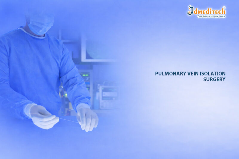
The Pulmonary Vein Isolation (PVI) procedure is a breakthrough in modern cardiac care. It is widely used to treat atrial fibrillation (AF), which is one of the most common heart rhythm disorders.

Atrial fibrillation can lead to serious complications, including stroke and heart failure. Therefore, early and effective treatment is essential. The Pulmonary Vein Isolation (PVI) procedure helps restore a normal heart rhythm by targeting the source of abnormal electrical signals.

What is the Pulmonary Vein Isolation (PVI) Procedure?

The Pulmonary Vein Isolation (PVI) procedure is a minimally invasive catheter-based technique. It focuses on isolating abnormal electrical signals originating from the pulmonary veins.

These veins connect the lungs to the left atrium of the heart. In patients with atrial fibrillation, they often trigger irregular electrical activity.

During the Pulmonary Vein Isolation (PVI) procedure, doctors use radiofrequency or cryoablation energy to create scar tissue. As a result, this scar tissue blocks abnormal signals and restores a stable heart rhythm.

Why is the Pulmonary Vein Isolation (PVI) Procedure Needed?

The Pulmonary Vein Isolation (PVI) procedure is recommended for patients who:

  • Have persistent or paroxysmal atrial fibrillation
  • Do not respond well to medications
  • Experience severe symptoms such as palpitations or fatigue
  • Are at increased risk of stroke

Moreover, this procedure is often preferred because it offers long-term relief compared to drug therapy.

How the Pulmonary Vein Isolation (PVI) Procedure Works

The Pulmonary Vein Isolation (PVI) procedure involves several precise steps:

1. Catheter Insertion

First, a thin catheter is inserted through a vein, usually in the groin. It is then guided to the heart.

2. Mapping the Heart

Next, advanced imaging systems map the electrical activity of the heart. This step helps identify abnormal pathways.

3. Ablation Process

After that, radiofrequency or cryoenergy is applied. This creates controlled scar tissue around the pulmonary veins.

4. Isolation of Signals

Finally, the abnormal signals are blocked. As a result, the heart can maintain a normal rhythm.

Types of Pulmonary Vein Isolation (PVI) Procedure

There are two main types of the Pulmonary Vein Isolation (PVI) procedure:

Radiofrequency Ablation
  • Uses heat energy
  • Highly precise and widely used
  • Suitable for complex cases
Cryoablation
  • Uses cold energy (freezing)
  • Faster procedure time
  • Lower risk of certain complications

Both methods are effective, and the choice depends on patient condition and physician expertise.

Benefits of the Pulmonary Vein Isolation (PVI) Procedure

The Pulmonary Vein Isolation (PVI) procedure offers several advantages:

  • Restores normal heart rhythm
  • Reduces symptoms of atrial fibrillation
  • Lowers the risk of stroke
  • Minimally invasive with faster recovery
  • Improves overall quality of life

In addition, many patients experience long-term success after the procedure.

Risks and Considerations

Although the Pulmonary Vein Isolation (PVI) procedure is generally safe, some risks may include:

  • Bleeding or infection at the catheter site
  • Blood clots
  • Damage to heart tissue
  • Rare complications like pulmonary vein stenosis

However, with advanced technology and experienced specialists, these risks are minimized.

Recovery After the Pulmonary Vein Isolation (PVI) Procedure

Recovery from the Pulmonary Vein Isolation (PVI) procedure is usually quick.

  • Patients may stay in the hospital for 1–2 days
  • Light activities can resume within a few days
  • Full recovery typically occurs within a week

Furthermore, follow-up care is important to monitor heart rhythm and ensure long-term success.

Future of Pulmonary Vein Isolation (PVI) Procedure

The Pulmonary Vein Isolation (PVI) procedure continues to evolve with technological advancements.

Innovations such as 3D mapping systems, robotic navigation, and improved catheter designs are enhancing precision and safety. As a result, the procedure is becoming more effective and accessible worldwide.

Conclusion

The Pulmonary Vein Isolation (PVI) procedure represents a major advancement in cardiac care. It offers a safe, effective, and minimally invasive solution for treating atrial fibrillation.

By restoring normal heart rhythm and reducing complications, the Pulmonary Vein Isolation (PVI) procedure significantly improves patient outcomes. Therefore, it remains a preferred choice for both patients and cardiologists.


Explore More Product >>

Get Connected:

+91 79909 93062 | +91 63513 72032 | exports@jdmeditech.com

Screw Dilator for Urethral Dilation – Precision & Safety

The Screw Dilator for Urethral Dilation is a precision-engineered urology instrument used for controlled urethral expansion. It plays a vital role in treating urethral strictures. These strictures can restrict urine flow and reduce patient comfort.

Moreover, the Screw Dilator for Urethral Dilation allows gradual expansion instead of forceful dilation. As a result, it improves safety and reduces complications. Therefore, many urologists prefer this advanced tool over traditional dilators.

What is a Screw Dilator for Urethral Dilation?

A Screw Dilator for Urethral Dilation is a specialized instrument with a threaded mechanism. This design enables controlled and step-by-step dilation of the urethra.

In addition, the screw mechanism allows precise adjustment during the procedure. Because of this, clinicians can achieve accurate results with minimal effort.

Key Features
  • Threaded design for gradual dilation
  • Smooth surface for reduced friction
  • High precision control
  • Ergonomic grip for better handling

Thus, the Screw Dilator for Urethral Dilation ensures both safety and efficiency.

Why Gradual Urethral Dilation Matters

Gradual dilation is essential in urology procedures. Sudden expansion can cause injury. However, controlled dilation reduces risks significantly.

For instance, the Screw Dilator for Urethral Dilation ensures uniform expansion. As a result, it protects delicate urethral tissues.

Benefits of Gradual Dilation
  • Reduces tissue trauma
  • Improves patient comfort
  • Ensures consistent results
  • Minimizes procedural risks

Therefore, gradual dilation leads to better clinical outcomes.

Clinical Applications

The Screw Dilator for Urethral Dilation is widely used in modern urology. It supports both therapeutic and diagnostic procedures.

Common Uses
  • Treatment of urethral strictures
  • Pre-procedural dilation
  • Post-surgical maintenance
  • Improved access for instruments

Additionally, doctors prefer the Screw Dilator for Urethral Dilation when precision is critical.

Advantages Over Traditional Dilators

Although traditional dilators are common, advanced tools provide better performance. In comparison, the Screw Dilator for Urethral Dilation offers superior control.

1. Precise Control

The screw mechanism allows fine adjustments. Therefore, surgeons can control dilation accurately.

2. Reduced Trauma

Gradual expansion lowers the risk of injury. As a result, patients recover faster.

3. Better Comfort

Because the process is smooth, patients experience less discomfort.

4. Consistent Results

Uniform dilation ensures reliable outcomes.

Design and Material

The Screw Dilator for Urethral Dilation is manufactured using high-quality materials. These materials ensure durability and safety.

Key Design Elements
  • Medical-grade stainless steel
  • Corrosion-resistant surface
  • Smooth polished finish
  • Sterilizable and reusable

Furthermore, these features improve hygiene and long-term performance.

Procedure Overview

The procedure using a Screw Dilator for Urethral Dilation is simple and controlled. However, trained professionals must perform it.

Steps Involved
  1. Apply lubrication
  2. Insert the dilator gently
  3. Rotate gradually for expansion
  4. Monitor patient response
  5. Remove after desired dilation

As a result, the Screw Dilator for Urethral Dilation ensures a safe and effective procedure.

Safety Considerations

Safety is critical in urology procedures. Therefore, proper handling is essential.

Important Guidelines
  • Always maintain sterility
  • Avoid excessive force
  • Monitor patient comfort
  • Follow clinical protocols

Consequently, proper use of the Screw Dilator for Urethral Dilation ensures optimal results.

Why Choose JDMeditech?

At JDMeditech, we deliver high-quality urology instruments designed for precision and reliability.

Our Advantages
  • Advanced manufacturing standards
  • Consistent product quality
  • Competitive pricing
  • Global supply capability
Conclusion

The Screw Dilator for Urethral Dilation is a reliable solution for controlled urethral expansion. It offers precision, safety, and improved patient comfort.

In conclusion, this instrument helps achieve better outcomes in urological procedures. Therefore, it remains an essential tool for modern urology practice.


Explore More Product >>

Get Connected:

+91 79909 93062 | +91 63513 72032 | exports@jdmeditech.com

Nitinol Guide Wires in Endoscopy – Precision GI Access Tool

In modern gastrointestinal procedures, precision and control are essential for successful outcomes. Among the most critical tools that support these goals are Nitinol Guide Wires in Endoscopy. These advanced devices provide exceptional flexibility, strength, and navigation capabilities.

Moreover, as endoscopic techniques continue to evolve, the demand for high-performance guide wires has increased significantly. Therefore, Nitinol guide wires have become the preferred choice for gastroenterologists worldwide.

What Are Nitinol Guide Wires?

Nitinol guide wires are specialized medical devices made from a unique alloy of nickel and titanium. This material, known as Nitinol, offers remarkable properties such as shape memory and superelasticity.

As a result, these guide wires can easily navigate through complex anatomical pathways while maintaining their original shape. In addition, their flexibility ensures minimal trauma during insertion.

Key Features of Nitinol Guide Wires in Endoscopy
1. Superior Flexibility and Elasticity

Nitinol guide wires can bend significantly without breaking. Consequently, they are ideal for navigating tortuous GI tracts.

2. Shape Memory Effect

Even after deformation, the wire returns to its original shape. Therefore, it ensures consistent performance during procedures.

3. High Torque Control

Physicians can precisely control the wire’s movement. As a result, this improves procedural accuracy.

4. Kink Resistance

Unlike traditional wires, Nitinol guide wires resist kinking. This ensures uninterrupted access and smooth device delivery.

5. Hydrophilic Coating Options

Many Nitinol guide wires come with hydrophilic coatings. Thus, they reduce friction and enhance maneuverability.

Applications in Endoscopic Procedures
1. ERCP (Endoscopic Retrograde Cholangiopancreatography)

Nitinol guide wires are widely used for biliary and pancreatic duct access. They help in accurate cannulation and stent placement.

2. Esophageal Dilation

These wires guide dilation devices safely through strictures. Consequently, they reduce procedural complications.

3. Gastrointestinal Stenting

Nitinol guide wires support precise placement of self-expanding stents in the GI tract.

4. Foreign Body Removal

They assist in guiding retrieval tools during complex endoscopic interventions.

Advantages Over Conventional Guide Wires

Compared to stainless steel guide wires, Nitinol Guide Wires in Endoscopy offer several advantages:

  • Greater flexibility for complex anatomy
  • Reduced risk of perforation
  • Enhanced durability and longevity
  • Improved patient safety
  • Better procedural success rates

Furthermore, these benefits contribute to shorter procedure times and improved patient outcomes.

Why Nitinol Guide Wires Improve GI Access

Efficient GI access is critical in endoscopy. Nitinol guide wires play a vital role by:

  • Providing smooth navigation through narrow pathways
  • Maintaining stability during device exchange
  • Enhancing control for precise interventions

Therefore, they significantly improve both clinical efficiency and safety.

Choosing the Right Nitinol Guide Wire

When selecting a guide wire, consider the following factors:

  • Diameter and length based on procedure type
  • Tip configuration (straight, angled, or J-tip)
  • Coating type (hydrophilic or PTFE)
  • Stiffness level for support vs flexibility

By choosing the correct specifications, clinicians can achieve optimal results.

Safety Considerations

Although Nitinol guide wires are highly reliable, proper handling is essential. For instance:

  • Avoid excessive force during insertion
  • Use fluoroscopic guidance when necessary
  • Ensure compatibility with other devices

Thus, adherence to best practices ensures maximum safety.

Future Trends in Guide Wire Technology

The future of Nitinol Guide Wires in Endoscopy looks promising. Innovations include:

  • Enhanced coating technologies
  • Improved tip designs for atraumatic access
  • Integration with advanced imaging systems

As a result, these advancements will further improve procedural precision.

Conclusion

In conclusion, Nitinol Guide Wires in Endoscopy have revolutionized gastrointestinal procedures by offering unmatched flexibility, durability, and control. Moreover, they enable safe and efficient GI access, which is essential for successful outcomes.

Therefore, investing in high-quality Nitinol guide wires is crucial for modern endoscopy practices.


Explore More Product >>

Get Connected:

+91 79909 93062 | +91 63513 72032 | exports@jdmeditech.com

Nottingham Dilator – Progressive Urethral Dilation Tool

The Nottingham Dilator plays a crucial role in modern urology. It is specifically designed for progressive urethral dilation, ensuring safe and controlled enlargement of the urethral passage. As a result, urologists rely on this instrument to manage urethral strictures effectively.

Moreover, with advancements in medical technology, the Nottingham Dilator offers improved precision, patient comfort, and procedural efficiency. Therefore, it has become an essential tool in both diagnostic and therapeutic urological procedures.

What is a Nottingham Dilator?

A Nottingham Dilator is a specialized medical instrument used to gradually dilate the urethra. Typically, it consists of a set of smooth, tapered rods available in multiple sizes.

In addition, these dilators are designed to allow step-by-step dilation, which minimizes trauma and reduces the risk of complications. Because of this, they are widely used in hospitals, urology clinics, and surgical centers.

Key Features of Nottingham Dilator
1. Graduated Sizing for Precision

Each dilator comes in incremental sizes. Therefore, clinicians can progressively increase the diameter with accuracy.

2. Smooth Surface Finish

The polished surface ensures minimal friction. As a result, it enhances patient comfort during insertion.

3. High-Quality Medical Material

Manufactured from premium-grade materials, the Nottingham Dilator ensures durability and safety.

4. Ergonomic Design

The instrument allows easy handling. Consequently, surgeons can perform procedures with better control.

5. Sterilizable and Reusable

Most Nottingham dilators are autoclavable. Hence, they maintain hygiene standards and reduce long-term costs.

Clinical Applications

The Nottingham Dilator is widely used in various urological conditions. For instance:

  • Urethral Stricture Management – Gradual dilation helps restore normal urine flow
  • Post-Surgical Care – Prevents recurrence of urethral narrowing
  • Diagnostic Procedures – Assists in evaluating urethral patency
  • Pre-Endoscopic Preparation – Facilitates easier instrument insertion

In addition, it is commonly used in both male and female patients, depending on clinical requirements.

Procedure Overview: How Nottingham Dilator Works

The use of a Nottingham Dilator involves a systematic approach:

  1. Initially, the smallest size dilator is selected
  2. Then, it is gently inserted into the urethra
  3. Subsequently, progressively larger sizes are introduced
  4. Finally, the desired dilation is achieved safely

Because the process is gradual, it significantly reduces tissue damage and discomfort.

Benefits of Using Nottingham Dilator
1. Controlled and Gradual Dilation

The stepwise approach ensures safety and precision.

2. Reduced Risk of Trauma

Smooth design minimizes injury to delicate urethral tissues.

3. Improved Patient Comfort

Patients experience less pain compared to aggressive dilation techniques.

4. Cost-Effective Solution

Reusable nature makes it economical for healthcare facilities.

5. Reliable Clinical Outcomes

Consistent results make it a preferred choice among urologists.

Comparison with Other Urethral Dilators

While several dilation tools exist, the Nottingham Dilator stands out due to its:

  • Superior control during progressive dilation
  • Enhanced patient safety features
  • Versatility across different procedures

For example, compared to balloon dilators, Nottingham dilators offer more tactile feedback. Therefore, clinicians can better assess resistance during the procedure.

Maintenance and Sterilization

Proper care is essential to maintain performance. Therefore:

  • Clean thoroughly after each use
  • Sterilize using autoclave methods
  • Store in a dry, sterile environment

By following these steps, the longevity and safety of the Nottingham Dilator are ensured.

Why Choose Nottingham Dilator from JDMeditech?

At JDMeditech, we prioritize quality and reliability. Our Nottingham Dilator range offers:

  • Precision-engineered designs
  • High-quality materials
  • Compliance with medical standards
  • Competitive pricing

Therefore, healthcare providers trust our products for consistent performance and safety.

Conclusion

The Nottingham Dilator is an indispensable tool in urology. It provides safe, controlled, and effective progressive urethral dilation, making it ideal for managing strictures and improving patient outcomes.

In conclusion, choosing a high-quality Nottingham Dilator ensures better procedural success and long-term reliability. Hence, it remains a preferred choice for urologists worldwide.


Explore More Product >>

Get Connected:

+91 79909 93062 | +91 63513 72032 | exports@jdmeditech.com

Heart Valve Repair Surgery – Precision Cardiac Treatment

Heart valve repair surgery is an advanced cardiac procedure that restores the normal function of damaged heart valves. Unlike replacement, this approach preserves the patient’s natural valve. As a result, patients often experience better long-term outcomes.

The heart contains four valves—mitral, aortic, tricuspid, and pulmonary—that regulate blood flow. However, when any of these valves become damaged, blood flow becomes inefficient. Consequently, the heart must work harder, which may lead to serious complications.

Therefore, heart valve repair surgery plays a critical role in modern cardiac care. In addition, it helps reduce the need for artificial implants and long-term medication.

Why Heart Valve Repair Surgery is Needed

Heart valve disorders can significantly affect overall heart function. For instance, when a valve fails to open or close properly, blood flow becomes disrupted. In such cases, doctors often recommend heart valve repair surgery.

Common Conditions That Require Repair
  • Valve Regurgitation : Blood flows backward due to improper closure
  • Valve Stenosis : Narrowed valves restrict blood flow
  • Congenital Defects : Structural abnormalities present from birth
  • Infective Endocarditis : Infection damages valve tissues

Moreover, early diagnosis ensures timely heart valve repair surgery. As a result, patients can avoid severe complications and improve their quality of life.

Types of Heart Valve Repair Techniques

Heart valve repair surgery includes several techniques. However, the choice depends on the severity and type of valve damage.

1. Annuloplasty

In this technique, surgeons place a ring around the valve. Consequently, the valve regains its proper shape and closes effectively.

2. Leaflet Repair

Surgeons reshape or remove damaged parts of the valve leaflets. As a result, blood flow improves significantly.

3. Chordal Repair

Doctors repair or replace the supporting chordae tendineae. Therefore, the valve opens and closes smoothly.

4. Patch Repair

A patch is used to repair holes or tears. In turn, this restores the valve’s structural integrity.

5. Minimally Invasive Repair

This approach uses smaller incisions. As a result, patients experience less pain and faster recovery. Furthermore, it reduces surgical risks.

Procedure of Heart Valve Repair Surgery

Heart valve repair surgery follows a precise and step-by-step process. First, doctors conduct a detailed evaluation. Then, they proceed with the surgery.

Step-by-Step Process
  1. Preoperative Evaluation : Doctors perform imaging tests such as echocardiography
  2. Anesthesia : The patient is placed under general anesthesia
  3. Surgical Access : Surgeons choose open-heart or minimally invasive methods
  4. Valve Repair : The damaged valve is repaired using appropriate techniques
  5. Closure and Monitoring : Finally, the incision is closed, and the patient is monitored

Furthermore, modern surgical tools enhance the accuracy of heart valve repair surgery. As a result, outcomes continue to improve.

Benefits of Heart Valve Repair Surgery

Heart valve repair surgery offers several advantages. Therefore, it is often preferred over valve replacement.

  • Preserves the natural valve
  • Reduces the risk of infection
  • Eliminates the need for lifelong blood thinners (in most cases)
  • Improves heart efficiency
  • Enhances overall quality of life

In addition, patients typically recover faster compared to replacement procedures.

Risks and Complications

Although heart valve repair surgery is generally safe, certain risks may occur. However, these risks are relatively low.

  • Bleeding
  • Infection
  • Blood clots
  • Irregular heartbeat
  • Rare need for reoperation

Nevertheless, advancements in technology have significantly reduced these complications. Therefore, heart valve repair surgery remains a reliable option.

Recovery After Heart Valve Repair Surgery

Recovery is an essential phase of heart valve repair surgery. After the procedure, patients require proper care and monitoring.

Recovery Timeline
  • Hospital Stay : 5 to 7 days
  • Initial Recovery : 2 to 4 weeks
  • Full Recovery : 6 to 12 weeks
Post-Surgery Care

To ensure a smooth recovery, patients should:

  • Attend regular follow-up visits
  • Take medications as prescribed
  • Maintain a heart-healthy diet
  • Gradually resume physical activity

Additionally, cardiac rehabilitation programs provide structured recovery support. As a result, patients regain strength more efficiently.

Who is a Suitable Candidate?

Heart valve repair surgery is suitable for patients with specific conditions. However, doctors carefully evaluate each case.

  • Moderate to severe valve damage
  • Repairable valve structure
  • Good overall health

Therefore, a detailed medical assessment is essential before proceeding with heart valve repair surgery.

Heart Valve Repair vs Replacement

Understanding the difference between these options is important. While both treatments are effective, they serve different needs.

Feature Repair Surgery Replacement Surgery
Valve Type Natural Valve Artificial Valve
Medication Usually Not Required Often Required
Infection Risk Lower Higher
Outcome More Natural Depends on Valve Type

Thus, heart valve repair surgery is often the preferred choice when feasible.

Latest Innovations in Heart Valve Repair Surgery

Technological advancements continue to improve heart valve repair surgery. As a result, procedures are now safer and more efficient.

  • Robotic-assisted surgery for enhanced precision
  • 3D imaging for accurate diagnosis
  • Minimally invasive techniques for faster recovery
  • Transcatheter procedures like MitraClip for high-risk patients

Consequently, these innovations have transformed cardiac care.

Conclusion

Heart valve repair surgery is a precise and effective solution for treating valve disorders. By preserving the natural valve, it improves heart function and reduces long-term risks.

Most importantly, early diagnosis and timely treatment lead to better outcomes. Therefore, patients should consult a cardiac specialist as soon as symptoms appear.


Explore More Product >>

Get Connected:

+91 79909 93062 | +91 63513 72032 | exports@jdmeditech.com


Heart Valve Replacement Surgery – Benefits & Recovery

Heart valve replacement surgery is a life-saving procedure that restores proper blood flow in patients with severe valve disease. When heart valves fail to open or close correctly, the heart struggles to pump blood efficiently. As a result, patients experience fatigue, breathlessness, and serious complications.

However, heart valve replacement surgery provides an effective and reliable solution. Moreover, modern advancements have made heart valve replacement surgery safer and more successful than ever before.

What is Heart Valve Replacement Surgery?

Heart valve replacement surgery is a procedure in which a damaged heart valve is removed and replaced with a new valve. This new valve can be mechanical or biological.

In addition, heart valve replacement surgery helps restore normal circulation and reduces strain on the heart. Doctors usually recommend heart valve replacement surgery when the valve condition becomes severe and symptoms worsen.

Types of Valves Used in Heart Valve Replacement Surgery
1. Mechanical Valves

Mechanical valves are strong and long-lasting. Therefore, they are ideal for younger patients. However, patients need lifelong blood-thinning medication after heart valve replacement surgery.

2. Biological Valves

Biological valves come from animal or human tissue. In contrast, they offer more natural function. Although they may wear out over time, they usually do not require long-term medication after heart valve replacement surgery.

When Do You Need Heart Valve Replacement Surgery?

Doctors recommend heart valve replacement surgery when valve damage becomes severe. For example, common conditions include:

  • Aortic stenosis
  • Mitral regurgitation
  • Congenital valve defects
  • Endocarditis

Furthermore, symptoms that indicate the need for heart valve replacement surgery include:

  • Shortness of breath
  • Chest pain
  • Fatigue
  • Irregular heartbeat

Therefore, early diagnosis plays a crucial role in successful heart valve replacement surgery.

Procedure of Heart Valve Replacement Surgery
Preoperative Preparation

Before heart valve replacement surgery, doctors perform tests such as echocardiography, blood tests, and imaging scans. These tests help plan the procedure effectively.

Surgical Process

During heart valve replacement surgery, the surgeon removes the damaged valve and replaces it with a new one. The procedure may involve open-heart surgery or minimally invasive techniques.

Advanced Techniques

Today, techniques like TAVR (Transcatheter Aortic Valve Replacement) make heart valve replacement surgery less invasive. As a result, patients recover faster and face fewer complications.

Benefits of Heart Valve Replacement Surgery

Heart valve replacement surgery offers several important benefits. For instance:

  • Improves blood flow
  • Reduces symptoms quickly
  • Enhances quality of life
  • Increases survival rate

In addition, heart valve replacement surgery allows patients to return to normal daily activities with improved energy levels.

Risks of Heart Valve Replacement Surgery

Although heart valve replacement surgery is safe, some risks may occur. These include:

  • Infection
  • Bleeding
  • Blood clots
  • Valve-related complications

However, experienced surgeons and advanced technology significantly reduce these risks during heart valve replacement surgery.

Recovery After Heart Valve Replacement Surgery

Recovery after heart valve replacement surgery is gradual but steady.

Hospital Stay

Patients usually stay in the hospital for 5–10 days after heart valve replacement surgery.

Rehabilitation

Cardiac rehabilitation helps patients regain strength. Moreover, it improves heart health after heart valve replacement surgery.

Lifestyle Changes

Patients should follow a healthy lifestyle after heart valve replacement surgery, including:

  • Balanced diet
  • Regular exercise
  • Avoiding smoking

Consequently, these steps ensure long-term success.

Latest Innovations in Heart Valve Replacement Surgery

Modern medicine continues to improve heart valve replacement surgery. For example:

  • Minimally invasive procedures
  • TAVR technology
  • Advanced imaging systems
  • Durable valve materials

Therefore, heart valve replacement surgery has become safer, faster, and more effective.

Conclusion

Heart valve replacement surgery is a proven and life-saving solution for patients with severe heart valve disease. It restores normal heart function and improves quality of life.

In conclusion, heart valve replacement surgery offers excellent outcomes when performed at the right time. With proper care and follow-up, patients can live longer, healthier lives.


Explore More Product >>

Get Connected:

+91 79909 93062 | +91 63513 72032 | exports@jdmeditech.com

Angioplasty (PCI) for Heart Artery Blockages

CABG surgery is one of the most trusted treatments for severe coronary artery disease. It helps restore healthy blood flow to the heart when the coronary arteries become blocked or narrow. Because of that, CABG surgery often reduces chest pain, improves daily function, and lowers the risk of serious cardiac events.

Doctors usually suggest CABG surgery when medicines, lifestyle changes, or angioplasty cannot provide enough relief. In many cases, this procedure offers better long-term outcomes for patients with multiple arterial blockages. Therefore, understanding CABG surgery can help patients and families feel more prepared and confident.

What Is CABG Surgery?

CABG surgery, or coronary artery bypass grafting, is a heart operation that creates a new path for blood to flow around blocked coronary arteries. During CABG surgery, the surgeon uses a healthy blood vessel from another part of the body and attaches it to bypass the blocked section.

These grafts may come from:

  • the leg vein
  • the internal mammary artery in the chest
  • the radial artery in the arm

As a result, the heart muscle receives better oxygen-rich blood. This improved circulation helps reduce symptoms and supports better heart function after CABG surgery.

Why Is CABG Surgery Needed?

Doctors recommend CABG surgery for patients with severe coronary artery disease. This condition develops when fatty deposits build up inside the coronary arteries and restrict blood flow.

Common reasons for CABG surgery include:
  • severe chest pain or angina
  • multiple blocked coronary arteries
  • left main coronary artery disease
  • failed stenting or angioplasty
  • high risk of heart attack
  • reduced heart function due to poor blood flow

In these situations, CABG surgery can become the most reliable treatment option. Moreover, it may improve survival in selected high-risk patients.

How CABG Surgery Works

The main goal of CABG surgery is to bypass blocked arteries and restore blood flow to the heart muscle. Instead of removing the blockage directly, the surgeon creates a new route around it.

For example, when one artery is blocked, a single graft may be enough. However, if several arteries are blocked, the surgeon may perform double, triple, or even quadruple bypass CABG surgery.

Because every patient has a different condition, the exact surgical plan may vary. Still, the basic purpose of CABG surgery remains the same: improve circulation and protect the heart.

Types of CABG Surgery

There are several approaches to CABG surgery, and the doctor chooses the best one based on the patient’s health and heart condition.

1. Traditional On-Pump CABG Surgery

In this method, the surgeon temporarily stops the heart and uses a heart-lung machine during the procedure.

2. Off-Pump CABG Surgery

In off-pump CABG surgery, the heart continues beating while the surgeon performs the bypass. This method may reduce certain complications in selected patients.

3. Minimally Invasive CABG Surgery

This approach uses smaller incisions instead of opening the full chest. Therefore, recovery may be faster for suitable patients.

Step-by-Step CABG Surgery Procedure

Understanding the CABG surgery process can reduce fear and improve patient awareness.

The usual CABG surgery steps are:
  1. The patient receives general anesthesia.
  2. The surgeon makes an incision in the chest.
  3. A healthy blood vessel is collected from the leg, chest, or arm.
  4. The surgeon attaches the graft above and below the blocked artery.
  5. Blood flow is redirected through the new pathway.
  6. The chest is closed carefully after the bypass is completed.

Most CABG surgery procedures take between 3 and 6 hours. However, the total time depends on the number of grafts and the complexity of the case.

Benefits of CABG Surgery

CABG surgery offers many important short-term and long-term benefits. That is why it remains a widely used cardiac procedure across the world.

Major benefits of CABG surgery:
  • improved blood flow to the heart
  • reduced chest pain
  • lower risk of future heart attack
  • better exercise tolerance
  • improved quality of life
  • better long-term results in complex coronary artery disease

In many patients, CABG surgery allows a return to more active and comfortable daily living.

Risks of CABG Surgery

Like any major operation, CABG surgery has some risks. However, experienced cardiac teams manage these risks carefully.

Possible risks of CABG surgery include:
  • infection
  • bleeding
  • irregular heartbeat
  • stroke
  • kidney problems
  • temporary memory or concentration issues

Even so, many patients complete CABG surgery safely and recover well with proper medical care.

Recovery After CABG Surgery

Recovery after CABG surgery takes time, but most patients improve steadily with the right support and follow-up care.

Typical CABG surgery recovery timeline:
  • hospital stay: 5 to 7 days
  • early recovery: 6 to 8 weeks
  • fuller recovery: around 2 to 3 months

After CABG surgery, patients should follow their doctor’s instructions closely. In addition, family support and cardiac rehabilitation can make recovery easier.

Helpful recovery tips after CABG surgery:
  • take medicines on time
  • walk regularly as advised
  • eat a heart-friendly diet
  • avoid smoking
  • get enough rest
  • attend follow-up appointments
  • join a cardiac rehabilitation program

These steps help patients heal better after CABG surgery and reduce future heart problems.

Lifestyle Changes After CABG Surgery

Although CABG surgery improves blood flow, patients must still protect their heart health after the operation. Long-term results depend greatly on healthy daily habits.

Important lifestyle changes after CABG surgery:
  • eat low-fat and low-salt foods
  • control blood pressure and cholesterol
  • exercise regularly
  • maintain a healthy weight
  • manage diabetes carefully
  • reduce stress
  • avoid tobacco completely

Therefore, lifestyle improvement remains essential even after successful CABG surgery.

Conclusion

CABG surgery is a proven and highly effective treatment for severe coronary artery disease. It improves blood flow, relieves symptoms, and supports better heart health in patients with serious arterial blockages.

Because heart disease can become life-threatening, early diagnosis and timely treatment matter a lot. CABG surgery continues to save lives and improve long-term outcomes for patients around the world. With expert care and healthy lifestyle changes, patients can enjoy a stronger recovery and a better quality of life after CABG surgery.

Explore More Product >>

Get Connected:

+91 79909 93062 | +91 63513 72032 | exports@jdmeditech.com

Meatal Dilator for Urethral Meatus Dilation | JDMeditech

The Meatal Dilator is an essential urological instrument designed to gently widen the urethral meatus. This tool plays a critical role in managing urethral narrowing (meatal stenosis) and improving urinary flow.

Moreover, healthcare professionals widely use the Meatal Dilator in both diagnostic and therapeutic procedures. Because of its simple design and reliable performance, it ensures safe and controlled dilation.

What is a Meatal Dilator?

A Meatal Dilator is a specialized medical device used to dilate the external opening of the urethra. Typically, it features a smooth, tapered structure that allows gradual expansion without causing trauma.

In addition, manufacturers produce these dilators in various sizes to accommodate different patient needs. As a result, clinicians can perform progressive dilation with precision and comfort.

Why is Meatal Dilation Necessary?

Meatal dilation becomes necessary when the urethral opening narrows, which can lead to discomfort and urinary complications. Therefore, the Meatal Dilator helps restore normal function efficiently.

Common Indications
  • Meatal stenosis
  • Difficulty in urination
  • Weak urinary stream
  • Post-surgical urethral narrowing
  • Pediatric and adult urological conditions

Furthermore, early intervention with a Meatal Dilator prevents complications and improves patient quality of life.

Key Features of Meatal Dilator

The Meatal Dilator offers several features that enhance safety and effectiveness:

1. Smooth Tapered Design

First, the rounded and polished tip ensures gentle insertion. Consequently, it minimizes discomfort and reduces the risk of injury.

2. High-Quality Material

Most Meatal Dilators are made from medical-grade stainless steel or biocompatible plastic. Therefore, they provide durability and easy sterilization.

3. Multiple Size Options

Additionally, the availability of graduated sizes allows step-by-step dilation. This feature ensures controlled expansion.

4. Ergonomic Handling

The instrument provides a firm grip. Hence, clinicians can perform procedures with better control and accuracy.

Benefits of Using a Meatal Dilator

Using a Meatal Dilator offers numerous advantages in urological care.

  • Ensures safe and gradual dilation
  • Improves urinary flow effectively
  • Reduces patient discomfort during procedures
  • Minimizes the risk of urethral trauma
  • Supports both diagnostic and therapeutic applications

As a result, the Meatal Dilator becomes a reliable tool for urologists worldwide.

Procedure: How Meatal Dilation is Performed

The procedure using a Meatal Dilator is simple yet requires clinical expertise.

  1. First, the clinician applies a sterile lubricant.
  2. Then, they gently insert the smallest size dilator.
  3. After that, they gradually increase the size step by step.
  4. Finally, they achieve the desired dilation safely.

Because of this controlled approach, the Meatal Dilator ensures minimal discomfort and effective results.

Applications in Urology

The Meatal Dilator finds use in various urological settings.

  • Hospitals and surgical centers
  • Urology clinics
  • Pediatric urology procedures
  • Post-operative care

In addition, it supports routine examinations and specialized interventions.

Safety and Sterilization

Proper handling of the Meatal Dilator is essential for patient safety.

  • Always use sterile equipment
  • Follow hospital sterilization protocols
  • Inspect for damage before use
  • Use appropriate lubrication

Therefore, maintaining hygiene ensures optimal performance and reduces infection risks.

Choosing the Right Meatal Dilator

Selecting the appropriate Meatal Dilator is crucial for effective treatment.

  • Consider material quality
  • Choose the correct size range
  • Ensure compliance with medical standards
  • Prefer trusted manufacturers like JDMeditech

As a result, the right choice improves procedural outcomes and patient safety.

Conclusion

The Meatal Dilator is a vital instrument for managing urethral meatus narrowing. Not only does it provide safe and controlled dilation, but it also improves patient comfort and urinary function.

Furthermore, its versatile design and ease of use make it an indispensable tool in modern urology. Therefore, healthcare providers rely on the Meatal Dilator for effective and reliable results.


Explore More Product >>

Get Connected:

+91 79909 93062 | +91 63513 72032 | exports@jdmeditech.com

Endovascular Aneurysm Repair Procedure – Complete Lifesaving Guide

The aneurysm repair procedure is a critical intervention in modern cardiology. It is specifically designed to treat weakened and bulging blood vessels, known as aneurysms. If left untreated, aneurysms can rupture and cause life-threatening complications. Therefore, timely diagnosis and treatment are essential.

In this blog, we will explain the aneurysm repair procedure, its types, benefits, risks, and recovery process. Moreover, we will highlight why this procedure plays a vital role in saving lives.

What is an Aneurysm?

An aneurysm occurs when a section of a blood vessel wall weakens and bulges outward. Most commonly, aneurysms develop in:

  • The aorta (aortic aneurysm)
  • Brain (cerebral aneurysm)
  • Peripheral arteries

However, aortic aneurysms are the most common and dangerous type. As a result, they often require an aneurysm repair procedure to prevent rupture.

What is an Aneurysm Repair Procedure?

The aneurysm repair procedure is a medical treatment used to fix or reinforce a weakened artery. It aims to prevent rupture and restore normal blood flow.

There are two primary types of aneurysm repair:

1. Open Surgical Repair

In this traditional approach, surgeons make an incision to access the affected artery. Then, they replace the weakened section with a synthetic graft.

Key advantages:

  • Durable long-term results
  • Suitable for complex aneurysms
2. Endovascular Aneurysm Repair (EVAR)

This is a minimally invasive aneurysm repair procedure. Doctors insert a catheter through a small incision and place a stent graft inside the artery.

Key advantages:

  • Faster recovery
  • Less pain
  • Reduced hospital stay
When is an Aneurysm Repair Procedure Needed?

Doctors recommend an aneurysm repair procedure when:

  • The aneurysm is large or growing rapidly
  • There is a high risk of rupture
  • The patient experiences symptoms like pain
  • Imaging tests show structural weakness

Therefore, early intervention significantly improves patient outcomes.

Benefits of Aneurysm Repair Procedure

The aneurysm repair procedure offers several life-saving benefits:

  • Prevents rupture and internal bleeding
  • Restores proper blood circulation
  • Reduces risk of sudden death
  • Improves long-term survival

Additionally, minimally invasive methods like EVAR enhance patient comfort and recovery.

Risks and Complications

Although the aneurysm repair procedure is generally safe, some risks may occur:

  • Infection
  • Bleeding
  • Blood clots
  • Graft leakage (in EVAR)
  • Heart or lung complications

However, with advanced technology and skilled surgeons, these risks are minimized.

Recovery After Aneurysm Repair Procedure

Recovery depends on the type of aneurysm repair procedure performed.

After Open Surgery:
  • Hospital stay: 5–10 days
  • Full recovery: 6–12 weeks
After EVAR:
  • Hospital stay: 1–3 days
  • Recovery: 2–4 weeks

Furthermore, patients should follow medical advice, maintain a healthy lifestyle, and attend regular follow-ups.

Technological Advancements in Aneurysm Repair

Modern cardiology has significantly improved the aneurysm repair procedure. Innovations include:

  • Advanced stent grafts
  • Imaging-guided precision
  • Minimally invasive techniques

As a result, outcomes are safer and more effective than ever before.

Why Choose High-Quality Medical Devices?

The success of an aneurysm repair procedure depends on high-quality medical equipment. Reliable devices ensure:

  • Accurate placement
  • Better durability
  • Reduced complications

At JDMeditech, we provide advanced cardiology devices designed to support safe and effective procedures.

Conclusion

The aneurysm repair procedure is a vital, life-saving treatment in cardiology. It not only prevents fatal complications but also improves patient quality of life. While both open surgery and EVAR have their benefits, the choice depends on the patient’s condition.

In conclusion, early diagnosis, timely intervention, and high-quality medical devices are key to successful outcomes.


Explore More Product >>

Get Connected:

+91 79909 93062 | +91 63513 72032 | exports@jdmeditech.com

Savary Gilliard Guide Wire – Reliable Navigation in Endoscopy

In modern gastrointestinal endoscopy, precision and control are critical for successful outcomes. Among the essential tools that enable smooth and safe procedures, the Savary Gilliard Guide Wire stands out as a trusted solution. It plays a vital role in guiding dilators and other devices through complex anatomical pathways with accuracy.

Because of its excellent flexibility and strength, the Savary Gilliard Guide Wire has become a preferred choice for gastroenterologists worldwide. Whether used in esophageal dilation or advanced therapeutic procedures, this guide wire ensures reliable navigation and improved patient safety.

What is a Savary Gilliard Guide Wire?

The Savary Gilliard Guide Wire is a specialized medical device used in endoscopic procedures to guide dilation instruments safely through the gastrointestinal tract. It is commonly used during esophageal dilation to treat strictures and obstructions.

Designed with a flexible yet durable structure, this guide wire allows clinicians to navigate challenging anatomies without causing trauma.

Key Features of Savary Gilliard Guide Wire
1. High Flexibility for Smooth Navigation

The wire offers excellent flexibility, allowing it to pass easily through curved and narrow pathways.

2. Optimal Stiffness for Support

While flexible, the Savary Gilliard Guide Wire maintains sufficient stiffness to support dilators during procedures.

3. Atraumatic Tip Design

Its soft, atraumatic tip minimizes the risk of mucosal injury, ensuring patient safety.

4. Radiopaque Markings

Radiopaque properties enable clear visualization under fluoroscopy, improving procedural accuracy.

5. Durable Construction

Manufactured using high-quality materials, the guide wire resists kinking and ensures long-lasting performance.

Clinical Applications in Endoscopy

The Savary Gilliard Guide Wire is widely used in various gastrointestinal procedures, including:

  • Esophageal dilationfor strictures
  • Treatment of dysphagia
  • Guiding bougies and dilators
  • Support in complex GI interventions

Because of its versatility, it is an indispensable tool in both diagnostic and therapeutic endoscopy.

Advantages of Using Savary Gilliard Guide Wire
Enhanced Procedural Safety

The atraumatic tip and controlled navigation reduce the risk of complications.

Improved Accuracy

Radiopaque visibility ensures precise placement during fluoroscopic guidance.

Time Efficiency

Smooth passage and reliable support help reduce procedure time.

Versatility

Compatible with a wide range of endoscopic devices and dilation systems.

Why Choose Savary Gilliard Guide Wire from JDMeditech?

At JDMeditech, we prioritize quality, precision, and reliability. Our Savary Gilliard Guide Wire is designed to meet international standards and deliver consistent performance in demanding clinical environments.

Our Key Benefits:
  • Strict quality control and sterile packaging
  • Designed for optimal flexibility and strength
  • Competitive pricing without compromising quality
  • Trusted by healthcare professionals
Best Practices for Safe Usage

To maximize the performance of the Savary Gilliard Guide Wire, clinicians should:

  • Ensure proper lubrication before insertion
  • Use fluoroscopic guidance when necessary
  • Handle the wire gently to avoid damage
  • Follow standard sterilization and handling protocols
Conclusion

The Savary Gilliard Guide Wire plays a crucial role in ensuring safe, efficient, and precise navigation during endoscopic procedures. Its combination of flexibility, strength, and atraumatic design makes it an essential tool for gastroenterologists.

As endoscopic techniques continue to evolve, reliable accessories like the Savary Gilliard Guide Wire remain fundamental to achieving successful clinical outcomes.


Explore More Product >>

Get Connected:

+91 79909 93062 | +91 63513 72032 | exports@jdmeditech.com