Top-Quality Dialysis Chairs – Comfort & Support for Every Dialysis Session

Introduction

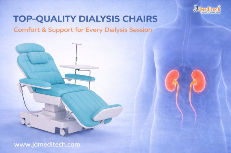
Dialysis is a life-saving procedure for patients with kidney failure. However, long treatment sessions can be physically and mentally exhausting. This is where top-quality dialysis chairs play a crucial role. These specialized chairs are designed to provide maximum comfort, safety, and support throughout every dialysis session.

In this blog, we will explore the importance, features, benefits, and selection criteria for dialysis chairs, helping healthcare providers make informed decisions.

What Are Top-Quality Dialysis Chairs?

Top-quality dialysis chairs are ergonomically designed medical chairs used during dialysis treatments. Patients often remain seated for several hours, so these chairs are built to ensure comfort, proper posture, and easy accessibility for healthcare professionals.

They are commonly used in:

  • Hospitals
  • Dialysis centers
  • Specialty clinics
Why Comfort Matters in Dialysis Sessions

Dialysis sessions typically last 3–5 hours. During this time, patients must remain still. Poor seating can lead to:

  • Back pain and discomfort
  • Poor blood circulation
  • Increased anxiety or restlessness

By using top-quality dialysis chairs, healthcare facilities can significantly enhance patient comfort and overall treatment experience.

Key Features of Top-Quality Dialysis Chairs
1. Ergonomic Design for Patient Comfort

Modern dialysis chairs are designed to support the natural body posture. Adjustable backrests, armrests, and footrests ensure optimal comfort.

2. Electric Adjustment Mechanism

High-end chairs come with motorized controls that allow smooth adjustments. This helps both patients and caregivers.

3. Durable and Hygienic Materials

These chairs are made with easy-to-clean, antimicrobial upholstery, ensuring hygiene and infection control.

4. Safety Features

Top-quality dialysis chairs include:

  • Lockable wheels
  • Stable base structure
  • Emergency positioning
5. Accessibility for Medical Procedures

The chair design allows easy access for healthcare professionals during dialysis procedures.

Benefits of Using Top-Quality Dialysis Chairs
Enhanced Patient Comfort

Patients experience less fatigue during long sessions, improving their overall well-being.

Improved Clinical Efficiency

Adjustable features help medical staff perform procedures more efficiently.

Better Patient Compliance

Comfortable patients are more likely to complete their sessions without interruption.

Increased Safety

Advanced safety mechanisms reduce the risk of accidents.

How to Choose the Best Dialysis Chair

Selecting the right dialysis chair is essential for both patient satisfaction and clinical performance.

Consider Patient Comfort

Look for chairs with cushioning, adjustability, and ergonomic support.

Evaluate Build Quality

Choose durable materials that withstand frequent use.

Check for Advanced Features

Electric controls, memory positions, and safety locks are important.

Ensure Easy Maintenance

Hygienic surfaces and easy cleaning are critical in healthcare settings.

Applications of Dialysis Chairs in Healthcare

Top-quality dialysis chairs are widely used in:

  • Hemodialysis units
  • Oncology treatment centers
  • Blood transfusion units
  • Daycare surgical centers

Their versatility makes them a valuable asset in modern healthcare facilities.

Maintenance Tips for Long-Term Use

To ensure longevity and performance:

  • Clean the chair regularly with approved disinfectants
  • Inspect mechanical and electrical components
  • Perform routine servicing
  • Train staff on proper usage
Why Choose JDMeditech Dialysis Chairs?

At JDMeditech, we provide top-quality dialysis chairs designed to meet international healthcare standards. Our chairs offer:

  • Superior comfort and ergonomic design
  • Advanced safety features
  • Durable and hygienic materials
  • Reliable performance for long-term use
Conclusion

Investing in top-quality dialysis chairs is essential for improving patient comfort, safety, and treatment efficiency. With the right chair, healthcare providers can deliver better care while enhancing operational performance.

If you are looking for reliable and high-performance dialysis chairs, JDMeditech offers solutions tailored to your needs.


Explore More Product >>

Get Connected:

+91 79909 93062 | +91 63513 72032 | exports@jdmeditech.com


Precision Cutting with Bugbee Electrodes – Ultimate Electrosurgical Tool

Precision Cutting with Bugbee Electrodes – The Ultimate Electrosurgical Tool

Introduction

Precision cutting with Bugbee electrodes has transformed modern electrosurgical procedures. Surgeons rely on this advanced tool for accurate tissue cutting and effective coagulation, especially in urology and endoscopy.

Bugbee electrodes provide excellent control, minimal tissue damage, and improved surgical outcomes. As a result, they have become an essential instrument in operating rooms worldwide.

What Are Bugbee Electrodes?

Bugbee electrodes are flexible, monopolar electrosurgical instruments designed for precise cutting and coagulation of soft tissues. They are commonly used in minimally invasive procedures, particularly in urology.

These electrodes consist of a thin wire tip that delivers controlled electrical energy. This energy allows surgeons to cut tissue while simultaneously controlling bleeding.

Why Precision Cutting with Bugbee Electrodes Matters

Precision cutting with Bugbee electrodes ensures clean incisions and reduces unnecessary tissue damage. This improves patient recovery and minimizes complications.

Key Benefits:
  • High precision during surgery
  • Minimal thermal spread
  • Reduced bleeding
  • Enhanced visibility in the surgical field
  • Faster healing time

Because of these advantages, surgeons prefer Bugbee electrodes for delicate procedures.

Applications of Bugbee Electrodes in Medical Procedures

Bugbee electrodes are widely used across various medical specialties. However, they are most popular in urology and endoscopic surgeries.

1. Urological Procedures

Surgeons use Bugbee electrodes during cystoscopy to remove small tumors, coagulate bleeding areas, and treat bladder conditions.

2. Endoscopic Surgeries

These electrodes allow precise cutting in minimally invasive procedures, reducing trauma to surrounding tissues.

3. Tumor Resection

Bugbee electrodes help in removing small growths with accuracy while maintaining hemostasis.

4. Coagulation of Bleeding Vessels

They effectively control bleeding by coagulating blood vessels during surgery.

How Bugbee Electrodes Work

Bugbee electrodes operate using monopolar electrosurgical energy. The electrical current passes through the electrode tip to the targeted tissue.

This process creates heat, which enables:

  • Cutting of tissue
  • Coagulation of blood vessels
  • Controlled surgical precision

The surgeon can adjust the energy level depending on the procedure, ensuring safe and effective outcomes.

Advantages of Using Bugbee Electrodes

Precision cutting with Bugbee electrodes offers several advantages over traditional surgical tools.

Improved Surgical Accuracy

The fine tip allows surgeons to perform delicate procedures with maximum control.

Reduced Tissue Damage

Minimal thermal spread protects surrounding tissues.

Enhanced Efficiency

Surgeries become quicker due to simultaneous cutting and coagulation.

Cost-Effective Solution

Reusable options make Bugbee electrodes a budget-friendly choice for hospitals.

Safety Considerations

Although Bugbee electrodes are highly effective, proper usage is essential for safety.

  • Always use appropriate power settings
  • Ensure proper grounding during monopolar procedures
  • Regularly inspect the electrode for damage
  • Follow sterilization protocols

By following these guidelines, healthcare professionals can ensure safe and successful procedures.

Choosing the Right Bugbee Electrode

Selecting the correct Bugbee electrode depends on the surgical requirement.

Factors to Consider:
  • Electrode length and flexibility
  • Tip size and shape
  • Compatibility with electrosurgical units
  • Reusable or disposable type

Choosing the right instrument enhances both precision and safety.

Future of Electrosurgical Tools

The demand for precision cutting with Bugbee electrodes continues to grow. Innovations in electrosurgical technology are improving efficiency, safety, and ease of use.

Modern designs now focus on better insulation, enhanced flexibility, and improved energy control. These advancements will further improve surgical outcomes in the future.

Conclusion

Precision cutting with Bugbee electrodes has become a cornerstone of modern electrosurgery. Their ability to deliver accurate cuts with minimal tissue damage makes them the ultimate electrosurgical tool.

From urology to endoscopic procedures, Bugbee electrodes provide unmatched performance and reliability. As technology advances, their role in surgery will only continue to expand.


Explore More Product >>

Get Connected:

+91 79909 93062 | +91 63513 72032 | exports@jdmeditech.com


ERCP Pancreatic Baskets: Uses, Types, Procedure & Benefits

What Are ERCP Pancreatic Baskets?

ERCP pancreatic baskets are small wire devices. Doctors use them to catch and remove stones from the pancreatic duct.

They pass through an endoscope during the ERCP procedure. These baskets are strong, flexible, and easy to control.

Key Features:
  • Flexible design for narrow ducts
  • Strong wires for firm grip
  • Smooth opening and closing system
  • Available in different sizes
Why Are ERCP Pancreatic Baskets Used?

Pancreatic stones can block the duct. Because of this, patients may feel severe pain. In some cases, it can lead to pancreatitis.

ERCP pancreatic baskets help to:
  • Remove stones safely
  • Improve enzyme flow
  • Reduce pain quickly
  • Prevent further complications
Types of ERCP Pancreatic Baskets

Different baskets are used for different cases. Therefore, doctors choose them based on the situation.

1. Four-Wire Basket

This is the most common type. It works well for medium-sized stones.

2. Eight-Wire Basket

It offers a stronger grip. So, it is useful for multiple or uneven stones.

3. Helical Basket

This basket moves easily in curved ducts. Hence, it is ideal for complex cases.

4. Nitinol (Memory) Basket

This basket keeps its shape. As a result, it works well even after repeated use.

ERCP Procedure Using Pancreatic Baskets

The procedure is simple and minimally invasive.

Steps:
  1. The doctor inserts an endoscope through the mouth
  2. The pancreatic duct is located
  3. A dye is used to view the duct clearly
  4. The basket is passed into the duct
  5. The stone is captured
  6. The basket is pulled out with the stone

This process is quick and effective.

Benefits of ERCP Pancreatic Baskets

These devices offer many benefits.

1. Minimally Invasive

No major surgery is needed. Therefore, recovery is faster.

2. High Success Rate

Most stones can be removed easily.

3. Better Control

Doctors can handle stones with precision.

4. Fewer Complications

There is less risk compared to open surgery.

5. Cost-Effective

Short hospital stays reduce overall cost.

Applications in Medical Practice

Doctors use ERCP pancreatic baskets in many situations.

  • Pancreatic stone removal
  • Chronic pancreatitis treatment
  • ERCP procedures
  • Endoscopic stone extraction
Risks and Considerations

The procedure is safe. However, some risks may occur.

Possible Risks:
  • Pancreatitis
  • Infection
  • Minor duct injury
  • Rare basket trapping

Even so, trained doctors can manage these risks well.

How to Choose the Right Basket

Choosing the right basket is important.

Doctors consider:

  • Stone size
  • Stone shape
  • Duct structure
  • Material type

Therefore, high-quality baskets give better results.

Future Developments

Medical tools are improving every year. As a result, ERCP baskets are becoming more advanced.

New Improvements:
  • Better flexibility
  • Stronger materials
  • Single-use options
  • Easy handling design

These changes help improve safety and performance.

Conclusion

ERCP Pancreatic Baskets are key tools for removing pancreatic stones. They are safe, effective, and easy to use.

Because they are minimally invasive, patients recover faster. In addition, new designs continue to improve results. Therefore, these baskets remain a top choice in modern ERCP procedures.


Explore More Product >>

Get Connected:

+91 79909 93062 | +91 63513 72032 | exports@jdmeditech.com


Embolectomy Catheter – Uses, Procedure & Benefits Explained

An embolectomy catheter is a medical device used to remove blood clots from blood vessels. These clots can block blood flow and cause serious health problems. Therefore, doctors must remove them quickly.

In many cases, this device helps restore circulation within minutes. As a result, it reduces the risk of stroke, tissue damage, and organ failure.

Today, the embolectomy catheter is widely used in cardiovascular procedures. It offers a fast, safe, and minimally invasive solution.

What is an Embolectomy Catheter?

An embolectomy catheter is a thin and flexible tube. Doctors insert it into a blocked artery or vein.

At the tip, there is a small balloon. First, the catheter reaches the clot. Then, the balloon inflates. After that, the doctor pulls the catheter back. As a result, the clot is removed from the vessel.

Because of this simple method, blood flow returns quickly.

Why is an Embolectomy Catheter Used?

Blood clots can be dangerous. In some cases, they can be life-threatening. Therefore, quick treatment is important.

Doctors use an embolectomy catheter in the following conditions:

  • Acute arterial blockage
  • Deep vein thrombosis (DVT)
  • Pulmonary embolism
  • Stroke-related clots
  • Peripheral artery disease

In addition, this device is useful in emergency situations. It helps save time and improves survival.

Types of Embolectomy Catheters

There are different types of embolectomy catheter devices. Each type serves a specific purpose.

Balloon Embolectomy Catheter

This is the most common type. It uses a balloon to capture and remove the clot.

Fogarty Catheter

Doctors often use this catheter in vascular surgery. It is reliable and easy to use.

Aspiration Catheter

This type removes clots using suction instead of a balloon.

Mechanical Devices

These devices break the clot into small pieces. Then, they remove it from the vessel.

Embolectomy Procedure

The procedure is simple and quick. Moreover, it is usually safe.

Step 1: Preparation

First, the patient receives anesthesia.

Step 2: Insertion

Next, the doctor inserts the embolectomy catheter into the blood vessel.

Step 3: Positioning

Then, imaging helps guide the catheter to the clot.

Step 4: Removal

After that, the balloon inflates and captures the clot. The doctor removes it carefully.

Step 5: Completion

Finally, blood flow returns to normal.

Benefits of Embolectomy Catheter

The embolectomy catheter offers many benefits.

  • Fast clot removal
  • Minimally invasive procedure
  • Quick recovery time
  • Lower risk of complications
  • High success rate

Most importantly, it helps prevent serious damage to tissues and organs.

Risks and Complications

Like any procedure, there are some risks. However, they are usually rare.

  • Bleeding
  • Infection
  • Vessel injury
  • Partial clot removal

Even so, experienced doctors manage these risks effectively.

Role in Cardiovascular Procedures

The embolectomy catheter plays a key role in modern medicine.

Doctors use it in:

  • Emergency vascular treatment
  • Stroke management
  • Cardiac procedures
  • Peripheral artery care

Therefore, it is an important tool in saving lives.

How to Choose the Right Embolectomy Catheter

Choosing the correct device is important. Doctors consider several factors.

  • Size of the vessel
  • Location of the clot
  • Type of blockage
  • Patient condition

As a result, the right choice improves treatment success.

Future of Embolectomy Catheters

Medical technology is improving every year. Therefore, newer devices are more advanced.

For example:

  • Better flexibility
  • Improved precision
  • Smart balloon systems
  • Combined clot removal techniques

In the future, these devices will become even safer and more effective.

Conclusion

The embolectomy catheter is a powerful tool for clot removal. It helps restore blood flow quickly and safely.

Because it is minimally invasive, patients recover faster. In addition, it reduces the risk of serious complications.

Overall, this device plays a vital role in cardiovascular procedures.


Explore More Product >>

Get Connected:

+91 79909 93062 | +91 63513 72032 | exports@jdmeditech.com

Functional Endoscopic Sinus Surgery (FESS)

Functional Endoscopic Sinus Surgery (FESS) is a minimally invasive surgical procedure used to treat chronic sinus problems. It helps restore normal drainage of the sinuses and improves breathing.

Today, FESS is one of the most advanced and commonly performed procedures for patients suffering from chronic sinusitis, nasal blockages, and sinus infections.

In this blog, we will explain everything about Functional Endoscopic Sinus Surgery, including its procedure, benefits, risks, and recovery.

What is Functional Endoscopic Sinus Surgery (FESS)?

Functional Endoscopic Sinus Surgery (FESS) is a modern surgical technique that uses a thin, flexible endoscope inserted through the nose to treat sinus issues.

Unlike traditional surgery, FESS:

  • Does not require external cuts
  • Is less painful
  • Offers faster recovery

This procedure focuses on restoring the natural function of the sinuses rather than removing large amounts of tissue.

Why is FESS Needed?

Doctors recommend Functional Endoscopic Sinus Surgery when medications fail to treat sinus conditions.

Common Conditions Treated:
  • Chronic sinusitis
  • Nasal polyps
  • Deviated nasal septum (in some cases)
  • Sinus infections
  • Blocked sinus drainage pathways
  • Fungal sinusitis

If you experience persistent nasal congestion, facial pain, or breathing difficulty, FESS may be the right solution.

Functional Endoscopic Sinus Surgery Procedure

The FESS procedure is safe and usually performed under general anesthesia.

Step-by-Step Procedure:
1. Preparation

The patient is given anesthesia to ensure comfort.

2. Endoscope Insertion

A small camera (endoscope) is inserted through the nostrils.

3. Identifying Blockages

The surgeon carefully examines sinus passages.

4. Removing Obstructions

Blocked tissues, polyps, or bone are removed to open sinus pathways.

5. Restoring Drainage

Normal airflow and sinus drainage are restored.

The entire procedure typically takes 1 to 3 hours, depending on the condition.

Benefits of Functional Endoscopic Sinus Surgery

FESS offers several advantages compared to traditional sinus surgery.

Key Benefits:
  • Minimally invasive procedure
  • No external scars
  • Faster recovery time
  • Reduced pain and bleeding
  • Improved breathing
  • Long-term relief from sinus symptoms

As a result, patients often experience a significant improvement in quality of life.

Risks and Complications of FESS

Although Functional Endoscopic Sinus Surgery is generally safe, some risks may occur.

Possible Risks:
  • Bleeding
  • Infection
  • Recurrence of sinus problems
  • Damage to surrounding structures (rare)
  • Vision-related complications (very rare)

However, choosing an experienced surgeon greatly reduces these risks.

Recovery After Functional Endoscopic Sinus Surgery

Recovery from FESS is usually quick and smooth.

What to Expect:
  • Mild discomfort for a few days
  • Nasal congestion or slight bleeding
  • Return to normal activities within 1–2 weeks
Post-Surgery Care Tips:
  • Avoid blowing your nose forcefully
  • Use prescribed nasal sprays
  • Attend follow-up appointments
  • Keep your nasal passages clean

Following proper care ensures faster healing and better results.

Who is a Good Candidate for FESS?

You may be a suitable candidate for Functional Endoscopic Sinus Surgery if:

  • You have chronic sinusitis lasting more than 12 weeks
  • Medications are not effective
  • You have nasal polyps or structural blockage
  • You frequently experience sinus infections

Consult an ENT specialist for proper evaluation.

Conclusion

Functional Endoscopic Sinus Surgery (FESS) is a safe, effective, and minimally invasive solution for chronic sinus problems. It helps restore normal sinus function, improves breathing, and enhances overall quality of life.

If you are struggling with long-term sinus issues, consult your doctor to see if FESS is the right option for you.

Explore More Product >>


Get Connected:

+91 79909 93062 | +91 63513 72032 | exports@jdmeditech.com

Pathology: Types, Importance & Role in Disease Diagnosis

Pathology is a vital branch of medical science that focuses on the study of diseases. It helps doctors understand the causes, development, and effects of various illnesses. Without pathology, accurate diagnosis and effective treatment would not be possible.

In modern healthcare, pathology plays a crucial role in detecting diseases early. As a result, it improves patient outcomes and supports life-saving treatments.

What is Pathology?

Pathology is the scientific study of disease through the examination of organs, tissues, blood, and bodily fluids. Pathologists analyze samples to identify abnormalities and determine the exact nature of a disease.

In simple terms, pathology answers key questions like:

  • What disease is present?
  • How severe is it?
  • What is causing it?

Therefore, it acts as the foundation of medical diagnosis.

Types of Pathology

Pathology is divided into several specialized branches. Each type focuses on a different aspect of disease analysis.

1. Clinical Pathology

Clinical pathology involves the analysis of blood, urine, and other body fluids. It includes:

  • Hematology (study of blood)
  • Microbiology (study of infections)
  • Biochemistry (study of chemical processes in the body)
2. Anatomical Pathology

Anatomical pathology focuses on examining tissues and organs. It includes:

  • Histopathology (study of tissues under a microscope)
  • Cytopathology (study of individual cells)
3. Molecular Pathology

Molecular pathology studies diseases at a genetic and molecular level. It is widely used in cancer diagnosis and personalized medicine.

4. Forensic Pathology

Forensic pathology determines the cause of death, especially in cases of accidents or criminal investigations.

Importance of Pathology in Healthcare

Pathology is essential in every stage of healthcare. It not only helps in diagnosis but also guides treatment decisions.

1. Accurate Disease Diagnosis

Pathology provides precise information about diseases. As a result, doctors can make correct diagnoses and avoid unnecessary treatments.

2. Early Detection of Diseases

Many diseases, including cancer, can be detected early through pathology tests. Early detection significantly improves survival rates.

3. Treatment Planning

Pathology reports help doctors choose the best treatment options. For example, cancer treatments often depend on biopsy results.

4. Monitoring Disease Progress

Pathology tests are used to track how a disease is progressing or responding to treatment.

Common Pathology Tests

Pathology laboratories perform a wide range of tests. Some of the most common include:

  • Blood tests (CBC, glucose levels)
  • Urine analysis
  • Biopsy tests
  • Pap smear tests
  • Culture tests for infections

These tests provide essential insights into a patient’s health.

Role of Pathologists

Pathologists are highly trained medical professionals who specialize in diagnosing diseases. Although they usually work behind the scenes, their role is critical.

They:

  • Analyze lab results
  • Interpret biopsy samples
  • Provide diagnostic reports
  • Support clinicians in treatment decisions

Therefore, pathologists are key contributors to patient care.

How Pathology Supports Modern Medicine

Pathology is the backbone of modern medicine. It supports advanced medical practices such as:

  • Cancer diagnosis and staging
  • Genetic testing and personalized medicine
  • Infectious disease detection
  • Organ transplant compatibility testing

As technology advances, pathology continues to evolve with digital pathology and AI-based diagnostics.

Future of Pathology

The future of pathology is rapidly advancing. New technologies are improving accuracy and efficiency.

Some key trends include:

  • Digital pathology systems
  • Artificial intelligence in diagnostics
  • Automated laboratory testing
  • Precision medicine

These innovations will make diagnosis faster and more reliable.

Conclusion

Pathology is a fundamental part of healthcare that ensures accurate diagnosis and effective treatment. From routine blood tests to advanced molecular analysis, pathology provides critical insights into diseases.

As healthcare continues to evolve, pathology will remain at the center of medical innovation and patient care.

Explore More Product >>


Get Connected:

+91 79909 93062 | +91 63513 72032 | exports@jdmeditech.com


Anesthesia: Types, Procedure, Risks, Benefits & Recovery Guide

Anaesthesia is a medical treatment that prevents pain during surgery or medical procedures. It works by blocking nerve signals in the body or inducing a temporary loss of sensation or consciousness.

A trained specialist called an anaesthesiologist administers anaesthesia and monitors the patient throughout the procedure.

Types of Anaesthesia

Different types of anaesthesia are used depending on the procedure, patient condition, and duration of surgery.

1. General Anaesthesia

General anaesthesia makes the patient completely unconscious.

Used for:

  • Major surgeries (heart surgery, brain surgery)
  • Long-duration procedures

Effects:

  • No awareness
  • No pain sensation
  • Temporary loss of reflexes
2. Regional Anaesthesia

Regional anaesthesia blocks pain in a specific part of the body.

Examples:

  • Spinal anaesthesia
  • Epidural anaesthesia

Used for:

  • Childbirth (C-section)
  • Orthopedic surgeries
3. Local Anaesthesia

Local anaesthesia numbs a small area of the body.

Used for:

  • Dental procedures
  • Minor surgeries

Benefits:

  • Patient remains awake
  • Quick recovery
4. Sedation (Conscious Sedation)

Sedation helps patients relax while remaining partially awake.

Used for:

  • Endoscopy
  • Minor procedures
How Does Anaesthesia Work?

Anaesthesia works by interrupting nerve signals in the brain and body. As a result, the patient does not feel pain during the procedure.

There are three main mechanisms:

  • Blocking pain signals
  • Inducing unconsciousness
  • Relaxing muscles
Anaesthesia Procedure: Step-by-Step
1. Pre-Anaesthesia Evaluation

Before surgery, the anaesthesiologist:

  • Reviews medical history
  • Checks allergies
  • Evaluates vital signs
2. Administration of Anaesthesia

Anaesthesia is given through:

  • Injection
  • Inhalation (gas)
  • Intravenous (IV)
3. Monitoring During Surgery

Doctors continuously monitor:

  • Heart rate
  • Blood pressure
  • Oxygen levels
4. Recovery Phase

After surgery, the patient is moved to a recovery room where medical staff monitor vital signs until the effects wear off.

Benefits of Anaesthesia

Anaesthesia offers several advantages:

  • Pain-free procedures
  • Improved surgical precision
  • Reduced anxiety
  • Controlled muscle relaxation
  • Safer complex surgeries
Risks and Side Effects of Anaesthesia

Although anaesthesia is generally safe, some risks may occur.

Common Side Effects
  • Nausea and vomiting
  • Drowsiness
  • Sore throat
  • Dizziness
Rare Risks
  • Allergic reactions
  • Breathing problems
  • Nerve damage (very rare)

However, modern techniques and monitoring systems significantly reduce these risks.

Recovery After Anaesthesia

Recovery depends on the type of anaesthesia used.

General Recovery Tips:
  • Follow doctor’s instructions
  • Avoid driving for 24 hours
  • Stay hydrated
  • Rest properly

Most patients recover quickly, especially after local or regional anesthesia.

Who Administers Anaesthesia?

Anaesthesia is administered by trained professionals such as:

  • Anaesthesiologists
  • Nurse anaesthetists
  • Anaesthesia assistants

They ensure patient safety before, during, and after the procedure.

When is Anaesthesia Required?

Anaesthesia is used in various medical procedures, including:

  • Surgical operations
  • Dental treatments
  • Diagnostic procedures
  • Cosmetic surgeries
Conclusion

Anaesthesia is a cornerstone of modern healthcare, making surgeries safe, painless, and effective. With advancements in medical technology, anaesthesia has become more precise and safer than ever before.

If you are scheduled for a procedure, understanding anaesthesia can help reduce anxiety and prepare you for a smooth experience.

Explore More Product >>


Get Connected:

+91 79909 93062 | +91 63513 72032 | exports@jdmeditech.com


Maintaining Your Surgical Instruments

Surgical instruments are the backbone of any healthcare facility. Therefore, maintaining your surgical instruments is essential for ensuring patient safety, improving surgical outcomes, and extending the lifespan of expensive tools.

Proper care not only reduces the risk of infection but also prevents instrument damage. In this guide, you will learn the best practices, step-by-step cleaning methods, and expert tips to maintain surgical instruments effectively.

Why Maintaining Your Surgical Instruments is Important

First and foremost, well-maintained instruments ensure precision during surgery. On the other hand, poorly maintained tools can lead to complications.

Key Benefits:
  • Enhances patient safety
  • Prevents surgical site infections (SSIs)
  • Increases instrument lifespan
  • Reduces replacement costs
  • Ensures compliance with healthcare standards
Types of Surgical Instruments That Require Maintenance

Different instruments need different care methods. Therefore, understanding categories is crucial.

1. Cutting Instruments
  • Scalpels
  • Scissors
  • Bone cutters
2. Grasping Instruments
  • Forceps
  • Clamps
3. Retracting Instruments
  • Surgical retractors
4. Specialized Instruments
  • Endoscopic tools
  • Microsurgical instruments
Step-by-Step Guide to Maintaining Surgical Instruments
1. Immediate Cleaning After Use

After surgery, instruments should be cleaned immediately. This step prevents blood and tissue from drying.

Tips:

  • Use lukewarm water
  • Avoid saline (it causes corrosion)
  • Wipe visible debris
2. Proper Cleaning Techniques

Cleaning is the most critical step. Therefore, it must be done carefully.

Manual Cleaning
  • Use a soft brush
  • Clean joints and hinges thoroughly
  • Use neutral pH detergents
Ultrasonic Cleaning
  • Removes microscopic debris
  • Ideal for complex instruments
  • Saves time and effort
3. Rinsing and Drying

After cleaning, rinse instruments with distilled water. Then, dry them completely.

Why this matters:
  • Prevents rust formation
  • Avoids water stains
4. Inspection and Function Testing

Next, inspect each instrument carefully.

Check for:
  • Cracks or corrosion
  • Loose joints
  • Sharpness of cutting tools

Replace or repair damaged instruments immediately.

5. Lubrication

Lubrication ensures smooth movement.

Best practices:
  • Use water-based lubricants
  • Apply to hinges and joints
  • Avoid oil-based products
6. Sterilization

Sterilization is essential to eliminate microorganisms.

Common Methods:
  • Autoclaving (steam sterilization)
  • Ethylene oxide (ETO)
  • Plasma sterilization

Always follow manufacturer guidelines for best results.

7. Proper Storage

Finally, store instruments correctly.

Storage tips:
  • Use dry, clean environments
  • Avoid overcrowding
  • Use protective trays
Common Mistakes to Avoid

Even small errors can damage instruments. Therefore, avoid these common mistakes:

  • Delaying cleaning after surgery
  • Using harsh chemicals
  • Skipping inspection
  • Improper sterilization techniques
  • Storing instruments while wet
Best Practices for Long-Term Maintenance

To maintain surgical instruments effectively, consistency is key.

Follow These Practices:
  • Train staff regularly
  • Use instrument tracking systems
  • Follow manufacturer instructions
  • Schedule routine maintenance checks
How Often Should You Maintain Surgical Instruments?

Maintenance should be a continuous process.

  • After every use: Cleaning and sterilization
  • Daily: Inspection and drying
  • Weekly: Deep cleaning and lubrication
  • Monthly: Detailed inspection and servicing
Latest Innovations in Instrument Maintenance

Modern technology has improved maintenance processes significantly.

New Trends:
  • Automated washer-disinfectors
  • Smart tracking systems
  • Anti-corrosion coatings
  • AI-based instrument monitoring

These innovations help reduce human error and improve efficiency.

Conclusion

Maintaining your surgical instruments is not just a routine task—it is a critical responsibility. Proper cleaning, sterilization, and storage ensure safety, efficiency, and cost savings.

By following the steps and best practices in this guide, healthcare facilities can significantly improve surgical outcomes while extending the life of their instruments.

Explore More Product >>


Get Connected:

+91 79909 93062 | +91 63513 72032 | exports@jdmeditech.com

Laryngoscopy: Procedure, Types, Uses, Risks & Recovery

Laryngoscopy is a medical procedure used to examine the larynx (voice box) and surrounding structures. Doctors commonly use it to diagnose throat problems, voice disorders, and breathing issues.

In addition, this procedure helps detect conditions such as infections, tumors, and vocal cord abnormalities. Because of its importance, laryngoscopy plays a key role in ENT (Ear, Nose, and Throat) care.

What is Laryngoscopy?

Laryngoscopy is a diagnostic and sometimes therapeutic procedure. It allows doctors to view the throat using a special instrument called a laryngoscope.

This procedure can be done in different ways. Therefore, the method used depends on the patient’s condition and the purpose of the examination.

Types of Laryngoscopy

There are three main types of laryngoscopy. Each type serves a specific purpose.

1. Indirect Laryngoscopy

This is the simplest method. Doctors use a small mirror or fiber-optic scope to examine the throat.

  • Quick and non-invasive
  • Performed in a clinic
  • No anaesthesia required
2. Direct Laryngoscopy

In this method, a rigid or flexible scope is inserted into the throat.

  • Provides a clear and detailed view
  • Often done under anesthesia
  • Used for diagnosis and minor procedures
3. Fiberoptic (Flexible) Laryngoscopy

This is the most commonly used modern technique.

  • Uses a thin, flexible tube with a camera
  • Inserted through the nose
  • Comfortable and widely used
Why is Laryngoscopy Done?

Doctors recommend laryngoscopy for several reasons. It helps diagnose and treat many conditions.

Common Uses Include:
  • Persistent sore throat
  • Hoarseness or voice changes
  • Difficulty swallowing (dysphagia)
  • Chronic cough
  • Breathing problems
  • Suspected throat cancer
  • Removal of foreign objects

Moreover, it helps evaluate vocal cord movement and detect abnormalities early.

Laryngoscopy Procedure

The procedure varies depending on the type. However, most steps remain similar.

Before the Procedure
  • The doctor reviews your medical history
  • You may be asked to avoid eating for a few hours
  • A local anaesthetic spray may be used
During the Procedure
  • A laryngoscope is gently inserted
  • The doctor examines the throat and vocal cords
  • The process usually takes 5 to 30 minutes
After the Procedure
  • Mild throat discomfort may occur
  • Avoid eating until numbness wears off
  • Most patients resume normal activities quickly
Benefits of Laryngoscopy

Laryngoscopy offers several advantages. As a result, it is widely used in medical practice.

  • Quick and effective diagnosis
  • Minimally invasive procedure
  • Helps detect serious conditions early
  • Can assist in minor treatments
Risks and Complications

.

Possible Risks:
  • Mild throat irritation
  • Gagging sensation
  • Bleeding (rare)
  • Infection (very rare)
  • Reaction to anaesthesia

However, serious complications are uncommon when performed by experienced professionals.

Recovery After Laryngoscopy

Recovery is usually quick and simple.

  • Most patients recover within a few hours
  • Drink warm fluids to soothe the throat
  • Avoid smoking or irritants
  • Follow doctor’s advice for medications

If symptoms such as severe pain or bleeding occur, seek medical attention immediately.

When to See a Doctor

You should consult a doctor if you experience:

  • Persistent hoarseness
  • Difficulty breathing
  • Pain while swallowing
  • Unexplained throat symptoms

Early diagnosis improves treatment outcomes significantly.

Conclusion

Laryngoscopy is an essential procedure for diagnosing and treating throat-related conditions. It is safe, effective, and widely used in modern healthcare.

By understanding the procedure, its types, and benefits, patients can feel more confident and prepared. If you have ongoing throat symptoms, consult an ENT specialist for proper evaluation.

Explore More Product >>


+91 79909 93062 | +91 63513 72032 | exports@jdmeditech.com

Importance of CSSD in Hospitals | Sterilization & Infection Control Guide

The importance of CSSD in hospitals cannot be overstated. CSSD, or Central Sterile Supply Department, plays a critical role in maintaining hygiene, preventing infections, and ensuring safe surgical procedures.

Every hospital relies on sterile instruments. Without proper sterilization, patients face serious risks such as hospital-acquired infections (HAIs). Therefore, CSSD acts as the backbone of infection control and patient safety.

In this blog, we will explore why CSSD is essential, how it functions, and its impact on healthcare quality.

What is CSSD in Hospitals?

CSSD stands for Central Sterile Supply Department. It is a specialized unit responsible for:

  • Cleaning medical instruments
  • Disinfecting equipment
  • Sterilizing surgical tools
  • Storing sterile supplies
  • Distributing sterile items across departments

This department ensures that all reusable medical devices are safe for patient use.

Importance of CSSD in Hospitals
1. Ensures Patient Safety

The primary importance of CSSD in hospitals is patient safety. Proper sterilization removes harmful microorganisms, reducing the risk of infections during medical procedures.

As a result, patients receive safe and reliable treatment.

2. Prevents Hospital-Acquired Infections (HAIs)

Hospital-acquired infections can be life-threatening. However, CSSD helps prevent these infections by:

  • Using advanced sterilization techniques
  • Maintaining strict hygiene protocols
  • Monitoring sterilization processes

Therefore, CSSD significantly reduces infection rates.

3. Maintains Quality Healthcare Standards

Hospitals must follow strict healthcare standards. CSSD ensures compliance with:

  • Infection control guidelines
  • Sterilization protocols
  • Regulatory standards

This improves overall healthcare quality and hospital reputation.

4. Supports Surgical Efficiency

Surgical procedures require sterile instruments at all times. CSSD ensures:

  • Timely availability of instruments
  • Proper packaging and labeling
  • Organized workflow

Because of this, surgeries can proceed without delays.

5. Extends Equipment Lifespan

Proper cleaning and sterilization help maintain medical instruments. CSSD:

  • Prevents damage caused by improper handling
  • Uses appropriate cleaning techniques
  • Ensures correct storage

As a result, hospitals save costs on equipment replacement.

6. Reduces Cross-Contamination

Cross-contamination can occur if instruments are not properly sterilized. CSSD minimizes this risk by:

  • Separating clean and contaminated areas
  • Following strict workflows
  • Using standardized procedures

Thus, it protects both patients and healthcare workers.

Key Functions of CSSD
Cleaning and Decontamination

All used instruments are thoroughly cleaned to remove biological material.

Disinfection

Chemical or thermal methods are used to reduce microorganisms.

Sterilization

High-pressure steam (autoclaving) or other methods ensure complete sterilization.

Storage

Sterile items are stored in controlled environments to maintain sterility.

Distribution

Sterile instruments are supplied to various hospital departments.

CSSD Workflow in Hospitals

The CSSD workflow follows a structured process:

  1. Collection of used instruments
  2. Cleaning and decontamination
  3. Inspection and packaging
  4. Sterilization
  5. Storage
  6. Distribution

This systematic approach ensures efficiency and safety.

Modern Technologies Used in CSSD

Modern CSSD units use advanced technologies such as:

  • Autoclaves
  • Ethylene Oxide (ETO) sterilizers
  • Plasma sterilization
  • Ultrasonic cleaners
  • Washer-disinfectors

These technologies improve accuracy and effectiveness.

Challenges Faced by CSSD

Despite its importance, CSSD faces several challenges:

  • High workload and time pressure
  • Need for skilled staff
  • Strict compliance requirements
  • Equipment maintenance

However, proper management can overcome these challenges.

Best Practices for Effective CSSD Management

To maximize the importance of CSSD in hospitals, follow these best practices:

  • Train staff regularly
  • Maintain strict hygiene protocols
  • Use advanced sterilization equipment
  • Monitor quality through audits
  • Ensure proper documentation

These steps enhance efficiency and safety.

Conclusion

The importance of CSSD in hospitals lies in its ability to ensure patient safety, prevent infections, and maintain high healthcare standards. It is a vital department that supports every medical procedure.

Without CSSD, hospitals cannot deliver safe and effective treatment. Therefore, investing in a well-equipped and properly managed CSSD is essential for any healthcare facility.


Explore More Product >>


Get Connected:

+91 79909 93062 | +91 63513 72032 | exports@jdmeditech.com