Kirschner Wires (K-Wires)

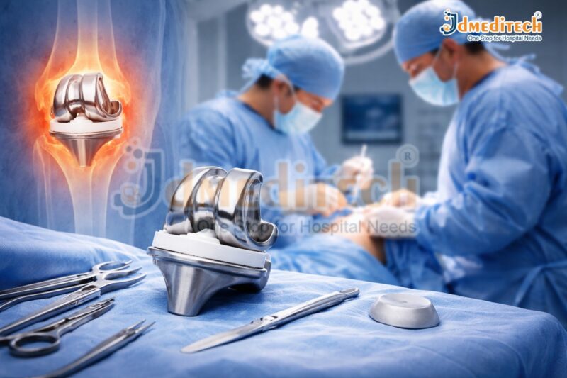
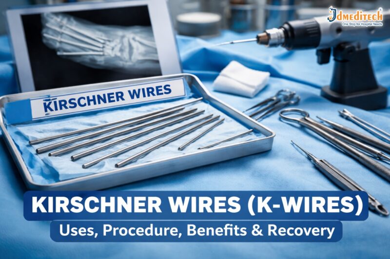
Kirschner wires (K-wires) are thin, smooth stainless steel pins used in orthopedic surgery to stabilize broken bones. These wires are highly effective for treating small bone fractures, pediatric injuries, and temporary fixation during complex procedures.

In this guide, you will learn everything about Kirschner wires, including their uses, procedure, advantages, risks, and recovery process.

What Are Kirschner Wires (K-Wires)?

Kirschner wires, also known as K-wires, are thin metal pins used by orthopedic surgeons to hold bone fragments in proper alignment during healing.

They are:
  • Made from surgical-grade stainless steel
  • Available in different sizes
  • Suitable for various types of fractures
How Are Kirschner Wires Inserted?

Kirschner wires can be inserted using:

  • Percutaneous fixation (through the skin)
  • Open surgical procedures
Uses of Kirschner Wires

Kirschner wires are widely used in orthopedic treatments:

1. Fracture Fixation
  • Wrist fractures (distal radius)
  • Finger and hand fractures
  • Foot and ankle fractures
2. Pediatric Orthopedics
  • Growth plate injuries
  • Bone alignment in children
3. Temporary Stabilization
  • Before placing plates or screws
  • Maintaining alignment during surgery
4. Joint Stabilization
  • Dislocations
  • Ligament injuries

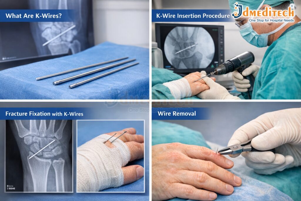
Kirschner Wires Procedure

The Kirschner wires procedure is simple and minimally invasive:

  1. Anesthesia – Local or general anesthesia is given
  2. Insertion – The wire is inserted using X-ray or fluoroscopy guidance
  3. Positioning – The wire stabilizes the fractured bone
  4. Fixation – Ends may remain outside or inside the skin
  5. Immobilization – A cast or splint is applied
Advantages of Kirschner Wires

Using Kirschner wires offers several benefits:

  • Minimally invasive technique
  • Shorter surgery time
  • Cost-effective treatment
  • Effective for small bones
  • Easy removal after healing
Risks and Complications of Kirschner Wires
  • Infection at the insertion site
  • Wire loosening or migration
  • Skin irritation
  • Damage to nearby tissues
  • Delayed healing (rare)

Proper care reduces these complications.

Recovery After Kirschner Wires Fixation

Recovery after Kirschner wires fixation depends on the fracture:

  • Immobilization: 3–6 weeks with cast or splint
  • Follow-ups: Regular X-rays
  • Wire removal: After healing
  • Physiotherapy: Restores movement and strength
Post-Procedure Care for Kirschner Wires

Follow these tips after Kirschner wires surgery:

  • Keep the area clean and dry
  • Avoid touching exposed wires
  • Watch for infection signs
  • Follow your doctor’s advice
Conclusion

Kirschner wires (K-wires) are an effective and minimally invasive solution for fracture stabilization. With proper care and follow-up, Kirschner wires treatment ensures excellent recovery outcomes.

Explore More Product >>

Contact Us
+91 79909 93062 | +91 63513 72032 | exports@jdmeditech.com


Kidney Stones: Definition, Facts, Causes & Prevention Guide

Kidney stones are a common yet painful urological condition affecting millions of people worldwide. Understanding what kidney stones are, how they form, and how to prevent them can help reduce the risk and avoid complications.

What Are Kidney Stones?

Kidney stones, medically known as renal calculi or nephrolithiasis, are hard deposits made of minerals and salts that form inside the kidneys. These stones develop when substances like calcium, oxalate, and uric acid become highly concentrated in urine, leading to crystal formation.

Over time, these crystals stick together and grow into solid stones that may stay in the kidney or travel through the urinary tract.

Key Facts About Kidney Stones

Here are some important and interesting facts about kidney stones:

  • Common Condition: Around 1 in 10 people will develop kidney stones at some point in their life.
  • Highly Painful: Passing a kidney stone can cause severe pain, often described as one of the most intense pains.
  • More Common in Men: Men are more likely to develop kidney stones than women.
  • Recurrence Rate: Nearly 50% of people who have had kidney stones may develop them again within 5 years.
  • Size Varies: Stones can be as small as a grain of sand or as large as a golf ball.
  • Climate Factor: People living in hot climates are at higher risk due to dehydration.
  • Diet Matters: High salt, sugar, and protein intake can increase risk.
Types of Kidney Stones

Kidney stones are classified based on their composition:

1. Calcium Stones
  • Most common type (about 80%)
  • Usually calcium oxalate
  • Linked to diet and metabolic factors
2. Uric Acid Stones
  • Form in people who don’t drink enough fluids
  • Common in high-protein diets
3. Struvite Stones
  • Associated with urinary tract infections (UTIs)
  • Can grow quickly and become large
4. Cystine Stones
  • Rare and genetic in origin
  • Caused by a hereditary disorder
Causes of Kidney Stones

Kidney stones form due to several factors:

  • Dehydration – Not drinking enough water
  • Dietary Habits – High intake of salt, sugar, and animal protein
  • Obesity – Linked to metabolic changes
  • Medical Conditions – Diabetes, gout, and UTIs
  • Family History – Genetic predisposition
  • Certain Medications – Can increase stone formation risk
Symptoms of Kidney Stones

Small stones may not cause symptoms, but larger stones can lead to:

  • Severe pain in the back or side (renal colic)
  • Pain during urination
  • Blood in urine (hematuria)
  • Nausea and vomiting
  • Frequent urge to urinate
  • Cloudy or foul-smelling urine
How Kidney Stones Are Diagnosed

Doctors use several methods to diagnose kidney stones:

  • Ultrasound
  • CT Scan (most accurate)
  • Urine Tests
  • Blood Tests
Prevention Tips for Kidney Stones

Preventing kidney stones is possible with simple lifestyle changes:

Stay Hydrated

Drink at least 2.5–3 liters of water daily.

Follow a Balanced Diet
  • Reduce salt intake
  • Limit animal protein
  • Avoid excessive oxalate-rich foods (spinach, nuts)
Maintain Healthy Weight

Obesity increases the risk of stone formation.

Limit Sugary Drinks

Avoid sodas and high-fructose beverages.

Take Medical Advice

Follow your doctor’s guidance if you have a history of stones.

When to See a Doctor

Seek medical attention if you experience:

  • Severe, persistent pain
  • Blood in urine
  • Fever and chills
  • Difficulty passing urine
Conclusion

Kidney stones are a common but preventable condition. By understanding their definition, causes, and key facts, you can take proactive steps to protect your kidney health. Staying hydrated, maintaining a balanced diet, and seeking timely medical care are essential for prevention and management.

Explore More Product >>

Get Connected:

+91 79909 93062 | +91 63513 72032 | exports@jdmeditech.com


Setting Up a Hospital

Setting Up a Hospital: Complete Guide to Planning, Licensing, and Success

Introduction

Setting up a hospital is a complex but rewarding venture that requires strategic planning, regulatory compliance, financial investment, and a patient-centric approach. With the rising demand for quality healthcare in India and globally, establishing a well-equipped hospital can be both impactful and profitable.

This comprehensive guide covers everything you need to know—from planning and infrastructure to licensing, staffing, and marketing—ensuring your hospital is built on a strong foundation.


1.Planning and Feasibility Study

Before starting, conduct a detailed feasibility study:

  • Location Analysis: Choose an area with high demand and limited competition.
  • Target Audience: Identify whether you will serve urban, semi-urban, or rural populations.
  • Specialty Selection: General hospital or specialized (urology, cardiology, etc.).
  • Market Research: Analyze nearby hospitals, pricing, and services.


2.Business Plan Development

A solid hospital business plan should include:

  • Vision & mission
  • Services offered
  • Capital investment estimation
  • Revenue model
  • Break-even analysis
  • Expansion strategy


3. Legal Requirements and Licensing in India

Setting up a hospital requires multiple approvals:

  • Clinical Establishment Act Registration
  • NABH Accreditation (Recommended for credibility)
  • Biomedical Waste Management Authorization
  • Fire Safety NOC
  • Pollution Control Board Clearance
  • Drug License (if pharmacy included)
  • Local Municipal Permissions

👉 Ensure compliance with state-specific regulations.


4.Hospital Infrastructure Planning

Space Planning

  • OPD (Outpatient Department)
  • IPD (Inpatient wards)
  • ICU & Emergency
  • Operation Theatres (OT)
  • Diagnostic labs
  • Pharmacy

Design Considerations

  • Patient flow efficiency
  • Infection control zones
  • Accessibility (ramps, lifts)
  • Ventilation and lighting


5.Medical Equipment Procurement

Equip your hospital with modern technology:

  • Diagnostic tools (X-ray, Ultrasound, CT scan)
  • Surgical equipment (laparoscopy, endoscopy systems)
  • ICU equipment (ventilators, monitors)
  • Sterilization units

👉 Choose cost-effective, high-quality equipment suppliers to optimize ROI.


6.Hiring Qualified Staff

Your hospital’s success depends on skilled professionals:

  • Doctors & Surgeons
  • Nurses & Technicians
  • Administrative staff
  • Housekeeping & support staff

👉 Focus on training, patient care, and communication skills.


7. IT Infrastructure & Hospital Management System (HMS)

Implement digital systems for efficiency:

  • Electronic Medical Records (EMR)
  • Appointment scheduling
  • Billing and insurance processing
  • Inventory management

👉 A good HMS improves patient experience and operational efficiency.


8. Financial Planning & Investment

Cost Breakdown

  • Land & construction
  • Equipment purchase
  • Licensing & legal fees
  • Staff salaries
  • Marketing budget

Funding Options

  • Bank loans
  • Private investors
  • Government healthcare schemes


9.Hospital Branding and Marketing

Digital Marketing Strategies

  • SEO-optimized website
  • Google My Business listing
  • Social media presence
  • Patient testimonials

Offline Marketing

  • Health camps
  • Local partnerships
  • Referral networks

👉 Focus on trust-building and patient satisfaction.


10. Quality Standards and Accreditation

Achieving NABH accreditation improves:

  • Patient safety
  • Hospital credibility
  • Insurance partnerships


11. Challenges in Setting Up a Hospital
  • High initial investment
  • Regulatory complexities
  • Staff retention
  • Competition

👉 Overcome these with proper planning and expert consultation.

Conclusion

Setting up a hospital is a significant investment that requires careful planning, compliance, and commitment to quality healthcare. By focusing on infrastructure, skilled staff, modern technology, and patient care, you can build a successful and sustainable healthcare institution.


Explore More Product >>


Get Connected:

+91 79909 93062 | +91 63513 72032 | exports@jdmeditech.com


Top Current Trends in Medical Device Innovation (2026 Guide)

Top Current Trends in Medical Device Innovation (2026 Guide)

Introduction

The medical device industry is undergoing rapid transformation driven by technological advancements, patient-centered care, and the demand for minimally invasive treatments. From AI-powered diagnostics to wearable health monitoring devices, innovation is reshaping how healthcare is delivered globally.

In this blog, we explore the top current trends in medical device innovation that are revolutionizing patient care, improving outcomes, and driving the future of healthcare.

1. Artificial Intelligence (AI) & Machine Learning Integration

Artificial Intelligence is at the forefront of medical device innovation. AI-powered systems are enhancing diagnostic accuracy, predicting disease progression, and assisting clinicians in decision-making.

Key Applications:
  • AI-based imaging tools (radiology, pathology)
  • Predictive analytics for early disease detection
  • Smart surgical assistance systems
Benefits:
  • Faster and more accurate diagnoses
  • Reduced human error
  • Improved patient outcomes
2. Wearable Medical Devices & Remote Monitoring

Wearable technology has become a major trend, allowing continuous health monitoring outside hospital settings.

Popular Devices:
  • Smartwatches with ECG and oxygen monitoring
  • Continuous glucose monitors (CGMs)
  • Blood pressure monitoring wearables
Why It Matters:
  • Enables real-time patient tracking
  • Reduces hospital visits
  • Supports telemedicine growth
3. Minimally Invasive & Robotic Surgery Devices

Patients and healthcare providers prefer minimally invasive procedures due to faster recovery and reduced complications.

Innovations Include:
  • Robotic-assisted surgical systems
  • Advanced laparoscopic tools
  • Miniaturized surgical instruments
Advantages:
  • Smaller incisions
  • Less pain and scarring
  • Shorter hospital stays
4. Internet of Medical Things (IoMT)

IoMT refers to interconnected medical devices that communicate through the internet.

Examples:
  • Smart infusion pumps
  • Connected imaging devices
  • Remote patient monitoring systems
Impact:
  • Improved data sharing between providers
  • Real-time monitoring and alerts
  • Enhanced clinical decision-making
5. 3D Printing in Medical Devices

3D printing is transforming how medical devices and implants are designed and manufactured.

Applications:
  • Customized prosthetics
  • Patient-specific implants
  • Surgical planning models
Benefits:
  • Personalized treatment
  • Faster production
  • Cost efficiency
6. Smart Implants & Bioelectronics

Next-generation implants are becoming “smart” with integrated sensors and connectivity.

Examples:
  • Smart orthopedic implants
  • Neurostimulators
  • Cardiac monitoring implants
Key Advantages:
  • Continuous internal monitoring
  • Early detection of complications
  • Improved long-term outcomes
7. Digital Therapeutics (DTx)

Digital therapeutics are software-based medical interventions used to prevent, manage, or treat diseases.

Use Cases:
  • Diabetes management apps
  • Mental health treatment platforms
  • Rehabilitation programs
Why It’s Growing:
  • Non-invasive treatment option
  • Personalized care plans
  • Data-driven therapy
8. Point-of-Care Diagnostics

Rapid diagnostic tools are making testing faster and more accessible.

Examples:
  • Portable ultrasound devices
  • Rapid blood testing kits
  • Handheld diagnostic tools
Benefits:
  • Immediate results
  • Improved emergency care
  • Better rural healthcare access
9. Cybersecurity in Medical Devices

As devices become more connected, cybersecurity is a critical focus area.

Key Concerns:
  • Data breaches
  • Device hacking
  • Patient privacy
Solutions:
  • Advanced encryption
  • Secure device software updates
  • Regulatory compliance
10. Sustainable & Eco-Friendly Medical Devices

Sustainability is becoming a priority in healthcare innovation.

Trends:

  • Reusable devices
  • Eco-friendly materials
  • Reduced medical waste
Impact:
  • Lower environmental footprint
  • Cost savings
  • Compliance with global sustainability goals
Future Outlook

The future of medical device innovation will be driven by personalization, connectivity, and automation. Emerging technologies such as nanotechnology, augmented reality (AR), and blockchain are expected to further revolutionize the industry.


Conclusion

Medical device innovation is transforming healthcare by making it more efficient, accessible, and patient-centered. Staying updated with these trends is essential for healthcare providers, manufacturers, and patients alike.

As technology continues to evolve, the integration of AI, IoMT, and personalized devices will define the next generation of healthcare solutions.

Explore More Product >>

Get Connected:

+91 79909 93062 | +91 63513 72032 | exports@jdmeditech.com


Cystoscopy

Cystoscopy: A Complete Guide to Procedure, Uses, and Recovery

Introduction

Cystoscopy is a common medical procedure used to examine the inside of the bladder and urethra. It helps doctors diagnose, monitor, and treat various urinary tract conditions. With advancements in medical technology, cystoscopy has become safer, quicker, and more comfortable for patients.

In this blog, we will explain everything you need to know about cystoscopy, including why it is performed, how the procedure works, preparation, risks, and recovery.

What is Cystoscopy?

Cystoscopy is a diagnostic and sometimes therapeutic procedure in which a doctor uses a thin tube with a camera and light, called a cystoscope, to view the inside of the urethra and bladder.

There are two types of cystoscopes:

  • Flexible cystoscope – Thin and bendable, usually used for diagnostic purposes.
  • Rigid cystoscope – Slightly thicker and straight, often used for surgical procedures.
Why is Cystoscopy Done?

Cystoscopy is recommended for several urinary tract conditions, including:

1. Diagnosing Problems
  • Blood in urine (hematuria)
  • Frequent urinary tract infections (UTIs)
  • Painful urination
  • Difficulty in passing urine
  • Overactive bladder symptoms
2. Detecting Diseases
  • Bladder cancer
  • Bladder stones
  • Urethral strictures
  • Enlarged prostate (in men)
3. Treatment Purposes
  • Removing small bladder tumors
  • Taking biopsy samples
  • Removing stones
  • Placing stents in ureters
How is Cystoscopy Performed?

The procedure is usually done in a clinic or hospital and typically takes 5–20 minutes for diagnostic purposes.

Step-by-Step Procedure:

  1. The patient lies on the examination table.
  2. The genital area is cleaned and sterilized.
  3. A local anesthetic gel is applied to the urethra.
  4. The cystoscope is gently inserted into the urethra and advanced into the bladder.
  5. Sterile water or saline is used to fill the bladder for better visibility.
  6. The doctor examines the bladder lining and may take samples if needed.

In some cases, spinal or general anesthesia may be used, especially for rigid cystoscopy.

Preparation for Cystoscopy

Proper preparation helps ensure a smooth procedure.

  • You may be asked to give a urine sample before the test.
  • Inform your doctor about medications you are taking.
  • Antibiotics may be prescribed to prevent infection.
  • In most cases, fasting is not required (unless general anesthesia is planned).


What to Expect After the Procedure

After cystoscopy, patients can usually go home the same day.

Common Symptoms:
  • Mild burning sensation while urinating
  • Slight blood in urine
  • Increased frequency of urination

These symptoms usually resolve within 24–48 hours.

Recovery Tips:
  • Drink plenty of water
  • Avoid strenuous activity for 24 hours
  • Take prescribed medications if advised
Risks and Complications
  • Urinary tract infection (UTI)
  • Bleeding
  • Bladder irritation
  • Rarely, injury to the urethra or bladder

Contact your doctor if you experience:

  • Persistent heavy bleeding
  • Fever or chills
  • Severe pain
  • Difficulty urinating
Benefits of Cystoscopy
  • Accurate diagnosis of urinary problems
  • Minimally invasive procedure
  • Quick recovery time
  • Can be used for both diagnosis and treatment
Conclusion

Cystoscopy is a valuable and widely used procedure in urology that allows doctors to directly examine the urinary tract. Whether for diagnosis or treatment, it plays a crucial role in managing bladder and urethral conditions effectively.

If you are experiencing urinary symptoms, consult a urologist to determine whether cystoscopy is the right option for you.


Explore More Product >>


Get Connected:

+91 79909 93062 | +91 63513 72032 | exports@jdmeditech.com


Uroflowmetry


Uroflowmetry: A Simple and Effective Test for Urinary Flow Analysis

Uroflowmetry is a non-invasive diagnostic test used to measure the flow rate of urine. It is commonly used to evaluate urinary tract function and detect problems related to bladder or urethra. This simple yet important test plays a key role in diagnosing various urological conditions.

What is Uroflowmetry?

Uroflowmetry is a test that records the speed and volume of urine flow during urination. The patient urinates into a special device called a uroflowmeter, which measures how quickly urine is released and how much is passed.

This test helps doctors understand how well the bladder and urinary tract are functioning.

How the Test Works

The procedure is simple and painless:

  • The patient is asked to urinate into a uroflowmetry machine
  • The device records urine flow rate and total volume
  • Data is displayed in the form of a graph
  • Doctors analyze the results to identify abnormalities

The test usually takes only a few minutes and does not require any special preparation.

Key Benefits of Uroflowmetry
  • Non-invasive and painless procedure
  • Quick and easy to perform
  • Helps in early detection of urinary problems
  • Provides accurate measurement of urine flow rate
  • Useful for monitoring treatment progress
When is Uroflowmetry Recommended?

Uroflowmetry is commonly recommended for patients experiencing:

  • Weak or slow urine stream
  • Difficulty in urination
  • Frequent urination
  • Incomplete bladder emptying
  • Suspected urinary obstruction or enlarged prostate
Importance in Urology

Uroflowmetry is widely used in diagnosing conditions such as:

  • Benign Prostatic Hyperplasia (BPH)
  • Urethral stricture
  • Bladder dysfunction
  • Urinary tract obstruction

It is often combined with other diagnostic tests for a complete evaluation.

Conclusion

Uroflowmetry is a reliable and efficient test for assessing urinary flow and detecting potential urological issues. Its simplicity, accuracy, and non-invasive nature make it an essential tool in modern urology for improving patient care and diagnosis.


Explore More Product >> 


Get Connected:

+91 79909 93062 | +91 63513 72032 | exports@jdmeditech.com


Intracorporeal Pneumatic Lithotripsy

Intracorporeal Pneumatic Lithotripsy: Advanced Solution for Kidney Stone Treatment

Intracorporeal Pneumatic Lithotripsy is a highly effective and widely used technique for the treatment of urinary stones, including kidney, ureter, and bladder stones. This modern procedure has become an essential part of urological care due to its precision, safety, and cost-effectiveness.

What is Intracorporeal Pneumatic Lithotripsy?

Intracorporeal Pneumatic Lithotripsy is a minimally invasive procedure that uses pneumatic (compressed air) energy to break hard stones into smaller fragments. These fragments can then be removed or naturally passed through the urinary tract.

The procedure is performed using an endoscope, allowing direct visualization of the stone and accurate targeting during fragmentation.

How the Procedure Works

During the procedure:

  • A ureteroscope or endoscope is inserted into the urinary tract
  • A pneumatic lithotripter probe is positioned against the stone
  • Mechanical energy is delivered to fragment the stone
  • Broken pieces are removed or flushed out

This method is especially effective for hard and large stones that are difficult to treat with other techniques.


Key Benefits of Pneumatic Lithotripsy
  • High efficiency in breaking hard stones
  • Minimally invasive with faster recovery time
  • Cost-effective treatment option
  • Safe and reliable procedure
  • Suitable for ureteric and bladder stones
Applications in Modern Urology

Intracorporeal Pneumatic Lithotripsy is commonly used in:

  • Ureteroscopy procedures
  • Treatment of ureteral stones
  • Management of bladder stones
  • Selected kidney stone cases

It remains a preferred choice in many healthcare settings due to its affordability and consistent results.

Growing Demand for Advanced Stone Treatment

With the increasing prevalence of kidney stones, there is a rising demand for advanced and efficient treatment methods. Intracorporeal Pneumatic Lithotripsy continues to play a vital role in modern healthcare by offering a dependable and accessible solution.

Conclusion

Intracorporeal Pneumatic Lithotripsy is a proven and effective method for treating urinary stones. Its minimally invasive approach, combined with high success rates, makes it an important technique in modern urology. As healthcare technology advances, this procedure continues to support better patient outcomes and improved quality of care.


Explore More Product >>

Get Connected:
+91 79909 93062 | +91 63513 72032 | exports@jdmeditech.com


TURP Surgery

TURP Surgery: Effective Treatment for Enlarged Prostate (BPH)

Transurethral Resection of the Prostate (TURP) is a widely performed surgical procedure used to treat urinary problems caused by an enlarged prostate, also known as Benign Prostatic Hyperplasia (BPH). It is considered one of the most effective and reliable treatments in modern urology.

What is TURP Surgery?

TURP surgery is a minimally invasive procedure in which excess prostate tissue is removed to relieve pressure on the urethra. This helps restore normal urine flow and reduces symptoms such as difficulty urinating, weak stream, and frequent urination.

The procedure is performed using a special instrument called a resectoscope, which is inserted through the urethra—eliminating the need for external incisions.

How the Procedure Works

The TURP procedure involves:

  • Insertion of a resectoscope through the urethra
  • Visualization of the prostate using a camera
  • Removal of excess prostate tissue using an electrical loop
  • Clearing the urinary passage to improve urine flow

The removed tissue is flushed out, and a catheter may be placed temporarily to aid recovery.

Key Benefits of TURP Surgery
  • Effective relief from urinary symptoms
  • Minimally invasive (no external cuts)
  • Improved urine flow and bladder control
  • Shorter hospital stay and recovery time
  • Proven and widely used procedure
Who Needs TURP Surgery?

TURP is recommended for patients experiencing:

  • Frequent or urgent need to urinate
  • Difficulty starting urination
  • Weak urine stream
  • Incomplete bladder emptying
  • Complications from enlarged prostate

It is typically advised when medications are not effective.

Risks and Considerations

Like any surgical procedure, TURP carries some risks, including:

  • Temporary urinary discomfort
  • Infection or bleeding
  • Retrograde ejaculation
  • Rare complications such as incontinence

However, it is generally considered safe with a high success rate.

Importance in Modern Healthcare

With the increasing prevalence of prostate enlargement in aging men, TURP surgery continues to be a gold standard treatment. Its effectiveness, safety, and long-term results make it a preferred choice for urologists worldwide.

Conclusion

TURP surgery is a trusted and effective solution for managing symptoms of an enlarged prostate. With its minimally invasive approach and proven outcomes, it significantly improves quality of life for patients experiencing urinary difficulties.

Explore More Product >>

Get Connected:

+91 79909 93062 | +91 63513 72032 | exports@jdmeditech.com

Hospital Medical Equipment

Hospital medical equipment plays a crucial role in delivering safe, accurate, and efficient healthcare services. From diagnosis to treatment and patient monitoring, advanced medical devices are essential for improving patient outcomes and supporting medical professionals.

Importance of Hospital Medical Equipment

Modern healthcare relies on a wide range of equipment such as patient monitors, ventilators, diagnostic machines, and surgical instruments. These tools help ensure accurate diagnosis, effective treatment, and continuous monitoring of patients.

Key Benefits
  • Improved accuracy in diagnosis and treatment
  • Enhanced patient safety and care
  • Increased efficiency for healthcare professionals
  • Support for critical and emergency care
Growing Demand in Healthcare

With the advancement of technology, the demand for high-quality medical equipment is increasing. Hospitals and clinics require reliable and modern devices to meet healthcare standards and provide better services.

Conclusion

Hospital medical equipment is the backbone of modern healthcare systems. Investing in high-quality and reliable equipment is essential for delivering better patient care and improving overall healthcare outcomes.

Explore More Product >>

Get Connected:
+91 79909 93062 | +91 63513 72032 | exports@jdmeditech.com


Knee Replacement Surgery

Knee pain is one of the most common joint problems affecting people of all ages, especially older adults. When the pain becomes severe and limits everyday activities, knee replacement surgery can offer a long-term solution.

This procedure has helped millions of people worldwide regain mobility, reduce pain, and improve their quality of life.

What is Knee Replacement Surgery?

Knee replacement surgery, also known as knee arthroplasty, is a surgical procedure in which damaged parts of the knee joint are replaced with artificial components made of metal and plastic.

These artificial joints are designed to replicate the natural movement of the knee, allowing smoother and pain-free motion.

Who Needs Knee Replacement Surgery?

Doctors may recommend knee joint replacement if you experience:

  • Persistent knee pain even at rest
  • Difficulty walking, climbing stairs, or bending
  • Swelling and stiffness in the knee
  • Limited mobility affecting daily life
  • No relief from medications or physiotherapy

The most common causes include:

  • Osteoarthritis
  • Rheumatoid arthritis
  • Knee injuries or trauma
Types of Knee Replacement Surgery
  1. Total Knee Replacement (TKR)

The entire knee joint is replaced. This is the most commonly performed procedure.

  1. Partial Knee Replacement (PKR)

Only the damaged portion of the knee is replaced, preserving healthy tissue

  1. Revision Knee Replacement

A follow-up surgery done when a previous knee replacement needs correction or replacement.


The Procedure :-

Knee replacement surgery typically involves:

  1. Removing damaged cartilage and bone
  2. Preparing the bone surfaces
  3. Placing the artificial implant
  4. Ensuring proper alignment and movement

The surgery usually takes about 1 to 2 hours and is performed under anesthesia.


Recovery and Rehabilitation

Recovery is a critical part of the success of knee replacement surgery.

  • Patients usually start walking within 1–2 days
  • Physical therapy begins soon after surgery
  • Most people resume daily activities within 6–12 weeks
  • Full recovery may take a few months

Consistent rehabilitation helps restore strength and mobility.

Benefits of Knee Replacement Surgery
  • Significant reduction in pain
  • Improved joint function and flexibility
  • Better quality of life
  • Ability to return to normal daily activities
Risks and Complications

Like any surgical procedure, knee replacement carries some risks:

  • Infection
  • Blood clots
  • Implant wear or loosening over time
  • Temporary stiffness

However, modern techniques have made this surgery safe and highly successful.

Conclusion

Knee replacement surgery is a proven and effective solution for individuals suffering from severe knee pain and mobility issues. With proper care, rehabilitation, and medical guidance, patients can return to an active and pain-free life.

If knee pain is affecting your daily routine, consulting an orthopedic specialist can help determine whether this procedure is the right option for you.

Explore More Product >>

Get Connected:
+91 79909 93062 | +91 63513 72032 | exports@jdmeditech.com