Hybrid Coronary Revascularization – Combined Treatment

Hybrid Coronary Revascularization is an advanced cardiac treatment that combines the strengths of coronary artery bypass surgery and percutaneous coronary intervention (PCI). This innovative approach offers an effective solution for patients with complex coronary artery disease, especially when traditional methods alone may not provide optimal results. By integrating surgical precision with minimally invasive stent placement, Hybrid Coronary Revascularization improves patient outcomes while reducing recovery time.

What is Hybrid Coronary Revascularization?

Hybrid Coronary Revascularization is a treatment strategy that involves performing a minimally invasive coronary artery bypass graft (CABG) on critical arteries, typically the left anterior descending artery (LAD), along with stenting of other blocked coronary arteries using PCI. This combination allows cardiologists and cardiac surgeons to tailor treatment based on the patient’s specific condition.

Unlike conventional open-heart surgery, this approach avoids full sternotomy in many cases. As a result, patients benefit from less trauma, reduced hospital stay, and faster recovery.

Why Hybrid Coronary Revascularization is Needed

Coronary artery disease can affect multiple vessels with varying levels of blockage. In many cases, one artery may require surgical bypass for long-term durability, while others can be effectively treated with stents. Therefore, Hybrid Coronary Revascularization offers a balanced and patient-specific solution.

Moreover, this method is particularly useful for:

  • Patients with multi-vessel coronary artery disease
  • Individuals at high risk for traditional open-heart surgery
  • Patients requiring faster recovery due to lifestyle or medical conditions
  • Cases where minimally invasive options are preferred
Procedure Overview

The Hybrid Coronary Revascularization procedure is typically performed in two stages or sometimes in a single hybrid operating room.

First, a cardiac surgeon performs a minimally invasive bypass graft, usually using the internal mammary artery to bypass the LAD artery. This step ensures long-term patency and excellent blood flow.

Next, an interventional cardiologist performs angioplasty and places stents in other affected coronary arteries. These stents help restore blood flow without the need for extensive surgery.

Because both techniques are used, the procedure maximizes the advantages of each method while minimizing their limitations.

Benefits of Hybrid Coronary Revascularization

Hybrid Coronary Revascularization offers several advantages over traditional treatments. Firstly, it reduces surgical trauma since it avoids full chest opening in many cases. Secondly, patients experience shorter hospital stays and quicker return to normal activities.

In addition, this approach provides:

  • Improved long-term outcomes for critical arteries like the LAD
  • Lower risk of complications compared to full CABG
  • Reduced blood loss and infection risk
  • Enhanced cosmetic results due to smaller incisions
  • Faster recovery and rehabilitation
Risks and Considerations

Although Hybrid Coronary Revascularization is highly effective, it still carries some risks. These may include bleeding, infection, or complications related to stent placement. However, with experienced medical teams and proper patient selection, these risks remain minimal.

Furthermore, not all patients are suitable candidates. Therefore, thorough evaluation by a multidisciplinary heart team is essential before proceeding with this treatment.

Recovery and Post-Procedure Care

Recovery after Hybrid Coronary Revascularization is generally faster compared to traditional open-heart surgery. Most patients can return to daily activities within a few weeks.

However, proper post-procedure care is crucial. Patients are advised to follow prescribed medications, maintain a heart-healthy diet, and engage in regular physical activity. In addition, routine follow-ups with cardiologists help ensure long-term success.

Advanced Technology Supporting Hybrid Procedures

Modern cardiac procedures rely on advanced medical equipment for precision and safety. Devices such as Electrosurgical Units play a vital role in minimally invasive surgeries by enabling precise tissue cutting and coagulation.

Future of Hybrid Coronary Revascularization

Hybrid Coronary Revascularization represents the future of cardiac care. As technology continues to evolve, more hospitals are adopting hybrid operating rooms that allow seamless collaboration between surgeons and cardiologists.

Furthermore, advancements in imaging, robotic surgery, and stent technology will continue to enhance the effectiveness of this approach. Consequently, patients can expect even better outcomes and safer procedures in the coming years.

Conclusion

Hybrid Coronary Revascularization is a breakthrough in the treatment of coronary artery disease. By combining surgical bypass with stent-based intervention, it offers a personalized, less invasive, and highly effective solution.

As a result, patients benefit from improved recovery times, reduced risks, and long-term cardiovascular health. Therefore, this combined approach is becoming an increasingly preferred option in modern cardiac care.

Explore More Product >>

Get Connected:
+91 79909 93062 | +91 63513 72032 | exports@jdmeditech.com 

Penile Clamp – Effective Solution for Male Urinary Incontinence

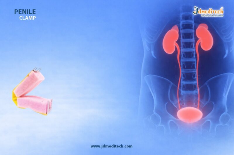
Penile Clamp is a simple yet highly effective device designed to manage male urinary incontinence. This condition, which involves involuntary leakage of urine, can significantly affect a patient’s confidence and quality of life. However, with the use of a Penile Clamp, patients can regain better control over urinary leakage in a non-invasive and convenient way.

Male urinary incontinence may occur due to prostate surgery, neurological disorders, aging, or bladder dysfunction. Therefore, finding a reliable management solution becomes essential. The Penile Clamp offers a practical approach by applying gentle pressure to the urethra, thereby preventing unwanted urine flow while allowing controlled release when needed.

What is a Penile Clamp?

A Penile Clamp is a small external medical device that fits around the penis to control urine leakage. It works by compressing the urethra, which stops urine from passing involuntarily. Importantly, the device is adjustable, comfortable, and reusable, making it suitable for long-term use under medical guidance.

Moreover, modern penile clamps are made using soft medical-grade materials to ensure patient safety and comfort. They are lightweight, discreet, and easy to use, which makes them a preferred choice for many patients dealing with urinary incontinence.

How Does a Penile Clamp Work?

The Penile Clamp functions by applying controlled pressure to the urethra. As a result, it effectively blocks the flow of urine. However, when the patient needs to urinate, the clamp can be easily released, allowing normal urination.

In addition, the device is designed to distribute pressure evenly. This prevents discomfort or tissue damage when used correctly. Most importantly, proper usage and regular adjustments are necessary to maintain blood circulation and avoid complications.

Key Features of Penile Clamp
  • Adjustable pressure control for personalized comfort
  • Soft padding for enhanced safety and reduced irritation
  • Lightweight and discreet design for daily use
  • Reusable and cost-effective solution
  • Easy to apply and remove without assistance
Benefits of Using a Penile Clamp

Using a Penile Clamp provides several advantages for patients suffering from male urinary incontinence.

Firstly, it offers immediate control over urine leakage, which improves confidence in social and professional settings. Secondly, it eliminates the constant need for absorbent pads or diapers, reducing long-term costs. Furthermore, it allows patients to maintain an active lifestyle without fear of embarrassment.

Additionally, the device is non-invasive, meaning it does not require surgery or medication. Therefore, it is an excellent option for patients looking for a simple and effective management solution.

Applications of Penile Clamp

The Penile Clamp is widely used in various clinical and home care settings.

  • Post-prostate surgery incontinence
  • Stress urinary incontinence in men
  • Neurological conditions affecting bladder control
  • Temporary urine control during rehabilitation
Safety and Usage Guidelines

While the Penile Clamp is highly effective, proper usage is essential to ensure safety.

Patients should not wear the clamp continuously for extended periods. Instead, it is recommended to release the clamp every 1–2 hours to restore normal blood flow. Additionally, the device should be positioned correctly to avoid excessive pressure.

Furthermore, patients must consult a healthcare professional before using a Penile Clamp, especially if they have sensitive skin or circulatory issues.

Why Choose JDMeditech Penile Clamp?

JDMeditech offers high-quality Penile Clamp solutions designed with precision and patient comfort in mind. Our products are manufactured using advanced materials to ensure durability, safety, and optimal performance.

With a focus on innovation and reliability, JDMeditech provides medical devices that meet international quality standards. As a result, healthcare professionals and patients trust our solutions for effective urinary incontinence management.

Conclusion

Penile Clamp is a practical and effective solution for managing male urinary incontinence. It provides a non-invasive, affordable, and reliable way to control urine leakage while improving quality of life. With proper usage and medical guidance, patients can achieve better comfort, confidence, and independence in their daily lives.

Explore More Product >>

Colostomy Surgery – Effective Treatment for Intestinal Conditions

Colostomy Surgery is a life-saving medical procedure used to treat severe intestinal conditions when normal bowel function becomes impossible. In this procedure, surgeons create an opening (stoma) in the abdominal wall to divert waste into a colostomy bag. As a result, patients can safely eliminate waste while protecting the affected portion of the colon.

Moreover, Colostomy Surgery plays a crucial role in managing conditions such as colorectal cancer, bowel obstruction, trauma, and inflammatory bowel disease. Therefore, understanding this procedure helps patients and caregivers make informed decisions.

What is Colostomy Surgery?

Colostomy Surgery involves bringing one end of the large intestine (colon) through the abdominal wall to form a stoma. This opening allows waste to exit the body into a disposable pouch.

Additionally, the surgery may be temporary or permanent depending on the underlying condition. While temporary colostomies allow the colon to heal, permanent ones are necessary when part of the colon is removed.

Types of Colostomy Surgery

There are different types of Colostomy Surgery, depending on the location of the stoma:

  • Ascending Colostomy– Located on the right side of the abdomen
  • Transverse Colostomy– Positioned across the upper abdomen
  • Descending Colostomy– Found on the left side
  • Sigmoid Colostomy– Most common type, near the sigmoid colon

Each type of Colostomy Surgery affects stool consistency and output frequency.

Indications for Colostomy Surgery

Doctors recommend Colostomy Surgery in several serious conditions. For example:

  • Colorectal cancer
  • Severe intestinal injury or trauma
  • Bowel obstruction
  • Diverticulitis complications
  • Inflammatory bowel diseases (Crohn’s disease, ulcerative colitis)
  • Congenital defects

Therefore, Colostomy Surgery ensures safe waste elimination when the colon cannot function normally.

Procedure of Colostomy Surgery

The process of Colostomy Surgery involves several steps:

  1. Administration of general anesthesia
  2. Making an incision in the abdomen
  3. Identifying and preparing the colon
  4. Creating a stoma on the abdominal wall
  5. Attaching a colostomy bag

Furthermore, surgeons carefully position the stoma to ensure comfort and easy management for the patient.

Benefits of Colostomy Surgery

Colostomy Surgery offers multiple advantages:

  • Relieves severe intestinal blockage
  • Prevents infection and complications
  • Improves quality of life
  • Supports healing of damaged intestines
  • Provides a safe and controlled waste removal system

As a result, patients experience significant relief and improved health outcomes.

Risks and Complications

Although Colostomy Surgery is generally safe, some risks may occur:

  • Infection
  • Stoma irritation or swelling
  • Bleeding
  • Hernia near the stoma
  • Blockage of the stoma

However, proper post-operative care reduces these risks significantly.

Recovery and Post-Surgical Care

Recovery after Colostomy Surgery requires careful attention. Initially, patients stay in the hospital for monitoring. After discharge:

  • Learn proper stoma care
  • Maintain hygiene around the stoma
  • Follow a balanced diet
  • Avoid heavy lifting
  • Attend regular follow-ups

In addition, healthcare providers guide patients on using and changing colostomy bags effectively.

Lifestyle After Colostomy Surgery

Living with a colostomy may require adjustments. However, most patients return to normal activities after recovery.

  • Maintain a healthy diet
  • Stay physically active
  • Manage stress effectively
  • Use appropriate colostomy products

Consequently, Colostomy Surgery does not prevent individuals from leading a fulfilling life.

Conclusion

Colostomy Surgery – Effective Treatment for Severe Intestinal Conditions is a vital procedure that saves lives and improves patient well-being. By diverting waste safely, it allows the colon to heal or compensates for lost function.

Therefore, with proper care, guidance, and support, patients undergoing Colostomy Surgery can achieve a comfortable and healthy lifestyle.


Explore More Product >>

Get Connected:
+91 79909 93062 | +91 63513 72032 | exports@jdmeditech.com 

Introducing Needle for Guidewire Placement – Clinical Use

Introduction to Introducing Needle for Guidewire Placement

The introducing needle for guidewire placement plays a crucial role in modern interventional procedures. It enables clinicians to achieve accurate vascular or cavity access before advancing a guidewire or catheter. Because precision matters in every step, the introducing needle for guidewire placement ensures smooth entry while minimizing tissue trauma.

Moreover, this specialized needle is widely used in cardiology, radiology, urology, and critical care. As a result, healthcare professionals rely on the introducing needle for guidewire placement to improve procedural success rates and patient safety.

What is an Introducing Needle?

An introducing needle for guidewire placement is a hollow, sharp medical instrument designed to puncture tissue and create an access pathway. After insertion, a guidewire passes through the needle lumen, allowing subsequent placement of catheters or other devices.

In addition, the introducing needle for guidewire placement features a beveled tip for easy penetration. It also includes a hub that ensures secure handling during delicate procedures. Therefore, it provides both control and accuracy in clinical settings.

Key Features of Introducing Needle for Guidewire Placement

The introducing needle for guidewire placement offers several advanced features that enhance its performance:

  • Sharp beveled tip for smooth and precise entry
  • Optimal lumen size for easy guidewire passage
  • Ergonomic hub for better grip and control
  • Medical-grade stainless steel construction
  • High resistance to bending and deformation
  • Available in multiple gauges and lengths

Furthermore, these features ensure that the introducing needle for guidewire placement performs efficiently across various procedures.

Applications in Medical Procedures

The introducing needle for guidewire placement is essential in a wide range of procedures. For example:

  • Central venous catheter insertion
  • Arterial access procedures
  • Interventional cardiology techniques
  • Urological interventions
  • Drainage procedures
  • Dialysis catheter placement

Because of its versatility, the introducing needle for guidewire placement becomes a standard tool in many operating rooms and diagnostic centers.

Benefits of Using Introducing Needle for Guidewire Placement

There are several advantages to using the introducing needle for guidewire placement:

Firstly, it ensures accurate initial access, which reduces procedural complications. Secondly, it supports smooth guidewire insertion, improving efficiency. Additionally, it minimizes patient discomfort due to its sharp and refined design.

As a result, the introducing needle for guidewire placement enhances both clinical outcomes and practitioner confidence.

Safety and Handling Considerations

Proper handling of the introducing needle for guidewire placement is essential. Clinicians must always maintain sterility during use. In addition, they should confirm correct positioning before advancing the guidewire.

Furthermore, using imaging guidance such as ultrasound or fluoroscopy improves accuracy. Therefore, combining technique and technology maximizes the effectiveness of the introducing needle for guidewire placement.

Why Choose JDMeditech Introducing Needle?

At JDMeditech, we focus on quality, precision, and reliability. Our introducing needle for guidewire placement is manufactured using advanced technology and strict quality standards.

Moreover, we offer a wide range of sizes to meet diverse clinical needs. Because we prioritize performance and safety, our products deliver consistent results in demanding environments.

Conclusion

The introducing needle for guidewire placement is an essential component in modern medical procedures. It provides precise access, supports smooth device insertion, and enhances procedural safety. Therefore, choosing a high-quality introducing needle for guidewire placement is critical for achieving optimal outcomes.

In conclusion, as medical technology continues to evolve, the role of the introducing needle for guidewire placement remains fundamental in ensuring accuracy, efficiency, and patient care excellence.


Explore More Product >>

Get Connected:
+91 79909 93062 | +91 63513 72032 | exports@jdmeditech.com

Endoscopic Ultrasound for GI Evaluation

Endoscopic Ultrasound is a highly advanced diagnostic procedure that combines endoscopy and ultrasound imaging to evaluate the gastrointestinal (GI) tract and surrounding organs. This modern technique allows doctors to obtain highly detailed images of internal structures, including the esophagus, stomach, pancreas, bile ducts, and nearby lymph nodes.

Unlike traditional imaging methods, Endoscopic Ultrasound provides real-time, high-resolution visualization, which significantly improves diagnostic accuracy. As a result, it has become an essential tool in gastroenterology for both diagnosis and staging of various diseases.

What is Endoscopic Ultrasound?

Endoscopic Ultrasound is a minimally invasive procedure that uses a flexible endoscope equipped with an ultrasound probe. The device is inserted through the mouth or rectum to capture detailed images of the digestive tract walls and adjacent organs.

Because the ultrasound probe is placed close to the target organs, Endoscopic Ultrasound produces clearer and more precise images than external ultrasound techniques. Therefore, it is widely preferred for evaluating complex GI conditions.

How Endoscopic Ultrasound Works

During an Endoscopic Ultrasound procedure, the physician gently guides the endoscope into the digestive tract. The ultrasound component emits sound waves that create detailed images of internal tissues.

Additionally, Endoscopic Ultrasound allows for fine needle aspiration (FNA), enabling tissue sampling for biopsy. This feature makes it highly valuable for diagnosing tumors, cysts, and other abnormalities.

Key Advantages of Endoscopic Ultrasound

Endoscopic Ultrasound offers several benefits that make it a preferred diagnostic tool:

  • High-resolution imaging of GI organs
  • Accurate detection of tumors and lesions
  • Ability to perform guided biopsies
  • Minimally invasive with low risk
  • Real-time visualization for precise diagnosis

Moreover, Endoscopic Ultrasound helps physicians detect diseases at an early stage, which improves treatment outcomes.

Applications of Endoscopic Ultrasound

Endoscopic Ultrasound is widely used in various medical conditions, including:

  • Evaluation of pancreatic diseases such as pancreatitis and pancreatic cancer
  • Assessment of esophageal and gastric tumors
  • Diagnosis of bile duct disorders
  • Staging of gastrointestinal cancers
  • Detection of submucosal lesions

In addition, Endoscopic Ultrasound plays a crucial role in guiding therapeutic procedures, such as cyst drainage and targeted drug delivery.

Procedure and Patient Experience

The Endoscopic Ultrasound procedure is usually performed under sedation to ensure patient comfort. The process typically takes 30 to 60 minutes.

Before the procedure, patients may need to fast for several hours. Afterward, mild throat discomfort or bloating may occur, but these symptoms usually resolve quickly.

Because Endoscopic Ultrasound is minimally invasive, recovery time is short, and most patients can return to normal activities within a day.

Safety and Risks

Endoscopic Ultrasound is considered a safe procedure when performed by trained professionals. However, like any medical procedure, it carries minimal risks such as:

  • Mild bleeding
  • Infection (rare)
  • Reaction to sedation

Nevertheless, complications are uncommon, and the benefits of Endoscopic Ultrasound far outweigh the risks.

Why Endoscopic Ultrasound is Important in Modern Medicine

Endoscopic Ultrasound has revolutionized the way gastrointestinal diseases are diagnosed and managed. It bridges the gap between imaging and intervention, providing both diagnostic clarity and therapeutic capability.

Furthermore, its ability to combine imaging with biopsy makes Endoscopic Ultrasound a powerful tool in early disease detection and cancer staging.

Conclusion

Endoscopic Ultrasound stands as a modern, reliable, and highly effective technique for GI evaluation. With its superior imaging capabilities and minimally invasive approach, it continues to play a vital role in improving patient care and clinical outcomes.

As medical technology advances, Endoscopic Ultrasound will remain at the forefront of gastrointestinal diagnostics, offering precision, safety, and efficiency.

Explore More Product >>

Get Connected:
+91 79909 93062 | +91 63513 72032 | exports@jdmeditech.com


Cardiac Biopsy – Accurate Heart Diagnosis Procedure

What is Cardiac Biopsy?

Cardiac Biopsy is a specialized procedure that helps doctors diagnose heart conditions by examining a small tissue sample. In this process, doctors collect heart tissue and analyze it under a microscope. As a result, Cardiac Biopsy reveals conditions that imaging tests may miss.

Moreover, Cardiac Biopsy provides direct and reliable information about heart health. Therefore, doctors often use Cardiac Biopsy when they need a clear and accurate diagnosis. In addition, this procedure supports better clinical decision-making.

Importance of Cardiac Biopsy in Diagnosis

Cardiac Biopsy plays a crucial role in identifying serious heart conditions. Because it allows direct tissue evaluation, doctors can confirm diagnoses with greater confidence. Consequently, Cardiac Biopsy improves treatment accuracy.

Doctors commonly use Cardiac Biopsy to:

  • Diagnose myocarditis
  • Detect cardiomyopathy
  • Monitor heart transplant rejection
  • Identify infections in heart tissue
  • Evaluate unexplained heart failure

Furthermore, Cardiac Biopsy helps doctors choose the most effective treatment plan. As a result, patients receive more targeted care.

Procedure of Cardiac Biopsy

Doctors perform Cardiac Biopsy in a controlled clinical setting. First, they prepare the patient and apply local anesthesia. Then, they insert a catheter with a biopsy tool through a vein.

Next, doctors guide the catheter into the heart using imaging support. After reaching the target area, they collect small tissue samples. Finally, specialists analyze these samples in a laboratory.

Steps involved in Cardiac Biopsy:

  1. Prepare the patient and apply anesthesia
  2. Insert the catheter
  3. Guide it to the heart
  4. Collect tissue samples
  5. Analyze the samples

Because doctors use a minimally invasive approach, Cardiac Biopsy allows quick recovery.

Benefits of Cardiac Biopsy

Cardiac Biopsy offers multiple advantages in heart disease diagnosis. Most importantly, it delivers precise and reliable results.

Key benefits include:

  • Provides accurate diagnosis
  • Detects diseases at an early stage
  • Supports better treatment planning
  • Helps monitor transplant patients
  • Reduces the need for major surgery

Additionally, Cardiac Biopsy improves diagnostic confidence. Therefore, many specialists prefer Cardiac Biopsy in complex cases.

Risks and Safety of Cardiac Biopsy

Cardiac Biopsy remains a safe procedure when trained professionals perform it. However, like any medical procedure, it involves minor risks.

Possible risks include:

  • Bleeding at the insertion site
  • Temporary irregular heartbeat
  • Infection
  • Rare complications such as perforation

Even so, modern techniques significantly reduce these risks. Therefore, Cardiac Biopsy continues to be a reliable diagnostic method.

Applications of Cardiac Biopsy in Modern Medicine

Cardiac Biopsy supports many areas of cardiology. For example, doctors use it in both diagnosis and research.

Common applications include:

  • Monitoring heart transplants
  • Diagnosing inflammatory heart diseases
  • Evaluating drug-related heart damage
  • Supporting clinical research

As technology advances, Cardiac Biopsy continues to expand its role. Consequently, it remains essential in modern healthcare.

Advanced Equipment for Cardiac Biopsy

High-quality instruments play a key role in Cardiac Biopsy procedures. Precision-designed biopsy tools allow doctors to collect accurate samples with minimal discomfort.

At JDMeditech, we provide advanced medical devices that support Cardiac Biopsy procedures. Moreover, our products meet international standards for safety and performance. Therefore, healthcare professionals can rely on our solutions.

Conclusion

Cardiac Biopsy provides a powerful way to diagnose heart conditions accurately. Because it offers direct tissue analysis, Cardiac Biopsy helps doctors make informed decisions.

In conclusion, Cardiac Biopsy remains an essential tool in modern cardiology. As a result, it improves diagnosis, enhances treatment outcomes, and supports better patient care.


Explore More Product >>

Get Connected:
+91 79909 93062 | +91 63513 72032 | exports@jdmeditech.com

Chiba Needle – Precision Tool for Biopsy and Diagnostic Procedures

The Chiba Needle is a widely used medical instrument for biopsy and diagnostic procedures. It helps doctors collect tissue or fluid samples safely and accurately. Because early diagnosis is critical in many diseases, the Chiba Needle plays an important role in modern healthcare.

Moreover, this needle supports minimally invasive techniques. As a result, patients experience less pain, faster recovery, and fewer complications. For these reasons, healthcare professionals rely on the Chiba Needle in hospitals, imaging centers, and diagnostic labs.

What is a Chiba Needle?

The Chiba Needle is a thin, flexible needle designed for fine needle aspiration (FNA) and percutaneous biopsy. Doctors use it along with imaging techniques such as ultrasound or CT scan to reach targeted areas inside the body.

In addition, its slim design allows easy access to deep or sensitive organs. Therefore, it is highly effective for precise sample collection without major surgical procedures.

Key Features of Chiba Needle
Slim and Patient-Friendly Design

The Chiba Needle has a very thin structure. Because of this, it reduces discomfort and minimizes tissue damage during insertion.

Sharp and Accurate Tip

Its beveled tip ensures smooth entry into the body. This improves accuracy and reduces procedure time.

Flexible Yet Durable

The needle offers a good balance of flexibility and strength. As a result, doctors can navigate complex anatomical pathways with better control.

High Visibility Under Imaging

The Chiba Needle is clearly visible under ultrasound or CT guidance. This helps doctors place it precisely at the target site.

Multiple Size Options

It is available in different gauges and lengths. Therefore, it can be used for a wide range of clinical applications.

Applications of Chiba Needle
Biopsy Procedures

Doctors use the Chiba Needle to collect tissue samples from organs such as the liver, lungs, kidneys, and pancreas.

Fine Needle Aspiration (FNA)

It is commonly used to remove cells or fluid from suspicious lumps or masses for testing.

Imaging-Guided Diagnosis

When used with imaging systems, the Chiba Needle allows highly accurate targeting of abnormal areas.

Oncology Use

The Chiba Needle helps in diagnosing cancer by collecting samples from tumors.

Fluid Aspiration

It is also used to drain fluid from cysts or abscesses for both diagnosis and treatment.

Advantages of Using Chiba Needle
High Accuracy

The Chiba Needle ensures precise targeting, which improves diagnostic results.

Minimally Invasive

Since it is very thin, it avoids the need for major surgical procedures.

Less Pain for Patients

Patients experience less discomfort due to its small size.

Faster Results

Quick sample collection leads to faster diagnosis and treatment planning.

Versatile Use

It is suitable for multiple medical fields, including radiology and oncology.

Procedure Overview
Step 1: Preparation

The patient is positioned properly, and the target area is identified.

Step 2: Imaging Guidance

Doctors use ultrasound or CT scan to locate the exact position.

Step 3: Needle Insertion

The Chiba Needle is gently inserted into the target area.

Step 4: Sample Collection

Tissue or fluid is collected using a syringe.

Step 5: Completion

The needle is removed, and the area is monitored for safety.

Safety Considerations

The Chiba Needle is safe when used correctly. However, trained professionals should always perform the procedure. Proper imaging guidance is also necessary to avoid complications such as bleeding or infection.

Why Choose JDMeditech Chiba Needle?

JDMeditech offers high-quality Chiba Needle products designed for precision and safety. Our needles are made from medical-grade materials and meet international standards.

Key benefits include:

  • Reliable performance
  • Sterile and ready-to-use design
  • Wide range of sizes
  • Trusted by healthcare professionals
Conclusion

The Chiba Needle is a precision tool that supports accurate and safe biopsy procedures. It offers excellent performance with minimal discomfort to patients. Because of its versatility and reliability, it has become an essential instrument in modern diagnostics.

For healthcare providers looking for trusted quality, JDMeditech delivers dependable Chiba Needle solutions for every clinical need.


Explore More Product >>

Get Connected:
+91 79909 93062 | +91 63513 72032 | exports@jdmeditech.com 

Esophagogastroduodenoscopy – Upper GI Procedure

Esophagogastroduodenoscopy is a widely used diagnostic and therapeutic procedure that allows doctors to examine the upper gastrointestinal tract with precision. It plays a critical role in identifying conditions affecting the esophagus, stomach, and duodenum. Because early diagnosis leads to better outcomes, Esophagogastroduodenoscopy has become an essential tool in modern gastroenterology.

Moreover, this procedure is minimally invasive, safe, and highly effective. As a result, healthcare professionals rely on Esophagogastroduodenoscopy to diagnose and treat a variety of digestive disorders.

What is Esophagogastroduodenoscopy?

Esophagogastroduodenoscopy is an endoscopic procedure in which a flexible tube with a camera, known as an endoscope, is inserted through the mouth. This allows direct visualization of the esophagus, stomach, and duodenum.

In addition, Esophagogastroduodenoscopy helps physicians detect abnormalities such as inflammation, ulcers, tumors, or bleeding. Therefore, it is considered one of the most accurate diagnostic methods for upper GI conditions.

Why is Esophagogastroduodenoscopy Performed?

Doctors recommend Esophagogastroduodenoscopy for several reasons. It not only helps diagnose symptoms but also allows therapeutic interventions.

Common indications include:

  • Persistent abdominal pain
  • Difficulty swallowing (dysphagia)
  • Chronic acid reflux or GERD
  • Unexplained nausea or vomiting
  • Gastrointestinal bleeding
  • Suspected ulcers or tumors

Furthermore, Esophagogastroduodenoscopy enables biopsy collection, which is crucial for confirming diagnoses such as cancer or infections.

How the Esophagogastroduodenoscopy Procedure Works

The Esophagogastroduodenoscopy procedure is typically performed in a hospital or endoscopy center.

  1. The patient receives mild sedation to ensure comfort.
  2. The endoscope is gently inserted through the mouth.
  3. The camera transmits real-time images to a monitor.
  4. The doctor examines the lining of the upper GI tract.
  5. If necessary, tissue samples or treatments are performed.

Because the procedure is quick and efficient, most patients can return home the same day.

Benefits of Esophagogastroduodenoscopy

Esophagogastroduodenoscopy offers multiple advantages, making it a preferred diagnostic method.

  • Provides direct visualization of internal organs
  • Enables early detection of serious diseases
  • Allows biopsy and treatment in a single procedure
  • Minimally invasive with low risk
  • Fast recovery time

In addition, Esophagogastroduodenoscopy reduces the need for more invasive surgical procedures.

Risks and Safety Considerations

Although Esophagogastroduodenoscopy is generally safe, some risks may occur in rare cases.

  • Mild throat discomfort
  • Bleeding after biopsy
  • Reaction to sedation
  • Perforation (very rare)

However, trained professionals perform Esophagogastroduodenoscopy with strict safety protocols, which significantly minimizes complications.

Preparation for Esophagogastroduodenoscopy

Proper preparation ensures accurate results during Esophagogastroduodenoscopy.

  • Avoid eating or drinking for 6–8 hours before the procedure
  • Inform your doctor about medications
  • Follow specific medical instructions carefully

By preparing correctly, patients can ensure a smooth and effective Esophagogastroduodenoscopy experience.

Recovery After Esophagogastroduodenoscopy

Recovery from Esophagogastroduodenoscopy is usually quick and uncomplicated.

Patients may experience mild drowsiness due to sedation. However, normal activities can typically resume within 24 hours. Doctors provide specific post-procedure guidelines to ensure safe recovery.

Role of Advanced Medical Devices in Esophagogastroduodenoscopy

High-quality endoscopic instruments are essential for successful Esophagogastroduodenoscopy procedures. Precision-engineered devices enhance visualization, improve maneuverability, and ensure accurate diagnosis.

At JDMeditech, we provide advanced gastroenterology devices designed to support procedures like Esophagogastroduodenoscopy with reliability and performance.

Conclusion

Esophagogastroduodenoscopy is a trusted and effective procedure for diagnosing and treating upper gastrointestinal conditions. Because of its accuracy, safety, and versatility, it remains a cornerstone in modern digestive healthcare.

As technology continues to evolve, Esophagogastroduodenoscopy will become even more efficient and patient-friendly. Therefore, both healthcare providers and patients benefit greatly from this advanced diagnostic approach.

Explore More Product >>

Get Connected:
+91 79909 93062 | +91 63513 72032 | exports@jdmeditech.com 


Epicardial Ablation Treatment for Cardiac Arrhythmia

Introduction to Epicardial Ablation

Epicardial ablation is a cutting-edge procedure in cardiac electrophysiology used to treat complex heart rhythm disorders. Unlike traditional endocardial approaches, epicardial ablation targets the outer surface of the heart. As a result, it provides an effective solution for arrhythmias that do not respond to standard treatments.

Moreover, epicardial ablation plays a critical role in managing ventricular tachycardia and other life-threatening arrhythmias. Because of its precision, cardiologists increasingly rely on this technique to improve patient outcomes.

What is Epicardial Ablation?

Epicardial ablation is a minimally invasive procedure that involves accessing the epicardial surface of the heart through a small puncture below the sternum. Then, specialized catheters deliver controlled energy to destroy abnormal electrical pathways.

In addition, this procedure allows electrophysiologists to map arrhythmogenic areas that are otherwise difficult to reach. Therefore, epicardial ablation becomes essential when endocardial ablation fails or proves insufficient.

Why Epicardial Ablation is Needed

Epicardial ablation is particularly useful in patients with complex arrhythmias. For instance, it is commonly recommended in cases such as:

  • Ventricular tachycardia originating from the outer heart layer
  • Arrhythmias associated with scar tissue
  • Failed previous catheter ablation procedures
  • Cardiomyopathies affecting electrical conduction

Furthermore, epicardial ablation offers a targeted approach, which reduces recurrence rates and enhances treatment success.

Procedure of Epicardial Ablation

The epicardial ablation procedure involves several precise steps. Initially, the patient receives local or general anesthesia. After that, the electrophysiologist gains access to the pericardial space using a needle.

Next, mapping catheters identify abnormal electrical signals. Once the target area is confirmed, radiofrequency energy or cryoablation is applied to eliminate the faulty pathways.

Finally, the physician carefully monitors the heart rhythm to ensure the success of epicardial ablation. Consequently, this structured approach ensures safety and effectiveness.

Benefits of Epicardial Ablation

Epicardial ablation offers multiple advantages over traditional techniques. Some key benefits include:

  • Enhanced access to difficult arrhythmia sources
  • Higher success rate in complex cases
  • Reduced need for repeat procedures
  • Minimally invasive approach with faster recovery

Additionally, epicardial ablation improves long-term rhythm control, making it a preferred option in advanced electrophysiology.

Risks and Considerations

Although epicardial ablation is generally safe, certain risks exist. These may include bleeding, infection, or injury to surrounding structures. However, with experienced specialists and modern imaging techniques, complications remain minimal.

Therefore, careful patient selection and proper planning are essential for successful epicardial ablation outcomes.

Role of Technology in Epicardial Ablation

Technological advancements have significantly improved epicardial ablation procedures. For example, 3D mapping systems provide accurate visualization of cardiac structures. Similarly, advanced catheter designs enhance precision and control.

As a result, epicardial ablation continues to evolve, offering safer and more effective treatments.

Future of Epicardial Ablation

The future of epicardial ablation looks promising. Researchers are developing innovative tools and techniques to further improve outcomes. In addition, integration with AI-driven mapping systems may enhance procedural accuracy.

Consequently, epicardial ablation is expected to become a standard approach for complex arrhythmias worldwide.

Conclusion

Epicardial ablation represents a major breakthrough in cardiac electrophysiology. By targeting arrhythmias from the outer heart surface, it offers a highly effective solution for difficult cases.

In conclusion, epicardial ablation continues to transform the treatment landscape, providing hope and improved quality of life for patients with serious heart rhythm disorders.


Explore More Product >>

Get Connected:
+91 79909 93062 | +91 63513 72032 | exports@jdmeditech.com

Aortic Dissection Repair Surgery – Life-Saving Cardiac Treatment

Aortic dissection repair surgery is a critical, life-saving cardiac procedure performed to treat a tear in the inner layer of the aorta. This condition, known as aortic dissection, allows blood to flow between the layers of the aortic wall, causing them to separate. Without immediate intervention, aortic dissection can lead to severe complications or even death. Therefore, aortic dissection repair surgery plays a vital role in restoring normal blood circulation and preventing rupture.

Moreover, advancements in cardiac care have significantly improved the success rate of aortic dissection repair surgery. Today, surgeons use advanced imaging, precision tools, and innovative techniques to ensure better patient outcomes.

What is Aortic Dissection?

Aortic dissection occurs when a tear develops in the inner lining of the aorta. As a result, blood surges through the tear and separates the layers of the vessel wall. This condition often presents suddenly and requires urgent medical attention.

In addition, aortic dissection is classified into two main types:

  • Type A : Involves the ascending aorta and requires immediate surgical intervention
  • Type B : Involves the descending aorta and may be managed with medication or surgery

Because of its rapid progression, timely diagnosis followed by aortic dissection repair surgery is essential.

Why is Aortic Dissection Repair Surgery Necessary?

Aortic dissection repair surgery becomes necessary when the condition threatens blood flow to vital organs or risks rupture. Furthermore, untreated dissections can lead to stroke, heart attack, organ failure, or sudden death.

The main objectives of aortic dissection repair surgery include:

  • Removing the damaged section of the aorta
  • Preventing further tearing
  • Restoring normal blood flow
  • Protecting vital organs

Thus, aortic dissection repair surgery remains the most effective solution for severe cases.

Types of Aortic Dissection Repair Surgery

There are two primary approaches to aortic dissection repair surgery:

  1. Open Surgical Repair
    In this traditional method, surgeons remove the damaged portion of the aorta and replace it with a synthetic graft. This approach is commonly used for Type A dissections.
  2. Endovascular Repair (TEVAR)
    This minimally invasive technique involves placing a stent graft inside the aorta through a catheter. It is often used for Type B dissections.

Both methods of aortic dissection repair surgery are highly effective when selected based on patient condition.

Procedure of Aortic Dissection Repair Surgery

The procedure of aortic dissection repair surgery varies depending on the type and severity of the condition. However, the general steps include:

  • Administration of general anesthesia
  • Opening the chest (for open surgery) or accessing through arteries (for endovascular repair)
  • Identification and removal or sealing of the damaged aortic segment
  • Placement of a graft or stent
  • Restoration of blood flow

Afterward, patients are closely monitored in intensive care to ensure stability.

Benefits of Aortic Dissection Repair Surgery

Aortic dissection repair surgery offers several life-saving benefits:

  • Prevents aortic rupture
  • Restores proper blood circulation
  • Reduces risk of organ damage
  • Improves survival rate significantly

Additionally, early aortic dissection repair surgery leads to faster recovery and better long-term outcomes.

Risks and Complications

Although aortic dissection repair surgery is essential, it carries some risks, such as:

  • Bleeding
  • Infection
  • Stroke
  • Kidney complications

However, with modern advancements, the risks associated with aortic dissection repair surgery have decreased considerably.

Recovery After Aortic Dissection Repair Surgery

Recovery from aortic dissection repair surgery depends on the type of procedure performed. Typically, hospital stays range from one to two weeks.

Patients are advised to:

  • Follow a heart-healthy diet
  • Avoid heavy physical activity initially
  • Take prescribed medications regularly
  • Attend follow-up appointments

Consequently, proper care ensures long-term success after aortic dissection repair surgery.

Role of Advanced Medical Equipment in Surgery

High-quality surgical instruments and advanced medical devices play a crucial role in aortic dissection repair surgery. Precision tools help surgeons perform complex procedures with greater accuracy and safety.

At JDMeditech, we provide a wide range of high-quality medical devices designed to support cardiac and surgical procedures. Our products meet international standards, ensuring reliability and performance in critical operations like aortic dissection repair surgery.

Conclusion

Aortic dissection repair surgery is a life-saving intervention that requires immediate attention and expert care. With the right diagnosis, timely treatment, and advanced surgical techniques, patients can achieve excellent recovery outcomes.

As healthcare continues to evolve, aortic dissection repair surgery remains a cornerstone in managing one of the most dangerous cardiovascular emergencies.


Explore More Product >>

Get Connected:
+91 79909 93062 | +91 63513 72032 | exports@jdmeditech.com