Automatic Biopsy Gun – Accurate Tissue Sampling Technology

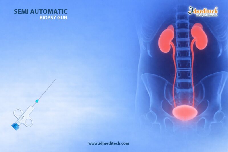
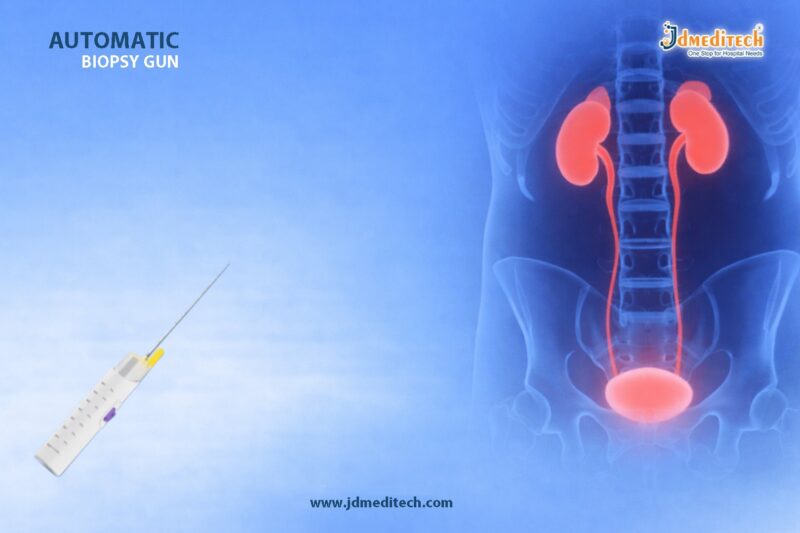
The Automatic Biopsy Gun is a highly advanced medical device designed to obtain accurate tissue samples for diagnostic purposes. Today, healthcare professionals rely on the Automatic Biopsy Gun for its speed, precision, and reliability. Moreover, it plays a crucial role in detecting diseases such as cancer, infections, and other abnormal tissue conditions.

In modern medicine, accurate diagnosis is essential. Therefore, the Automatic Biopsy Gun has become an indispensable tool in hospitals, diagnostic centers, and pathology labs.

What is an Automatic Biopsy Gun?

An Automatic Biopsy Gun is a spring-loaded device used to collect tissue samples quickly and efficiently. It works by inserting a biopsy needle into the targeted area and retrieving a small tissue sample for laboratory analysis.

Unlike manual methods, the Automatic Biopsy Gun offers consistent performance. As a result, it reduces human error and improves diagnostic accuracy.

Key Features of Automatic Biopsy Gun
High-Speed Operation

The Automatic Biopsy Gun operates with a rapid firing mechanism. Consequently, it minimizes patient discomfort and ensures quick sample collection.

Precision Sampling

It allows accurate targeting of tissues. Therefore, doctors can obtain reliable samples even from deep or sensitive areas.

Ergonomic Design

The device features a user-friendly design. As a result, clinicians can handle it easily during procedures.

Adjustable Penetration Depth

Many Automatic Biopsy Guns come with adjustable depth settings. This feature helps customize procedures based on patient requirements.

Compatibility with Biopsy Needles

The device supports various biopsy needles. Hence, it is suitable for multiple medical applications.

Applications of Automatic Biopsy Gun

The Automatic Biopsy Gun is widely used across different medical fields. For instance:

Oncology

Doctors use it to detect tumors and confirm cancer diagnoses.

Urology

It helps in prostate biopsies for detecting abnormalities.

Gastroenterology

It assists in sampling tissues from the liver or gastrointestinal tract.

Gynecology

It is useful for breast and uterine tissue sampling.

Advantages of Automatic Biopsy Gun

The Automatic Biopsy Gun offers several benefits that enhance medical procedures.

Improved Accuracy

It ensures precise tissue collection. Therefore, diagnostic results become more reliable.

Reduced Procedure Time

The fast mechanism reduces overall procedure time. As a result, patient comfort improves.

Minimal Trauma

It causes less tissue damage compared to traditional methods.

Consistency

The automated system provides uniform results in every procedure.

How Does an Automatic Biopsy Gun Work?

The working principle of the Automatic Biopsy Gun is simple yet effective.

First, the clinician positions the needle at the target site. Then, the device is triggered, which quickly inserts and retracts the needle. As a result, a tissue sample is captured inside the needle chamber.

Finally, the sample is sent to the laboratory for further examination.

Safety and Sterilization

Safety is a top priority when using an Automatic Biopsy Gun. Most devices are designed for single-use or easy sterilization. Therefore, they help prevent infections and ensure patient safety.

Additionally, clinicians follow strict hygiene protocols during biopsy procedures.

Why Choose Automatic Biopsy Gun from JDMeditech?

At JDMeditech, we provide high-quality Automatic Biopsy Guns that meet international healthcare standards. Our products ensure durability, precision, and superior performance.

Furthermore, we focus on innovation and reliability. As a result, healthcare professionals trust JDMeditech for advanced medical solutions.

Conclusion

The Automatic Biopsy Gun has revolutionized tissue sampling in modern medicine. With its advanced technology, it ensures accurate diagnosis, faster procedures, and improved patient comfort.

As healthcare continues to evolve, devices like the Automatic Biopsy Gun will remain essential in delivering precise and effective diagnostic outcomes.


Explore More Product >>

Get Connected:
+91 79909 93062 | +91 63513 72032 | exports@jdmeditech.com

Electrophysiology Study – Understanding Heart Rhythm Diagnosis

What is an Electrophysiology Study

Electrophysiology Study is a specialized cardiac test used to evaluate the electrical activity of the heart. It helps doctors understand abnormal heart rhythms, also known as arrhythmias. As cardiovascular diseases continue to rise globally, Electrophysiology Study plays a critical role in early diagnosis and targeted treatment.

Unlike standard ECG or Holter monitoring, Electrophysiology Study provides detailed internal mapping of the heart’s electrical pathways. Therefore, it is considered one of the most accurate diagnostic tools in modern cardiology.

Why Electrophysiology Study is Important
Role in Diagnosing Arrhythmias

Electrophysiology Study is essential when patients experience symptoms such as palpitations, dizziness, or unexplained fainting. These symptoms often indicate irregular heart rhythms that require deeper investigation.

Moreover, Electrophysiology Study helps identify conditions like atrial fibrillation, ventricular tachycardia, and supraventricular tachycardia. As a result, doctors can design precise treatment plans.

Guiding Treatment Decisions

Another important benefit of Electrophysiology Study is its ability to guide treatment. For instance, it helps determine whether a patient needs medication, catheter ablation, or device implantation such as a pacemaker.

Because of this, Electrophysiology Study not only diagnoses but also directly influences treatment strategy.

How Electrophysiology Study is Performed
Step-by-Step Procedure

Electrophysiology Study is typically performed in a specialized cardiac catheterization lab. First, the patient receives local anesthesia and mild sedation to ensure comfort.

Next, thin flexible tubes called catheters are inserted through a vein, usually in the groin or neck. These catheters are carefully guided to the heart using imaging technology.

Once in position, the electrodes on the catheters record electrical signals from different areas of the heart. In addition, doctors may stimulate the heart to reproduce abnormal rhythms. This allows precise identification of the problem area.

Duration and Monitoring

Electrophysiology Study usually takes between one to four hours. During the procedure, the patient’s heart rhythm, blood pressure, and oxygen levels are continuously monitored. Therefore, safety remains a top priority throughout the process.

Benefits of Electrophysiology Study
Accurate Diagnosis

Electrophysiology Study provides unmatched accuracy in diagnosing complex arrhythmias. Because it directly studies the heart’s electrical system, it delivers highly reliable results.

Minimally Invasive Approach

Although it involves catheter insertion, Electrophysiology Study is considered minimally invasive. As a result, recovery time is relatively short compared to open-heart procedures.

Immediate Treatment Possibility

In many cases, doctors can perform catheter ablation during the same Electrophysiology Study session. This eliminates the need for a second procedure and improves patient outcomes.

Risks and Complications of Electrophysiology Study
Possible Side Effects

Electrophysiology Study is generally safe; however, like any medical procedure, it carries some risks. These may include bleeding, infection, or minor damage to blood vessels.

Rare Complications

In rare cases, complications such as heart perforation or serious arrhythmias may occur. Nevertheless, experienced cardiologists take extensive precautions to minimize these risks.

Recovery After Electrophysiology Study
Post-Procedure Care

After Electrophysiology Study, patients are usually monitored for a few hours. The insertion site is checked for bleeding, and patients are advised to rest.

Return to Normal Activities

Most individuals can return to normal activities within one to two days. However, heavy physical activity should be avoided for a short period to ensure proper healing.

Who Needs an Electrophysiology Study
Common Indications

Doctors recommend Electrophysiology Study for patients with unexplained fainting, rapid heartbeats, or irregular ECG results. It is also used for individuals at risk of sudden cardiac arrest.

High-Risk Patients

Patients with structural heart disease or previous heart attacks may benefit significantly from Electrophysiology Study. Early detection through this procedure can be life-saving.

Future of Electrophysiology Study
Advancements in Cardiac Mapping

With technological advancements, Electrophysiology Study is becoming more precise and efficient. Modern 3D mapping systems allow cardiologists to visualize the heart in real-time.

Integration with AI

Artificial intelligence is increasingly being integrated into Electrophysiology Study. This enhances accuracy and helps predict arrhythmia patterns more effectively.

Conclusion
A Key Tool in Modern Cardiology

Electrophysiology Study remains a cornerstone in diagnosing and managing heart rhythm disorders. Because it offers precise insights and immediate treatment options, it significantly improves patient care.

As healthcare continues to evolve, Electrophysiology Study will play an even greater role in ensuring better cardiac outcomes worldwide.


Explore More Product >>

Get Connected:
+91 79909 93062 | +91 63513 72032 | exports@jdmeditech.com 

Semi Automatic Biopsy Gun for Tissue Sampling

The semi automatic biopsy gun plays an important role in modern diagnostic procedures. It helps doctors collect tissue samples quickly and accurately, which is essential for detecting diseases at an early stage. Because of its precision and ease of use, the semi automatic biopsy gun is widely used in hospitals, diagnostic centers, and pathology laboratories.
What is a Semi Automatic Biopsy Gun?

A semi automatic biopsy gun is a medical device designed to collect tissue samples from the body using a spring-loaded mechanism. Unlike fully automatic systems, this device requires manual preparation before use. As a result, doctors gain better control during the procedure, which improves accuracy and reduces the chances of tissue damage.

Key Features of Semi Automatic Biopsy Gun

The semi automatic biopsy gun offers several features that enhance its performance. It has a lightweight and ergonomic design, which makes it easy to handle during procedures. In addition, it provides adjustable penetration depth, allowing precise targeting of the required tissue. The device is also compatible with different biopsy needles, making it suitable for various medical applications.

Working Mechanism

The working process of the semi automatic biopsy gun is straightforward. First, the device is manually cocked to prepare the internal spring mechanism. Next, the doctor positions the needle at the targeted area. Finally, the trigger is pressed, releasing the needle to capture the tissue sample. This quick action ensures accurate sampling while minimizing discomfort for the patient.

Applications in Medical Practice

The semi automatic biopsy gun is used in many medical fields. For example, it is commonly used in oncology to diagnose tumors. It is also widely used in nephrology for kidney biopsies and in hepatology for liver tissue sampling. Furthermore, doctors use it for breast biopsies and soft tissue examinations, making it a versatile diagnostic tool.

Advantages of Semi Automatic Biopsy Gun
The semi automatic biopsy gun offers several benefits. Most importantly, it provides high precision during tissue sampling. Because of its controlled mechanism, it reduces the risk of complications. In addition, the procedure is minimally invasive, which helps patients recover faster. It is also more cost-effective than fully automatic devices, making it a practical choice for many healthcare facilities.
Semi Automatic vs Fully Automatic Biopsy Guns

Both semi automatic and fully automatic biopsy guns are used for tissue sampling. However, the semi automatic biopsy gun gives better control to the user, which is important in delicate procedures. On the other hand, fully automatic devices work faster but may offer less control in certain situations. Therefore, the choice depends on the doctor’s preference and the clinical requirement.

Safety and Handling Considerations

Proper handling of the semi automatic biopsy gun is essential to ensure patient safety. Doctors must follow strict sterilization procedures before and after use. It is also important to use compatible needles and check the device regularly. These steps help maintain performance and reduce the risk of errors during the procedure.

Importance in Accurate Diagnosis

Accurate diagnosis depends on high-quality tissue samples. The semi automatic biopsy gun helps achieve this by collecting precise and reliable samples. These samples are then analyzed in laboratories to detect diseases. As a result, early diagnosis becomes possible, which improves treatment outcomes.

Conclusion

The semi automatic biopsy gun is a reliable and efficient tool for tissue sampling. Its precision, safety, and versatility make it an essential device in modern healthcare. As diagnostic technologies continue to advance, the importance of the semi automatic biopsy gun will continue to grow.


Explore More Product >>

Get Connected:
+91 79909 93062 | +91 63513 72032 | exports@jdmeditech.com 


Stone Cone Basket – Prevent Stone Migration in Urology

The Stone Cone Basket is an essential device in modern urology. It helps prevent stone migration during procedures like ureteroscopy and lithotripsy. When treating kidney or ureteral stones, migration can complicate the process. Therefore, using a Stone Cone Basket improves both safety and efficiency.

Moreover, urologists rely on this tool to ensure better clinical outcomes. As minimally invasive procedures grow in popularity, the importance of the Stone Cone Basket continues to increase.

What is a Stone Cone Basket?

A Stone Cone Basket is a specialized urological device designed to block the upward movement of stones during fragmentation. It is typically inserted into the ureter before lithotripsy begins.

In addition, the Stone Cone Basket forms a helical or cone-shaped barrier. This structure prevents fragments from migrating toward the kidney.

Why Stone Migration is a Problem

Stone migration is a common challenge in urology. When stones move upward during treatment, it can:

  • Increase procedure time
  • Require additional interventions
  • Reduce treatment success rate
  • Cause patient discomfort

However, by using a Stone Cone Basket, surgeons can effectively prevent these issues. As a result, the procedure becomes more controlled and predictable.

Key Features of Stone Cone Basket

The Stone Cone Basket offers several advanced features that enhance performance:

  • Flexible Design : Allows smooth navigation through the ureter
  • Nitinol Construction : Ensures durability and shape memory
  • Helical Coil Structure : Prevents stone migration effectively
  • Radiopaque Markers : Improve visibility under imaging
  • Single-use Sterility : Maintains hygiene and reduces infection risk

Because of these features, the Stone Cone Basket is widely preferred in urological procedures.

How Stone Cone Basket Works

The working principle of a Stone Cone Basket is simple yet highly effective.

First, the device is inserted past the stone. Then, it is deployed to form a cone-shaped barrier. After that, lithotripsy is performed to break the stone into smaller fragments.

Consequently, the Stone Cone Basket traps the fragments and prevents them from moving upward. This ensures complete and efficient stone removal.

Applications in Urology

The Stone Cone Basket is commonly used in various procedures, including:

  • Ureteroscopy (URS)
  • Laser lithotripsy
  • Pneumatic lithotripsy
  • Kidney and ureteral stone management

Furthermore, it plays a crucial role in minimally invasive urological surgeries.

Benefits of Using Stone Cone Basket

Using a Stone Cone Basket provides multiple benefits:

  • Prevents stone migration
  • Improves stone-free rates
  • Reduces need for repeat procedures
  • Saves procedural time
  • Enhances patient safety

Therefore, it is considered a must-have tool in modern urology.

Stone Cone Basket vs Traditional Methods

Traditional methods often fail to control stone migration effectively. In contrast, the Stone Cone Basket provides a reliable mechanical barrier.

Additionally, it reduces dependence on complex techniques. This makes the procedure simpler and more efficient for surgeons.

Choosing the Right Stone Cone Basket

When selecting a Stone Cone Basket, consider the following:

  • Size compatibility with ureter
  • Material quality (preferably nitinol)
  • Flexibility and deployment mechanism
  • Sterility and packaging

Choosing a high-quality Stone Cone Basket ensures better performance and safety.

Why Choose JDMeditech Stone Cone Basket?

At JDMeditech, we provide premium-quality Stone Cone Basket solutions designed for precision and reliability.

Our products offer:

  • High-grade nitinol construction
  • Excellent flexibility and control
  • Strict quality standards
  • Competitive pricing
Conclusion

The Stone Cone Basket is a vital innovation in urology. It effectively prevents stone migration, ensuring smoother and safer procedures.

As a result, it enhances surgical efficiency and patient outcomes. For healthcare professionals, investing in a reliable Stone Cone Basket is a smart decision.


Explore More Product >>

Get Connected:

+91 79909 93062 | +91 63513 72032 | exports@jdmeditech.com

Colon Polypectomy Surgery – Minimally Invasive Cancer Prevention

Colon polypectomy surgery plays a crucial role in preventing colorectal cancer. It is a minimally invasive procedure that removes abnormal growths, known as polyps, from the colon lining. These polyps can become cancerous over time. Therefore, early removal through colon polypectomy surgery significantly reduces cancer risk.

Moreover, doctors commonly perform this procedure during a colonoscopy. As a result, patients benefit from early detection and immediate treatment in one session. Because of its effectiveness and safety, colon polypectomy surgery has become a standard preventive measure in modern gastroenterology.

What is Colon Polypectomy Surgery?

Colon polypectomy surgery is a medical procedure used to remove polyps from the inner wall of the colon. These polyps vary in size and shape. While some remain harmless, others can develop into colorectal cancer if left untreated.

During colon polypectomy surgery, a gastroenterologist inserts a colonoscope into the colon. Then, specialized tools remove the polyp safely. This approach avoids open surgery and ensures faster recovery.

Types of Colon Polyps

Understanding polyp types helps determine the need for colon polypectomy surgery:

  • Adenomatous Polyps : These are precancerous and require removal
  • Hyperplastic Polyps : Usually benign but monitored carefully
  • Inflammatory Polyps : Often linked to inflammatory bowel disease
  • Sessile Serrated Polyps : Higher cancer risk if untreated

Because some polyps carry cancer potential, doctors strongly recommend colon polypectomy surgery for timely removal.

Why is Colon Polypectomy Surgery Important?

Colon polypectomy surgery offers multiple benefits. Most importantly, it prevents cancer before it develops. In addition, it supports early diagnosis and improves long-term outcomes.

Key Benefits:
  • Prevents colorectal cancer
  • Removes precancerous growths early
  • Reduces need for major surgery
  • Provides quick recovery
  • Enhances digestive health

Therefore, regular screening combined with colon polypectomy surgery ensures proactive healthcare management.

Procedure of Colon Polypectomy Surgery

The procedure is simple, safe, and minimally invasive. Doctors usually perform it during a colonoscopy.

Step-by-Step Process:
  1. Preparation : The patient undergoes bowel cleansing
  2. Sedation : Mild sedation ensures comfort
  3. Colonoscope Insertion : A flexible tube examines the colon
  4. Polyp Removal : Tools like snares or forceps remove polyps
  5. Inspection : The doctor checks for additional abnormalities

Because the procedure is minimally invasive, colon polypectomy surgery does not require large incisions.

Techniques Used in Colon Polypectomy Surgery

Doctors use different techniques depending on polyp size and type:

  • Snare Polypectomy : Uses a wire loop to cut polyps
  • Hot Biopsy Forceps : Applies heat to remove small polyps
  • Endoscopic Mucosal Resection (EMR) : Removes larger polyps
  • Endoscopic Submucosal Dissection (ESD) : For complex lesions

Each technique ensures precise removal during colon polypectomy surgery.

Recovery After Colon Polypectomy Surgery

Recovery is usually quick and smooth. Most patients return home the same day.

Post-Procedure Care:
  • Mild bloating or discomfort may occur
  • Avoid heavy meals for a short period
  • Follow doctor’s dietary advice
  • Monitor for unusual symptoms

Because of its minimally invasive nature, colon polypectomy surgery allows faster healing compared to traditional surgery.

Risks and Complications

Although generally safe, colon polypectomy surgery carries minor risks:

  • Bleeding at removal site
  • Perforation (rare)
  • Infection (very rare)

However, experienced professionals significantly minimize these risks.

Who Should Consider Colon Polypectomy Surgery?

Doctors recommend colon polypectomy surgery for:

  • Patients with detected colon polyps
  • Individuals over 45 undergoing screening
  • People with family history of colorectal cancer
  • Patients with gastrointestinal symptoms

Early intervention through colon polypectomy surgery improves outcomes and reduces complications.

Role of Advanced Endoscopic Devices

Modern endoscopic tools improve the safety and efficiency of colon polypectomy surgery. High-quality snares, biopsy forceps, and electrosurgical units ensure precise removal.

Conclusion

Colon polypectomy surgery is a powerful preventive procedure in modern medicine. It removes potentially dangerous polyps before they turn cancerous. Moreover, its minimally invasive approach ensures safety, comfort, and quick recovery.

Therefore, regular screening combined with timely colon polypectomy surgery can save lives. Investing in early detection is the key to long-term health and cancer prevention.


Explore More Product >>

Get Connected:

+91 79909 93062 | +91 63513 72032 | exports@jdmeditech.com

Patent Ductus Arteriosus Closure – Minimally Invasive Heart Repair

Patent Ductus Arteriosus Closure is a modern and minimally invasive procedure used to treat a common congenital heart defect. In this condition, a blood vessel called the ductus arteriosus fails to close after birth. As a result, abnormal blood flow occurs between the aorta and pulmonary artery.

Fortunately, Patent Ductus Arteriosus Closure helps restore normal circulation effectively. Moreover, it reduces complications and improves long-term heart health. Today, cardiologists widely prefer this procedure because it offers faster recovery and minimal discomfort.

What is Patent Ductus Arteriosus (PDA)?

Patent Ductus Arteriosus is a condition in which the ductus arteriosus remains open after birth. Normally, this vessel closes within a few days of life. However, when it stays open, it causes excess blood flow to the lungs.

As a result, the heart and lungs experience increased workload. Therefore, timely Patent Ductus Arteriosus Closure becomes essential to prevent complications such as heart failure or pulmonary hypertension.

Why is Patent Ductus Arteriosus Closure Needed?

Doctors recommend Patent Ductus Arteriosus Closure when the condition leads to noticeable symptoms or complications.

Common Reasons Include:
  • Abnormal heart murmur
  • Rapid breathing or shortness of breath
  • Poor weight gain in infants
  • Frequent respiratory infections
  • Risk of heart enlargement

In addition, early Patent Ductus Arteriosus Closure helps avoid severe cardiovascular issues later in life.

Types of Patent Ductus Arteriosus Closure Procedures

There are two main methods for performing Patent Ductus Arteriosus Closure. However, minimally invasive techniques are now the preferred choice.

1. Transcatheter Closure (Minimally Invasive)

This is the most common method of Patent Ductus Arteriosus Closure.

  • A catheter is inserted through a blood vessel, usually in the groin
  • A closure device is guided to the heart
  • The device seals the open ductus arteriosus
Advantages:
  • No major surgery required
  • Faster recovery
  • Minimal pain
  • Short hospital stay
2. Surgical Closure

In some cases, especially in premature infants, surgery may be required.

  • A small incision is made in the chest
  • The ductus arteriosus is tied or clipped

Although effective, surgical Patent Ductus Arteriosus Closure involves longer recovery compared to catheter-based procedures.

Procedure of Patent Ductus Arteriosus Closure

The process of Patent Ductus Arteriosus Closure is straightforward and safe.

Step-by-Step Overview:
  1. The patient receives local or general anesthesia
  2. A catheter is inserted into a blood vessel
  3. The catheter is guided to the heart using imaging techniques
  4. A closure device is deployed to seal the duct
  5. Blood flow returns to normal

Typically, the procedure takes about 1 to 2 hours.

Benefits of Patent Ductus Arteriosus Closure

Patent Ductus Arteriosus Closure offers multiple advantages for patients of all ages.

  • Improves heart efficiency
  • Prevents long-term complications
  • Reduces symptoms quickly
  • Minimally invasive with small or no scars
  • Short recovery time

Therefore, it has become a preferred solution in modern cardiology.

Risks and Complications

Although Patent Ductus Arteriosus Closure is generally safe, some risks may occur.

  • Bleeding at the catheter insertion site
  • Device displacement (rare)
  • Infection
  • Allergic reaction to contrast dye

However, with experienced cardiologists and advanced equipment, complications remain minimal.

Recovery After Patent Ductus Arteriosus Closure

Recovery after Patent Ductus Arteriosus Closure is usually quick and smooth.

  • Most patients return home within 24 hours
  • Normal activities can resume within a few days
  • Follow-up visits ensure proper healing

In addition, patients often experience immediate improvement in symptoms.

Who Needs Patent Ductus Arteriosus Closure?

Not all cases require immediate treatment. However, doctors recommend Patent Ductus Arteriosus Closure for:

  • Infants with significant PDA
  • Children with symptoms
  • Adults with untreated PDA
  • Patients at risk of complications

Early diagnosis and treatment improve outcomes significantly.

Conclusion

Patent Ductus Arteriosus Closure is a highly effective and minimally invasive solution for treating congenital heart defects. It not only restores normal blood flow but also prevents serious complications.

With modern techniques like transcatheter closure, patients benefit from faster recovery and improved quality of life. Therefore, timely Patent Ductus Arteriosus Closure plays a crucial role in ensuring long-term heart health.


Explore More Product >>

Get Connected:

+91 79909 93062 | +91 63513 72032 | exports@jdmeditech.com

Paw Basket Retrieval – Efficient Stone Removal in Urology

PAW BASKEY-urology products jdmeditech

Urological procedures have evolved significantly with the introduction of advanced tools designed for precision and patient safety. Among these innovations, the Paw Basket stone retrieval device stands out as a reliable solution for removing urinary stones effectively.

In modern endourology, surgeons require instruments that offer both control and efficiency. Therefore, the Paw Basket stone retrieval system plays a crucial role in improving outcomes during procedures like ureteroscopy and cystoscopy.

What is a Paw Basket?

A Paw Basket stone retrieval device is a specialized urological instrument used to capture and remove stones from the urinary tract. It typically features flexible wires arranged in a basket-like structure, which allows it to securely grasp stones.

Moreover, the design of the Paw Basket stone retrieval system ensures minimal trauma to surrounding tissues. As a result, it becomes an essential tool for urologists performing minimally invasive procedures.

Key Features of Paw Basket Stone Retrieval Devices

The Paw Basket stone retrieval device offers several advanced features that enhance its performance:

  • Flexible Wire Design : Ensures smooth navigation through the urinary tract
  • Strong Gripping Mechanism : Allows secure stone capture
  • Atraumatic Tip : Minimizes mucosal injury
  • High Durability : Suitable for repeated clinical use
  • Precise Control Handle : Provides better maneuverability

Additionally, these features collectively improve procedural efficiency and patient safety.

Applications in Urological Procedures

The Paw Basket stone retrieval device is widely used in various urological interventions. Some of the key applications include:

1. Ureteroscopy (URS)

During ureteroscopy, the Paw Basket stone retrieval tool helps in capturing and extracting stones from the ureter.

2. Kidney Stone Removal

It is also used in procedures involving renal stones, ensuring safe removal without fragmentation.

3. Cystoscopy

In bladder stone management, the Paw Basket stone retrieval device assists in retrieving smaller calculi effectively.

4. Post-Lithotripsy Fragment Removal

After lithotripsy, this device helps in clearing residual stone fragments.

Benefits of Using Paw Basket Stone Retrieval

Using the Paw Basket stone retrieval device offers multiple advantages:

  • Minimally Invasive Approach : Reduces patient discomfort
  • Improved Success Rate : Ensures complete stone removal
  • Reduced Procedure Time : Enhances surgical efficiency
  • Lower Risk of Complications : Minimizes tissue damage
  • Better Visualization : Works effectively with endoscopic systems

Furthermore, these benefits make it a preferred choice among urologists worldwide.

How Paw Basket Enhances Surgical Precision

Precision is critical in urology. The Paw Basket stone retrieval device provides excellent control during procedures.

Because of its ergonomic handle and flexible structure, surgeons can easily navigate complex anatomical pathways. Consequently, this leads to improved accuracy and reduced risk of stone migration.

Materials and Design Considerations

The Paw Basket stone retrieval device is usually made from high-quality materials such as stainless steel or nitinol. These materials provide flexibility and strength.

In addition, the basket configuration is designed to adapt to different stone sizes. Therefore, it ensures efficient capture without slipping.

Why Choose JDMeditech Paw Basket?

At JDMeditech, we focus on delivering high-performance urological instruments. Our Paw Basket stone retrieval devices are designed to meet international quality standards.

Key Advantages:
  • Premium-grade materials
  • Precision-engineered design
  • Consistent performance
  • Cost-effective solutions
  • Trusted by healthcare professional
Best Practices for Using Paw Basket Stone Retrieval

To achieve optimal results, follow these best practices:

  • Always ensure proper visualization before capturing the stone
  • Use gentle movements to avoid tissue injury
  • Select the appropriate basket size
  • Maintain sterility throughout the procedure

Additionally, proper training and handling improve overall effectiveness.

Future Trends in Stone Retrieval Technology

The future of Paw Basket stone retrieval devices looks promising. Innovations are focusing on improved flexibility, enhanced grip strength, and better compatibility with advanced endoscopic systems.

As technology advances, these devices will continue to improve patient outcomes and surgical efficiency.

Conclusion

The Paw Basket stone retrieval device has become an indispensable tool in modern urology. Its ability to provide safe, efficient, and minimally invasive stone removal makes it highly valuable in clinical practice.

By choosing high-quality solutions like those offered by JDMeditech, healthcare providers can ensure better procedural success and patient satisfaction.


Explore More Product >>

Get Connected:

+91 79909 93062 | +91 63513 72032 | exports@jdmeditech.com

Esophageal Dilation Surgery for Swallowing Disorders

Esophageal Dilation Surgery is a proven treatment for swallowing disorders, also known as dysphagia. When the esophagus becomes narrow, patients often struggle to swallow food and liquids. Therefore, Esophageal Dilation Surgery helps restore normal swallowing by widening the narrowed passage.

Moreover, this procedure improves patient comfort and overall quality of life. Because of its effectiveness, doctors widely recommend Esophageal Dilation Surgery for long-term relief.

What is Esophageal Dilation Surgery?

Esophageal Dilation Surgery is a minimally invasive procedure that expands a narrowed esophagus. In most cases, doctors use endoscopic techniques to perform Esophageal Dilation Surgery safely and accurately.

During the procedure, specialists insert a dilator or balloon into the esophagus. Then, they gradually widen the affected area. As a result, Esophageal Dilation Surgery allows food to pass smoothly.

Causes of Swallowing Disorders

Several conditions can lead to esophageal narrowing. Therefore, identifying the cause is important before performing Esophageal Dilation Surgery.

Common causes include:

  • Acid reflux (GERD) leading to strictures
  • Esophageal rings and webs
  • Scar tissue formation
  • Radiation damage
  • Eosinophilic esophagitis
  • Tumors or abnormal growth

Because these conditions restrict the esophagus, Esophageal Dilation Surgery becomes necessary in many cases.

When is Esophageal Dilation Surgery Required?

Doctors recommend Esophageal Dilation Surgery when symptoms persist despite medication. In addition, early treatment prevents complications.

Typical symptoms include:

  • Difficulty swallowing
  • Food getting stuck in the throat
  • Chest discomfort during meals
  • Weight loss
  • Frequent regurgitation

If these symptoms worsen, Esophageal Dilation Surgery provides effective relief.

Techniques Used in Esophageal Dilation Surgery

Doctors use different methods in Esophageal Dilation Surgery depending on the condition.

Balloon Dilation

First, a balloon is inserted into the narrowed area. Then, it is inflated to widen the esophagus evenly.

Bougie Dilators

In this method, doctors pass flexible tubes of increasing size. As a result, Esophageal Dilation Surgery gradually expands the esophagus.

Guidewire-Assisted Dilation

Here, a guidewire ensures precise placement of dilators. Therefore, Esophageal Dilation Surgery becomes safer and more controlled.

Procedure Steps

Understanding the process helps patients feel more confident about Esophageal Dilation Surgery.

  1. First, the patient receives sedation
  2. Then, an endoscope is inserted
  3. The doctor identifies the narrowed section
  4. A dilator or balloon is positioned
  5. The esophagus is gently widened
  6. Finally, the instruments are removed

Overall, Esophageal Dilation Surgery usually takes less than one hour.

Benefits of Esophageal Dilation Surgery

Esophageal Dilation Surgery offers several important benefits. For example:

  • Immediate improvement in swallowing
  • Minimally invasive approach
  • Quick recovery time
  • Better nutrition and hydration
  • Reduced discomfort

Therefore, Esophageal Dilation Surgery is a preferred treatment option for dysphagia.

Risks of Esophageal Dilation Surgery

Although Esophageal Dilation Surgery is safe, some risks may occur. However, complications remain rare.

Possible risks include:

  • Mild throat pain
  • Minor bleeding
  • Esophageal perforation (very rare)
  • Recurrence of narrowing

Even so, experienced doctors perform Esophageal Dilation Surgery with high success rates.

Recovery After Esophageal Dilation Surgery

Recovery after Esophageal Dilation Surgery is usually fast. In most cases, patients return home the same day.

After the procedure:

  • Patients start with soft foods
  • They avoid spicy or hot meals
  • Doctors may prescribe medications
  • Follow-up visits ensure proper healing

As a result, Esophageal Dilation Surgery leads to quick improvement.

Long-Term Results

Esophageal Dilation Surgery provides long-lasting relief in many patients. However, some patients may require repeat procedures.

With proper care, Esophageal Dilation Surgery helps maintain normal swallowing function. Therefore, regular monitoring ensures better outcomes.

Role of Medical Devices

High-quality instruments play a key role in Esophageal Dilation Surgery. Advanced dilators and guidewires improve precision and safety.

At JDMeditech, we provide reliable medical devices that support Esophageal Dilation Surgery and enhance clinical performance.

Conclusion

Esophageal Dilation Surgery is a safe and effective solution for swallowing disorders. By widening the esophagus, it restores normal function and improves patient comfort.

If swallowing problems continue, Esophageal Dilation Surgery offers a dependable path to relief and better health.


Explore More Product >>

Get Connected:

+91 79909 93062 | +91 63513 72032 | exports@jdmeditech.com

Gastrojejunostomy – Surgical Bypass for Gastric Obstruction Explained

Gastrojejunostomy surgical bypass for gastric obstruction plays a vital role in modern gastrointestinal surgery. Doctors use this procedure to create a direct connection between the stomach and the jejunum, effectively bypassing a blocked or narrowed part of the digestive tract.

In many cases, gastric obstruction results from conditions such as tumors, peptic ulcers, or severe inflammation. Therefore, gastrojejunostomy provides an effective solution that restores digestive flow and improves patient comfort.

What is Gastrojejunostomy?

Gastrojejunostomy is a surgical procedure in which a surgeon connects the stomach directly to the jejunum, which is the middle section of the small intestine. As a result, food bypasses the obstructed part of the stomach or duodenum.

This gastrojejunostomy surgical bypass for gastric obstruction is commonly performed when normal gastric emptying becomes impossible.

Why is Gastrojejunostomy Performed?

Doctors recommend gastrojejunostomy in several clinical situations. Most importantly, it helps when other treatments fail or when obstruction becomes severe.

Common Indications
  • Gastric outlet obstruction
  • Stomach or pancreatic cancer
  • Chronic peptic ulcer disease
  • Duodenal strictures
  • Congenital abnormalities

Moreover, surgeons often choose this gastrojejunostomy surgical bypass for gastric obstruction to provide palliative relief in advanced cancer cases.

Types of Gastrojejunostomy

Different surgical techniques exist depending on the patient’s condition and surgeon’s expertise.

1. Open Gastrojejunostomy

Surgeons perform this procedure through a larger abdominal incision. Although it allows direct access, it usually requires longer recovery.

2. Laparoscopic Gastrojejunostomy

This minimally invasive approach uses small incisions and a camera. As a result, patients experience less pain and faster healing.

3. Endoscopic Gastrojejunostomy

In selected cases, doctors use endoscopic techniques to create the bypass without major surgery. Therefore, it reduces hospital stay significantly.

Step-by-Step Procedure

Understanding the procedure helps patients feel more confident and prepared.

  1. First, the patient receives general anesthesia.
  2. Next, the surgeon accesses the stomach and jejunum.
  3. Then, a connection (anastomosis) is created between them.
  4. After that, the surgeon ensures proper blood flow and alignment.
  5. Finally, the incision is closed carefully.

This structured approach ensures that the gastrojejunostomy surgical bypass for gastric obstruction works effectively.

Benefits of Gastrojejunostomy

This procedure offers multiple clinical advantages.

  • Restores normal food passage
  • Relieves symptoms like vomiting and nausea
  • Improves nutritional intake
  • Enhances quality of life
  • Provides palliative care in cancer patients

Additionally, laparoscopic methods reduce complications and speed up recovery.

Risks and Complications

Although gastrojejunostomy is generally safe, certain risks may occur.

  • Infection
  • Bleeding
  • Anastomotic leakage
  • Delayed gastric emptying
  • Dumping syndrome

However, experienced surgeons minimize these risks through proper technique and care.

Recovery and Aftercare

Recovery depends on the surgical method used.

  • Patients usually stay in the hospital for a few days
  • Doctors gradually reintroduce liquids and soft foods
  • Regular follow-ups ensure proper healing
  • Patients must follow dietary guidelines strictly

Furthermore, early mobilization and proper nutrition support faster recovery after gastrojejunostomy surgical bypass for gastric obstruction.

Role of Medical Devices in Gastrojejunostomy

High-quality surgical instruments play a crucial role in successful outcomes.

At JDMeditech, we provide precision-engineered medical devices designed for gastrointestinal procedures. These include:

  • Surgical staplers
  • Anastomosis devices
  • Endoscopic accessories
  • Guidewires and dilation tools

Reliable instruments improve surgical accuracy and patient safety.

Conclusion

Gastrojejunostomy surgical bypass for gastric obstruction remains a highly effective solution for managing complex gastrointestinal conditions. Not only does it restore digestive function, but it also significantly improves patient quality of life.

As surgical techniques continue to advance, minimally invasive approaches are making this procedure safer and more efficient. Therefore, healthcare providers increasingly rely on this method for both curative and palliative treatment.


Explore More Product >>

Get Connected:

+91 79909 93062 | +91 63513 72032 | exports@jdmeditech.com

Amplatz Renal Dilator for Minimally Invasive Access

The Amplatz Renal Dilator plays a vital role in modern urology. Surgeons use it in minimally invasive procedures such as Percutaneous Nephrolithotomy (PCNL) to create safe access to the kidney.

Today, doctors prefer minimally invasive techniques because they reduce complications and speed up recovery. Therefore, the Amplatz Renal Dilator helps improve surgical precision and patient outcomes.

What is an Amplatz Renal Dilator?

The Amplatz Renal Dilator is a specialized medical instrument that expands the nephrostomy tract during kidney procedures.

Surgeons pass the Amplatz Renal Dilator over a guidewire in a controlled sequence. This method allows them to widen the tract gradually. Its rigid structure provides stability and ensures precise dilation.

Why Do Surgeons Use Amplatz Renal Dilator?

Surgeons use the Amplatz Renal Dilator to create direct and safe access to the kidney. They commonly use it in:

  • Percutaneous Nephrolithotomy (PCNL)
  • Kidney stone removal procedures
  • Nephrostomy tract dilation
  • Endourological surgeries

In addition, it helps protect surrounding tissues and allows smooth instrument insertion.

Key Features of Amplatz Renal Dilator
1. Step-by-Step Dilation

The Amplatz Renal Dilator comes in multiple sizes. Surgeons increase the size gradually, which improves safety and control.

2. Strong and Durable Material

Manufacturers use high-quality medical-grade materials to produce the Amplatz Renal Dilator. This ensures durability and consistent performance.

3. Smooth Surface Design

The smooth surface allows easy insertion. It also reduces friction and minimizes tissue damage.

4. Guidewire Compatibility

The Amplatz Renal Dilator works seamlessly with standard guidewires. This compatibility supports efficient workflow during procedures.

5. Easy Handling

The ergonomic design gives surgeons better grip and control. As a result, they perform procedures with greater accuracy.

How Does Amplatz Renal Dilator Work?

Surgeons follow a clear and controlled process when using the Amplatz Renal Dilator:

  1. First, they insert a guidewire into the kidney.
  2. Then, they introduce a small-sized Amplatz Renal Dilator.
  3. Next, they use larger dilators step by step.
  4. Finally, they achieve the required tract size.

This gradual method ensures safe and effective dilation.

Benefits of Amplatz Renal Dilator

The Amplatz Renal Dilator offers several important advantages:

  • Provides minimally invasive kidney access
  • Reduces tissue trauma
  • Improves surgical precision
  • Speeds up patient recovery
  • Lowers the risk of complications

Additionally, it helps surgeons complete procedures more efficiently.

Clinical Applications

Doctors use the Amplatz Renal Dilator in many urological procedures, including:

  • PCNL for kidney stone removal
  • Renal access creation
  • Advanced endourological procedures

It performs well in both routine and complex cases.

Why Surgeons Prefer Amplatz Renal Dilator

Surgeons prefer the Amplatz Renal Dilator because it:

  • Delivers consistent dilation
  • Provides strong stability
  • Allows smooth instrument access
  • Reduces procedural risks

Because of these advantages, it has become a trusted choice in urology.

Maintenance and Sterilization

Proper maintenance keeps the Amplatz Renal Dilator in optimal condition:

  • Clean it immediately after use
  • Use approved cleaning solutions
  • Follow standard sterilization protocols
  • Inspect it before every procedure

These steps help maintain safety and extend the instrument’s lifespan.

Conclusion

The Amplatz Renal Dilator remains a key instrument in minimally invasive kidney procedures. It allows surgeons to create precise and controlled access while protecting surrounding tissues.

As medical technology advances, surgeons will continue to rely on the Amplatz Renal Dilator for safe and effective outcomes. High-quality instruments always lead to better patient care.


Explore More Product >>

Get Connected:

+91 79909 93062 | +91 63513 72032 | exports@jdmeditech.com