An intrauterine contraceptive device, also known as an IUCD or IUD, is a small device that provides long-term contraception. Most IUCDs have thin threads that allow a trained healthcare professional to remove the device during a routine appointment.
However, removal may become more difficult when the threads are not visible, the device has shifted, or part of the device has become embedded in the uterine wall.
In selected cases, hysteroscopy provides a direct view of the uterine cavity. As a result, a gynecologist can locate the device and assess a suitable removal approach.
This JDMeditech guide explains hysteroscopic IUCD removal surgery, its instruments, clinical indications, benefits, risks, recovery, and equipment selection factors.
What Is Hysteroscopic IUCD Removal Surgery?
Hysteroscopic IUCD Removal Surgery is a procedure in which a gynecologist uses a hysteroscope to examine the uterine cavity and remove a suitable intrauterine contraceptive device.
The hysteroscope is a narrow viewing instrument that enters through the cervix. It allows the specialist to examine the inside of the uterus without making an abdominal incision.
Furthermore, compatible grasping forceps or other retrieval instruments may help remove the IUCD when simpler methods fail.
However, not every patient with missing IUCD threads needs hysteroscopy. The specialist must first assess the device’s location and the patient’s clinical condition.
Understanding Intrauterine Contraceptive Devices
An IUCD is a small contraceptive device that a trained healthcare professional places inside the uterus.
Depending on its type, the device may contain copper or release a hormone called levonorgestrel.
Both options provide long-term contraception. However, their duration of use, effects on menstrual bleeding, and other features differ.
Most IUCDs have retrieval threads that extend through the cervix. These threads usually help clinicians remove the device when necessary.
Nevertheless, the threads may retract into the cervical canal or uterine cavity. Therefore, missing threads do not always mean that the IUCD has moved out of position.
Why Is Hysteroscopic IUCD Removal Performed?
Most IUCD removals are straightforward and do not require surgery.
However, certain clinical situations make removal more challenging. For example, the retrieval threads may not appear during a speculum examination.
In other cases, a specialist may encounter resistance while attempting removal.
Consequently, the healthcare team may use ultrasound or other imaging to confirm the IUCD’s location before choosing the next treatment.
If simpler retrieval methods fail, hysteroscopic removal may offer a suitable option for selected patients.
Common Indications for Hysteroscopic IUCD Removal
1. Missing IUCD Threads
Missing IUCD threads are a common reason for further examination.
However, the device may still remain in its correct position inside the uterus.
Therefore, clinicians first exclude pregnancy when appropriate and confirm the device’s location before attempting intrauterine retrieval.
2. Retained IUCD
A retained IUCD is a device that remains inside the uterus when removal is required or requested.
For example, a patient may need removal because the device has reached its recommended replacement time.
If routine removal proves difficult, the specialist may consider additional techniques.
3. Embedded IUCD
Sometimes, part of an IUCD becomes embedded in the endometrium or the uterine muscle.
As a result, routine removal may cause resistance or discomfort.
In selected cases, hysteroscopy allows the specialist to assess the device under direct visualization.
4. Difficult IUCD Removal
Some devices cannot be removed easily with standard instruments.
Consequently, the clinician may refer the patient to an experienced gynecologist.
Depending on the situation, ultrasound-guided retrieval or hysteroscopy may be appropriate.
5. Malpositioned IUCD
An IUCD may move away from its intended position within the uterus.
However, management depends on the type of device, its location, symptoms, and pregnancy risk.
When removal is necessary and routine methods fail, hysteroscopy may help in selected cases.
6. Suspected IUCD Fragment
Occasionally, part of an IUCD may break during removal.
Therefore, the healthcare team may use imaging or hysteroscopy to investigate a suspected retained fragment.
However, treatment depends on the fragment’s location and the risks of further intervention.
7. Persistent Symptoms With a Difficult-to-Remove IUCD
Some patients experience pelvic pain, abnormal bleeding, or other symptoms while using an IUCD.
Nevertheless, these symptoms can have several causes.
For this reason, clinicians must evaluate the patient before deciding whether device removal or another treatment is appropriate.
Common Instruments Used in Hysteroscopic IUCD Removal Surgery
Different clinical situations require different retrieval instruments. Moreover, equipment selection depends on the device’s position, uterine anatomy, and available hysteroscopy system.
The following instruments may support selected IUCD removal procedures. However, they do not represent a confirmed JDMeditech surgical-set packing list.
1. Hysteroscope Telescope
A hysteroscope telescope provides a direct view of the uterine cavity.
Furthermore, it helps the specialist identify the IUCD, its threads, and nearby uterine structures.
Hospitals must select a telescope that matches the intended sheath and imaging system.
2. Diagnostic Hysteroscopy Sheath
A diagnostic sheath provides access for the hysteroscope.
Depending on its design, the sheath may also support fluid flow or compatible working instruments.
Therefore, hospitals should verify its diameter and channel configuration.
3. Hysteroscopy Grasping Forceps
Compatible grasping forceps can help the specialist hold IUCD threads or accessible parts of the device.
For example, some hysteroscopy systems support slim forceps that pass through a working channel.
However, the surgeon must select the appropriate instrument for the device and clinical situation.
4. Alligator Grasping Forceps
Alligator-type forceps may support selected IUCD retrieval procedures.
Their jaws allow a trained clinician to grasp suitable device components.
Nevertheless, the exact forceps design must match the intended procedure and equipment.
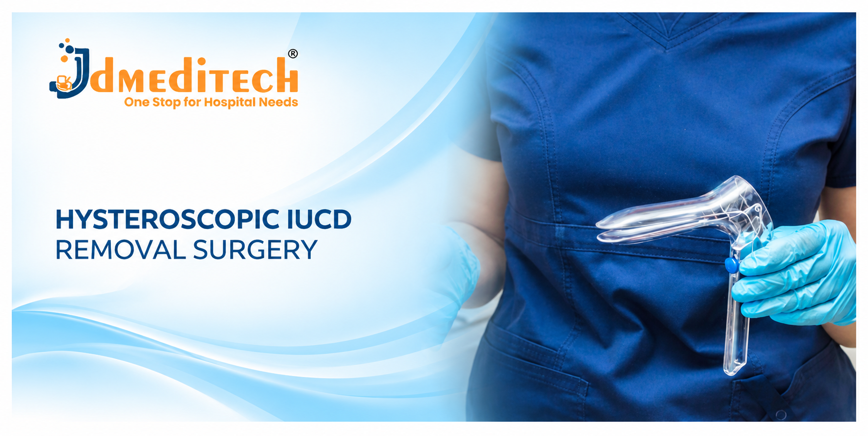
5. Gynecological Speculum
A speculum allows the healthcare professional to view the cervix during a gynecological examination.
Additionally, it may support selected IUCD removal procedures.
However, a speculum alone does not provide visualization inside the uterine cavity.
6. Hysteroscopy Camera System
A compatible camera system displays the hysteroscopic view on a medical monitor.
As a result, the specialist can assess the operative field during the procedure.
7. Medical Light Source
A medical light source illuminates the uterine cavity through a compatible telescope.
Therefore, the light cable and connector must match the selected hysteroscopy system.
8. Hysteroscopy Fluid Management System
A suitable fluid system helps maintain a clear view of the uterine cavity.
Moreover, the healthcare team monitors fluid use and fluid deficit during procedures that require uterine distension.
9. Ultrasound Equipment
Ultrasound helps clinicians identify the IUCD’s position inside the uterus.
In addition, ultrasound guidance may support selected retrieval attempts.
However, ultrasound equipment is separate from the hysteroscopic instrument set.
10. Additional Retrieval Instruments
Depending on the clinical findings, specialists may require compatible thread retrievers, scissors, or other instruments.
Nevertheless, instrument selection must follow clinical requirements and approved manufacturer instructions.
Hysteroscopic IUCD Removal Instruments and Functions
| Instrument | Primary Function |
|---|---|
| Hysteroscope Telescope | Provides a direct view of the uterine cavity |
| Diagnostic Hysteroscopy Sheath | Supports hysteroscope access and selected working instruments |
| Grasping Forceps | Helps retrieve suitable IUCD components |
| Alligator Forceps | Supports selected device retrieval procedures |
| Gynecological Speculum | Allows visualization of the cervix |
| Camera System | Displays the hysteroscopic view |
| Light Source | Provides illumination |
| Fluid Management System | Supports cavity distension and fluid monitoring |
| Ultrasound Equipment | Helps locate the IUCD and guide selected retrieval attempts |
| Additional Retrieval Instruments | Support specific retrieval requirements |
Importantly, hospitals must verify the actual equipment configuration and compatibility before use.
Types of IUCD Removal Methods
1. Routine IUCD Removal
Routine removal is the usual approach when the clinician can access the retrieval threads.
In most uncomplicated cases, the healthcare professional can remove the device during an office visit.
2. IUCD Thread Retrieval
Sometimes, the retrieval threads retract into the cervical canal.
After confirming the clinical situation, an appropriately trained clinician may use a suitable instrument to retrieve them.
3. Ultrasound-Guided IUCD Removal
Ultrasound guidance may help specialists remove selected IUCDs with missing threads.
Furthermore, imaging can provide useful information about the device’s position.
4. Hysteroscopic IUCD Removal
Hysteroscopy provides direct visualization inside the uterine cavity.
Therefore, specialists may use it when simpler retrieval methods fail or when device position makes removal difficult.
5. Surgical Removal of an Extrauterine IUCD
In rare cases, an IUCD may perforate the uterine wall and move outside the uterus.
However, hysteroscopy alone may not provide access to an extrauterine device.
Consequently, a gynecological specialist may consider laparoscopic or another suitable surgical approach.
How Is Hysteroscopic IUCD Removal Surgery Performed?
A qualified gynecologist performs hysteroscopic IUCD removal in an appropriate medical setting.
First, the healthcare team reviews the patient’s medical history, symptoms, and reason for removal.
Next, the specialist assesses the device’s position using suitable examination and imaging methods.
When necessary, the clinician excludes pregnancy and evaluates other possible causes of missing threads.
During hysteroscopy, a compatible telescope provides a direct view of the uterine cavity.
The specialist then assesses the IUCD and uses suitable retrieval instruments when removal is appropriate.
Throughout the procedure, the healthcare team monitors patient comfort and safety.
After removal, the clinician checks the retrieved device and evaluates whether further examination is necessary.
Finally, the patient receives recovery instructions and advice about future contraception.
However, the exact technique depends on the patient’s condition and device location. This overview does not replace professional procedural training.
Potential Benefits of Hysteroscopic IUCD Removal
1. Direct Visualization
Hysteroscopy allows the specialist to examine the uterine cavity directly.
As a result, the clinician can identify accessible IUCD components and assess the surrounding tissue.
2. Support for Difficult Removal
Some IUCDs resist standard removal methods.
In selected cases, hysteroscopy offers another approach after appropriate assessment.
3. No Abdominal Incision for Intrauterine Retrieval
Hysteroscopy usually provides access through the cervix.
Consequently, removal of a suitable intrauterine IUCD generally does not require an abdominal incision.
4. Assessment of Device Position
Direct visualization can help the specialist assess an IUCD’s location within the uterine cavity.
Moreover, it may reveal tissue changes that affect removal.
5. Potential Outpatient Treatment
Some specialists perform hysteroscopic IUCD removal in an outpatient setting.
However, the appropriate setting depends on patient comfort, device position, and procedural complexity.
6. Individualized Treatment
Hysteroscopy provides an additional treatment option for selected difficult cases.
Nevertheless, clinicians must weigh its benefits against simpler retrieval methods and possible complications.
Routine IUCD Removal vs Hysteroscopic IUCD Removal
| Feature | Routine IUCD Removal | Hysteroscopic IUCD Removal |
|---|---|---|
| Typical Indication | Accessible IUCD threads | Selected difficult retrieval cases |
| Visualization | Cervical examination | Direct uterine cavity visualization |
| Common Instruments | Speculum and suitable forceps | Hysteroscope and compatible retrieval instruments |
| Imaging | Not routinely required | Often useful before difficult retrieval |
| Clinical Setting | Usually office-based | Office or operating room |
| Complexity | Usually straightforward | Depends on device position and clinical findings |
Importantly, most IUCD removals do not require hysteroscopy. Specialists reserve more complex methods for suitable clinical situations.
Hysteroscopic IUCD Removal vs Ultrasound-Guided Removal
| Feature | Hysteroscopic Removal | Ultrasound-Guided Removal |
|---|---|---|
| Guidance | Direct endoscopic view | Ultrasound imaging |
| Common Equipment | Hysteroscope and retrieval instruments | Ultrasound system and suitable instruments |
| Uterine Cavity View | Direct visualization | Imaging-based assessment |
| Potential Use | Selected difficult or embedded IUCD cases | Selected devices with missing threads |
| Selection | Depends on clinical findings | Depends on clinical findings |
Both approaches have important clinical roles. Therefore, the treating specialist must choose the most suitable method for each patient.
Who May Need Additional Assessment Before IUCD Removal?
Patients with missing IUCD threads require appropriate evaluation before clinicians attempt intrauterine retrieval.
For example, the healthcare team may need to exclude pregnancy and confirm the device’s location.
If imaging suggests that the device lies outside the uterus, the patient requires specialist assessment.
Similarly, suspected infection, severe pelvic pain, or abnormal bleeding may require additional investigation.
Therefore, clinicians should not attempt difficult IUCD removal without suitable training and diagnostic information.
Risks and Complications of Hysteroscopic IUCD Removal
Hysteroscopic IUCD removal is an invasive procedure with potential complications.
For example, patients may experience temporary pelvic discomfort, cramping, or light bleeding.
In addition, hysteroscopy carries risks of infection, cervical injury, and uterine perforation.
Difficult retrieval may also cause device breakage or injury to nearby tissue.
Furthermore, procedures that use uterine distension fluid require appropriate fluid monitoring.
Consequently, specialist experience, careful patient selection, and suitable equipment are essential.
Recovery After Hysteroscopic IUCD Removal
Recovery depends on the procedure, anesthesia, and patient’s clinical condition.
Some patients experience mild cramping or spotting after treatment.
However, symptoms and recovery time vary between individuals.
The healthcare team provides advice about pain relief, daily activities, and follow-up.
Patients should seek prompt medical attention if they develop severe pain, heavy bleeding, fever, or unusual discharge.
Contraception After IUCD Removal
Fertility can return quickly after IUCD removal.
Therefore, patients who do not wish to become pregnant should discuss another contraceptive method before removal.
Furthermore, recent sexual activity may affect the timing of removal and the need for additional contraceptive advice.
If the patient wishes to conceive, the clinician can provide suitable preconception counseling.
How to Choose Hysteroscopic IUCD Removal Instruments
Hospitals and gynecology departments should identify the intended clinical application before selecting hysteroscopy instruments.
First, determine whether the requirement involves diagnostic hysteroscopy, device retrieval, or another operative procedure.
Next, review the telescope, sheath, forceps, camera, and fluid-system compatibility.
Important Equipment Selection Factors
- Approved intended use
- Hysteroscope telescope diameter
- Telescope viewing angle
- Diagnostic or operative sheath configuration
- Working channel diameter
- Grasping forceps design
- Alligator forceps compatibility
- Instrument working length
- Camera and light source compatibility
- Fluid inflow and outflow connections
- Fluid management requirements
- Instrument material
- Cleaning and sterilization instructions
- Reusable or single-use classification
- Actual instrument-set contents
- Manufacturer instructions for use
- Applicable regulatory documentation
Furthermore, purchasing teams should request the exact technical datasheet and packing list before ordering.
Hysteroscopy Instruments by JDMeditech
JDMeditech welcomes inquiries from hospitals, gynecology departments, surgical centers, and international medical distributors regarding hysteroscopy instruments and related surgical accessories.
For example, customers can inquire about available hysteroscopy telescopes, diagnostic sheaths, grasping forceps, alligator graspers, scissors, and other compatible instruments.
In addition, the JDMeditech team can confirm available product configurations, technical specifications, and applicable documentation for individual inquiries.
For product information and quotations, visit www.jdmeditech.com.
Frequently Asked Questions About Hysteroscopic IUCD Removal Surgery
1. What is hysteroscopic IUCD removal surgery?
It is a gynecological procedure in which a specialist uses a hysteroscope and suitable instruments to remove certain IUCDs under direct visualization.
2. Why is hysteroscopic IUCD removal necessary?
Specialists may consider hysteroscopy when routine removal fails or when the device’s position makes retrieval difficult.
3. What happens when IUCD threads are missing?
Clinicians assess the patient and confirm the device’s location. Depending on the findings, they may recommend observation, simpler retrieval methods, or specialist removal.
4. Can ultrasound help locate an IUCD?
Yes. Ultrasound commonly helps clinicians confirm an IUCD’s position inside the uterus.
5. What instruments are used during hysteroscopic IUCD removal?
Common equipment includes a hysteroscope, suitable sheath, grasping forceps, camera system, light source, and fluid management equipment.
6. Is an abdominal incision required?
Generally, no. Hysteroscopic removal uses access through the cervix. However, an IUCD outside the uterus may require a different surgical approach.
7. Can an embedded IUCD be removed by hysteroscopy?
In selected cases, yes. However, the specialist must assess the degree of embedment, device position, and risks before choosing treatment.
8. Is hysteroscopic IUCD removal painful?
Patients may experience discomfort or cramping. Therefore, the healthcare team should discuss suitable pain-management options according to the clinical setting.
9. What are the risks of hysteroscopic IUCD removal?
Potential complications include bleeding, infection, cervical injury, uterine perforation, and device breakage.
10. Where can hospitals inquire about JDMeditech hysteroscopy instruments?
Hospitals and medical distributors can visit JDMeditech to request product details and quotations.
Conclusion
Hysteroscopic IUCD Removal Surgery offers a useful treatment option for selected patients with difficult-to-remove intrauterine contraceptive devices.
Moreover, hysteroscopes, compatible grasping forceps, imaging systems, and other gynecological instruments support appropriate retrieval procedures.
However, successful treatment requires accurate device localization, careful patient assessment, specialist expertise, and suitable equipment.
Therefore, hospitals should select instruments according to clinical requirements, professional guidelines, and approved manufacturer instructions.
Contact JDMeditech for hysteroscopy instrument inquiries, gynecological surgical accessories, and international medical device requirements.
Medical Disclaimer: This article provides general educational information only. It does not replace professional medical advice or surgical training. Qualified healthcare professionals must make clinical and equipment decisions according to the patient’s condition, applicable guidelines, and approved manufacturer instructions.