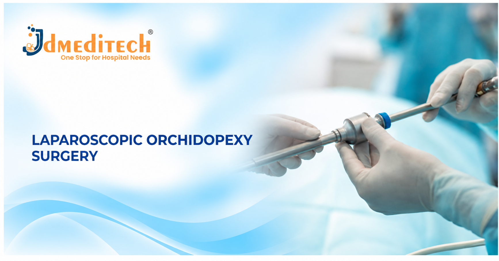
Introduction to Laparoscopic Orchidopexy Surgery
Laparoscopic Orchidopexy Surgery is a minimally invasive urological procedure used to locate and reposition selected undescended testes into the scrotum. The procedure is particularly valuable when a testis cannot be felt during physical examination and may be located inside the abdomen.
An undescended testis, medically known as cryptorchidism, is a condition in which one or both testicles do not reach their normal position in the scrotum. Although spontaneous descent can occur during early infancy, persistent undescended testes usually require specialist evaluation.
For selected patients, laparoscopic surgery provides direct visualization of the abdominal cavity. Furthermore, it helps surgeons assess testicular location, blood supply, and anatomical structures before planning treatment.
However, not every undescended testis requires a laparoscopic approach. Therefore, pediatric urologists choose the procedure according to clinical examination, testicular position, and individual circumstances.
In this guide, JDMeditech explains laparoscopic orchidopexy surgery, its indications, surgical techniques, instruments, potential benefits, risks, recovery, and frequently asked questions.
What Is Laparoscopic Orchidopexy Surgery?
Laparoscopic orchidopexy, also called laparoscopic orchiopexy, is a surgical technique that uses a camera and specialized instruments to manage selected undescended testes.
During the procedure, the surgeon introduces a laparoscope through a small abdominal incision. The camera provides images of the internal anatomy on a surgical monitor.
Next, the surgeon examines the testis, spermatic vessels, vas deferens, and surrounding structures. If appropriate, the surgical team mobilizes the testis and positions it within the scrotum.
However, some high intra-abdominal testes cannot safely reach the scrotum during one operation. Consequently, the surgeon may recommend a staged procedure.
Understanding Undescended Testis and Cryptorchidism
Normally, the testes develop inside the abdomen during fetal life and descend toward the scrotum before birth. However, one or both testes may remain outside their expected scrotal position.
This condition is called cryptorchidism. It may affect one testis or both testes.
Furthermore, an undescended testis may be located in the abdomen, inguinal canal, or another abnormal position.
Therefore, careful examination is necessary to determine whether the testis is palpable, nonpalpable, retractile, or ectopic.
Types of Undescended Testes
1. Palpable Undescended Testis
A palpable undescended testis can be felt during physical examination, usually in the groin.
In most cases, surgeons manage these testes through a conventional inguinal or scrotal orchidopexy rather than laparoscopy.
2. Nonpalpable Undescended Testis
A nonpalpable testis cannot be located by routine physical examination.
Consequently, examination under anesthesia and diagnostic laparoscopy may help determine whether the testis is intra-abdominal, located near the internal inguinal ring, or absent.
3. Intra-Abdominal Testis
An intra-abdominal testis remains within the abdominal cavity instead of descending into the scrotum.
Therefore, laparoscopic examination and treatment may be appropriate.
4. Ectopic Testis
An ectopic testis lies outside the normal pathway of descent. However, the surgical approach depends on its actual location.
5. Retractile Testis
A retractile testis can move between the scrotum and groin because of an active cremasteric reflex.
Importantly, retractile testes generally do not require immediate surgery. Nevertheless, regular examination is recommended because secondary ascent can occur.
What Causes Undescended Testes?
The development of cryptorchidism involves several biological and anatomical factors. However, a single cause cannot always be identified.
For example, hormonal influences, fetal development, and anatomical factors may affect testicular descent.
Additionally, premature birth is associated with a higher frequency of undescended testes.
Nevertheless, parents should understand that ordinary childcare activities do not cause this condition.
Why Is Orchidopexy Surgery Important?
Persistent undescended testes are associated with increased risks of impaired fertility and testicular malignancy later in life.
Furthermore, an undescended testis may be more difficult to examine when it remains outside the scrotum.
Therefore, timely orchidopexy aims to place a viable testis in an appropriate scrotal position and support future monitoring.
However, surgery does not completely eliminate fertility concerns or cancer risk.
When Should Orchidopexy Be Performed?
Spontaneous testicular descent becomes uncommon after approximately six months of corrected age.
Therefore, international pediatric urology guidance recommends timely specialist management when a testis remains undescended at this age.
Ideally, surgical treatment should be completed by 12 months of age and no later than 18 months.
However, children who present later still require appropriate assessment and management.
Who May Require Laparoscopic Orchidopexy?
Laparoscopic orchidopexy is particularly relevant for selected patients with a nonpalpable or intra-abdominal testis.
- Children with a persistent nonpalpable testis
- Patients with a confirmed intra-abdominal testis
- Selected patients with a testis near the internal inguinal ring
- Patients requiring laparoscopic anatomical assessment
- Selected patients requiring staged orchidopexy
- Patients referred for specialist management after previous evaluation
Importantly, the surgical team must confirm that the testis is present and suitable for repositioning.
How Is an Undescended Testis Diagnosed?
Clinical history and physical examination are the primary methods used to evaluate an undescended testis.
First, the pediatric urologist examines the scrotum and groin. Next, the clinician assesses whether the testis is palpable and whether it can remain within the scrotum.
However, routine ultrasound or MRI is generally not recommended before referral for an otherwise uncomplicated undescended testis because imaging cannot reliably establish whether a nonpalpable testis is present.
In selected patients with bilateral nonpalpable testes or atypical genital findings, additional endocrine or genetic evaluation may be necessary.
Furthermore, examination under anesthesia may identify a testis that was previously nonpalpable.
How Should Patients Prepare for Laparoscopic Orchidopexy?
Preparation depends on the child’s age, medical condition, and planned procedure.
First, the surgical team reviews the medical history and confirms the intended treatment.
Next, the pediatric anesthesia team assesses the patient and provides appropriate fasting instructions.
Additionally, parents or caregivers receive information about expected benefits, potential complications, postoperative pain management, and follow-up.
Finally, the surgeon obtains informed consent before proceeding.
How Is Laparoscopic Orchidopexy Surgery Performed?
A qualified pediatric urologist performs laparoscopic orchidopexy in an appropriately equipped operating room.
First, the anesthesia team provides general anesthesia. Next, the surgeon performs an examination to reassess testicular location.
When laparoscopy remains indicated, the surgeon introduces a laparoscope through a small abdominal incision.
Subsequently, carbon dioxide insufflation creates an operative space for visualization. The surgical team then examines the abdominal cavity to locate the testis and assess its anatomical relationships.
If the testis is viable and can be safely mobilized, the surgeon may reposition it into the scrotum.
However, a high intra-abdominal testis may require a staged procedure. Furthermore, some patients have an absent or severely atrophic testis, which changes the treatment plan.
Finally, the surgical team completes the operation, closes the incisions, and provides postoperative care instructions.
Types of Laparoscopic Orchidopexy Procedures
1. Single-Stage Laparoscopic Orchidopexy
A single-stage procedure may be appropriate when the testis can reach the scrotum without excessive tension.
Consequently, the surgeon can complete repositioning during one operation.
2. Two-Stage Fowler-Stephens Orchiopexy
Selected high intra-abdominal testes require a staged approach because the available spermatic vessel length may be insufficient.
In Fowler-Stephens orchidopexy, the surgical strategy relies on collateral blood supply to the testis. Therefore, preservation of adequate circulation is essential.
However, this approach carries a risk of testicular atrophy and requires specialist judgment.
3. Laparoscopic Traction Technique
Selected surgeons use a staged traction-based technique, sometimes called the Shehata technique, for certain high intra-abdominal testes.
This approach aims to achieve additional testicular mobility while preserving the spermatic vessels.
Nevertheless, technique selection depends on anatomy, surgeon experience, and clinical suitability.
4. Diagnostic Laparoscopy for Nonpalpable Testis
Diagnostic laparoscopy helps identify whether a nonpalpable testis is intra-abdominal, near the inguinal ring, or absent.
As a result, the surgeon can select the appropriate subsequent treatment.
Essential Instruments Used in Laparoscopic Orchidopexy Surgery
Laparoscopic orchidopexy instruments support abdominal visualization, tissue handling, controlled dissection, and surgical monitoring.
Furthermore, pediatric procedures require careful selection of instrument dimensions and compatible equipment.
The following list describes common instrument categories rather than a confirmed JDMeditech orchidopexy surgical kit.
1. Laparoscope Telescope
A laparoscope provides visualization of the abdominal cavity. Consequently, the surgeon can identify the testis and surrounding anatomical structures.
2. Laparoscopic Camera System
A compatible camera system transfers laparoscopic images to a surgical monitor.
3. Medical Light Source
A suitable light source illuminates the operative field through the laparoscope.
4. Fiber-Optic Light Cable
A compatible light cable transfers illumination from the light source to the laparoscope.
5. Laparoscopic Trocar and Cannula
Suitable trocars and cannulas provide access for the laparoscope and surgical instruments.
Importantly, pediatric instrument dimensions must be appropriate for the patient’s anatomy.
6. Laparoscopic Grasping Forceps
Grasping forceps support controlled tissue handling during selected laparoscopic procedures.
7. Atraumatic Laparoscopic Grasper
An atraumatic grasper supports delicate handling of suitable anatomical structures.
8. Laparoscopic Dissecting Forceps
Dissecting forceps support controlled tissue separation and surgical manipulation.
9. Laparoscopic Scissors
Laparoscopic scissors support selected tissue cutting tasks.
10. Laparoscopic Maryland Dissector
A compatible Maryland dissector supports selected fine dissection and tissue handling tasks.
11. Laparoscopic Electrosurgical Hook
A compatible electrosurgical hook may support selected dissection tasks when clinically appropriate.
However, energy devices require careful use near delicate anatomical structures.
12. Laparoscopic Bipolar Forceps
Bipolar forceps may support selected tissue coagulation tasks.
13. Laparoscopic Needle Holder
A needle holder supports laparoscopic suturing when required by the selected technique.
14. Laparoscopic Suction and Irrigation Instrument
A compatible suction and irrigation instrument helps manage fluid and maintain visualization.
15. CO2 Insufflator
A carbon dioxide insufflator supports controlled creation of the laparoscopic working space.
Furthermore, pressure and flow settings must be appropriate for pediatric patients.
16. Insufflation Tubing
Compatible tubing connects the insufflator to the selected access system.
17. Electrosurgical Generator
A compatible electrosurgical generator supplies energy to suitable instruments when required.
18. Surgical Monitor
A medical-grade monitor displays laparoscopic images during the procedure.
19. Surgical Sutures
Suitable sutures may support tissue closure or testicular fixation according to the surgeon’s chosen technique.
20. Sterile Surgical Accessories
Compatible drapes, tubing, gloves, and other sterile accessories support the planned procedure.
Laparoscopic Orchidopexy Instruments and Their Functions
| Instrument | Primary Function |
|---|---|
| Laparoscope Telescope | Provides abdominal visualization |
| Camera System | Displays operative images |
| Light Source | Illuminates the surgical field |
| Trocar and Cannula | Provide laparoscopic access |
| Grasping Forceps | Support controlled tissue handling |
| Atraumatic Grasper | Supports delicate tissue manipulation |
| Dissecting Forceps | Support tissue separation |
| Laparoscopic Scissors | Support selected tissue cutting |
| Maryland Dissector | Supports fine dissection |
| Electrosurgical Hook | Supports selected energy-based dissection |
| Bipolar Forceps | Support selected coagulation tasks |
| Needle Holder | Supports laparoscopic suturing |
| Suction and Irrigation Instrument | Helps maintain visualization |
| CO2 Insufflator | Creates the laparoscopic working space |
| Electrosurgical Generator | Supplies compatible surgical energy |
| Surgical Monitor | Displays laparoscopic images |
Benefits of Laparoscopic Orchidopexy Surgery
Laparoscopic orchidopexy offers several potential advantages for selected patients with nonpalpable or intra-abdominal testes.
1. Direct Visualization
Laparoscopy allows surgeons to examine abdominal anatomy directly.
2. Small Abdominal Incisions
The procedure uses small abdominal access incisions. Consequently, suitable patients may experience less abdominal wound discomfort than with some open abdominal approaches.
3. Diagnosis and Treatment
Diagnostic laparoscopy can help locate a nonpalpable testis. Furthermore, the surgeon may proceed with appropriate treatment during the same operation.
4. Testicular Repositioning
Successful orchidopexy places a viable testis within the scrotum.
5. Future Examination
A scrotal testis is generally easier to examine during later follow-up.
6. Individualized Surgical Planning
Laparoscopic findings help surgeons choose between single-stage, staged, or alternative management.
7. Potential Fertility Preservation
Timely treatment may support future fertility potential. However, orchidopexy cannot guarantee normal fertility.
Risks and Complications of Laparoscopic Orchidopexy
Although laparoscopic orchidopexy is an established procedure, complications can occur.
- Bleeding
- Wound infection
- Injury to nearby abdominal structures
- Injury to the vas deferens
- Damage to testicular blood vessels
- Testicular atrophy
- Testicular re-ascent
- Hematoma or swelling
- Incomplete repositioning
- Need for staged or additional surgery
- Anesthesia-related complications
Importantly, high intra-abdominal testes may carry greater risks of atrophy or retraction than testes treated through a standard inguinal approach.
Laparoscopic vs Open Orchidopexy
| Feature | Laparoscopic Orchidopexy | Open Orchidopexy |
|---|---|---|
| Typical Application | Selected nonpalpable or intra-abdominal testes | Most palpable undescended testes |
| Access | Small abdominal incisions, with additional scrotal access as needed | Inguinal or scrotal incision |
| Visualization | Laparoscopic camera | Direct surgical exposure |
| General Anesthesia | Usually required | Usually required |
| Staged Surgery | May be needed for high intra-abdominal testes | Depends on clinical circumstances |
| Technique Selection | Based on nonpalpable testis evaluation | Based on palpable testis location |
Recovery After Laparoscopic Orchidopexy Surgery
Recovery depends on the child’s age, surgical complexity, and anesthesia.
Many children return home after an uncomplicated procedure. However, some patients require additional observation or inpatient care.
Initially, mild discomfort, bruising, or swelling may occur near the incision sites.
Over time, these symptoms usually improve. Nevertheless, parents should follow instructions regarding wound care, bathing, activity restrictions, and prescribed pain relief.
Furthermore, follow-up appointments are important to assess the testicular position and postoperative healing.
Postoperative Care After Orchidopexy
- Follow the surgeon’s wound-care instructions
- Administer prescribed pain relief as directed
- Keep scheduled follow-up appointments
- Follow bathing and dressing instructions
- Observe for unusual swelling or bleeding
- Follow activity restrictions
- Report persistent or increasing pain
- Contact the surgical team about fever or wound discharge
- Discuss long-term testicular follow-up with the specialist
Warning Signs After Laparoscopic Orchidopexy
Parents and caregivers should seek prompt medical advice when concerning symptoms occur.
- Persistent or high fever
- Increasing wound redness or discharge
- Severe or worsening pain
- Significant swelling or bleeding
- Persistent vomiting
- Difficulty passing urine
- Unexpected scrotal discoloration
- Other serious postoperative symptoms
Severe bleeding, difficulty breathing, collapse, or other emergency symptoms require immediate medical care.
Orchidopexy and Future Fertility
Undescended testes are associated with impaired fertility potential, especially when both testes are affected.
Therefore, early treatment is important. However, even successful orchidopexy does not guarantee normal sperm production later in life.
Furthermore, long-term outcomes depend on testicular development, location, and other individual factors.
Orchidopexy and Testicular Cancer Risk
Cryptorchidism is associated with an increased risk of testicular cancer later in life.
Timely orchidopexy may reduce this risk compared with delayed treatment. Nevertheless, the risk is not completely eliminated.
Additionally, positioning the testis in the scrotum makes future examination easier.
Therefore, appropriate education and follow-up remain important as the patient grows older.
How to Choose Laparoscopic Orchidopexy Instruments
Hospitals and pediatric urology departments should carefully assess their surgical requirements before purchasing laparoscopic instruments.
First, purchasing teams should identify the intended procedures and patient age groups.
Next, they should review laparoscope diameter, trocar dimensions, instrument lengths, and compatibility with existing camera systems.
Furthermore, hospitals should verify the suitability of graspers, scissors, dissectors, and energy devices for delicate pediatric anatomy.
Insufflation equipment also requires careful consideration. Therefore, purchasing teams should confirm pediatric operating capabilities and manufacturer instructions.
Finally, hospitals should review sterilization requirements, reusable or single-use classification, and applicable regulatory documentation.
Important Equipment Selection Factors
- Approved intended use
- Pediatric instrument suitability
- Laparoscope diameter and viewing angle
- Trocar and cannula dimensions
- Instrument length and working diameter
- Atraumatic grasper specifications
- Dissector and scissors compatibility
- Camera system compatibility
- Light source compatibility
- CO2 insufflator capabilities
- Electrosurgical generator compatibility
- Cleaning and sterilization instructions
- Reusable or single-use classification
- Manufacturer technical documentation
- Applicable regulatory documentation
Laparoscopic Orchidopexy Instruments – JDMeditech
JDMeditech welcomes inquiries from hospitals, pediatric urology departments, surgical centers, healthcare institutions, and international medical distributors regarding laparoscopic instruments and related accessories.
For example, customers can inquire about available laparoscopic grasping forceps, scissors, trocars, needle holders, dissectors, and compatible surgical equipment.
Furthermore, the JDMeditech team can discuss available configurations, technical specifications, intended uses, and applicable documentation.
Therefore, hospitals and medical distributors can contact JDMeditech to discuss their laparoscopic urology equipment requirements.
For product information and international business inquiries, visit www.jdmeditech.com.
Frequently Asked Questions About Laparoscopic Orchidopexy Surgery
1. What is laparoscopic orchidopexy surgery?
It is a minimally invasive surgical procedure used to locate and reposition selected undescended testes into the scrotum.
2. What is an undescended testis?
An undescended testis is a testicle that has not reached its normal position in the scrotum.
3. What is cryptorchidism?
Cryptorchidism is the medical term for an undescended testis.
4. What is the difference between orchidopexy and orchiopexy?
Both terms refer to surgical repositioning and fixation of a testis.
5. When should orchidopexy be performed?
Guidelines recommend completing treatment ideally by 12 months of age and no later than 18 months when a testis remains undescended after six months of corrected age.
6. Is laparoscopy required for every undescended testis?
No. Most palpable undescended testes are treated using inguinal or scrotal surgery.
7. Why is laparoscopy used for a nonpalpable testis?
Laparoscopy helps determine whether the testis is located inside the abdomen, near the inguinal ring, or absent.
8. What is an intra-abdominal testis?
An intra-abdominal testis is a testicle that remains within the abdominal cavity.
9. What is Fowler-Stephens orchidopexy?
It is a specialized surgical technique used for selected high intra-abdominal testes when additional mobility is required.
10. Can orchidopexy require two operations?
Yes. Some high intra-abdominal testes require staged treatment.
11. What instruments are used in laparoscopic orchidopexy?
Common instruments include laparoscopes, trocars, graspers, scissors, dissectors, needle holders, and camera systems.
12. Is general anesthesia required?
Yes. Laparoscopic orchidopexy is generally performed under general anesthesia.
13. Does laparoscopic orchidopexy leave scars?
The procedure requires small abdominal incisions and may also involve scrotal access. Consequently, small scars can remain.
14. How long does laparoscopic orchidopexy take?
Operation time varies according to testicular location, anatomy, and surgical complexity.
15. What are the risks of orchidopexy?
Potential risks include bleeding, infection, testicular atrophy, re-ascent, and injury to nearby structures.
16. Can a testis become smaller after orchidopexy?
Yes. Testicular atrophy can occur if blood supply is inadequate or compromised.
17. Can orchidopexy improve fertility?
Timely treatment may support future fertility potential. However, normal fertility is not guaranteed.
18. Does orchidopexy eliminate testicular cancer risk?
No. Early treatment may reduce risk, but ongoing awareness and appropriate follow-up remain important.
19. How long is recovery after laparoscopic orchidopexy?
Recovery varies. Many children resume usual light activities relatively quickly after uncomplicated surgery, following their surgeon’s advice.
20. Where can hospitals inquire about JDMeditech laparoscopic instruments?
Hospitals and medical distributors can visit JDMeditech to inquire about available laparoscopic instruments and related accessories.
Conclusion
Laparoscopic Orchidopexy Surgery is an important treatment option for selected patients with nonpalpable or intra-abdominal undescended testes.
Furthermore, laparoscopic visualization helps surgeons evaluate testicular location and select an appropriate surgical approach.
However, successful treatment depends on timely diagnosis, appropriate patient selection, surgical expertise, and postoperative follow-up.
For laparoscopic orchidopexy instrument inquiries and international business requirements, contact JDMeditech – One Stop for Hospital Needs.
Website: www.jdmeditech.com
Medical Disclaimer: This article provides general educational information only. It does not replace professional medical advice, diagnosis, or surgical training. Qualified healthcare professionals must make treatment and equipment decisions according to current clinical guidance, local regulations, and approved manufacturer instructions.