Introduction to Laparoscopic Pelvic Lymph Node Dissection Surgery
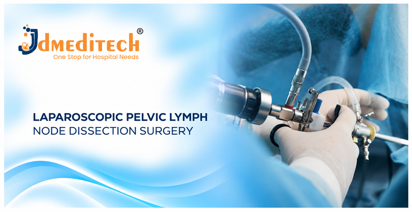
Laparoscopic Pelvic Lymph Node Dissection is a minimally invasive surgical procedure in which surgeons remove selected lymph nodes from the pelvic region. Doctors commonly use this procedure during the assessment and treatment of certain pelvic cancers.
During the operation, surgeons use a laparoscope, camera system, and specialized surgical instruments. As a result, the surgical team can examine the pelvic anatomy through small abdominal incisions.
Furthermore, pathological examination of the removed lymph nodes helps doctors determine whether cancer cells have spread. Therefore, lymph node assessment can provide important information for cancer staging and treatment planning.
However, not every patient requires extensive pelvic lymph node removal. In selected cases, doctors may recommend sentinel lymph node biopsy or another staging approach instead.
In this guide, JDMeditech explains the surgical procedure, clinical applications, laparoscopic instruments, possible benefits, risks, recovery, and frequently asked questions.
What Is Laparoscopic Pelvic Lymph Node Dissection?
Laparoscopic pelvic lymph node dissection, also called laparoscopic pelvic lymphadenectomy, involves removing lymph nodes from selected areas of the pelvis.
The lymphatic system helps the body transport lymph fluid and supports immune function. Additionally, lymph nodes filter lymph fluid and contain immune cells.
However, cancer cells can sometimes spread through lymphatic pathways. Consequently, doctors may examine pelvic lymph nodes to assess whether certain cancers have spread beyond their original location.
During surgery, the team removes lymphatic tissue according to the planned oncological procedure. Afterward, a pathologist examines the specimens under a microscope.
Where Are the Pelvic Lymph Nodes Located?
Pelvic lymph nodes lie near important blood vessels, nerves, and pelvic organs. Therefore, surgeons need detailed anatomical knowledge during lymph node dissection.
Important pelvic lymph node regions include:
- External iliac lymph nodes
- Internal iliac lymph nodes
- Obturator lymph nodes
- Common iliac lymph nodes
- Other pelvic nodal groups when clinically indicated
Furthermore, the planned surgical boundaries depend on the cancer type and treatment strategy. Surgeons must also protect nearby structures, including major blood vessels, ureters, and nerves.
Why Is Laparoscopic Pelvic Lymph Node Dissection Performed?
Doctors primarily use pelvic lymph node assessment to investigate possible cancer spread. Additionally, lymph node findings may help guide further treatment.
1. Prostate Cancer
Pelvic lymph node dissection may form part of surgical management for selected patients with prostate cancer. For example, doctors may recommend an extended pelvic lymph node dissection when the estimated risk of lymph node involvement justifies it.
However, the decision depends on cancer risk assessment, clinical guidelines, and individual patient factors.
2. Endometrial Cancer
Doctors assess lymph nodes in selected patients with endometrial cancer to determine the extent of disease. However, modern treatment guidelines increasingly support sentinel lymph node mapping for appropriate patients.
Therefore, systematic pelvic lymphadenectomy is not necessary for every patient with endometrial cancer.
3. Cervical Cancer
Pelvic lymph node assessment may play an important role in selected cervical cancer treatment plans. In addition, lymph node findings can influence staging and subsequent treatment decisions.
Nevertheless, doctors must follow current oncological guidance when selecting surgery, radiation therapy, or other treatments.
4. Bladder Cancer
Pelvic lymph node dissection commonly forms part of radical cystectomy for selected patients with bladder cancer. Furthermore, pathological findings help determine the extent of cancer spread.
5. Other Selected Pelvic Malignancies
Doctors may consider pelvic lymph node assessment for other cancers when clinical evidence supports it. However, the indication and surgical extent vary by cancer type.
Types of Pelvic Lymph Node Surgery
Different procedures remove different amounts of lymphatic tissue. Therefore, doctors select the appropriate approach according to the patient’s cancer diagnosis.
Sentinel Lymph Node Biopsy
Sentinel lymph node biopsy focuses on the first lymph node or group of nodes likely to receive lymphatic drainage from a tumor.
In selected cancers, this approach can provide staging information while removing fewer lymph nodes. Consequently, it may reduce some complications associated with extensive dissection.
Systematic Pelvic Lymph Node Dissection
Systematic dissection removes lymphatic tissue from specified pelvic regions. Surgeons follow a planned anatomical template according to the cancer type and relevant guidelines.
Extended Pelvic Lymph Node Dissection
Extended pelvic lymph node dissection covers a broader set of pelvic lymph node regions. For example, doctors may recommend it for selected patients undergoing prostate cancer surgery.
However, the precise boundaries depend on the oncological procedure and clinical guidance.
Laparoscopic vs Open Pelvic Lymph Node Dissection
Surgeons may use laparoscopic, robotic-assisted, or open techniques for pelvic lymph node assessment. Each approach has specific advantages and limitations.
| Feature | Laparoscopic Approach | Open Approach |
|---|---|---|
| Incisions | Several smaller abdominal incisions | A larger surgical incision |
| Visualization | Camera-assisted magnified view | Direct operative exposure |
| Early Recovery | May be faster in suitable cases | May require longer recovery |
| Hospital Stay | May be shorter in selected patients | Depends on surgical complexity |
| Suitability | Depends on diagnosis and expertise | May be preferable in selected complex cases |
Importantly, surgeons must prioritize oncological safety rather than choosing an approach based only on incision size.
How Is Laparoscopic Pelvic Lymph Node Dissection Performed?
A qualified surgical team performs laparoscopic pelvic lymph node dissection in an appropriately equipped operating room.
First, the team reviews the patient’s diagnosis, imaging findings, and treatment plan. Next, the anesthesia team prepares the patient for the operation.
During surgery, the surgeon establishes laparoscopic access and examines the pelvic anatomy through the camera system.
Then, the surgeon identifies the relevant lymph node regions and performs the planned dissection using compatible instruments. Throughout the operation, the team takes care to protect important blood vessels, nerves, and nearby organs.
After removing the selected lymphatic tissue, the surgeon places specimens into suitable containers or retrieval systems. Subsequently, the pathology department examines the specimens.
Finally, the team completes the procedure and closes the surgical access sites. However, the exact technique depends on the patient’s cancer type and the planned operation.
Common Instruments Used in Laparoscopic Pelvic Lymph Node Dissection
Laparoscopic pelvic lymph node dissection instruments support visualization, abdominal access, tissue handling, dissection, bleeding control, and specimen retrieval.
Furthermore, instrument selection depends on the surgeon’s technique and the equipment available in the operating room.
The following list describes common instrument categories. It does not represent a confirmed JDMeditech product set.
1. Laparoscope Telescope
A laparoscope provides magnified images of the pelvic cavity. As a result, surgeons can visualize relevant anatomical structures during the operation.
2. Laparoscopic Camera System
A camera system transfers images from the laparoscope to a surgical monitor. Additionally, suitable image quality supports clear visualization.
3. Medical Light Source
A medical light source provides illumination inside the operative field. Therefore, surgeons can observe pelvic structures more clearly.
4. Fiber-Optic Light Cable
A compatible light cable transfers illumination from the light source to the laparoscope.
5. Laparoscopic Trocar
A laparoscopic trocar forms part of the surgical access system. It supports the introduction of compatible instruments into the abdominal cavity.
6. Laparoscopic Cannula
A cannula maintains an access pathway for surgical instruments. Furthermore, its dimensions must match the planned procedure.
7. CO2 Insufflator
A carbon dioxide insufflator helps maintain the abdominal working space. Consequently, surgeons gain room for visualization and instrument movement.
8. Laparoscopic Grasping Forceps
Grasping forceps help surgeons manipulate suitable tissues during the procedure. Additionally, different jaw designs support different tasks.
9. Atraumatic Grasper
An atraumatic grasper supports controlled handling of delicate tissues. Therefore, surgeons may use it when gentle tissue manipulation is appropriate.
10. Maryland Dissector
A Maryland dissector supports fine tissue separation. Moreover, its design helps surgeons perform controlled dissection in suitable anatomical areas.
11. Laparoscopic Scissors
Laparoscopic scissors support tissue cutting during selected surgical tasks. Furthermore, surgeons choose suitable blade designs and working lengths.
12. Laparoscopic Needle Holder
A laparoscopic needle holder supports suturing when the operation requires tissue repair or closure.
13. Bipolar Forceps
Bipolar forceps support coagulation and bleeding control. Additionally, surgeons use them with compatible electrosurgical equipment.
14. Monopolar Electrosurgical Instrument
A suitable monopolar instrument supports selected cutting and coagulation tasks. However, safe use requires compatible equipment and appropriate surgical training.
15. Vessel-Sealing Device
A vessel-sealing device may support selected vessel and tissue management tasks. Therefore, the surgical team must verify the device’s approved intended use.
16. Laparoscopic Clip Applicator
A clip applicator supports the placement of compatible surgical clips when clinically appropriate.
17. Suction and Irrigation Cannula
Suction and irrigation instruments help manage fluids in the operative field. As a result, the team can maintain visibility during suitable surgical tasks.
18. Specimen Retrieval Bag
A specimen retrieval bag supports controlled removal of excised lymphatic tissue. Furthermore, correct specimen identification helps the pathology team assess the relevant lymph node regions.
19. Surgical Sutures
Surgeons use suitable sutures when the procedure requires tissue repair or incision closure.
20. Surgical Monitor
A medical-grade monitor displays laparoscopic images. Consequently, the surgical team can observe the operative field throughout the procedure.
21. Endoscopy Trolley
An endoscopy trolley supports organized placement of laparoscopic equipment. Additionally, a suitable equipment arrangement helps the team manage the surgical setup.
22. Sterile Surgical Accessories
Compatible drapes, tubing, dressings, and other sterile accessories support the planned procedure. Furthermore, the team follows appropriate infection-control practices.
Laparoscopic Pelvic Lymph Node Dissection Instruments and Functions
| Instrument | Primary Function |
|---|---|
| Laparoscope | Provides pelvic visualization |
| Camera System | Displays operative images |
| Light Source | Provides illumination |
| Trocar and Cannula | Support laparoscopic access |
| CO2 Insufflator | Maintains abdominal working space |
| Grasping Forceps | Support tissue handling |
| Maryland Dissector | Supports fine tissue dissection |
| Laparoscopic Scissors | Support controlled tissue cutting |
| Needle Holder | Supports suturing |
| Bipolar Forceps | Support coagulation |
| Vessel-Sealing Device | Supports selected vessel management |
| Clip Applicator | Supports selected clip placement |
| Suction and Irrigation | Support fluid management |
| Specimen Retrieval Bag | Supports tissue retrieval |
Potential Benefits of Laparoscopic Pelvic Lymph Node Dissection
Laparoscopic surgery may offer advantages for suitable patients. However, the clinical benefit depends on the underlying cancer and the planned treatment.
1. Smaller Surgical Incisions
Laparoscopic surgery generally uses smaller abdominal incisions than open surgery. As a result, patients may have smaller scars.
2. Magnified Visualization
The laparoscopic camera provides magnified views of pelvic structures. Therefore, surgeons can examine relevant anatomy during the operation.
3. Potentially Shorter Hospital Stay
Selected patients may require a shorter hospital stay than after comparable open surgery. However, recovery depends on the complete operation and any complications.
4. Faster Early Recovery
Some patients return to light daily activities sooner after minimally invasive surgery. Nevertheless, recovery varies between individuals.
5. Cancer Staging Information
Pathological examination of removed lymph nodes can reveal whether cancer cells have spread. Consequently, doctors may use these findings to guide further treatment.
Risks and Complications of Pelvic Lymph Node Dissection
Pelvic lymph node dissection carries important surgical risks. Therefore, patients should discuss the expected benefits and potential complications with their treating team.
- Bleeding
- Surgical-site infection
- Injury to nearby blood vessels
- Injury to the ureter or other pelvic structures
- Nerve injury
- Blood clots
- Anesthesia-related complications
- Lymphocele formation
- Lower-limb lymphedema
- Postoperative pain
- Possible conversion to open surgery
- Other procedure-specific complications
Importantly, lymphocele and lymphedema are particularly relevant to lymph node surgery because the operation can disrupt lymphatic drainage.
What Is a Lymphocele?
A lymphocele is a collection of lymphatic fluid that can develop after lymphatic vessels are disrupted during surgery.
Some lymphoceles cause no symptoms. However, larger or symptomatic collections may cause discomfort, infection, or pressure on nearby structures.
Therefore, doctors assess postoperative symptoms and recommend appropriate management when necessary.
What Is Lymphedema?
Lymphedema is swelling caused by impaired lymphatic drainage. After pelvic lymph node surgery, it may affect one or both legs.
For example, patients may notice persistent leg swelling, heaviness, or discomfort. Additionally, symptoms can develop months or years after treatment.
Therefore, patients should report new or persistent swelling to their healthcare team.
Recovery After Laparoscopic Pelvic Lymph Node Dissection
Recovery depends on the extent of lymph node removal and whether the surgeon performs other procedures during the same operation.
Initially, patients may experience abdominal discomfort, fatigue, or incision tenderness. Furthermore, the healthcare team monitors for bleeding, infection, and other complications.
Over time, patients gradually resume daily activities according to medical advice. However, recovery after major cancer surgery may take longer than recovery after an isolated laparoscopic procedure.
Postoperative Care After Pelvic Lymph Node Dissection
Proper postoperative care supports healing and helps identify complications early. Therefore, patients should follow their surgeon’s instructions.
- Take prescribed medicines as directed
- Keep surgical wounds clean
- Attend follow-up appointments
- Review pathology results with the treating doctor
- Follow activity and lifting restrictions
- Report persistent leg swelling
- Follow blood-clot prevention instructions
- Discuss any additional cancer treatment
Furthermore, patients should seek medical attention for persistent fever, worsening abdominal pain, unusual wound discharge, or new leg swelling.
Importantly, difficulty breathing, severe bleeding, or other serious symptoms require emergency medical care.
How Do Pathology Results Help Cancer Treatment?
After surgery, a pathologist examines the removed lymph nodes for cancer cells. The pathology report may describe the number of nodes examined and the number containing cancer.
For example, a positive lymph node indicates that the pathologist identified cancer cells within the node.
Consequently, the treating team may use these findings to refine cancer staging and discuss further treatment.
However, doctors interpret lymph node findings together with the original tumor diagnosis, imaging, and other clinical information.
How to Choose Laparoscopic Pelvic Lymph Node Dissection Instruments
Hospitals and surgical departments should evaluate their equipment requirements before purchasing laparoscopic instruments.
First, the purchasing team should identify the intended procedures and required instrument functions. Next, it should confirm compatibility with the existing camera, light source, insufflator, and electrosurgical equipment.
Furthermore, teams should review instrument working lengths, jaw designs, cleaning instructions, and approved intended uses.
Finally, buyers should verify the manufacturer’s technical documentation and applicable regulatory information.
Important Instrument Selection Factors
- Approved intended use
- Planned surgical application
- Laparoscope diameter and viewing angle
- Camera and monitor compatibility
- Medical light source compatibility
- Trocar and cannula dimensions
- CO2 insufflator compatibility
- Grasper jaw design
- Dissector working length
- Laparoscopic scissors configuration
- Needle holder specifications
- Electrosurgical compatibility
- Vessel-sealing device compatibility
- Clip applicator compatibility
- Suction and irrigation requirements
- Specimen retrieval requirements
- Reusable or single-use classification
- Cleaning and sterilization instructions
- Manufacturer technical documentation
- Applicable regulatory documentation
Laparoscopic Surgical Instruments – JDMeditech
JDMeditech welcomes inquiries from hospitals, oncology departments, urology centers, gynecological surgery departments, and international medical distributors regarding laparoscopic surgical instruments and related accessories.
For example, customers can inquire about available laparoscopic telescopes, grasping forceps, dissecting forceps, scissors, needle holders, and compatible accessories.
Furthermore, the JDMeditech team can discuss available product configurations, technical specifications, intended uses, and applicable documentation.
Therefore, hospitals and medical distributors can contact JDMeditech to discuss their surgical instrument requirements.
For product information and business inquiries, visit www.jdmeditech.com.
Frequently Asked Questions About Laparoscopic Pelvic Lymph Node Dissection
1. What is laparoscopic pelvic lymph node dissection?
It is a minimally invasive surgical procedure that removes selected lymph nodes from the pelvis. Doctors often use it during cancer staging or treatment.
2. What is pelvic lymphadenectomy?
Pelvic lymphadenectomy is another name for pelvic lymph node dissection. Therefore, both terms describe the removal of pelvic lymph nodes.
3. Why do surgeons remove pelvic lymph nodes?
Surgeons remove selected lymph nodes to assess possible cancer spread or meet other specific oncological treatment goals.
4. Which cancers may require pelvic lymph node assessment?
Selected prostate, bladder, cervical, and endometrial cancers may require pelvic lymph node assessment. However, the indication varies by diagnosis.
5. Is pelvic lymph node dissection always necessary?
No. In selected cases, doctors may recommend sentinel lymph node biopsy or another approach instead.
6. What is the difference between sentinel node biopsy and pelvic lymphadenectomy?
Sentinel node biopsy targets selected first-draining lymph nodes. In contrast, systematic pelvic lymphadenectomy removes lymphatic tissue from a broader planned region.
7. What instruments are used during the procedure?
Common instruments include laparoscopes, trocars, graspers, dissectors, scissors, needle holders, and suitable vessel-management devices.
8. Is laparoscopic pelvic lymph node dissection minimally invasive?
Yes. Surgeons generally use small incisions and a camera-assisted system. However, the procedure still carries important surgical risks.
9. What is a lymphocele?
A lymphocele is a collection of lymphatic fluid that may develop after lymph node surgery.
10. What is lymphedema?
Lymphedema is swelling caused by impaired lymph drainage. For example, it may affect the legs after pelvic lymph node removal.
11. Can pelvic lymph node surgery cause leg swelling?
Yes. Disruption of lymphatic drainage can cause swelling. Therefore, patients should report persistent or new leg swelling.
12. What are the main risks of pelvic lymphadenectomy?
Potential risks include bleeding, infection, vessel injury, blood clots, lymphocele, and lymphedema.
13. How long does the procedure take?
Operation time depends on the planned dissection and any additional surgery. Therefore, the surgical team provides an individual estimate.
14. How long does recovery take?
Recovery varies according to the patient’s condition and the extent of surgery. Furthermore, additional cancer procedures may lengthen recovery.
15. What happens to removed lymph nodes?
A pathology laboratory examines the removed tissue for cancer cells. Consequently, the findings can help doctors assess disease spread.
16. Can lymph node results affect cancer treatment?
Yes. Pathology findings may influence staging and decisions about additional treatment.
17. Can pelvic lymph node dissection be performed with prostate surgery?
Yes. Surgeons may perform pelvic lymph node dissection during radical prostatectomy for selected patients.
18. Can the procedure be performed during gynecological cancer surgery?
Yes. Pelvic lymph node assessment may form part of selected gynecological cancer operations.
19. Is open surgery sometimes necessary?
Yes. Surgeons may recommend open surgery for selected complex conditions or when a minimally invasive approach is unsuitable.
20. Where can hospitals inquire about JDMeditech laparoscopic instruments?
Hospitals and medical distributors can visit JDMeditech to inquire about available laparoscopic surgical instruments and accessories.
Conclusion
Laparoscopic Pelvic Lymph Node Dissection plays an important role in the assessment and management of selected pelvic cancers.
Furthermore, pathological examination of lymph nodes helps doctors understand the extent of disease and plan appropriate treatment.
However, systematic lymph node removal is not necessary for every patient. Therefore, surgeons must follow current cancer-specific guidance when choosing between sentinel lymph node biopsy, systematic dissection, and other approaches.
Successful care depends on appropriate patient selection, qualified surgical expertise, compatible medical equipment, and postoperative monitoring.
For laparoscopic surgical instrument inquiries and international business requirements, contact JDMeditech – One Stop for Hospital Needs.
Website: www.jdmeditech.com
Medical Disclaimer: This article provides general educational information only. It does not replace professional medical advice, oncology consultation, or surgical training. Qualified healthcare professionals must make treatment and equipment decisions according to current clinical guidance, local regulations, and approved manufacturer instructions.