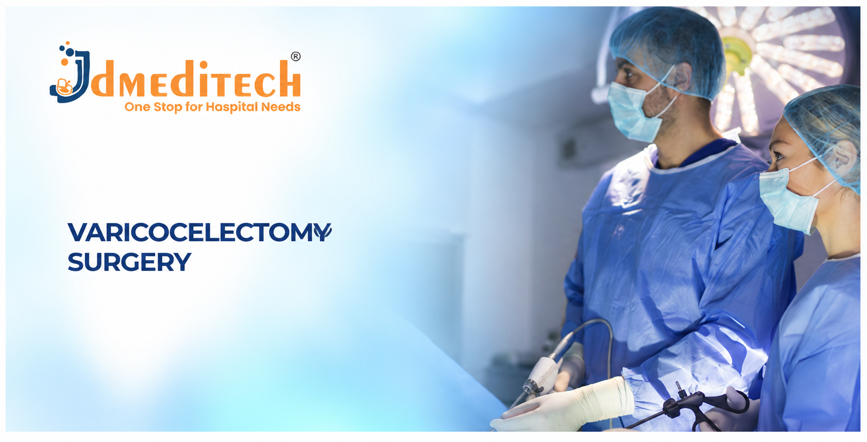
Introduction to Varicocelectomy Surgery
Varicocelectomy Surgery is a urological procedure that treats enlarged veins around the testicle. These abnormal veins, known as a varicocele, develop within the pampiniform plexus of the spermatic cord.
Although many varicoceles cause no symptoms, some patients experience scrotal discomfort, abnormal semen parameters, or changes in testicular development. Therefore, urologists may recommend treatment after evaluating the patient’s symptoms and reproductive goals.
During varicocelectomy, surgeons interrupt abnormal venous drainage while protecting important structures. Furthermore, microsurgical techniques use magnification to help identify small arteries, veins, and lymphatic vessels.
In this guide, JDMeditech explains varicocelectomy surgery, its indications, techniques, surgical instruments, benefits, complications, recovery, and frequently asked questions.
What Is Varicocelectomy Surgery?
Varicocelectomy is an operation that treats a varicocele by interrupting selected enlarged spermatic veins. Consequently, blood can drain through alternative venous pathways.
Surgeons may perform the procedure through an open, microsurgical, or laparoscopic approach. However, the preferred technique depends on the patient’s condition, available expertise, and treatment goals.
Importantly, varicocelectomy does not remove the testicle. Instead, the surgeon aims to preserve the testicle, testicular artery, vas deferens, and lymphatic vessels.
Understanding a Varicocele
A varicocele is an abnormal enlargement of veins within the pampiniform plexus. These veins help drain blood from the testicle.
Normally, venous valves and blood-flow patterns help direct blood away from the testicle. However, abnormal venous reflux can contribute to enlarged veins.
Furthermore, varicoceles occur more frequently on the left side because of anatomical differences in venous drainage.
Some patients describe the enlarged veins as feeling like a bag of worms. Nevertheless, many people notice no symptoms.
Types and Grades of Varicocele
1. Grade I Varicocele
The clinician can feel the varicocele only during a Valsalva maneuver, such as bearing down.
2. Grade II Varicocele
The clinician can feel the enlarged veins without a Valsalva maneuver. However, the veins are not clearly visible.
3. Grade III Varicocele
The enlarged veins are visible during physical examination. Consequently, the condition may cause noticeable scrotal swelling.
4. Subclinical Varicocele
Ultrasound detects this varicocele, but the clinician cannot feel it during examination. Importantly, infertility guidelines do not recommend routine surgical repair for ultrasound-only varicoceles.
5. Bilateral Varicocele
A bilateral varicocele affects both sides. Therefore, the urologist evaluates each side before planning treatment.
Common Symptoms of Varicocele
Many varicoceles cause no symptoms. However, some patients experience discomfort or reproductive concerns.
Possible symptoms and findings include:
- Dull scrotal pain
- A feeling of heaviness
- Visible enlarged scrotal veins
- Discomfort after prolonged standing
- Symptoms that improve while lying down
- Reduced testicular volume in selected patients
- Abnormal semen parameters
- Difficulty conceiving
Importantly, sudden severe testicular pain requires urgent medical assessment. Doctors must exclude testicular torsion and other emergencies.
What Causes a Varicocele?
Varicoceles develop when venous drainage around the testicle becomes abnormal. As a result, blood may collect within the pampiniform plexus.
Several factors may contribute, including venous anatomy, valve dysfunction, and increased venous pressure.
Furthermore, the left spermatic vein drains into the left renal vein. This anatomical arrangement contributes to the higher frequency of left-sided varicoceles.
However, a new, rapidly developing, or non-reducible varicocele may require additional evaluation for an underlying cause.
How Do Doctors Diagnose a Varicocele?
1. Medical History
First, the urologist reviews symptoms, fertility concerns, and previous medical conditions.
2. Physical Examination
Next, the doctor examines the scrotum while the patient stands. The examination may also include a Valsalva maneuver.
3. Scrotal Doppler Ultrasound
Ultrasound helps assess testicular volume, venous enlargement, and blood-flow reflux. Additionally, it can identify other scrotal conditions.
4. Semen Analysis
For patients experiencing infertility, semen analysis helps evaluate sperm concentration, movement, and other relevant parameters.
5. Hormonal Evaluation
Doctors may request testosterone or other reproductive hormone tests when clinical findings suggest hormonal abnormalities.
6. Additional Imaging
In selected circumstances, clinicians may investigate possible secondary causes. However, not every varicocele requires abdominal imaging.
When Is Varicocelectomy Surgery Recommended?
Not every varicocele requires surgery. In fact, many patients need only observation and periodic evaluation.
However, a urologist may recommend repair when the condition causes clinically significant problems.
Possible indications include:
- Infertility with a palpable varicocele and abnormal semen parameters
- Persistent scrotal discomfort despite appropriate conservative care
- Persistent testicular size reduction in selected adolescents
- Progressive testicular dysfunction in selected patients
- Other individualized reproductive considerations
Importantly, surgery does not guarantee pregnancy or complete pain relief. Therefore, patients should discuss expected outcomes and alternative treatments with their specialist.
Types of Varicocelectomy Surgery
1. Microsurgical Subinguinal Varicocelectomy
The surgeon accesses the spermatic cord through a small incision below the external inguinal ring. Then, an operating microscope helps identify important structures.
Furthermore, magnification supports careful preservation of arteries and lymphatic vessels. Consequently, experienced surgeons may achieve lower recurrence and complication rates with this approach.
2. Microsurgical Inguinal Varicocelectomy
This technique uses an inguinal incision and operating microscope. Similarly, the surgeon identifies abnormal veins while protecting the spermatic artery and other important structures.
3. Open Varicocelectomy
Open techniques provide direct access to the spermatic veins. However, the level of magnification and specific incision vary according to the selected method.
4. Laparoscopic Varicocelectomy
Laparoscopic varicocelectomy uses small abdominal incisions, a laparoscope, and compatible surgical instruments.
First, the surgeon visualizes the relevant abdominal anatomy. Next, the team identifies and treats selected spermatic veins.
However, laparoscopy requires abdominal access and carries technique-specific risks.
5. Varicocele Embolization
Varicocele embolization is a minimally invasive radiological alternative rather than a surgical varicocelectomy.
During this procedure, an interventional radiologist uses catheter-based techniques to occlude selected veins.
Therefore, treatment selection depends on patient factors, specialist expertise, and available facilities.
Comparison of Varicocele Treatment Methods
| Method | Approach | Important Consideration |
|---|---|---|
| Microsurgical Subinguinal Repair | Small groin incision with microscope | Precise identification of vessels and lymphatics |
| Microsurgical Inguinal Repair | Inguinal incision with microscope | Magnified dissection |
| Open Repair | Conventional surgical incision | Technique and outcomes vary |
| Laparoscopic Repair | Small abdominal incisions | Requires laparoscopic equipment |
| Embolization | Catheter-based radiological access | Alternative to surgery; radiation and contrast considerations |
Varicocelectomy and Male Fertility
A varicocele may affect sperm production and semen quality in some patients. For example, abnormal testicular temperature regulation and other biological mechanisms may contribute to impaired sperm function.
Consequently, repairing a clinically significant varicocele may improve semen parameters in appropriately selected patients.
However, improvement does not occur in every case. Furthermore, pregnancy depends on multiple reproductive factors affecting both partners.
Therefore, couples should discuss varicocele repair alongside a complete fertility evaluation.
Varicocelectomy in Adolescents
Doctors usually monitor many adolescent varicoceles rather than operating immediately.
However, treatment may become appropriate when a clinical varicocele accompanies persistent testicular size reduction or other important findings.
Furthermore, specialists may consider symptoms, bilateral disease, and testicular development.
Therefore, pediatric urologists should individualize treatment decisions and monitor growth over time.
Alternatives to Varicocelectomy Surgery
Some patients may manage symptoms without surgery. For example, a clinician may recommend supportive underwear or suitable pain relief.
Other management options include:
- Observation
- Periodic urological examinations
- Scrotal support
- Appropriate pain medication
- Varicocele embolization
- Fertility evaluation and assisted reproduction when appropriate
However, conservative care does not remove enlarged veins. Therefore, patients should review persistent symptoms with their urologist.
Preparation Before Varicocelectomy Surgery
Preparation begins with a clinical assessment. First, the urologist confirms the diagnosis and treatment indication.
Next, the team reviews the patient’s medical history, medications, and relevant investigations.
Additionally, fertility-related cases may require semen analysis and reproductive counseling.
Preparation may include:
- Physical examination
- Scrotal ultrasound when indicated
- Semen analysis
- Hormonal evaluation when appropriate
- Relevant laboratory tests
- Anesthesia assessment
- Medication review
- Discussion of treatment alternatives
- Explanation of complications
- Informed surgical consent
Finally, the healthcare team provides instructions about fasting, transportation, and postoperative care.
How Is Varicocelectomy Surgery Performed?
The exact procedure depends on whether the surgeon selects microsurgical, open, or laparoscopic repair.
First, the anesthesia team prepares the patient according to the planned approach.
Next, the surgeon gains access to the relevant spermatic veins.
During microsurgical repair, an operating microscope provides magnified visualization. Consequently, the surgeon can distinguish veins from nearby arteries and lymphatic vessels.
The surgeon then interrupts selected abnormal veins while preserving important structures according to the planned technique.
Afterward, the surgical team checks the operative field for bleeding and other concerns.
Finally, the surgeon closes the incision and provides postoperative instructions.
However, the exact method varies with anatomy, surgeon expertise, and clinical circumstances.
How Long Does Varicocelectomy Surgery Take?
Operation time varies according to the approach and whether the surgeon treats one or both sides.
For example, microsurgical repair may take approximately one to three hours. In contrast, some laparoscopic procedures may take less time.
However, anesthesia preparation and postoperative observation increase the total time spent in the surgical facility.
Essential Instruments Used in Varicocelectomy Surgery
Varicocelectomy surgical instruments support precise tissue handling, vascular identification, dissection, ligation, and wound closure.
However, microsurgical and laparoscopic approaches require different instrument setups.
The following list describes general instrument categories. It does not represent a verified JDMeditech varicocelectomy surgical kit.
1. Operating Microscope
An operating microscope provides magnified visualization during microsurgical varicocelectomy.
2. Microsurgical Needle Holder
A microsurgical needle holder supports fine suturing and delicate tissue management.
3. Micro Dissecting Forceps
Micro forceps support careful handling of small vessels and surrounding tissues.
4. Micro Scissors
Micro scissors support precise cutting during delicate dissection.
5. Micro Hemostatic Forceps
These instruments help surgeons manage selected small bleeding points.
6. Surgical Scalpel
The scalpel supports precise skin and tissue incisions.
7. Metzenbaum Scissors
Metzenbaum scissors support delicate soft-tissue dissection.
8. Mayo Scissors
Mayo scissors support selected cutting tasks involving suitable tissues or sutures.
9. Adson Tissue Forceps
Adson forceps support precise tissue handling during exposure and closure.
10. DeBakey Forceps
DeBakey forceps support delicate handling of appropriate tissues and vessels.
11. Mosquito Artery Forceps
Mosquito forceps help control selected small bleeding points.
12. Kelly Forceps
Kelly forceps support selected clamping and tissue-handling tasks.
13. Langenbeck Retractor
The Langenbeck retractor helps maintain exposure of the operative field.
14. Small Self-Retaining Retractor
A compatible self-retaining retractor may help maintain surgical exposure.
15. Vessel Loops
Vessel loops may assist with identifying and handling selected vascular structures.
16. Microvascular Doppler Probe
A suitable Doppler probe may help the surgeon identify and preserve the testicular artery during microsurgical repair.
17. Surgical Ligatures
Ligatures support secure closure of selected veins.
18. Surgical Clips and Clip Applicator
Compatible clips may support selected vessel ligation tasks when appropriate.
19. Electrosurgical Instrument
Electrosurgical instruments support selected hemostasis tasks.
20. Electrosurgical Generator
The generator supplies energy to compatible electrosurgical instruments.
21. Surgical Suction Instrument
Suction helps maintain visibility by removing appropriate fluids.
22. Laparoscope Telescope
A laparoscope provides visualization during laparoscopic varicocelectomy.
23. Laparoscopic Camera System
The camera transfers laparoscopic images to the surgical monitor.
24. Laparoscopic Trocar and Cannula
Trocars and cannulas provide access for compatible laparoscopic instruments.
25. Laparoscopic Grasping Forceps
Graspers support controlled manipulation of selected tissues during laparoscopic repair.
26. Laparoscopic Maryland Dissector
The Maryland dissector supports fine dissection in appropriate laparoscopic procedures.
27. Laparoscopic Scissors
These scissors support selected tissue cutting tasks during laparoscopic repair.
28. CO2 Insufflator
The insufflator creates the working space during laparoscopic surgery.
29. Surgical Sutures
Sutures support selected vessel management and wound closure tasks.
30. Sterile Surgical Drapes
Sterile drapes help establish the operative field.
Varicocelectomy Instruments and Their Functions
| Instrument | Primary Function | Typical Approach |
|---|---|---|
| Operating Microscope | Magnifies small vessels | Microsurgical |
| Micro Forceps | Supports delicate tissue handling | Microsurgical |
| Micro Scissors | Supports fine dissection | Microsurgical |
| Micro Needle Holder | Supports fine suturing | Microsurgical |
| Microvascular Doppler | Helps identify arterial flow | Selected microsurgical cases |
| Vessel Loops | Supports vessel identification | Open / Microsurgical |
| Metzenbaum Scissors | Supports delicate dissection | Open / Microsurgical |
| Mosquito Forceps | Supports selected hemostasis | Open / Microsurgical |
| Langenbeck Retractor | Supports surgical exposure | Open / Microsurgical |
| Laparoscope | Provides internal visualization | Laparoscopic |
| Trocar and Cannula | Provides instrument access | Laparoscopic |
| Maryland Dissector | Supports fine dissection | Laparoscopic |
| CO2 Insufflator | Creates the laparoscopic working space | Laparoscopic |
| Surgical Ligatures | Supports vessel ligation | Technique-dependent |
| Surgical Sutures | Supports closure | Technique-dependent |
Why Is Microsurgical Magnification Important?
Microsurgical magnification helps surgeons distinguish small spermatic veins from arteries and lymphatic vessels.
Furthermore, precise identification supports preservation of the testicular blood supply.
Additionally, lymphatic preservation helps reduce postoperative hydrocele formation.
Therefore, microsurgical techniques offer important advantages when experienced surgeons and appropriate equipment are available.
Why Is Lymphatic Preservation Important?
Lymphatic vessels help drain tissue fluid around the testicle.
However, damage to these vessels may increase the risk of hydrocele formation after varicocele repair.
Consequently, surgeons aim to preserve appropriate lymphatic structures whenever the technique allows.
Potential Benefits of Varicocelectomy Surgery
1. Improvement in Selected Semen Parameters
Repair may improve sperm concentration, movement, or other semen measures in appropriately selected patients.
2. Potential Improvement in Fertility Outcomes
Some couples may experience improved pregnancy prospects after clinically indicated repair. However, surgery cannot guarantee conception.
3. Reduction in Scrotal Discomfort
Selected patients may experience relief from persistent varicocele-related pain.
4. Support for Testicular Development
In selected adolescents, repair may support catch-up testicular growth.
5. Treatment of Enlarged Veins
Varicocelectomy interrupts abnormal venous pathways and redirects drainage.
6. Microsurgical Precision
Magnification helps surgeons identify small anatomical structures and manage them carefully.
Risks and Complications of Varicocelectomy Surgery
Although varicocelectomy is an established procedure, complications can occur.
For example, patients may develop bruising, swelling, or wound infection. Furthermore, some patients experience persistent pain or recurrent varicocele.
Potential complications include:
- Postoperative discomfort
- Bruising
- Bleeding
- Hematoma
- Wound infection
- Hydrocele formation
- Persistent scrotal pain
- Recurrent or persistent varicocele
- Injury to the testicular artery
- Rare testicular injury or atrophy
- Anesthesia-related complications
- Need for further treatment
Importantly, complication risks vary according to the technique, surgeon experience, and patient characteristics.
Recovery After Varicocelectomy Surgery
Recovery depends on the surgical approach and individual health factors.
Initially, patients may experience mild swelling, bruising, and groin discomfort.
However, many patients return home on the same day after uncomplicated surgery.
Furthermore, some patients resume light activities within several days. Nevertheless, complete recovery may take several weeks.
Therefore, patients should follow their surgeon’s instructions about work, exercise, lifting, and sexual activity.
Postoperative Care After Varicocelectomy
Appropriate postoperative care supports healing and helps patients identify potential complications.
- Take prescribed medicines as directed
- Follow wound-care instructions
- Wear scrotal support when recommended
- Gradually resume walking
- Avoid heavy lifting until cleared
- Follow exercise restrictions
- Attend follow-up appointments
- Report increasing pain or swelling
- Complete follow-up semen analysis when recommended
- Discuss persistent fertility concerns with the specialist
When Do Semen Parameters Improve After Varicocelectomy?
Changes in semen parameters take time because sperm production follows a biological cycle.
Therefore, clinicians commonly reassess semen quality after approximately three months, with additional follow-up when appropriate.
However, improvement varies among patients. Furthermore, not every patient experiences measurable fertility benefits.
Can Varicocele Return After Surgery?
Yes. A varicocele may persist or recur after treatment.
However, recurrence rates vary according to the surgical approach and individual anatomy.
Therefore, patients with recurrent symptoms should undergo specialist assessment rather than assuming they require another operation.
Does Varicocelectomy Affect Testosterone?
Some patients may experience changes in testosterone levels after varicocele repair.
However, results vary, and surgery does not guarantee an increase in testosterone.
Therefore, doctors should assess hormonal symptoms and laboratory findings individually.
Warning Signs After Varicocelectomy Surgery
Patients should seek medical advice if they develop concerning symptoms.
For example, increasing scrotal pain, fever, or wound discharge may indicate a complication.
- Persistent fever
- Severe or worsening pain
- Rapidly increasing scrotal swelling
- Heavy bleeding
- Increasing wound redness
- Wound discharge
- Persistent vomiting
- Difficulty passing urine
- New leg swelling
- Chest pain or breathing difficulty
Importantly, severe pain, collapse, or breathing difficulty requires urgent medical evaluation.
How to Choose Varicocelectomy Surgical Instruments
Hospitals and urology departments should evaluate their preferred surgical approach before purchasing varicocelectomy instruments.
First, purchasing teams should determine whether surgeons perform microsurgical, open, or laparoscopic repair.
Next, they should review microscope compatibility, instrument dimensions, and required forceps or scissors.
Furthermore, microsurgical teams should evaluate fine needle holders, vessel loops, and appropriate magnification equipment.
Additionally, laparoscopic departments should verify camera, trocar, insufflator, and electrosurgical compatibility.
Finally, hospitals should confirm approved intended uses, reprocessing instructions, and applicable regulatory documentation.
Important Equipment Selection Factors
- Approved intended use
- Surgical approach
- Operating microscope requirements
- Magnification quality
- Micro forceps dimensions
- Micro scissors specifications
- Needle holder design
- Vessel loop compatibility
- Microvascular Doppler compatibility
- Retractor dimensions
- Hemostatic instrument selection
- Laparoscope specifications when applicable
- Trocar and cannula compatibility
- Electrosurgical generator compatibility
- Reusable or single-use classification
- Cleaning and sterilization instructions
- Manufacturer technical documentation
- Applicable regulatory documentation
Varicocelectomy Surgical Instruments – JDMeditech
JDMeditech welcomes inquiries from hospitals, urology departments, surgical centers, healthcare institutions, and international medical distributors regarding urological surgical instruments and related equipment.
For example, customers can inquire about available forceps, scissors, needle holders, retractors, and other surgical accessories.
Furthermore, hospitals can discuss their requirements for microsurgical or laparoscopic instrument configurations with the JDMeditech team.
However, customers should confirm product availability, technical specifications, approved intended uses, and applicable regulatory documentation before ordering.
For product information and international business inquiries, visit www.jdmeditech.com.
Frequently Asked Questions About Varicocelectomy Surgery
1. What is varicocelectomy surgery?
Varicocelectomy is a procedure that treats enlarged spermatic veins around the testicle.
2. What is a varicocele?
A varicocele is an abnormal enlargement of veins within the pampiniform plexus.
3. Why do doctors perform varicocelectomy?
Doctors may recommend repair for selected fertility problems, persistent pain, or testicular development concerns.
4. Does every varicocele require surgery?
No. Many varicoceles cause no symptoms and require only observation.
5. What is microsurgical varicocelectomy?
It is a technique that uses an operating microscope to support precise vessel identification and repair.
6. What is subinguinal varicocelectomy?
It is an approach that accesses the spermatic cord through an incision below the external inguinal ring.
7. Can surgeons perform varicocelectomy laparoscopically?
Yes. Laparoscopic repair uses small abdominal incisions and compatible laparoscopic instruments.
8. What instruments do surgeons use for varicocelectomy?
Common instruments include fine forceps, scissors, needle holders, retractors, vessel loops, and technique-specific equipment.
9. What is a varicocelectomy surgical set?
It is a collection of compatible instruments and accessories for selected varicocele repair techniques.
10. Does varicocelectomy improve sperm count?
It may improve sperm concentration and other semen parameters in selected patients. However, results vary.
11. Can varicocelectomy improve fertility?
Yes, in appropriately selected patients. Nevertheless, surgery cannot guarantee pregnancy.
12. Does varicocelectomy require anesthesia?
Yes. The anesthesia team selects an appropriate method according to the surgical approach and patient needs.
13. How long does varicocelectomy take?
The duration varies by technique. For example, microsurgical repair may take approximately one to three hours.
14. Is varicocelectomy painful?
Anesthesia prevents pain during surgery. However, temporary postoperative discomfort may occur.
15. Can varicocele return after surgery?
Yes. Persistent or recurrent varicocele can occur after treatment.
16. Can varicocelectomy cause hydrocele?
Yes. Injury to lymphatic vessels may cause hydrocele formation. Therefore, surgeons aim to preserve these vessels.
17. Does varicocelectomy affect testosterone?
Some patients may experience hormonal changes. However, an increase in testosterone is not guaranteed.
18. How long is recovery after varicocelectomy?
Many patients resume light activities within several days. Nevertheless, complete recovery may take several weeks.
19. Is varicocele embolization the same as surgery?
No. Embolization uses catheter-based radiological techniques, whereas varicocelectomy uses a surgical approach.
20. Where can hospitals inquire about JDMeditech varicocelectomy instruments?
Hospitals and medical distributors can visit JDMeditech to inquire about available urological surgical instruments and accessories.
Conclusion
Varicocelectomy Surgery provides an established treatment option for selected patients with clinically significant varicoceles.
Depending on the approach, surgeons use microsurgical, open, or laparoscopic instruments to interrupt abnormal veins while protecting important reproductive structures.
Furthermore, treatment may improve selected semen parameters, reduce discomfort, or support testicular development.
However, not every varicocele requires repair, and treatment cannot guarantee fertility improvement.
For urological surgical instrument inquiries and international business requirements, contact JDMeditech – One Stop for Hospital Needs.
Website: www.jdmeditech.com
Medical Disclaimer: This article provides general educational information only. It does not replace professional medical advice, fertility evaluation, diagnosis, or surgical training. Qualified healthcare professionals must make treatment and equipment decisions according to current clinical guidance, applicable regulations, and approved manufacturer instructions.