Cystoscopy

Cystoscopy: A Complete Guide to Procedure, Uses, and Recovery

Introduction

Cystoscopy is a common medical procedure used to examine the inside of the bladder and urethra. It helps doctors diagnose, monitor, and treat various urinary tract conditions. With advancements in medical technology, cystoscopy has become safer, quicker, and more comfortable for patients.

In this blog, we will explain everything you need to know about cystoscopy, including why it is performed, how the procedure works, preparation, risks, and recovery.

What is Cystoscopy?

Cystoscopy is a diagnostic and sometimes therapeutic procedure in which a doctor uses a thin tube with a camera and light, called a cystoscope, to view the inside of the urethra and bladder.

There are two types of cystoscopes:

  • Flexible cystoscope – Thin and bendable, usually used for diagnostic purposes.
  • Rigid cystoscope – Slightly thicker and straight, often used for surgical procedures.
Why is Cystoscopy Done?

Cystoscopy is recommended for several urinary tract conditions, including:

1. Diagnosing Problems
  • Blood in urine (hematuria)
  • Frequent urinary tract infections (UTIs)
  • Painful urination
  • Difficulty in passing urine
  • Overactive bladder symptoms
2. Detecting Diseases
  • Bladder cancer
  • Bladder stones
  • Urethral strictures
  • Enlarged prostate (in men)
3. Treatment Purposes
  • Removing small bladder tumors
  • Taking biopsy samples
  • Removing stones
  • Placing stents in ureters
How is Cystoscopy Performed?

The procedure is usually done in a clinic or hospital and typically takes 5–20 minutes for diagnostic purposes.

Step-by-Step Procedure:

  1. The patient lies on the examination table.
  2. The genital area is cleaned and sterilized.
  3. A local anesthetic gel is applied to the urethra.
  4. The cystoscope is gently inserted into the urethra and advanced into the bladder.
  5. Sterile water or saline is used to fill the bladder for better visibility.
  6. The doctor examines the bladder lining and may take samples if needed.

In some cases, spinal or general anesthesia may be used, especially for rigid cystoscopy.

Preparation for Cystoscopy

Proper preparation helps ensure a smooth procedure.

  • You may be asked to give a urine sample before the test.
  • Inform your doctor about medications you are taking.
  • Antibiotics may be prescribed to prevent infection.
  • In most cases, fasting is not required (unless general anesthesia is planned).


What to Expect After the Procedure

After cystoscopy, patients can usually go home the same day.

Common Symptoms:
  • Mild burning sensation while urinating
  • Slight blood in urine
  • Increased frequency of urination

These symptoms usually resolve within 24–48 hours.

Recovery Tips:
  • Drink plenty of water
  • Avoid strenuous activity for 24 hours
  • Take prescribed medications if advised
Risks and Complications
  • Urinary tract infection (UTI)
  • Bleeding
  • Bladder irritation
  • Rarely, injury to the urethra or bladder

Contact your doctor if you experience:

  • Persistent heavy bleeding
  • Fever or chills
  • Severe pain
  • Difficulty urinating
Benefits of Cystoscopy
  • Accurate diagnosis of urinary problems
  • Minimally invasive procedure
  • Quick recovery time
  • Can be used for both diagnosis and treatment
Conclusion

Cystoscopy is a valuable and widely used procedure in urology that allows doctors to directly examine the urinary tract. Whether for diagnosis or treatment, it plays a crucial role in managing bladder and urethral conditions effectively.

If you are experiencing urinary symptoms, consult a urologist to determine whether cystoscopy is the right option for you.


Explore More Product >>


Get Connected:

+91 79909 93062 | +91 63513 72032 | exports@jdmeditech.com


Uroflowmetry


Uroflowmetry: A Simple and Effective Test for Urinary Flow Analysis

Uroflowmetry is a non-invasive diagnostic test used to measure the flow rate of urine. It is commonly used to evaluate urinary tract function and detect problems related to bladder or urethra. This simple yet important test plays a key role in diagnosing various urological conditions.

What is Uroflowmetry?

Uroflowmetry is a test that records the speed and volume of urine flow during urination. The patient urinates into a special device called a uroflowmeter, which measures how quickly urine is released and how much is passed.

This test helps doctors understand how well the bladder and urinary tract are functioning.

How the Test Works

The procedure is simple and painless:

  • The patient is asked to urinate into a uroflowmetry machine
  • The device records urine flow rate and total volume
  • Data is displayed in the form of a graph
  • Doctors analyze the results to identify abnormalities

The test usually takes only a few minutes and does not require any special preparation.

Key Benefits of Uroflowmetry
  • Non-invasive and painless procedure
  • Quick and easy to perform
  • Helps in early detection of urinary problems
  • Provides accurate measurement of urine flow rate
  • Useful for monitoring treatment progress
When is Uroflowmetry Recommended?

Uroflowmetry is commonly recommended for patients experiencing:

  • Weak or slow urine stream
  • Difficulty in urination
  • Frequent urination
  • Incomplete bladder emptying
  • Suspected urinary obstruction or enlarged prostate
Importance in Urology

Uroflowmetry is widely used in diagnosing conditions such as:

  • Benign Prostatic Hyperplasia (BPH)
  • Urethral stricture
  • Bladder dysfunction
  • Urinary tract obstruction

It is often combined with other diagnostic tests for a complete evaluation.

Conclusion

Uroflowmetry is a reliable and efficient test for assessing urinary flow and detecting potential urological issues. Its simplicity, accuracy, and non-invasive nature make it an essential tool in modern urology for improving patient care and diagnosis.


Explore More Product >> 


Get Connected:

+91 79909 93062 | +91 63513 72032 | exports@jdmeditech.com


Intracorporeal Pneumatic Lithotripsy

Intracorporeal Pneumatic Lithotripsy: Advanced Solution for Kidney Stone Treatment

Intracorporeal Pneumatic Lithotripsy is a highly effective and widely used technique for the treatment of urinary stones, including kidney, ureter, and bladder stones. This modern procedure has become an essential part of urological care due to its precision, safety, and cost-effectiveness.

What is Intracorporeal Pneumatic Lithotripsy?

Intracorporeal Pneumatic Lithotripsy is a minimally invasive procedure that uses pneumatic (compressed air) energy to break hard stones into smaller fragments. These fragments can then be removed or naturally passed through the urinary tract.

The procedure is performed using an endoscope, allowing direct visualization of the stone and accurate targeting during fragmentation.

How the Procedure Works

During the procedure:

  • A ureteroscope or endoscope is inserted into the urinary tract
  • A pneumatic lithotripter probe is positioned against the stone
  • Mechanical energy is delivered to fragment the stone
  • Broken pieces are removed or flushed out

This method is especially effective for hard and large stones that are difficult to treat with other techniques.


Key Benefits of Pneumatic Lithotripsy
  • High efficiency in breaking hard stones
  • Minimally invasive with faster recovery time
  • Cost-effective treatment option
  • Safe and reliable procedure
  • Suitable for ureteric and bladder stones
Applications in Modern Urology

Intracorporeal Pneumatic Lithotripsy is commonly used in:

  • Ureteroscopy procedures
  • Treatment of ureteral stones
  • Management of bladder stones
  • Selected kidney stone cases

It remains a preferred choice in many healthcare settings due to its affordability and consistent results.

Growing Demand for Advanced Stone Treatment

With the increasing prevalence of kidney stones, there is a rising demand for advanced and efficient treatment methods. Intracorporeal Pneumatic Lithotripsy continues to play a vital role in modern healthcare by offering a dependable and accessible solution.

Conclusion

Intracorporeal Pneumatic Lithotripsy is a proven and effective method for treating urinary stones. Its minimally invasive approach, combined with high success rates, makes it an important technique in modern urology. As healthcare technology advances, this procedure continues to support better patient outcomes and improved quality of care.


Explore More Product >>

Get Connected:
+91 79909 93062 | +91 63513 72032 | exports@jdmeditech.com


TURP Surgery

TURP Surgery: Effective Treatment for Enlarged Prostate (BPH)

Transurethral Resection of the Prostate (TURP) is a widely performed surgical procedure used to treat urinary problems caused by an enlarged prostate, also known as Benign Prostatic Hyperplasia (BPH). It is considered one of the most effective and reliable treatments in modern urology.

What is TURP Surgery?

TURP surgery is a minimally invasive procedure in which excess prostate tissue is removed to relieve pressure on the urethra. This helps restore normal urine flow and reduces symptoms such as difficulty urinating, weak stream, and frequent urination.

The procedure is performed using a special instrument called a resectoscope, which is inserted through the urethra—eliminating the need for external incisions.

How the Procedure Works

The TURP procedure involves:

  • Insertion of a resectoscope through the urethra
  • Visualization of the prostate using a camera
  • Removal of excess prostate tissue using an electrical loop
  • Clearing the urinary passage to improve urine flow

The removed tissue is flushed out, and a catheter may be placed temporarily to aid recovery.

Key Benefits of TURP Surgery
  • Effective relief from urinary symptoms
  • Minimally invasive (no external cuts)
  • Improved urine flow and bladder control
  • Shorter hospital stay and recovery time
  • Proven and widely used procedure
Who Needs TURP Surgery?

TURP is recommended for patients experiencing:

  • Frequent or urgent need to urinate
  • Difficulty starting urination
  • Weak urine stream
  • Incomplete bladder emptying
  • Complications from enlarged prostate

It is typically advised when medications are not effective.

Risks and Considerations

Like any surgical procedure, TURP carries some risks, including:

  • Temporary urinary discomfort
  • Infection or bleeding
  • Retrograde ejaculation
  • Rare complications such as incontinence

However, it is generally considered safe with a high success rate.

Importance in Modern Healthcare

With the increasing prevalence of prostate enlargement in aging men, TURP surgery continues to be a gold standard treatment. Its effectiveness, safety, and long-term results make it a preferred choice for urologists worldwide.

Conclusion

TURP surgery is a trusted and effective solution for managing symptoms of an enlarged prostate. With its minimally invasive approach and proven outcomes, it significantly improves quality of life for patients experiencing urinary difficulties.

Explore More Product >>

Get Connected:

+91 79909 93062 | +91 63513 72032 | exports@jdmeditech.com

Hospital Medical Equipment

Hospital medical equipment plays a crucial role in delivering safe, accurate, and efficient healthcare services. From diagnosis to treatment and patient monitoring, advanced medical devices are essential for improving patient outcomes and supporting medical professionals.

Importance of Hospital Medical Equipment

Modern healthcare relies on a wide range of equipment such as patient monitors, ventilators, diagnostic machines, and surgical instruments. These tools help ensure accurate diagnosis, effective treatment, and continuous monitoring of patients.

Key Benefits
  • Improved accuracy in diagnosis and treatment
  • Enhanced patient safety and care
  • Increased efficiency for healthcare professionals
  • Support for critical and emergency care
Growing Demand in Healthcare

With the advancement of technology, the demand for high-quality medical equipment is increasing. Hospitals and clinics require reliable and modern devices to meet healthcare standards and provide better services.

Conclusion

Hospital medical equipment is the backbone of modern healthcare systems. Investing in high-quality and reliable equipment is essential for delivering better patient care and improving overall healthcare outcomes.

Explore More Product >>

Get Connected:
+91 79909 93062 | +91 63513 72032 | exports@jdmeditech.com


Knee Replacement Surgery

Knee pain is one of the most common joint problems affecting people of all ages, especially older adults. When the pain becomes severe and limits everyday activities, knee replacement surgery can offer a long-term solution.

This procedure has helped millions of people worldwide regain mobility, reduce pain, and improve their quality of life.

What is Knee Replacement Surgery?

Knee replacement surgery, also known as knee arthroplasty, is a surgical procedure in which damaged parts of the knee joint are replaced with artificial components made of metal and plastic.

These artificial joints are designed to replicate the natural movement of the knee, allowing smoother and pain-free motion.

Who Needs Knee Replacement Surgery?

Doctors may recommend knee joint replacement if you experience:

  • Persistent knee pain even at rest
  • Difficulty walking, climbing stairs, or bending
  • Swelling and stiffness in the knee
  • Limited mobility affecting daily life
  • No relief from medications or physiotherapy

The most common causes include:

  • Osteoarthritis
  • Rheumatoid arthritis
  • Knee injuries or trauma
Types of Knee Replacement Surgery
  1. Total Knee Replacement (TKR)

The entire knee joint is replaced. This is the most commonly performed procedure.

  1. Partial Knee Replacement (PKR)

Only the damaged portion of the knee is replaced, preserving healthy tissue

  1. Revision Knee Replacement

A follow-up surgery done when a previous knee replacement needs correction or replacement.


The Procedure :-

Knee replacement surgery typically involves:

  1. Removing damaged cartilage and bone
  2. Preparing the bone surfaces
  3. Placing the artificial implant
  4. Ensuring proper alignment and movement

The surgery usually takes about 1 to 2 hours and is performed under anesthesia.


Recovery and Rehabilitation

Recovery is a critical part of the success of knee replacement surgery.

  • Patients usually start walking within 1–2 days
  • Physical therapy begins soon after surgery
  • Most people resume daily activities within 6–12 weeks
  • Full recovery may take a few months

Consistent rehabilitation helps restore strength and mobility.

Benefits of Knee Replacement Surgery
  • Significant reduction in pain
  • Improved joint function and flexibility
  • Better quality of life
  • Ability to return to normal daily activities
Risks and Complications

Like any surgical procedure, knee replacement carries some risks:

  • Infection
  • Blood clots
  • Implant wear or loosening over time
  • Temporary stiffness

However, modern techniques have made this surgery safe and highly successful.

Conclusion

Knee replacement surgery is a proven and effective solution for individuals suffering from severe knee pain and mobility issues. With proper care, rehabilitation, and medical guidance, patients can return to an active and pain-free life.

If knee pain is affecting your daily routine, consulting an orthopedic specialist can help determine whether this procedure is the right option for you.

Explore More Product >>

Get Connected:
+91 79909 93062 | +91 63513 72032 | exports@jdmeditech.com


Retinal Detachment Surgery

Retinal detachment is a serious eye condition where the retina—the thin layer of tissue at the back of the eye responsible for capturing light and sending visual signals to the brain—separates from its normal position. If left untreated, it can lead to permanent vision loss. Fortunately, retinal detachment surgery can restore vision and prevent further damage.

What is a retinal detachment?

Retinal detachment is a serious eye condition in which the retina — the thin layer of light-sensitive tissue at the back of the eye — separates from its normal position. The retina is responsible for capturing light and sending visual signals to the brain, allowing us to see. When it detaches, the retina cannot function properly, leading to vision loss.

Retinal detachment often occurs due to a tear or hole in the retina, allowing fluid to accumulate underneath and lift it away from the underlying tissue. If not treated promptly, it can result in permanent blindness in the affected eye.

Why Do I Have a Retinal Detachment?

Retinal detachment occurs when the retina — the light-sensitive layer at the back of your eye — separates from its normal position. Several factors can increase your risk:

  1. Aging and Changes in the Vitreous: As you age, the gel-like substance inside your eye (vitreous) can shrink or pull away, sometimes creating tears in the retina.
  2. Severe Nearsightedness (Myopia): People with high myopia have longer eyeballs, which stretches the retina and makes it more vulnerable to detachment.
  3. Eye Injuries or Trauma: A direct hit or injury to the eye can trigger a tear or detachment.
  4. Previous Eye Surgery: Surgeries such as cataract removal may slightly increase the risk of retinal detachment.
  5. Family History: Genetics can play a role, making some people more prone to retinal problems.
  6. Underlying Eye Conditions: Certain conditions, like diabetic retinopathy or retinal degenerations, can weaken the retina.

What Are the Benefits of Retina Surgery?

Retina surgery is performed to repair serious problems like retinal detachment, macular holes, vitreous bleeding, or other retinal disorders. The goal of surgery is to restore vision, prevent further damage, and maintain the overall health of the eye.

Key Benefits of Retina Surgery

  1. Prevents Permanent Vision Loss

Retinal conditions—especially retinal detachment—can cause irreversible blindness if untreated. Surgery helps reattach or repair the retina, protecting your vision.

  1. Restores Vision or Improves Blurred Vision

Many patients regain lost vision after successful retina surgery. Even if full vision doesn’t return, the procedure often significantly improves clarity and prevents further deterioration.

  1. Stops Progression of Retinal Damage

Retina surgery prevents small retinal tears, holes, or detachments from getting worse, avoiding more serious complications.

  1. Treats Floaters, Bleeding, or Scar Tissue

In procedures like vitrectomy, the surgeon removes blood, scar tissue, or floaters from the vitreous gel, improving vision quality.

  1. Improves Quality of Life

By restoring or stabilizing vision, patients can return to daily activities such as reading, driving, and working with greater confidence.

  1. Helps Manage Underlying Eye Conditions

Retina surgery can help control conditions like:

  • Diabetic retinopathy
  • Macular holes
  • Epiretinal membranes
  • Vitreous hemorrhage

Treating these issues early can prevent long-term complications.

  1. Highly Effective and Safe

Modern retinal surgeries use advanced technology, making the procedures safe, precise, and highly successful in preserving vision.

Conclusion

Retinal detachment is a medical emergency, but modern surgical techniques offer excellent outcomes when detected early. Prompt medical attention, proper surgical intervention, and diligent aftercare can preserve vision and significantly improve quality of life.

Explore More Product >>

Get Connected:
+91 79909 93062 | +91 63513 72032 | exports@jdmeditech.com

Knee Arthroscopy

Knee arthroscopy is a minimally invasive surgical procedure used to diagnose and treat various knee joint problems. It involves inserting a small camera (arthroscope) and miniature surgical instruments through tiny incisions to view and repair the inside of the knee. Because the cuts are small, recovery is faster, pain is less, and results are highly effective.

What Is Knee Arthroscopy?

Knee arthroscopy is commonly performed when patients experience knee pain, swelling, locking, instability, or movement difficulties. The arthroscope displays a clear image of the knee joint on a monitor, helping the surgeon identify problems such as torn cartilage, ligament injuries, or inflamed tissue.
It has become one of the most commonly performed orthopedic procedures due to its safety and quick recovery time.


Other important structures that make up the knee joint include:

  • The ends of the femur and tibia, and the back of the patella are covered with articular cartilage. This slippery substance helps your knee bones glide smoothly across each other as you bend or straighten your leg.
  • The knee joint is surrounded by a thin lining called synovium. This lining releases a fluid that lubricates the cartilage and reduces friction during movement.
  • Two wedge-shaped pieces of meniscal cartilage between the femur and tibia act as shock absorbers. Different from articular cartilage, the meniscus is tough and rubbery to help cushion and stabilize the joint.
  • Bones are connected to other bones by ligaments. The four main ligaments in your knee act like strong ropes to hold the bones together and keep your knee stable.

Who Needs Knee Arthroscopy?

Knee arthroscopy is recommended for people who have persistent knee problems that do not improve with medications, physiotherapy, rest, or other non-surgical treatments. Your doctor may suggest this procedure if you have:

  • Meniscus tears causing pain, catching, or locking
  • Ligament injuries such as ACL or PCL tears
  • Cartilage damage leading to stiffness and swelling
  • Loose bodies (small pieces of bone or cartilage floating inside the joint)
  • Knee swelling or synovitis that keeps returning
  • Patellar (kneecap) instability or misalignment
  • Early-stage knee arthritis that requires cleaning or smoothing of joint surfaces
  • Unexplained knee pain that needs accurate diagnosis
  • Knee infections that need cleaning (lavage)

You may be a good candidate for knee arthroscopy if:

  • Your knee pain affects daily activities
  • You experience clicking, locking, or giving-way sensations
  • You are unable to straighten or bend your knee fully
  • Imaging tests (MRI or X-ray) show a treatable internal knee injury

What Happens During Knee Arthroscopy?

Knee arthroscopy is a minimally invasive procedure performed through very small incisions. Here’s what typically happens during the surgery:

Anesthesia is Given

You may receive local, regional, or general anesthesia depending on your condition and the surgeon’s preference. This ensures you feel no pain during the procedure.

Small Incisions Are Made

The surgeon creates two or three tiny cuts around the knee. Through one incision, the arthroscope (a small camera) is inserted.

Visual Examination of the Joint

The arthroscope sends live images to a monitor, allowing the surgeon to clearly view the inside of your knee. This helps identify the exact problem — such as a torn meniscus, damaged cartilage, or loose fragments.

Surgical Instruments Are Inserted

Through the other incisions, miniature instruments are used to:

  • Trim or repair a meniscus tear
  • Remove loose bone or cartilage pieces
  • Smooth damaged cartilage
  • Treat inflamed tissue (synovectomy)
  • Repair ligament injuries
  • Clean the joint if infection is present

The surgeon performs both diagnosis and treatment during the same procedure.

Joint Is Flushed and Cleaned

A sterile solution may be used to wash the knee joint, improving visibility and removing debris.

Incisions Are Closed

Once the repair is complete, the instruments are removed, and the small incisions are closed with:

  • Sutures
  • Steri-strips
  • Small bandages
Recovery Begins

You will be moved to a recovery area where:

  • Your knee will be bandaged
  • Ice may be applied
  • You will receive instructions about walking, pain relief, and home care

What Are the Advantages of Knee Arthroscopy?

Knee arthroscopy offers several benefits compared to traditional open knee surgery. Because it uses tiny incisions and advanced instruments, patients experience faster healing and fewer complications. The major advantages include:

  1. Minimally Invasive Procedure

Only small cuts are made, causing less damage to surrounding tissues and muscles.

  1. Less Pain After Surgery

Smaller incisions mean reduced postoperative pain and discomfort.

  1. Faster Recovery Time

Most patients return to daily activities much sooner than with open surgery.

  1. Lower Risk of Complications

There is less chance of infection, bleeding, or significant scarring.

  1. Better Visualization of the Knee Joint

The arthroscope provides a clear, magnified view, helping the surgeon accurately diagnose and treat the problem.

  1. Outpatient Procedure

Knee arthroscopy is usually a day-care surgery, meaning you can go home the same day.

  1. Small Scars

Because the incisions are tiny, the scars are minimal and improve cosmetically over time.

  1. Effective for Multiple Knee Problems

It can treat various conditions such as meniscus tears, cartilage damage, loose bodies, ligament issues, and inflammation.

  1. Quicker Return to Sports and Work

Athletes and active individuals benefit from the shorter rehabilitation period.

Conclusion

Knee arthroscopy is a reliable, minimally invasive solution for diagnosing and treating knee joint problems. It offers fast recovery, less pain, and excellent long-term results when combined with proper rehabilitation. If you are experiencing knee pain or mobility issues, consult an orthopedic specialist to see if knee arthroscopy is the right option for you.

Explore More Product >>

Get Connected:
+91 79909 93062 | +91 63513 72032 | exports@jdmeditech.com

Glaucoma Surgery

Glaucoma is a group of eye diseases that damage the optic nerve, often due to increased intraocular pressure (IOP). Left untreated, glaucoma can lead to permanent vision loss and even blindness. While medications and laser treatments are common first-line therapies, surgery may be necessary when these options are not sufficient.

Who Needs Glaucoma Surgery?

Glaucoma surgery is typically recommended for patients who:

  • Have progressive optic nerve damage despite medication.
  • Cannot tolerate glaucoma medications due to side effects.
  • Have very high intraocular pressure that poses an immediate risk to vision.

What Happens During Glaucoma Surgery?

The exact steps depend on the type of glaucoma surgery being performed, but the main goal is the same: reduce intraocular pressure (IOP) to protect the optic nerve.

  1. Preparation
  • The patient is usually given local anesthesia to numb the eye.
  • In some cases, mild sedation may be used to help the patient relax.
  • The eye is cleaned and draped in a sterile manner.
  1. Surgical Procedure
  2. a) Trabeculectomy
  • A small flap is created in the white part of the eye (sclera).
  • A tiny opening is made under the flap to allow fluid (aqueous humor) to drain from inside the eye to a small reservoir under the conjunctiva (the thin membrane covering the eye).
  • This helps lower eye pressure.
  1. b) Glaucoma Drainage Device (Tube or Shunt)
  • A tiny tube or implant is placed to direct excess fluid out of the eye.
    • The device is positioned so that fluid drains safely, reducing pressure.
  1. c) Minimally Invasive Glaucoma Surgery (MIGS)
  • Small stents or micro-devices are inserted into the eye’s drainage channels.
  • These procedures are less invasive and usually combined with cataract surgery.
  1. d) Laser Surgery
  • A laser is used to improve fluid drainage:
    • Trabeculoplasty: Opens the drainage meshwork.
    • Iridotomy: Creates a tiny hole in the iris to relieve pressure in angle-closure glaucoma.
  1. Closing and Recovery
  • In traditional surgeries, the flap or incision is carefully closed.
  • Antibiotic or anti-inflammatory eye drops are applied.
  • A protective eye patch or shield may be used temporarily.
  1. After Surgery
  • Most patients go home the same day.
  • Eye drops are prescribed to prevent infection and control inflammation.
  • Follow-up visits are critical to monitor eye pressure and healing.

Types of Glaucoma Surgery

There are several surgical options available depending on the type and severity of glaucoma:

Trabeculectomy

  • Creates a small drainage flap to allow fluid to escape from the eye, reducing intraocular pressure.
  • One of the most common glaucoma surgeries with proven long-term effectiveness.

Glaucoma Drainage Implants (Shunts or Tubes)

  • Devices implanted to help drain excess eye fluid.
  • Usually considered for patients who have failed other treatments or have complex glaucoma.

Minimally Invasive Glaucoma Surgery (MIGS)

  • Newer procedures that are less invasive and have faster recovery times.
  • Often combined with cataract surgery.
  • Examples include iStent, Xen Gel Stent, and Hydrus microstent.

Laser Surgeries

  • Laser Trabeculoplasty: Helps improve drainage through the eye’s natural drainage system.
  • Laser Iridotomy: Primarily for angle-closure glaucoma, creating a small hole in the iris to improve fluid flow.

Before

Before undergoing surgery, the medical staff will prepare you mentally to ensure you’re comfortable. You will then go to the operation room, where you will lie on the operating table.

In the room, there will be an eye surgeon, a nurse, and surgical assistants.

Your eyes will be cleaned and your face covered, leaving only the infected eye exposed.

Your doctor will also install devices to monitor your heart rate, blood pressure, and oxygen levels throughout the procedure.

An anesthesiologist will also be present to administer sedatives to keep you relaxed during surgery.

After

After surgery, you’ll be allowed to rest your eyes for a moment.

During this time, your eye doctor will continuously monitor your eye pressure. If everything seems okay, you’ll be discharged.

However, before you leave, the eye doctor will instruct you on the dos and don’ts after glaucoma surgery.

You may also be required to schedule a follow-up appointment.

After glaucoma surgery, your eye will be patched up for some time. This may affect your ability to see. In addition, the effects of sedation also affect your ability to operate machinery.

Explore More Product >>

Get Connected:
+91 79909 93062 | +91 63513 72032 | exports@jdmeditech.com

Nutrition

What Is Nutrition?

Nutrition is the science that studies how food and nutrients affect the growth, health, and functioning of the human body. Every bite of food we eat contains nutrients that provide energy, build and repair tissues, support immunity, and keep our organs working properly. Good nutrition helps prevent disease, improves overall well-being, and supports a healthy, active life.

Why Is Nutrition Important?

Your body needs a balanced supply of nutrients every day. These nutrients include carbohydrates, proteins, fats, vitamins, minerals, fiber, and water. Each nutrient has a specific role:

  • Carbohydrates give energy.
  • Proteins help build and repair tissues.
  • Fats support brain health, hormone production, and energy storage.
  • Vitamins and minerals regulate body functions and strengthen immunity.
  • Fiber supports digestion.
  • Water keeps every cell of your body hydrated and functioning.

When you eat a balanced diet, your body receives everything it needs to perform at its best. Poor nutrition, on the other hand, can lead to fatigue, weakened immunity, slow healing, and increased risk of lifestyle diseases like diabetes, obesity, and heart disease.

Nutrition as a Daily Practice

Good nutrition isn’t about strict diets—it’s about making healthier choices every day. Including whole foods, fruits, vegetables, lean proteins, whole grains, and healthy fats in your meals can have a powerful impact on long-term health. Staying hydrated, eating mindfully, and maintaining balanced portions are also key parts of good nutrition.

What Are Macronutrients?

Macronutrients are the nutrients your body needs in large amounts to function properly. They provide energy (calories) and support essential processes such as growth, metabolism, immunity, and overall health. The three main macronutrients are carbohydrates, proteins, and fats.

  1. Carbohydrates

Carbohydrates are the body’s primary source of energy. They break down into glucose, which fuels your brain, muscles, and organs.

Types of carbohydrates:

  • Simple carbs: sugars found in fruits, milk, and processed foods
  • Complex carbs: whole grains, vegetables, beans, lentils
Benefits:
  • Provide quick and sustained energy
  • Support brain function
  • Help regulate digestion when taken as whole grains and fibre-rich foods
Healthy sources:
Whole wheat, brown rice, oats, millets, fruits, vegetables, and legumes.
  1. Proteins

Proteins are the building blocks of the body. They are essential for the growth, repair, and maintenance of tissues, muscles, hair, skin, and organs.

Why your body needs protein:

  • Builds and repairs tissues
  • Supports hormones and enzyme production
  • Strengthens the immune system
Healthy sources:
Eggs, fish, chicken, dairy, paneer, lentils, beans, chickpeas, soy products, nuts and seeds.
  1. Fats

Fats are a concentrated source of energy and play a vital role in brain health, cell structure, and hormone regulation.

Types of fats:
  • Healthy fats: unsaturated fats (olive oil, nuts, seeds, avocados, fatty fish)
  • Unhealthy fats: trans fats and excess saturated fats (fried foods, processed snacks)
Benefits of healthy fats:
  • Enhance brain function
  • Improve vitamin absorption (A, D, E, K)
  • Support heart and hormonal health

Why Macronutrients Matter

A balanced intake of macronutrients ensures your body gets the right fuel to function efficiently. The exact ratio depends on age, lifestyle, physical activity, and individual health goals, but a well-balanced plate typically includes:

  • Complex carbs
  • Lean proteins
  • Healthy fats

What Are Minerals?

Minerals are essential micronutrients that your body needs in small amounts to perform vital functions. Even though they are required in tiny quantities, minerals play a major role in keeping your bones strong, maintaining heart health, supporting nerve function, and helping your body produce energy.

Your body cannot make minerals on its own—they must come from the foods you eat.

Types of Minerals

  1. Macro Minerals (Major Minerals)

These are needed in larger amounts.

Key Macro Minerals & Their Functions:
  • Calcium—Builds strong bones and teeth; supports muscle function and nerve transmission.
    Sources: Milk, yogurt, paneer, leafy greens, almonds.
  • Phosphorus—Helps build bones and teeth; involved in energy production.
    Sources: Dairy, meat, fish, beans, nuts.
  • Magnesium—Supports muscle and nerve function; helps maintain heart rhythm.
    Sources: Nuts, seeds, whole grains, leafy greens.
  • Sodium—Helps maintain fluid balance and nerve function.
    Sources: Table salt, pickles, processed foods (consume in moderation).
  • Potassium—Regulates heart function, muscle contractions, and fluid balance.
    Sources: Bananas, oranges, potatoes, lentils.
  • Chloride—Works with sodium to maintain fluid balance and digestion.
    Sources: Salt, seaweed, tomatoes.
  • Sulfur—essential for protein synthesis and detoxification.
    Sources: Garlic, onions, eggs, legumes.


  1. Trace Minerals (Micro Minerals)

These are needed in very small amounts, but they are still crucial for good health.

Key Trace Minerals & Their Functions:
  • Iron—Helps form hemoglobin, which carries oxygen in the blood.
    Sources: Spinach, beans, red meat, lentils.
  • Zinc—Supports immunity, wound healing, and enzyme activity.
    Sources: Nuts, seeds, dairy, seafood.
  • Iodine—essential for thyroid hormone production.
    Sources: Iodized salt, seafood.
  • Selenium—Acts as an antioxidant, protecting cells from damage.
    Sources: Nuts (especially Brazil nuts), fish, and eggs.
  • Copper—Helps in red blood cell formation and immune function.
    Sources: Nuts, seeds, and whole grains.
  • Manganese – Aids metabolism and bone formation.
    Sources: Whole grains, nuts, leafy vegetables.
  • Fluoride—strengthens teeth and prevents cavities.
    Sources: Fluoridated water, tea, seafood.
Why Are Minerals Important?

Minerals help your body:
✔ Build strong bones and teeth
✔ Maintain a healthy heartbeat
✔ Support nerve and muscle function
✔ Produce energy
✔ Boost immunity
✔ Balance body fluids
✔ Support growth and repair

Conclusion

Good nutrition doesn’t have to be complicated. By embracing whole foods, balancing your meals, staying hydrated, eating mindfully and adopting sustainable habits, you can make significant improvements to your health and energy levels over time.

Explore More Product >>

Get Connected:
+91 79909 93062 | +91 63513 72032 | exports@jdmeditech.com

Hepatitis

Hepatitis is a medical condition characterized by inflammation of the liver. It can result from viral infections, excessive alcohol consumption, toxins, or autoimmune diseases. The liver plays a vital role in digestion, detoxification, and metabolism, so hepatitis can significantly impact overall health.

Types of Hepatitis

Understanding the different types of hepatitis is essential for proper diagnosis and treatment.

1. Hepatitis A (HAV)
  • Spread through contaminated food or water
  • Usually acute and short-term
  • Preventable with vaccination
2. Hepatitis B (HBV)
  • Transmitted through blood, semen, or body fluids
  • Can be acute or chronic
  • Preventable with vaccination
3. Hepatitis C (HCV)
  • Spread mainly through infected blood
  • Often becomes chronic
  • No vaccine available, but treatable
4. Hepatitis D (HDV)
  • Occurs only in people with Hepatitis B
  • Can worsen liver damage
5. Hepatitis E (HEV)
  • Spread through contaminated water
  • Common in developing countries
  • Usually acute
Common Causes of Hepatitis

Hepatitis can develop due to various reasons, including:

  • Viral infections (HAV, HBV, HCV, HDV, HEV)
  • Excessive alcohol consumption
  • Autoimmune disorders
  • Toxic substances and medications
  • Poor hygiene and contaminated food/water
Symptoms of Hepatitis

Symptoms may vary depending on the type and severity, but common signs include:

  • Fatigue and weakness
  • Loss of appetite
  • Nausea and vomiting
  • Abdominal pain (especially upper right side)
  • Dark urine
  • Pale stools
  • Jaundice (yellowing of skin and eyes)

Some people, especially in early stages, may show no symptoms.

How Hepatitis Affects the Body

The liver is essential for filtering toxins and producing important proteins. Hepatitis disrupts these functions, which may lead to:

  • Liver scarring (cirrhosis)
  • Liver failure
  • Increased risk of liver cancer
Diagnosis of Hepatitis

Doctors diagnose hepatitis using:

  • Blood tests (to detect virus and liver function)
  • Imaging tests (ultrasound, CT scan)
  • Liver biopsy (in severe cases)
Treatment Options for Hepatitis

Treatment depends on the type:

  • Hepatitis A & E : Usually resolve on their own with supportive care
  • Hepatitis B : Antiviral medications for chronic cases
  • Hepatitis C : Highly effective antiviral therapy available
  • Alcoholic Hepatitis : Lifestyle changes and medical support
  • Autoimmune Hepatitis : Immunosuppressive drugs
Prevention of Hepatitis

Prevention plays a crucial role in controlling hepatitis:

Vaccination
  • Available for Hepatitis A and B
Safe Hygiene Practices
  • Wash hands regularly
  • Drink clean, safe water
Safe Medical Practices
  • Use sterilized needles and equipment
  • Screen blood transfusions
Lifestyle Changes
  • Avoid excessive alcohol
  • Practice safe sex
  • Do not share personal items like razors or toothbrushes
When to See a Doctor

Seek medical attention if you experience:

  • Persistent fatigue
  • Yellowing of skin or eyes
  • Severe abdominal pain
  • Dark urine or unusual symptoms

Early diagnosis can prevent serious complications.

Conclusion

Hepatitis is a serious but often preventable and treatable condition. Understanding its types, causes, and symptoms can help in early detection and better management. With proper hygiene, vaccination, and medical care, the risks associated with hepatitis can be significantly reduced.


Explore More Product >>

Get Connected:
+91 79909 93062 | +91 63513 72032 | exports@jdmeditech.com

Breast Cancer

Breast cancer is one of the most common cancers affecting women worldwide. It develops when abnormal cells in the breast begin to grow uncontrollably, forming a tumor. While breast cancer mainly affects women, men can also develop it, though less commonly. Early detection and timely treatment significantly improve the chances of recovery.

What Is Breast Cancer?

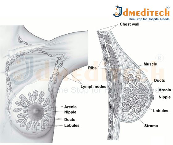
Breast cancer begins in the breast tissue, usually in the milk ducts (ductal cancer) or the milk-producing glands (lobular cancer). These abnormal cells can invade nearby tissues and, in advanced stages, spread to other parts of the body (metastasis).

Breast cancer can vary from slow-growing forms to aggressive types that spread quickly. Understanding your risk and recognizing early signs can help with early diagnosis and better treatment outcomes.

Symptoms of Breast Cancer

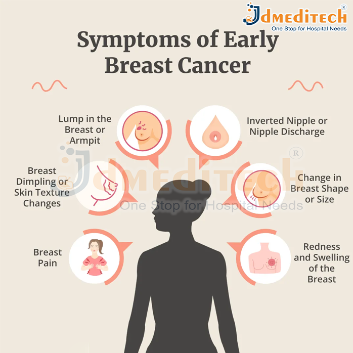
Breast cancer may not cause symptoms in its early stages. However, common signs include:

  • A lump or thickening in the breast or underarm
  • Changes in the size or shape of the breast
  • Nipple discharge (especially bloody)
  • Pain in any part of the breast
  • Skin changes such as redness, dimpling, or puckering
  • Inverted nipple or changes around the nipple area

How Does Breast Cancer Spread?

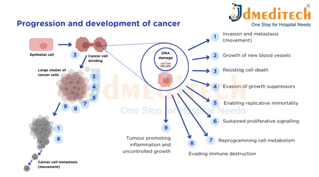
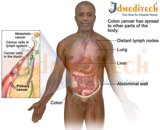
Breast cancer spreads when abnormal cells in the breast grow uncontrollably and begin to move beyond their original location. This process is known as metastasis. Cancer cells can break away from the primary tumor, travel through the body, and form new tumors in other organs. Breast cancer mainly spreads through the following pathways:

  1. Lymphatic System

The most common route of spread.
Cancer cells enter nearby lymph nodes—usually in the armpit (axillary nodes), collarbone, or chest—and from there, they travel to other parts of the body.

  1. Bloodstream

Cancer cells can also enter the blood vessels and circulate throughout the body. Through the bloodstream, breast cancer can reach major organs such as the lungs, liver, bones, and brain.

  1. Direct Spread to Nearby Tissues

As the tumor grows, it can invade nearby structures such as the skin, chest wall, or muscles around the breast.

When breast cancer spreads to distant organs, it is called metastatic or stage 4 breast cancer, even if the new tumors are found in other parts of the body. Early detection and treatment help stop or slow down the spread, improving survival and quality of life.

Types of breast cancer

  1. Ductal Carcinoma In Situ (DCIS)

A non-invasive cancer where abnormal cells are found in the lining of the milk ducts but haven’t spread outside the duct.
DCIS is considered an early form of breast cancer and is highly treatable.

  1. Invasive Ductal Carcinoma (IDC)

The most common type of breast cancer.
IDC begins in the milk ducts and then invades surrounding breast tissue. It may spread to lymph nodes or other parts of the body if untreated.

  1. Invasive Lobular Carcinoma (ILC)

Starts in the milk-producing glands (lobules) and spreads to nearby tissues.
ILC can be harder to detect on imaging because of its growth pattern.

  1. Lobular Carcinoma In Situ (LCIS)

LCIS is not considered true cancer but a marker of increased breast cancer risk.
Abnormal cells stay within the lobules without invading nearby tissues.

  1. Triple-Negative Breast Cancer (TNBC)

A fast-growing type that does not have estrogen, progesterone, or HER2 receptors.
TNBC is aggressive and often requires chemotherapy because hormone therapies do not work on it.

  1. HER2-Positive Breast Cancer

This type has high levels of the HER2 protein, causing rapid cancer cell growth.
Targeted therapies (like trastuzumab) are very effective for HER2-positive cancers.

  1. Inflammatory Breast Cancer (IBC)

A rare and aggressive form where cancer cells block lymph vessels in the breast skin.
It causes redness, swelling, and a warm feeling—often without a lump.

  1. Paget’s Disease of the Breast

A rare cancer that affects the nipple and areola.
It often appears as flaky, red, or irritated skin and is usually linked to underlying DCIS or IDC.

  1. Metastatic Breast Cancer (Stage 4)

Occurs when cancer spreads to other organs like the bones, liver, lungs, or brain.
It is not a different disease but an advanced stage of breast cancer.

  1. Recurrent Breast Cancer

Cancer that returns months or years after treatment.
It can come back in the breast (loc

al), nearby lymph nodes (regional), or distant organs (distant recurrence).

What Causes Breast Cancer?

The exact cause of breast cancer is not fully known. Breast cancer develops when cells in the breast begin to grow abnormally. These cells divide faster than healthy cells and can form a lump or mass. Over time, they may spread to nearby tissues or other parts of the body. While the specific trigger for this abnormal growth is still unclear, several factors can increase the likelihood of developing breast cancer.

  1. Genetic Changes

Certain inherited gene mutations—especially BRCA1 and BRCA2—significantly increase the risk of breast and ovarian cancer. A family history of breast cancer also suggests a higher chance of developing the disease.

  1. Hormonal Influence

Estrogen and progesterone levels in the body play a major role. Conditions or treatments that increase exposure to these hormones—such as early periods, late menopause, or hormone replacement therapy—can raise the risk.

  1. Age and Gender

Breast cancer is more common in women, and the risk increases with age, especially after 40.

  1. Lifestyle Factors

Certain habits or health conditions can contribute to breast cancer risk:

  • Obesity
  • Lack of physical activity
  • Smoking
  • Excessive alcohol consumption
  • High-fat or processed diet
  1. Radiation Exposure

Previous radiation therapy to the chest area—especially during childhood or teenage years—can increase breast cancer risk later in life.

  1. Reproductive Factors
  • Having the first child after age 30
  • Not breastfeeding
  • Never having a full-term pregnancy
    These can slightly increase the risk due to longer exposure to hormones.
  1. Dense Breast Tissue

Women with dense breasts have more glandular tissue than fatty tissue, which can make tumors harder to spot and slightly increase risk.

How Is Breast Cancer Diagnosed?

Diagnosing breast cancer involves a combination of physical examination, imaging tests, and tissue analysis. Early diagnosis is crucial because it increases the chances of successful treatment and better long-term outcomes. Here’s how doctors typically diagnose breast cancer:

  1. Clinical Breast Examination

A healthcare provider checks the breasts and underarm areas for lumps, swelling, skin changes, or any unusual signs. This is usually the first step if symptoms are present.

  1. Mammography

A mammogram is an X-ray of the breast and is the most common screening tool for early detection.
It helps identify tumors that may be too small to feel. If something suspicious is seen, additional imaging may be recommended.

  1. Breast Ultrasound

Ultrasound uses sound waves to create images of the breast.
It helps distinguish between:

  • Solid lumps (which may be cancerous)
  • Fluid-filled cysts (usually non-cancerous)

Ultrasound is especially helpful for women with dense breast tissue.

  1. Breast MRI (Magnetic Resonance Imaging)

MRI offers highly detailed images and is used for:

  • High-risk patients
  • Evaluating the extent of cancer
  • Clarifying unclear results from other imaging tests

It is often used along with mammography rather than as a standalone test.

  1. Biopsy (The Confirmatory Test)

A biopsy is the only way to definitively diagnose breast cancer.
A doctor removes a small sample of breast tissue and sends it to a laboratory to check for cancer cells. Types of biopsy include:

  • Fine-needle aspiration
  • Core needle biopsy
  • Surgical biopsy

The lab also determines the type, grade, and hormone receptor status of the cancer.

  1. Additional Tests After Diagnosis

If cancer is confirmed, further tests help determine the stage and spread, such as:

  • CT scans
  • PET scans
  • Bone scans
  • Blood tests

How Is Breast Cancer Treated?

Breast cancer treatment depends on the type, stage, and size of the tumor and whether it has spread to other parts of the body. Doctors also consider the patient’s overall health and personal preferences. Most treatment plans involve a combination of therapies to remove the cancer, stop its growth, and reduce the chance of it returning.

  1. Surgery

Surgery is often the first step in treating breast cancer. Common options include:

  • Lumpectomy: Removes only the tumor and a small margin of surrounding tissue.
  • Mastectomy: Removes the entire breast when the cancer is large or widespread.
  • Lymph Node Removal: Checks if cancer has spread to lymph nodes.
    Some patients may choose breast reconstruction surgery after a mastectomy.
  1. Radiation Therapy

Radiation uses high-energy beams to destroy remaining cancer cells after surgery.
It helps reduce the risk of the cancer coming back in the same area.

  1. Chemotherapy

Chemotherapy involves anti-cancer drugs given orally or through an IV.
It is used to:

  • Shrink tumors before surgery
  • Kill remaining cancer cells after surgery
  • Treat cancers that have spread

Chemotherapy is especially important for aggressive types like triple-negative breast cancer.

  1. Hormone (Endocrine) Therapy

Many breast cancers grow in response to hormones like estrogen or progesterone.
Hormone therapy blocks these hormones or lowers their levels.
It is effective for hormone receptor-positive cancers.

  1. Targeted Therapy

Targeted drugs focus on specific proteins or genes that help cancer grow.
For example, HER2-positive breast cancer is treated with medicines that block the HER2 protein.
These treatments usually cause fewer side effects than chemotherapy.

  1. Immunotherapy

Immunotherapy helps the body’s immune system recognize and attack cancer cells.
It is used for certain types of advanced or triple-negative breast cancer.

  1. Supportive & Palliative Care

Supportive treatments help manage symptoms, improve comfort, and maintain quality of life.
This may include pain management, counseling, nutrition support, and physical therapy.

Conclusion

Breast cancer is a serious but treatable condition when identified early. Awareness, routine screenings, and healthy lifestyle choices play a crucial role in prevention and early diagnosis. If you or someone you know is at risk, consult a healthcare provider for guidance on screenings and preventive measures.

Explore More Product >>

Get Connected:
+91 79909 93062 | +91 63513 72032 | exports@jdmeditech.com

Blood Donation

Blood donation is one of the simplest yet most powerful ways to make a positive impact on someone’s life. Every day, hospitals and medical centers rely on donated blood to help patients undergoing surgeries, treatments for chronic illnesses, or emergency care following accidents. Since blood cannot be manufactured, voluntary donations from healthy individuals are essential to meet the constant demand.

Donating blood is safe, quick, and often takes less than 30 minutes. Before donation, a brief health check ensures the donor is fit, making the process secure for both the donor and recipient. One unit of blood can save up to three lives, highlighting the immense difference a single donation can make.

Beyond saving lives, blood donation also offers benefits for the donor. It helps stimulate the production of new blood cells, supports healthy iron levels, and promotes a sense of fulfillment by contributing to the community. Regular donations can also strengthen awareness about personal health and encourage a culture of giving.

By choosing to donate blood, you are offering hope, health, and life to someone in need. It is a small gesture that carries the power to make an extraordinary difference. Be a hero—donate blood and help save lives today.

blood and help save lives today.

Who is a Suitable Candidate for Blood Donation?

A suitable candidate for blood donation is generally a healthy adult who meets specific criteria to ensure both the donor’s safety and the quality of the donated blood. Typical requirements include:

  • Age: Usually between 18 and 65 years old (varies slightly by country).
  • Weight: Minimum of 50 kg (110 lbs) to ensure safe donation.
  • Health: Free from infections or chronic illnesses at the time of donation.
  • Hemoglobin Levels: Sufficient hemoglobin to prevent anemia after donation.
  • Lifestyle Factors: No recent high-risk behaviors, infections, or travel to regions with certain infectious diseases.
  • Medications: Not taking medications that could affect blood safety.

Certain temporary conditions, such as recent illness, surgery, or tattooing, may require deferral until fully recovered. Pregnant or breastfeeding women are usually advised to wait before donating.

Donor Safety

Ensuring the safety of blood donors is a top priority in the blood donation process. Proper guidelines and procedures are followed to protect donors’ health and make the experience safe and comfortable.

  1. Health Screening: Before donating, donors undergo a brief medical check-up to assess blood pressure, hemoglobin levels, and overall health. This ensures only healthy individuals donate.
  2. Sterile Equipment: Single-use, sterile needles and blood bags are used for every donation, eliminating the risk of infection.
  3. Trained Staff: Qualified medical personnel supervise the donation process, monitoring donors for any discomfort or complications.
  4. Proper Technique: Blood is drawn slowly and carefully to minimize pain and prevent injury.
  5. Post-Donation Care: Donors are advised to rest, drink fluids, and eat light snacks to prevent dizziness or fatigue.
  6. Temporary Deferral: Certain conditions, like recent illness, medications, or travel to specific regions, may temporarily prevent donation, ensuring safety for both donor and recipient.

Why Is Blood Donation Conducted?

Blood donation is conducted to save lives and support the healthcare system in treating patients who need blood. It is a vital process because blood cannot be manufactured artificially—it can only come from generous donors. The main reasons for blood donation include:

  1. Emergency Situations: Accidents, natural disasters, or sudden medical emergencies often require immediate blood transfusions to save lives.
  2. Surgical Procedures: Many surgeries, including heart surgery, organ transplants, and major operations, require blood to ensure patient safety.
  3. Treatment of Chronic Illnesses: Patients with conditions such as anemia, hemophilia, kidney disease, or cancer may need regular blood transfusions.
  4. Supporting Medical Research: Donated blood is sometimes used for research to develop new treatments and improve medical care.
  5. Maintaining Blood Supply: Regular donations ensure a constant and safe blood supply in hospitals and blood banks.

Benefits of Blood Donation

Blood donation is not only a lifesaving act for recipients but also offers several benefits for donors:

  1. Saves Lives: One donation can help save up to three lives, making it one of the most impactful acts of kindness.
  2. Health Check-Up: Before donating, donors undergo a basic health screening, including blood pressure, hemoglobin levels, and overall health assessment.
  3. Maintains Healthy Iron Levels: Regular donation can help reduce excess iron in the blood, which may lower the risk of heart-related issues.
  4. Stimulates New Blood Cell Production: Donating blood encourages the body to produce fresh red blood cells, improving circulation and overall health.
  5. Promotes Emotional Wellbeing: The act of helping others fosters a sense of fulfillment and social responsibility.
  6. Strengthens Community Support: Blood donation encourages a culture of generosity, compassion, and solidarity within society.

Blood Typing

Blood typing is the process of determining a person’s blood group based on the presence or absence of specific antigens on the surface of red blood cells. It is a crucial step before blood donation or transfusion to ensure compatibility and prevent serious reactions.

Major Blood Groups

The most widely recognized blood group systems are:

  1. ABO System:
    • Type A: Has A antigens on red cells and anti-B antibodies in plasma.
    • Type B: Has B antigens and anti-A antibodies.
    • Type AB: Has both A and B antigens but no antibodies (universal recipient).
    • Type O: Has no antigens but both anti-A and anti-B antibodies (universal donor).
  2. Rh Factor:
    • Rh-positive (Rh+): Presence of the Rh antigen (D antigen).
    • Rh-negative (Rh−): Absence of the Rh antigen.
Importance of Blood Typing
  • Ensures safe blood transfusions and prevents life-threatening reactions.
  • Helps in pregnancy care, as Rh incompatibility between mother and baby can cause complications.
  • Aids in medical research and organizing blood supplies in blood banks.

Conclusion

Blood donation is more than a medical procedure—it is an act of kindness that can make a life-changing difference. By donating blood, you are giving hope, health, and life to someone in need. Every drop counts. Be a hero—donate blood today.

Explore More Product >>

Get Connected:
+91 79909 93062 | +91 63513 72032 | exports@jdmeditech.com

What Is Cancer?

What Is Cancer?

Cancer is a group of diseases in which abnormal cells grow uncontrollably and spread to other parts of the body. Normally, the cells in our body grow, divide, and die in a controlled way. But when this process breaks down, cells begin to grow even when they aren’t needed. These abnormal cells form a mass called a tumor, which can be harmless (benign) or dangerous (malignant).

Malignant tumors can invade nearby tissues and organs. They can also spread to distant parts of the body through the bloodstream or lymphatic system—a process known as metastasis. This uncontrolled growth and spread make cancer a serious and sometimes life-threatening condition.

Differences Between Cancer Cells and Normal Cells

Cancer cells differ from normal cells in many important ways that affect how they grow, function, and spread. Normal cells follow the body’s natural rules—they grow in an organized manner, repair themselves when possible, and die when they are old or damaged. This controlled behavior keeps tissues and organs healthy.

Cancer cells, however, do not follow these rules. They grow rapidly and divide without control, even when the body does not need more cells. They do not die when they should, allowing damaged and mutated cells to accumulate. These cells often look abnormal under a microscope, with irregular shapes and larger-than-normal nuclei. Unlike normal cells, cancer cells can invade nearby tissues and spread to distant organs through the bloodstream or lymphatic system, a process known as metastasis.

Cancer cells also have altered metabolism, meaning they use more energy to fuel their rapid growth. They ignore signals from surrounding cells, produce their own growth signals, and often evade the immune system. Because they do not perform any helpful function in the body, cancer cells disrupt normal organ activities and can form harmful tumors.

These key differences explain why cancer is so challenging to control and why early detection and treatment are important.

How Does Cancer Develop?

Cancer develops when normal cells in the body begin to grow abnormally due to damage in their DNA. In a healthy body, cells grow, divide, and die in a controlled way. But when the DNA inside a cell becomes damaged or mutated, the cell may start behaving differently.

Instead of repairing itself or dying, the damaged cell continues to divide uncontrollably. Each new cell carries the same mutation, causing a group of abnormal cells to form. Over time, these cells can grow into a tumor.

  1. DNA Damage or Mutation

Cancer usually starts when a cell’s DNA becomes damaged. This can happen due to:

  • Genetics
  • Smoking or tobacco use
  • Radiation
  • Harmful chemicals
  • Viral infections (like HPV)
  • Unhealthy lifestyle habits
  • Aging
  1. Uncontrolled Cell Growth

Once DNA is damaged, the cell ignores normal signals that regulate growth. It keeps dividing, even when the body does not need more cells.

  1. Formation of a Tumor

As abnormal cells multiply, they form a mass called a tumor.

  • Benign tumors stay in one place and are usually not dangerous.
  • Malignant tumors grow aggressively and can damage nearby tissues.
  1. Angiogenesis (New Blood Supply)

Cancer cells release signals to create new blood vessels. This gives the tumor oxygen and nutrients, helping it grow faster.

  1. Invasion and Metastasis

Advanced cancer cells can break away from the original tumor and travel through:

  • The bloodstream
  • The lymphatic system

They can form new tumors in other organs—a process called metastasis. This is what makes cancer life-threatening.

When Does Cancer Spread?

Cancer can become more dangerous when it spreads beyond its original location. This spreading process is known as metastasis. It begins when cancer cells break away from the primary tumor and invade nearby tissues. These cells can then enter the bloodstream or lymphatic system, which act like transportation networks in the body. Through these pathways, cancer cells travel to distant organs such as the lungs, liver, bones, or brain.

Once they settle in a new area, the cancer cells may start growing and form a secondary tumor. Even if the cancer spreads to another organ, it still remains the same type as the original cancer. For example, breast cancer that spreads to the lungs is still classified as breast cancer, not lung cancer.

The spread of cancer is serious because it interferes with the normal functioning of vital organs and makes treatment more complex. Metastatic cancer often requires stronger or combination treatments like chemotherapy, targeted therapy, immunotherapy, radiation, or specialized surgery. Detecting cancer early and starting treatment promptly can help reduce the chances of metastasis.

Types of Cancer

  1. Carcinomas
  2. Sarcomas
  3. Leukemias
  4. Lymphomas
  5. Myeloma (Multiple Myeloma)
  6. Melanoma
  7. Central Nervous System (CNS) Cancers
  8. Germ Cell Tumors
  9. Neuroendocrine Tumors

Conclusion

Cancer is a complex disease, but early detection, modern treatments, and healthy lifestyle choices can significantly improve outcomes. Understanding the basics of cancer empowers individuals to take charge of their health and seek timely medical help.

Explore More Product >>

Get Connected:
+91 79909 93062 | +91 63513 72032 | exports@jdmeditech.com

Tonsillectomy Surgery

A tonsillectomy is a surgical procedure to remove the tonsils, two small glands located at the back of the throat. These glands are part of the immune system and help fight infections. However, in some cases, they themselves can become frequently infected or cause other health problems, making their removal necessary.

Why Is a Tonsillectomy Done?

A tonsillectomy may be recommended for:

  • Recurrent tonsillitis: Frequent infections of the tonsils that do not respond well to medication.
  • Sleep apnea: Enlarged tonsils can block the airway, causing breathing difficulties during sleep.
  • Difficulty swallowing: Enlarged tonsils can interfere with normal swallowing.
  • Chronic tonsil infections: Persistent infections leading to abscesses or other complications.

How Is the Surgery Performed?

Your healthcare provider will give you general anesthesia to keep you asleep and comfortable during your procedure. Next, they’ll remove your tonsils. You won’t feel pain during this step. Surgeons use many methods to perform tonsillectomies, including :

  • Electrocautery: This method uses heat to remove the tonsils and stop any bleeding.
  • Cold knife (steel) dissection: A surgeon uses a scalpel (traditional surgical knife) to remove your tonsils. Then, they’ll stop the bleeding with electrocautery (extreme heat) or sutures.
  • Snare tonsillectomy: A surgeon uses a special surgical instrument called a snare, which has a thin wire loop at the end. Once your surgeon frees your tonsil, they’ll place this device around it to clamp it off. This helps reduce bleeding.
  • Harmonic scalpel: This method uses ultrasonic vibrations to remove your tonsils and stop bleeding at the same time.
  • Other methods include the use of radiofrequency ablation techniques, carbon dioxide lasers, and/or a microdebrider (which uses a combination of suction and cutting).

Before Tonsillectomy Surgery

Before undergoing a tonsillectomy, patients should prepare for the procedure to ensure safety and a smooth recovery:

  • Medical Evaluation: Your ENT doctor will review your medical history, current medications, and any allergies.
  • Fasting: You may be asked not to eat or drink for several hours before surgery.
  • Medications: Certain medications, especially blood thinners, may need to be paused before surgery.
  • Arrangements: Arrange for someone to accompany you to and from the hospital.
  • Expectations: Understand the procedure, risks, and recovery process by discussing them with your doctor.

After Tonsillectomy Surgery

Recovery after tonsillectomy is crucial for healing and minimizing complications:

  • Hospital Stay: Most patients are discharged the same day or after an overnight stay.
  • Pain Management: Throat pain is common; prescribed pain medications help manage discomfort.
  • Diet: Soft, cool foods like yogurt, ice cream, and soups are recommended. Avoid spicy or hard foods.
  • Hydration: Drink plenty of fluids to prevent dehydration.
  • Rest: Take adequate rest and avoid strenuous activity for 1–2 weeks.
  • Follow-Up: Attend follow-up appointments to monitor healing and address any concerns.

Benefits of Tonsillectomy

A tonsillectomy offers several health and lifestyle benefits, especially for individuals suffering from chronic tonsil problems:

  1. Reduces Frequent Infections
    Removing the tonsils can significantly decrease the number of throat infections, such as tonsillitis, leading to fewer sick days and less antibiotic use.
  2. Improves Sleep Quality
    Enlarged tonsils can block the airway, causing sleep apnea or snoring. Tonsil removal can improve breathing during sleep and enhance overall rest.
  3. Eases Swallowing and Eating
    For patients with enlarged tonsils, swallowing can be painful or difficult. A tonsillectomy makes eating and drinking more comfortable.
  4. Prevents Complications
    Chronic tonsil infections can lead to abscesses or spread infections to nearby tissues. Removing the tonsils reduces these risks.
  5. Boosts Overall Quality of Life
    Less throat pain, fewer infections, and improved sleep can result in better energy, concentration, and daily functioning, especially in children.


Conclusion

Tonsillectomy is an effective solution for chronic or severe tonsil problems and can significantly improve quality of life. If you or your child experience frequent tonsil infections, sleep disturbances, or difficulty swallowing, consult an ENT specialist to determine if a tonsillectomy is the right option.

Explore More Product >>

Get Connected:
+91 79909 93062 | +91 63513 72032 | exports@jdmeditech.com

Tympanoplasty

Tympanoplasty is a surgical procedure performed to repair a perforated or damaged eardrum (tympanic membrane). It helps restore hearing, prevents recurrent ear infections, and improves overall ear health. The surgery is usually recommended when the eardrum does not heal on its own or when chronic infections affect hearing ability.

What Is Tympanoplasty?

Tympanoplasty is a procedure in which the surgeon patches or reconstructs the torn eardrum using a tissue graft. The graft can be taken from the patient’s own tissue, such as the fascia (a thin layer from behind the ear) or cartilage. The goal is to close the perforation and improve the transmission of sound waves.

Types of Tympanoplasty

There are different types of tympanoplasty depending on the extent of damage:

  1. Type I (Myringoplasty)

Only the eardrum is repaired. The ossicles (hearing bones) are intact.

  1. Type II

The eardrum and part of the malleus (one of the ossicles) are repaired.

  1. Type III

The eardrum is connected to the stapes bone when other bones are damaged.

  1. Type IV

Repair is done when most ossicles are damaged, leaving only the stapes footplate.

  1. Type V

Used in complex cases with severe middle ear damage.

Why Is a Tympanoplasty Done?

Tympanoplasty is performed to repair a perforated (hole) or damaged eardrum that does not heal on its own. It helps restore normal hearing, prevent infections, and protect the middle ear from further damage. Doctors recommend tympanoplasty in the following situations:

  1. Persistent Eardrum Perforation

If the eardrum hole remains open for more than 3–6 months, surgery is needed to close it.

  1. Frequent Ear Infections

A damaged eardrum makes the ear more vulnerable to repeated infections. Tympanoplasty helps stop chronic ear discharge and prevents infection spread.

  1. Hearing Loss

A perforated eardrum reduces sound transmission, leading to reduced hearing. Repair improves hearing quality.

  1. Trauma or Injury

Sudden loud noises, accidents, or inserting objects into the ear can damage the eardrum. Tympanoplasty restores normal structure.

  1. Damage From Prior Ear Surgeries

Some previous procedures or untreated infections may weaken the eardrum, requiring surgical repair.

  1. Preventing Future Complications

Without treatment, chronic perforation can lead to cholesteatoma (dangerous skin growth in the middle ear), further hearing loss, or long-term infection. Tympanoplasty prevents these issues.

The Tympan Mastoidectomy Procedure

A tympanomastoidectomy procedure is performed in a hospital setting under general anesthesia and typically takes several hours. During the tympanomastoidectomy, incisions are made inside and behind the infected ear. The middle ear and mastoid bone are opened, and the infected tissue, or the cholesteatoma, is removed. The eardrum is repaired with muscle lining from behind the ear.

After the tympanomastoidectomy is completed, the surgeon will place packing inside the ear to keep tissues in place as they heal. Most tympanomastoidectomy patients can be released after an overnight observation, unless they are experiencing nausea or dizziness.

During Tympanoplasty Surgery

Tympanoplasty is a surgical procedure aimed at repairing a damaged or perforated eardrum. The surgery is usually performed under general anesthesia, although in some cases, local anesthesia may be used. Here’s what typically happens step by step:

  1. Preparation
  • The patient is given anesthesia for comfort and pain-free surgery.
  • The surgeon cleans and sterilizes the ear area to prevent infection.
  1. Incision
  • Depending on the location of the eardrum perforation, the surgeon may make an incision behind the ear or inside the ear canal.
  • This allows access to the eardrum and middle ear.
  1. Harvesting the Graft
  • A small piece of tissue, often fascia from behind the ear or cartilage, is harvested to repair the eardrum.
  • This graft acts as a patch over the perforation.
  1. Repairing the Eardrum
  • The surgeon carefully cleans the edges of the perforation.
  • The graft is placed over or under the eardrum and positioned to ensure proper healing.
  • In some cases, tiny hearing bones (ossicles) may also be repaired if damaged.
  1. Packing the Ear
  • The ear canal may be gently packed with absorbable material or gauze to support the graft and stabilize the eardrum.
  1. Closing the Incision
  • If an external incision was made, it is closed with fine sutures.
  • A protective dressing is placed behind the ear if necessary.
  1. Duration
  • The entire procedure typically lasts 1–2 hours.
  • Most patients can go home the same day, depending on the type of anesthesia used.

Conclusion

Tympanoplasty is a highly effective surgery for repairing a damaged eardrum and restoring hearing. With modern techniques, the procedure is safe, minimally invasive, and provides long-lasting relief from ear infections and hearing issues. If you have chronic ear discharge or hearing loss due to a perforated eardrum, consulting an ENT surgeon can help you determine whether tympanoplasty is the right option for you.

Explore More Product >>

Get Connected:
+91 79909 93062 | +91 63513 72032 | exports@jdmeditech.com

Bronchoscopy

A bronchoscopy is a medical procedure using a thin, lighted tube (bronchoscope) inserted via the nose or mouth to view, diagnose, and treat lung and airway problems like infections, tumors, and blockages or to take tissue samples (biopsy). Done with sedation, it allows doctors to see inside the lungs, take samples, clear mucus, or even place stents, often on an outpatient basis. Patients usually go home the same day but need someone to drive them due to sedation.

What Is Bronchoscopy?

Bronchoscopy is an endoscopic examination in which a bronchoscope is inserted through the nose or mouth, down the throat, and into the lungs. The device has a tiny camera and light that help doctors visualize the airway lining.
It can be performed using:

  • Flexible bronchoscopy: Most common; used for diagnosis and minor treatments.
  • Rigid bronchoscopy: Used for more complex situations, such as removing large obstructions or controlling heavy bleeding.

How Is Bronchoscopy Performed?

  1. Preparation

    The patient is asked to avoid food and drink for a few hours before the procedure. A local anesthetic is sprayed into the throat, and mild sedation is given.

  2. Procedure
  • The bronchoscope is gently inserted through the nose or mouth.
  • The doctor examines the windpipe and bronchi while watching the video monitor.
  • If required, samples of mucus or tissue are collected.
  1. Duration

    The procedure typically lasts 20–45 minutes, depending on the purpose.

What Happens During a Bronchoscopy?

During a bronchoscopy, a doctor examines your airways and lungs using a thin tube called a bronchoscope. The procedure is usually done in a hospital or clinic and takes about 20–45 minutes. Here’s what typically happens step-by-step:

  1. Preparation Before the Procedure
  • You will be asked not to eat or drink for a few hours before the bronchoscopy.
  • A local anesthetic is sprayed in your nose and throat to numb the area.
  • You may receive a mild sedative through an IV to help you relax.
  1. Inserting the Bronchoscope
  • The doctor gently inserts the bronchoscope through your nose or mouth.
  • It then moves down your throat, into the windpipe (trachea), and finally into the bronchi (airways of the lungs).
  1. Visual Examination
  • The bronchoscope has a tiny camera and light.
  • The doctor views the airways on a video screen, checking for:
    • Inflammation
    • Infection
    • Bleeding
    • Tumors or abnormal growths
    • Blockages
  1. Collecting Samples (If Needed)

The doctor may take:

  • Biopsy (small tissue samples)
  • Mucus or fluid samples (bronchoalveolar lavage)
  • Brushings from the airway walls

These samples help diagnose infections, cancer, inflammation, or other lung diseases.

  1. Treatment During Bronchoscopy (If Required)

In some cases, bronchoscopy is used to perform treatments such as:

  • Removing mucus plugs or foreign objects
  • Controlling bleeding
  • Opening narrow airways
  • Delivering medication directly into the lungs
  1. Completion of the Procedure
  • The bronchoscope is carefully removed.
  • You will be taken to a recovery area until the sedative wears off.
  • Throat numbness or mild coughing is normal for a few hours.

Conclusion

Bronchoscopy is a valuable diagnostic and therapeutic procedure that provides a clear view of the lungs and airways. It helps doctors diagnose complex respiratory issues and guide treatment effectively. If you have persistent breathing problems, coughing, or unexplained chest abnormalities, your doctor may recommend this safe and reliable procedure.

Explore More Product >>

Get Connected:
+91 79909 93062 | +91 63513 72032 | exports@jdmeditech.com

Urodynamic testing

What is urodynamic testing?

Urodynamic testing measures the function of your lower urinary tract. It evaluates the bladder’s ability to hold urine, empty efficiently, and the coordination of nerves and muscles involved in urination. This testing is particularly useful when symptoms are not explained by simpler evaluations like urine tests or imaging.

Why Doctors Recommend Urodynamic Testing

Doctors may recommend urodynamic testing if you experience:

  • Frequent urination or urgency
  • Difficulty starting urination
  • Weak urine flow or interrupted stream
  • Urinary incontinence (leakage)
  • Recurrent urinary tract infections
  • Symptoms after prostate surgery or pelvic surgery

This testing helps pinpoint the cause of urinary symptoms, ensuring the right treatment plan—whether medication, physical therapy, or surgery.

How Urodynamic Testing is Performed

The testing usually involves several procedures:

  1. Cystometry—Measures how much your bladder can hold and how it behaves when filling with liquid.
  2. Uroflowmetry—Assesses the strength and pattern of urine flow.
  3. Post-void Residual Measurement—Checks the amount of urine left in the bladder after urination.
  4. Pressure Flow Study – Evaluates bladder pressure during urination.
  5. Electromyography (EMG)—Measures the electrical activity of the muscles around the bladder and urethra.

During the tests, a thin catheter may be inserted into the bladder, and sensors may be placed on the pelvic area to measure muscle activity.

Risks of Urodynamic Testing

Urodynamic testing is generally safe, but some minor risks may include:

  • Temporary discomfort or burning sensation during urination
  • Mild urinary tract infection
  • Minor bladder irritation

Your doctor may prescribe antibiotics or recommend precautions to minimize these risks.

Preparing for Urodynamic Testing

Preparation may vary depending on the type of test, but common steps include:

  • Emptying your bladder before the test
  • Avoiding caffeine or alcohol
  • Informing your doctor about medications that may affect urination

Conclusion

Urodynamic testing provides valuable insights into the function of your bladder and urinary system. If you are experiencing urinary problems, consulting a urologist for these tests can help identify the cause and guide effective treatment. Early diagnosis can improve quality of life and prevent further complications.

Explore More Product >>

Get Connected:
+91 79909 93062 | +91 63513 72032 | exports@jdmeditech.com

Circumcision

Circumcision is a common surgical procedure in which the foreskin—the fold of skin covering the tip of the penis—is removed. It can be performed for medical, cultural, religious, or personal reasons. The surgery is usually quick and safe, and it can be done for newborns, children, and adults.

What Is Circumcision?

Circumcision is a minor surgical procedure that involves removing the foreskin from the glans (head) of the penis. The foreskin is a natural layer of skin that provides protection and lubrication. In some cases, it becomes tight or infected, leading to medical issues that may require circumcision.

Why Is Circumcision Done?

Circumcision may be recommended for:

  1. Medical Reasons
  • Phimosis – The foreskin becomes too tight and cannot be pulled back.
  • Recurrent Infections – Frequent foreskin or urinary infections.
  • Balanitis—Inflammation of the penis due to trapped bacteria.
  • Poor Hygiene – Difficulty cleaning under the foreskin.
  1. Cultural & Religious Reasons

Circumcision is a part of many cultural and religious traditions followed around the world.

  1. Personal Preference

Some adults choose circumcision for hygiene, appearance, or sexual comfort.

How Is Circumcision Performed?

Circumcision is typically a short procedure. The steps may vary depending on age:

  1. For Babies
  • Performed under local anesthesia.
  • A special clamp or device is used to safely remove the foreskin.
  • Takes around 10–15 minutes.
  1. For Children & Adults
  • Performed under local or general anesthesia.
  • The surgeon carefully removes the foreskin using surgical instruments.
  • Dissolvable stitches are used.
  • The procedure takes 30–45 minutes.

What are the benefits of circumcision?

Circumcision has several benefits. Not only does it help aspects of penis health, but it can improve hygiene. That’s because a circumcised penis is simpler to clean and wash, especially for children.

The specific health benefits of circumcision include a lower risk of:

  • Certain penis conditions: · These penile disorders include balanoposthitis (inflammation of the glans and foreskin), paraphimosis, and phimosis, when the foreskin gets stuck out of place.
  • Cervical cancer for partners: Female sex partners of circumcised men are less likely to get cervical cancer.
  • Penile cancer: Men who had circumcisions are less likely to get cancer of the penis.
  • Sexually transmitted infections: Men who have had circumcision have a lower risk of certain STIs, including HIV.
  • Urinary tract infections: UTIs are more common in uncircumcised males.

Conclusion

Circumcision is a safe, widely performed procedure with proven medical and hygiene benefits. Whether done for medical, cultural, or personal reasons, it can help prevent infections, improve comfort, and enhance overall penile health. With proper care and guidance from a qualified surgeon, recovery is usually quick and smooth.

Explore More Product >>

Get Connected:
+91 79909 93062 | +91 63513 72032 | exports@jdmeditech.com

Hernia Surgery

A hernia is usually treated with surgery. The three main types of hernia surgery are open repair, laparoscopic (minimally invasive) repair, and robotic repair. This article provides details of each procedure.

What Is a Hernia?

A hernia occurs when an internal organ or tissue pushes through a weak spot in the surrounding muscle or connective tissue. It commonly appears in areas like the abdomen, groin, or near previous surgical incisions. Hernias can cause discomfort, pain, or a visible bulge and often require surgery to prevent complications.

Why Is Hernia Surgery Needed?

Doctors recommend hernia surgery when:

  • The hernia causes pain or restricts daily activities
  • The bulge increases in size
  • There is risk of incarceration (trapped tissue) or strangulation (blocked blood supply), which can be life-threatening
  • Nonsurgical treatments no longer provide relief.

Types of Hernia Surgery

  1. Open Hernia Repair
  • A single larger cut is made near the hernia site
  • The bulging tissue is pushed back, and the weakened area is reinforced with sutures or a mesh
  • Recovery time is slightly longer but highly effective
  1. Laparoscopic Hernia Repair
  • Performed through 2–3 small incisions
  • A camera and surgical tools are inserted to repair the hernia using mesh
  • Less pain, faster recovery, and minimal scarring
  1. Robotic Hernia Repair
  • Similar to laparoscopy but performed using advanced robotic arms
  • Offers high precision and enhanced visualization
How to reduce and manage pain after hernia surgery?

Take your pain medicines on time.

– Use an ice pack for 15–20 minutes to reduce swelling.

– Avoid heavy lifting for 4–6 weeks.

– Walk gently every day to improve recovery.

– Keep the wound clean and dry.

– Support the area while coughing or getting up.

– Eat a fiber-rich diet to avoid constipation.

– Rest well and avoid strain.

– Call your doctor if pain worsens or you notice redness, fever, or pus.

Explore More Product >>

Get Connected:
+91 79909 93062 | +91 63513 72032 | exports@jdmeditech.com

Appendectomy Surgery

Appendicitis is an inflammation of the appendix, a finger-shaped pouch that projects from your colon on the lower right side of your abdomen.

Appendicitis causes pain in your lower right abdomen. However, in most people, pain begins around the navel and then moves. As inflammation worsens, appendicitis pain typically increases and eventually becomes severe.

Although anyone can develop appendicitis, most often it occurs in people between the ages of 10 and 30. Standard treatment is surgical removal of the appendix.

What Is an Appendectomy?

An appendectomy is a surgical procedure to remove the appendix—a small, finger-shaped organ located in the lower right side of the abdomen. It is most commonly performed to treat appendicitis, a condition where the appendix becomes inflamed, swollen, and infected. Since a burst appendix can cause serious complications, an appendectomy is usually considered an emergency surgery.

Why Is Appendectomy Needed?

Doctors recommend appendectomy when:

  • The appendix is inflamed (acute appendicitis)
  • There is a risk of appendix rupture
  • The patient experiences severe abdominal pain
  • Infection or pus is detected in the abdomen
  • Other treatments cannot control the condition

Types of Appendectomy Surgery

  1. Laparoscopic Appendectomy
  • Performed using small keyhole incisions
  • A camera (laparoscope) guides the surgeon
  • Less pain, faster recovery, and minimal scarring
  1. Open Appendectomy
  • A single, larger incision is made in the abdomen
  • Typically done in complicated cases (ruptured appendix, severe infection)
  • Allows better access to the abdominal cavity

How Is an Appendectomy Performed?

Laparoscopic Procedure:
  • The patient receives anesthesia
  • 2–3 small incisions made
  • Laparoscope and surgical tools inserted
  • Appendix located and removed
  • Incisions closed with sutures
Open Surgery Procedure:
  • A larger incision made on the lower right abdomen
  • Appendix removed directly
  • Abdominal cavity cleaned (if infected)
  • The wound was closed with stitches

Is appendectomy safe?

In most cases, appendectomy is the safest treatment for appendicitis in children and adults. There are a few exceptions — for example, some people might not qualify for surgery. Some might only need antibiotics if they have mild appendicitis or if they catch it early enough. Even so, these people have a higher risk of recurrent (returning) infection. Ultimately, the risk of rupture is much greater than the low risk of surgery. It’s safest to remove your inflamed appendix if it’s at risk of rupturing.

What happens before appendix removal?

Most appendectomies occur within 24 hours of your appendicitis diagnosis. Prior to your surgery, your healthcare team will place an IV line into a vein so they can give you antibiotics as soon as possible. They’ll continue antibiotic therapy until sometime after the surgery—usually one to seven days—depending on the severity of the infection. Your provider will check your response to the antibiotics to make sure you need surgery before proceeding.

Your healthcare provider may also need to run additional tests before your surgery, like a blood test or imaging scans. These tests can give them more information about the nature of your appendicitis. They’ll also need to review your medical history, including any current medications,  allergies or conditions. Based on these and other factors, they’ll explain the type of surgery they want to perform and ask for your consent.

Conclusion

Appendectomy is a safe and life-saving procedure that treats appendicitis effectively. With modern laparoscopic techniques, patients experience faster healing, minimal pain, and a quick return to daily life. If symptoms of appendicitis appear, immediate medical care is essential to avoid complications.

Explore More Product >>

Get Connected:
+91 79909 93062 | +91 63513 72032 | exports@jdmeditech.com

Urodynamic testing

What is urodynamic testing?

Urodynamic testing measures the function of your lower urinary tract. It evaluates the bladder’s ability to hold urine, empty efficiently, and the coordination of nerves and muscles involved in urination. This testing is particularly useful when symptoms are not explained by simpler evaluations like urine tests or imaging.

Why Doctors Recommend Urodynamic Testing

Doctors may recommend urodynamic testing if you experience:

  • Frequent urination or urgency
  • Difficulty starting urination
  • Weak urine flow or interrupted stream
  • Urinary incontinence (leakage)
  • Recurrent urinary tract infections
  • Symptoms after prostate surgery or pelvic surgery

This testing helps pinpoint the cause of urinary symptoms, ensuring the right treatment plan—whether medication, physical therapy, or surgery.

How Urodynamic Testing is Performed

The testing usually involves several procedures:

  1. Cystometry—Measures how much your bladder can hold and how it behaves when filling with liquid.
  2. Uroflowmetry—Assesses the strength and pattern of urine flow.
  3. Post-void Residual Measurement—Checks the amount of urine left in the bladder after urination.
  4. Pressure Flow Study – Evaluates bladder pressure during urination.
  5. Electromyography (EMG)—Measures the electrical activity of the muscles around the bladder and urethra.

During the tests, a thin catheter may be inserted into the bladder, and sensors may be placed on the pelvic area to measure muscle activity.

Risks of Urodynamic Testing

Urodynamic testing is generally safe, but some minor risks may include:

  • Temporary discomfort or burning sensation during urination
  • Mild urinary tract infection
  • Minor bladder irritation

Your doctor may prescribe antibiotics or recommend precautions to minimize these risks.

Preparing for Urodynamic Testing

Preparation may vary depending on the type of test, but common steps include:

  • Emptying your bladder before the test
  • Avoiding caffeine or alcohol
  • Informing your doctor about medications that may affect urination

Conclusion

Urodynamic testing provides valuable insights into the function of your bladder and urinary system. If you are experiencing urinary problems, consulting a urologist for these tests can help identify the cause and guide effective treatment. Early diagnosis can improve quality of life and prevent further complications.

Explore More Product >>

Get Connected:
+91 79909 93062 | +91 63513 72032 | exports@jdmeditech.com

ERCP Surgery

ERCP (endoscopic retrograde cholangiopancreatography) is a specialized procedure used to diagnose and treat problems in the bile ducts, pancreas, and gallbladder. It combines the use of endoscopy and X-ray imaging to visualize these organs and intervene when necessary.

What is ERCP?

ERCP is a minimally invasive procedure that allows doctors to examine the bile ducts, pancreatic ducts, and gallbladder for blockages, stones, tumors, or other abnormalities. Unlike standard imaging tests, ERCP can also be therapeutic, meaning it can treat issues during the same procedure.

Why is ERCP performed?

Doctors may recommend ERCP for:

  • Gallstones lodged in the bile duct
  • Bile duct strictures or narrowing
  • Pancreatic duct problems such as chronic pancreatitis
  • Tumors affecting the bile or pancreatic ducts
  • Leaking bile ducts after surgery

How is ERCP performed?

  1. Preparation: Patients are usually asked to fast for several hours before the procedure. Sedation or anesthesia is given for comfort.
  2. Endoscopy: A thin, flexible tube called an endoscope is inserted through the mouth into the stomach and small intestine.
  3. Contrast Injection: A special dye is injected into the bile and pancreatic ducts.
  4. X-ray Imaging: X-rays are taken to detect blockages, stones, or other abnormalities.
  5. Treatment (if needed): Stones may be removed, stents can be placed to open narrow ducts, or tissue samples may be taken for biopsy.

Benefits of ERCP

  • Minimally invasive, avoiding open surgery
  • Can both diagnose and treat problems in the same procedure
  • Shorter recovery time compared to traditional surgery

Equipment and Instrumentation Considerations

Successful ERCP relies on high‑quality endoscopes, X‑ray imaging systems, contrast injectors, ductal stents or stone‑retrieval tools, and sterile disposables. For a supplier/manufacturer like JDmeditech—which provides hospital instruments and endoscopic accessories—emphasis should be on:

  • Durable, high‑grade endoscopic instruments and accessories, including endoscopes, imaging light sources, irrigation pumps, contrast injectors, disposable catheters, stents, etc.
  • Reliable after‑sales support and maintenance, since ERCP instruments require careful sterilization, regular calibration, and careful handling.
  • Accessible consumables (disposables) to ensure smooth procedure flow and infection control.

Risks, Safety and Quality Requirements

As with any endoscopic procedure, ERCP carries certain risks: risk of post-ERCP pancreatitis, infections, bleeding, ductal or intestinal perforation, and reactions to contrast or sedation. For a hospital or surgical center, ensuring high‑quality instruments, properly maintained sterilization, trained staff, and adherence to safety protocols is key.

That’s why sourcing instruments and accessories only from reputable suppliers—with documented quality standards, reliable manufacturing, and after‑sales support—is crucial. At JDmeditech, we understand that “life is precious,” so we emphasize quality, reliability, and patient safety when supplying endoscopic and surgical instruments.

Recovery After ERCP

Most patients are observed for a few hours after the procedure and may go home the same day. Mild sore throat, bloating, or cramping may occur, but serious complications are rare. Doctors provide specific post-procedure instructions regarding diet and medications.

Conclusion

ERCP is a valuable procedure that helps doctors diagnose and treat conditions of the bile and pancreatic ducts without major surgery. Early intervention can prevent serious complications and improve patient outcomes. If you experience jaundice, abdominal pain, or unexplained digestive issues, consult a gastroenterologist to see if ERCP might be right for you.

Explore More Product >>

Get Connected:
+91 79909 93062 | +91 63513 72032 | exports@jdmeditech.com

Upper GI Endoscopy

The medical term for an upper endoscopy is esophagogastroduodenoscopy. You may have an upper endoscopy done in your health care provider’s office, an outpatient surgery center, or a hospital.

This procedure is done using a long, flexible tube called an endoscope. The tube has a tiny light and video camera on one end. The tube is put into your mouth and throat. Then it is slowly pushed through your esophagus and stomach and into your duodenum. Video images from the tube are seen on a monitor.

Why is it done?

1. Investigating Symptoms
  • Persistent heartburn or acid reflux
  • Difficulty swallowing or pain while swallowing
  • Nausea, vomiting, or unexplained weight loss
  • Upper abdominal pain or discomfort
2 Detecting Conditions
  • Gastroesophageal reflux disease (GERD)
  • Ulcers in the stomach or duodenum
  • Inflammation of the esophagus (esophagitis) or stomach (gastritis)
  • Polyps, tumors, or cancer
  • Infections, such as Helicobacter pylori
3 Monitoring and Screening
  • Follow-up for known digestive tract conditions
  • Screening for cancer or precancerous changes, especially in high-risk individuals
4 Treatment Purposes
  • Removing polyps or foreign objects
  • Stopping bleeding from ulcers
  • Dilating strictures (narrowed areas) in the esophagus

What Happens During an Upper GI Endoscopy?

1. Preparation
    • You will be asked to fast for 6–8 hours before the procedure to ensure your stomach is empty.
    • Your doctor may review your medications and health history.
  1. Sedation and Comfort
    • A mild sedative or local anesthetic spray for the throat is given to make you comfortable.
    • Most patients stay awake but relaxed, while some may receive stronger sedation to sleep lightly.
  2. Insertion of the Endoscope
    • A thin, flexible tube with a camera (endoscope) is gently passed through your mouth and throat into the esophagus, stomach, and duodenum.
    • You may feel some pressure or gagging, but discomfort is usually minimal.
  3. Examination
    • The doctor carefully examines the lining of your upper digestive tract on a monitor.
    • Images may be taken for records.
  4. Biopsy or Minor Treatment (if needed)
    • Small tissue samples (biopsies) can be taken for testing.
    • Minor procedures, like removing polyps, dilating narrowed areas, or stopping bleeding, can be performed through the endoscope.
  5. Completion and Recovery
    • The endoscope is gently removed, and you are monitored for a short period.
    • Most patients can go home the same day, though someone should accompany you if sedation was used.
    • You may feel a sore throat, bloating, or mild cramping, which usually goes away quickly.

Risks of Upper GI Endoscopy

  1. Bleeding—This may occur at the site where tissue samples (biopsies) are taken or polyps are removed. Usually, the bleeding is minor and stops on its own.
  2. Perforation—Rarely, a tear in the lining of the esophagus, stomach, or duodenum can happen. Some cases may require surgery to repair.
  3. Reaction to Sedation—Some patients may experience an adverse reaction to the sedative, affecting breathing or heart function. Doctors can manage this promptly with medications or IV fluids.

Conclusion

Upper GI endoscopy is a valuable diagnostic and therapeutic tool for various gastrointestinal conditions. Early detection through this procedure can prevent complications and guide effective treatment. If you experience persistent digestive issues, consult your doctor to see if an upper GI endoscopy is right for you.

Explore More Product >>

Get Connected:
+91 79909 93062 | +91 63513 72032 | exports@jdmeditech.com

Colonoscopy

A colonoscopy is a medical procedure used to examine the large intestine (colon) and rectum from the inside. It helps doctors detect problems like polyps, inflammation, bleeding, ulcers, or early signs of colorectal cancer.

How is virtual colonoscopy different from colonoscopy?

A colonoscopy is a procedure in which a doctor uses a flexible tube with a camera to examine the inside of the large intestine for polyps, inflammation, bleeding, or cancer. A virtual colonoscopy, on the other hand, uses CT scan images to create a 3D view of the colon without inserting a scope. Unlike a traditional colonoscopy, a virtual colonoscopy cannot remove polyps or take biopsies and is mainly used for screening.

Why do doctors use colonoscopy?

Doctors use colonoscopy because it is the most reliable way to check the health of the colon and rectum. It helps in both diagnosing and preventing diseases.

Main reasons doctors use colonoscopy
  1. To find the cause of symptoms
  • Blood in stool
  • Persistent abdominal pain
  • Chronic diarrhea or constipation
  • Sudden changes in bowel habits
  • Unexplained weight loss
  1. To screen for colorectal cancer
  • Recommended after age 45 (earlier if high-risk).
  • Helps detect cancer early when it is easiest to treat.
  1. To remove polyps
  • Polyps can turn into cancer over time.
  • During colonoscopy, they can be removed immediately.
  1. To diagnose diseases
  • Inflammatory bowel diseases (IBD) like Crohn’s disease or ulcerative colitis
  • Diverticulosis or diverticulitis
  • Infections or ulcers
  1. To take biopsies
  • Small tissue samples can be collected for accurate diagnosis.
  1. To treat certain problems
  • Stop bleeding
  • Remove foreign bodies
  • Dilate narrowed areas of the colon

How do doctors perform a colonoscopy?

During a colonoscopy, the doctor gently inserts a long, flexible tube called a colonoscope through the rectum to examine the entire large intestine. The scope has a tiny camera and light that display real-time images on a monitor. Air or carbon dioxide is used to slightly inflate the colon for better visibility. As the scope moves through the colon, the doctor can look for polyps, inflammation, bleeding, or other abnormalities. If needed, the doctor can remove polyps, take tissue biopsies, or treat bleeding during the same procedure. Most patients receive sedation, making the process comfortable and usually painless.

What are the risks of colonoscopy?

  • bleeding
  • perforation of the colon
  • a reaction to the sedative, including breathing or heart problems
  • severe pain in your abdomen
  • death, although this risk is rare

Colonoscopy is generally safe, but like any medical procedure, it carries some risks. These may include bleeding, especially if a polyp is removed; perforation (a small tear in the colon); reactions to sedatives; infection; and, rarely, complications from existing health conditions. Most complications are uncommon, and doctors take precautions to minimize them.

Conclusion

A colonoscopy is a safe and essential procedure for maintaining colon health. It not only helps detect and prevent serious conditions like colorectal cancer but also allows for the treatment of certain problems during the same procedure. Regular screenings and timely checkups can play a crucial role in ensuring a healthy digestive system.

Explore More Product >>

Get Connected:
+91 79909 93062 | +91 63513 72032 | exports@jdmeditech.com