Abdominal Hysterectomy Surgery for Uterine Disease Treatment

Abdominal hysterectomy is a well-established surgical procedure used to remove the uterus through an incision in the lower abdomen. It remains one of the most reliable treatment options for women suffering from severe gynecological conditions such as large uterine fibroids, chronic pelvic pain, abnormal uterine bleeding, adenomyosis, endometriosis, and certain gynecological cancers.

Although minimally invasive hysterectomy techniques have become increasingly popular, abdominal hysterectomy continues to play a critical role in complex cases where better surgical access and visualization are required. Furthermore, advances in surgical technology, anesthesia, and postoperative care have significantly improved patient safety and recovery outcomes. As a result, abdominal hysterectomy remains a trusted procedure in modern gynecological surgery.

What is Abdominal Hysterectomy Surgery?

Abdominal hysterectomy is a surgical procedure in which the uterus is removed through an incision made in the lower abdomen. Depending on the patient’s medical condition, the surgeon may also remove the cervix, fallopian tubes, ovaries, or surrounding tissues.

Unlike vaginal or laparoscopic hysterectomy, the abdominal approach provides direct access to the pelvic organs. Therefore, it is often preferred when treating large uterine masses, extensive pelvic adhesions, advanced endometriosis, or gynecological cancers.

Why is Abdominal Hysterectomy Performed?

Abdominal hysterectomy is recommended when other treatment methods fail to provide adequate symptom relief or when the underlying condition requires complete removal of the uterus.

Common Medical Conditions Treated
  • Large uterine fibroids
  • Chronic abnormal uterine bleeding
  • Severe endometriosis
  • Adenomyosis
  • Chronic pelvic pain
  • Uterine prolapse
  • Endometrial cancer
  • Cervical cancer
  • Ovarian cancer
  • Precancerous uterine conditions

Moreover, the procedure can provide permanent relief from symptoms that significantly affect a woman’s daily life and overall well-being.

Types of Abdominal Hysterectomy

The specific type of hysterectomy depends on the diagnosis and treatment objectives.

Total Abdominal Hysterectomy (TAH)

This procedure involves the removal of both the uterus and cervix. It is the most commonly performed type of abdominal hysterectomy.

Supracervical Hysterectomy

In this procedure, the uterus is removed while the cervix remains intact.

Radical Hysterectomy

A radical hysterectomy involves the removal of the uterus, cervix, surrounding tissues, and sometimes part of the vagina. It is typically performed for gynecological cancer treatment.

Hysterectomy with Bilateral Salpingo-Oophorectomy

This procedure includes removal of the uterus, fallopian tubes, and ovaries when medically necessary.

Benefits of Abdominal Hysterectomy

Despite the availability of minimally invasive procedures, abdominal hysterectomy offers several important advantages, especially in complex surgical cases.

Key Benefits
  • Complete removal of diseased uterine tissue
  • Effective treatment for large fibroids
  • Better access to pelvic organs
  • Suitable for cancer management
  • Long-term symptom relief
  • Reduced risk of disease recurrence
  • Reliable surgical outcomes
  • Improved quality of life

Furthermore, the procedure allows surgeons to manage multiple pelvic conditions during a single operation.

Preoperative Evaluation and Preparation

Before surgery, patients undergo a comprehensive medical assessment to determine surgical suitability and optimize treatment planning.

Common Diagnostic Tests
  • Pelvic examination
  • Ultrasound imaging
  • MRI scanning
  • CT imaging when required
  • Blood investigations
  • Endometrial biopsy
  • Pap smear
  • Cancer screening evaluations

As a result, surgeons can accurately assess the condition and develop an individualized surgical approach.

Procedure of Abdominal Hysterectomy Surgery

The surgery is usually performed under general anesthesia in a hospital operating room. Depending on the complexity of the condition, the procedure typically takes between one and three hours.

Step-by-Step Surgical Procedure
  1. Administration of general anesthesia.
  2. Preparation and sterilization of the surgical site.
  3. Creation of a lower abdominal incision.
  4. Careful identification of pelvic structures.
  5. Separation of the uterus from supporting tissues and blood vessels.
  6. Removal of the uterus.
  7. Removal of additional organs if required.
  8. Control of bleeding and inspection of the surgical area.
  9. Closure of the abdominal incision.
  10. Transfer to the recovery unit for postoperative monitoring.

Throughout the procedure, advanced surgical techniques help minimize blood loss and improve patient safety.

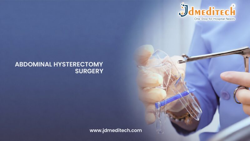
Surgical Instruments Used in Abdominal Hysterectomy

Successful hysterectomy procedures depend on high-quality surgical instruments and operating room equipment.

Common Instruments
  • Surgical retractors
  • Tissue forceps
  • Hemostatic clamps
  • Needle holders
  • Surgical scissors
  • Electrosurgical generators
  • Vessel sealing systems
  • Suction and irrigation devices
  • Surgical staplers
  • Operating room illumination systems

Additionally, modern energy-based devices help surgeons achieve precise tissue dissection and effective bleeding control.

Recovery After Abdominal Hysterectomy

Recovery following abdominal hysterectomy generally takes longer than minimally invasive hysterectomy procedures because of the abdominal incision. However, most patients experience gradual improvement over several weeks.

Recovery Guidelines
  • Follow medical advice carefully.
  • Take medications as prescribed.
  • Avoid heavy lifting during recovery.
  • Maintain proper wound care.
  • Attend all follow-up appointments.
  • Resume normal activities gradually.

Furthermore, maintaining a healthy diet and staying physically active within recommended limits can support faster healing.

Possible Risks and Complications

Abdominal hysterectomy is considered a safe procedure when performed by experienced surgeons. However, like any major surgery, it carries some potential risks.

Potential Risks
  • Bleeding
  • Infection
  • Blood clot formation
  • Injury to surrounding organs
  • Wound complications
  • Urinary tract injury
  • Anesthesia-related complications

Nevertheless, advances in surgical care have significantly reduced complication rates and improved patient outcomes.

Importance of Advanced Surgical Technology

Modern surgical technology plays a vital role in improving the safety and efficiency of abdominal hysterectomy procedures.

Features of Advanced Surgical Systems
  • Enhanced surgical precision
  • Better visualization of pelvic anatomy
  • Improved bleeding control
  • Reduced tissue trauma
  • Increased patient safety
  • More efficient operating room workflow

Consequently, healthcare facilities worldwide continue to invest in advanced gynecological surgery equipment to improve treatment outcomes.

Conclusion

Abdominal hysterectomy remains one of the most effective and reliable treatment options for women with serious uterine and gynecological conditions. Whether treating large fibroids, chronic abnormal bleeding, severe endometriosis, adenomyosis, or gynecological cancers, the procedure provides long-term symptom relief and improved quality of life.

Because abdominal hysterectomy offers excellent surgical access and proven clinical outcomes, it continues to be an important part of modern gynecological care. Ultimately, the combination of experienced surgeons, advanced surgical technology, and comprehensive postoperative care helps ensure safe procedures and successful patient outcomes.


Explore More Product >>

Get Connected:
+91 79909 93062 | +91 63513 72032 | exports@jdmeditech.com 

Hysteroscopic Foreign Body Removal Surgery for Uterine Cavity Cleaning

Hysteroscopic foreign body removal is a minimally invasive gynecological procedure used to locate and remove foreign objects from the uterine cavity. Foreign bodies may include retained intrauterine devices (IUDs), broken contraceptive components, retained surgical materials, fetal bone fragments, or other intrauterine objects that can cause pain, abnormal bleeding, infection, infertility, and reproductive complications.

Fortunately, modern hysteroscopic technology allows gynecologists to remove foreign bodies with exceptional precision while preserving healthy uterine tissue. As a result, patients experience faster recovery, improved reproductive health, and reduced risk of long-term complications. Furthermore, direct visualization of the uterine cavity ensures accurate diagnosis and complete removal of the foreign material.

What is Hysteroscopic Foreign Body Removal?

Hysteroscopic foreign body removal is a surgical procedure performed using a hysteroscope and specialized operative instruments. During the procedure, the hysteroscope is inserted through the cervix into the uterine cavity, providing a clear and magnified view of the uterus.

Once the foreign object is identified, the surgeon carefully removes it using dedicated hysteroscopic instruments. Unlike blind procedures, hysteroscopy enables direct visualization throughout the surgery. Therefore, the risk of incomplete removal is significantly reduced while patient safety is enhanced.

Understanding Foreign Bodies in the Uterus

A foreign body inside the uterus refers to any object that remains within the uterine cavity when it should not be present. In some cases, foreign bodies may remain unnoticed for extended periods. However, they can eventually cause symptoms and complications.

Common Types of Intrauterine Foreign Bodies
  • Retained intrauterine devices (IUDs)
  • Broken IUD fragments
  • Retained fetal bone fragments
  • Retained surgical materials
  • Retained products of conception
  • Foreign tissue fragments
  • Embedded contraceptive devices

Consequently, early diagnosis and treatment are important to prevent infection, pain, and reproductive difficulties.

Symptoms Associated with Uterine Foreign Bodies

The symptoms depend on the type, size, and location of the foreign body. While some patients remain asymptomatic, others may experience significant gynecological problems.

Common Symptoms
  • Abnormal uterine bleeding
  • Pelvic pain
  • Chronic pelvic discomfort
  • Irregular menstruation
  • Vaginal discharge
  • Recurrent uterine infections
  • Infertility
  • Difficulty conceiving
  • Pain during intercourse

Therefore, patients with persistent symptoms should undergo appropriate gynecological evaluation.

Why is Hysteroscopic Foreign Body Removal Performed?

Hysteroscopic foreign body removal is recommended when a retained object causes symptoms, reproductive issues, or potential health risks. In addition, the procedure may be performed when imaging studies identify a foreign body inside the uterus.

Common Indications
  • Retained intrauterine device (IUD)
  • Embedded contraceptive devices
  • Retained fetal bone fragments
  • Retained surgical materials
  • Chronic pelvic pain
  • Recurrent uterine infections
  • Abnormal uterine bleeding
  • Infertility associated with uterine foreign bodies
  • Recurrent implantation failure

Moreover, removing foreign objects helps restore normal uterine anatomy and function.

How are Uterine Foreign Bodies Diagnosed?

Before surgery, physicians perform diagnostic evaluations to determine the location and nature of the foreign body.

Common Diagnostic Methods
  • Diagnostic hysteroscopy
  • Pelvic ultrasound
  • Transvaginal ultrasound
  • Sonohysterography
  • X-ray imaging
  • Magnetic resonance imaging (MRI)

As a result, surgeons can accurately plan the removal procedure and minimize treatment-related risks.

Procedure of Hysteroscopic Foreign Body Removal

The procedure is typically performed in a hospital or specialized gynecological surgery center. Depending on the complexity of the case, local, regional, or general anesthesia may be used.

Step-by-Step Procedure
  1. Patient preparation and anesthesia administration.
  2. Introduction of the hysteroscope through the cervix.
  3. Expansion of the uterine cavity using sterile distension media.
  4. Identification of the foreign body.
  5. Assessment of its location and attachment.
  6. Removal using specialized hysteroscopic instruments.
  7. Retrieval of the foreign material.
  8. Final inspection of the uterine cavity.
  9. Postoperative monitoring and recovery.

Furthermore, most procedures are completed within 20 to 60 minutes. Consequently, many patients can return home on the same day.

Instruments Used in Hysteroscopic Foreign Body Removal

The success of foreign body removal depends on advanced visualization systems and specialized surgical instruments.

Common Instruments
  • Operative hysteroscope
  • Hysteroscopy sheath
  • Grasping forceps
  • Retrieval forceps
  • Hysteroscopic scissors
  • Bipolar electrodes
  • Monopolar electrodes
  • Resectoscope
  • Endoscopic camera system
  • LED light source

Additionally, high-definition imaging systems help surgeons perform accurate and safe procedures.

Benefits of Hysteroscopic Foreign Body Removal

Compared with conventional methods, hysteroscopic removal offers several important advantages.

Key Benefits
  • Minimally invasive procedure
  • Direct visualization of the uterine cavity
  • Accurate foreign body localization
  • Complete object removal
  • Reduced tissue trauma
  • Faster recovery
  • Preservation of fertility
  • Lower complication rates
  • Reduced infection risk
  • Improved patient comfort

As a result, hysteroscopic foreign body removal has become the preferred treatment method in modern gynecology.

How Foreign Body Removal Improves Reproductive Health

Foreign bodies within the uterine cavity can interfere with implantation, increase inflammation, and contribute to infertility. Consequently, reproductive outcomes may be negatively affected.

By removing the foreign object and restoring the normal uterine environment, hysteroscopic surgery helps improve fertility potential and uterine health. Furthermore, many patients experience relief from symptoms such as abnormal bleeding and pelvic discomfort following treatment.

Is Hysteroscopic Foreign Body Removal Safe?

Hysteroscopic foreign body removal is considered a safe and effective procedure when performed by experienced gynecological surgeons. Moreover, advances in hysteroscopic technology have significantly enhanced safety and precision.

Possible Risks
  • Mild cramping
  • Temporary spotting
  • Infection
  • Cervical injury
  • Fluid overload
  • Uterine perforation (rare)

Nevertheless, serious complications remain uncommon when modern surgical techniques and equipment are used.

Recovery After Hysteroscopic Foreign Body Removal

Recovery is generally quick and uncomplicated. Most patients return to normal daily activities within a few days after the procedure.

Recovery Recommendations
  • Follow postoperative instructions carefully.
  • Take prescribed medications as directed.
  • Avoid strenuous activities if advised.
  • Attend follow-up appointments.
  • Monitor for unusual symptoms.

In addition, proper postoperative care supports optimal healing and long-term treatment success.

Importance of Advanced Hysteroscopy Equipment

Modern hysteroscopy systems play a critical role in successful foreign body removal procedures. Therefore, healthcare facilities increasingly rely on advanced endoscopic technologies.

Features of Modern Hysteroscopy Systems
  • High-definition visualization
  • Superior image quality
  • Enhanced procedural precision
  • Improved patient safety
  • Efficient fluid management
  • Better clinical workflow

Consequently, advanced hysteroscopy equipment helps achieve accurate diagnosis, complete foreign body removal, and improved patient outcomes.

Conclusion

Hysteroscopic foreign body removal is a safe, effective, and minimally invasive solution for removing retained objects from the uterine cavity. Because the procedure provides direct visualization and precise surgical control, patients benefit from faster recovery, reduced complications, and improved reproductive health.

Whether addressing retained IUD fragments, foreign tissue, surgical materials, or other intrauterine objects, hysteroscopic foreign body removal offers excellent clinical outcomes. Ultimately, the combination of skilled surgical expertise and advanced hysteroscopy technology helps restore uterine health and enhance overall patient well-being.


Explore More Product >>

Get Connected:
+91 79909 93062 | +91 63513 72032 | exports@jdmeditech.com 

Hysteroscopic Endometrial Ablation Surgery for Heavy Menstrual Bleeding

Hysteroscopic endometrial ablation is a minimally invasive gynecological procedure designed to treat abnormal uterine bleeding and heavy menstrual periods by destroying the endometrial lining of the uterus. Excessive menstrual bleeding can significantly affect a woman’s physical health, emotional well-being, and quality of life. Fortunately, hysteroscopic endometrial ablation offers an effective, uterus-preserving alternative to more invasive surgical treatments.

Over the years, advancements in hysteroscopic technology have revolutionized the management of abnormal uterine bleeding. As a result, gynecologists can perform precise endometrial ablation procedures with enhanced safety and accuracy. Furthermore, the procedure helps many women achieve long-term relief from excessive menstrual bleeding while avoiding major surgery.

What is Hysteroscopic Endometrial Ablation?

Hysteroscopic endometrial ablation is a surgical procedure that uses a hysteroscope and specialized energy-based instruments to destroy the endometrial lining inside the uterus. During the procedure, the hysteroscope is inserted through the cervix, allowing direct visualization of the uterine cavity.

Once the uterine lining is identified, the surgeon uses electrical, thermal, or other energy sources to ablate the endometrial tissue. Consequently, menstrual bleeding is significantly reduced, and in some cases, menstruation may stop completely. Unlike hysterectomy, endometrial ablation preserves the uterus and generally requires a shorter recovery period.

Understanding Abnormal Uterine Bleeding

Abnormal uterine bleeding is one of the most common gynecological conditions affecting women worldwide. Although hormonal imbalances frequently contribute to the condition, structural and endometrial abnormalities may also play a role.

Common Symptoms
  • Heavy menstrual bleeding
  • Prolonged menstrual periods
  • Frequent menstrual cycles
  • Irregular bleeding patterns
  • Fatigue caused by blood loss
  • Iron-deficiency anemia
  • Reduced quality of life

Therefore, women experiencing persistent or excessive bleeding should seek medical evaluation and treatment.

Why is Hysteroscopic Endometrial Ablation Performed?

Hysteroscopic endometrial ablation is recommended for women who suffer from heavy menstrual bleeding that does not respond adequately to medication or hormonal therapy. In addition, the procedure is often considered for patients who wish to avoid hysterectomy.

Common Indications
  • Heavy menstrual bleeding (menorrhagia)
  • Abnormal uterine bleeding
  • Chronic excessive menstrual blood loss
  • Bleeding unresponsive to medical treatment
  • Endometrial disorders without malignancy
  • Perimenopausal bleeding problems
  • Quality-of-life impairment due to excessive bleeding

Moreover, the procedure offers a minimally invasive solution that can significantly improve daily comfort and overall health.

Who is a Suitable Candidate for Endometrial Ablation?

Patient selection is critical for achieving successful treatment outcomes. Therefore, gynecologists perform detailed evaluations before recommending the procedure.

Suitable Candidates
  • Women with completed childbearing plans
  • Patients with heavy menstrual bleeding
  • Women seeking minimally invasive treatment
  • Individuals wishing to avoid hysterectomy
  • Patients without suspected uterine cancer
  • Women with normal or appropriately evaluated uterine cavities

Furthermore, comprehensive diagnostic assessment helps determine whether endometrial ablation is the most appropriate treatment option.

Preoperative Evaluation

Before surgery, physicians conduct several diagnostic tests to identify the cause of abnormal bleeding and confirm treatment eligibility.

Common Diagnostic Methods
  • Diagnostic hysteroscopy
  • Pelvic ultrasound
  • Transvaginal ultrasound
  • Endometrial biopsy
  • Sonohysterography
  • Blood investigations

As a result, clinicians can accurately diagnose underlying conditions and create an individualized treatment plan.

Procedure of Hysteroscopic Endometrial Ablation

The procedure is usually performed in a hospital or specialized gynecological center. Depending on patient needs, local, regional, or general anesthesia may be used.

Step-by-Step Procedure
  1. Patient preparation and anesthesia administration.
  2. Introduction of the hysteroscope through the cervix.
  3. Expansion of the uterine cavity using sterile distension media.
  4. Examination of the endometrial lining.
  5. Application of energy to ablate the endometrial tissue.
  6. Controlled treatment of the uterine cavity.
  7. Final inspection of the treatment area.
  8. Completion of the procedure and postoperative observation.

Furthermore, the surgery is generally completed within 30 to 60 minutes. Consequently, most patients can return home the same day.

Instruments Used in Hysteroscopic Endometrial Ablation

The effectiveness of endometrial ablation depends on advanced hysteroscopy systems and specialized surgical instruments.

Common Instruments
  • Operative hysteroscope
  • Resectoscope
  • Hysteroscopy sheath
  • Rollerball electrodes
  • Loop electrodes
  • Bipolar ablation systems
  • Monopolar energy systems
  • Endoscopic camera system
  • LED light source
  • Fluid management system

Additionally, high-definition visualization systems improve treatment precision and procedural safety.

Benefits of Hysteroscopic Endometrial Ablation

Compared with traditional surgical approaches, hysteroscopic endometrial ablation offers numerous benefits.

Key Benefits
  • Minimally invasive procedure
  • No abdominal incisions
  • Effective bleeding control
  • Preservation of the uterus
  • Reduced need for hysterectomy
  • Shorter hospital stay
  • Faster recovery
  • Reduced postoperative discomfort
  • Improved quality of life
  • High patient satisfaction

As a result, the procedure has become a widely accepted treatment option for abnormal uterine bleeding.

How Endometrial Ablation Controls Heavy Menstrual Bleeding

The endometrium is the tissue that sheds during menstruation. When this lining becomes excessively thick or abnormal, menstrual bleeding may become heavy and prolonged.

By destroying the endometrial tissue, hysteroscopic endometrial ablation reduces or eliminates the source of excessive bleeding. Consequently, many women experience lighter periods, while some may stop menstruating entirely. Furthermore, reduced blood loss often leads to improvements in anemia, fatigue, and daily functioning.

Is Hysteroscopic Endometrial Ablation Safe?

Hysteroscopic endometrial ablation is considered a safe and effective procedure when performed by experienced gynecological surgeons. Moreover, modern hysteroscopic technologies have significantly enhanced procedural accuracy and patient safety.

Possible Risks
  • Mild cramping
  • Temporary vaginal discharge
  • Light spotting
  • Infection
  • Fluid overload
  • Uterine perforation (rare)

Nevertheless, serious complications remain uncommon when established surgical protocols are followed.

Recovery After Hysteroscopic Endometrial Ablation

Recovery is generally quick, and most patients return to routine activities within a few days. However, recovery times may vary depending on individual circumstances.

Recovery Recommendations
  • Follow all postoperative instructions carefully.
  • Take prescribed medications as directed.
  • Avoid strenuous activities if advised.
  • Attend scheduled follow-up appointments.
  • Report unusual symptoms promptly.

In addition, proper postoperative care helps ensure successful healing and long-term treatment effectiveness.

Importance of Advanced Hysteroscopy Equipment

The success of hysteroscopic endometrial ablation depends significantly on the quality of visualization systems and surgical instruments. Therefore, healthcare providers increasingly invest in advanced gynecological endoscopy technologies.

Features of Modern Hysteroscopy Systems
  • High-definition visualization
  • Superior image quality
  • Enhanced treatment precision
  • Improved patient safety
  • Efficient fluid management
  • Better clinical workflow

Consequently, advanced hysteroscopy systems contribute to effective bleeding control and improved patient outcomes.

Conclusion

Hysteroscopic endometrial ablation is an advanced endoscopic procedure that provides effective and long-lasting control of abnormal uterine bleeding. Because the treatment is minimally invasive and uterus-preserving, patients benefit from faster recovery, reduced discomfort, and significant improvements in quality of life.

Whether managing menorrhagia, chronic abnormal bleeding, or bleeding disorders that do not respond to medication, hysteroscopic endometrial ablation offers excellent clinical outcomes. Ultimately, the combination of experienced surgical expertise and advanced hysteroscopy technology supports better women’s health and improved long-term well-being.


Explore More Product >>

Get Connected:
+91 79909 93062 | +91 63513 72032 | exports@jdmeditech.com 

Hysteroscopic Endometrial Resection Surgery for Heavy Menstrual Bleeding

Hysteroscopic endometrial resection is a minimally invasive gynecological procedure designed to treat abnormal uterine bleeding by removing or reducing the endometrial lining of the uterus. Excessive menstrual bleeding, also known as menorrhagia, can significantly affect a woman’s quality of life, causing fatigue, anemia, discomfort, and daily activity limitations. Fortunately, hysteroscopic endometrial resection provides an effective alternative to more invasive surgical procedures.

Over the years, advances in hysteroscopic technology have transformed the management of abnormal uterine bleeding. As a result, gynecologists can now perform precise endometrial tissue removal while preserving the uterus and minimizing patient recovery time. Furthermore, the procedure offers long-term symptom relief and improved overall well-being for many women.

What is Hysteroscopic Endometrial Resection?

Hysteroscopic endometrial resection is a surgical procedure performed using a hysteroscope and specialized resection instruments. During the procedure, the surgeon inserts the hysteroscope through the cervix into the uterine cavity, allowing direct visualization of the endometrial lining.

The abnormal or excessive endometrial tissue is then carefully removed using a resectoscope or specialized electrodes. Therefore, menstrual blood loss can be significantly reduced, and in some cases, menstruation may stop altogether. Unlike hysterectomy, this procedure preserves the uterus and generally involves a shorter recovery period.

Understanding Abnormal Uterine Bleeding

Abnormal uterine bleeding is one of the most common gynecological conditions affecting women of reproductive and perimenopausal age. Although hormonal changes are often responsible, structural abnormalities and endometrial disorders may also contribute.

Common Symptoms
  • Heavy menstrual bleeding
  • Prolonged menstrual periods
  • Frequent menstrual cycles
  • Irregular uterine bleeding
  • Anemia caused by blood loss
  • Fatigue and weakness
  • Reduced quality of life

Consequently, women experiencing persistent symptoms should seek medical evaluation and appropriate treatment.

Why is Hysteroscopic Endometrial Resection Performed?

Hysteroscopic endometrial resection is typically recommended when conservative treatments such as medications fail to control abnormal uterine bleeding. In addition, it may be considered for women who wish to avoid hysterectomy while achieving effective symptom relief.

Common Indications
  • Heavy menstrual bleeding (menorrhagia)
  • Abnormal uterine bleeding
  • Endometrial hyperplasia without malignancy
  • Bleeding unresponsive to medical therapy
  • Perimenopausal bleeding disorders
  • Recurrent excessive menstrual bleeding
  • Quality-of-life impairment due to bleeding

Moreover, the procedure provides a uterus-preserving treatment option that can significantly improve patient comfort and daily functioning.

Who is an Ideal Candidate for Endometrial Resection?

Not every patient with abnormal uterine bleeding is a suitable candidate for hysteroscopic endometrial resection. Therefore, proper evaluation is essential before treatment planning.

Suitable Candidates
  • Women with completed childbearing plans
  • Patients with heavy menstrual bleeding
  • Women seeking minimally invasive treatment
  • Patients wishing to avoid hysterectomy
  • Individuals without suspected uterine malignancy

Furthermore, detailed diagnostic assessment helps ensure optimal patient selection and treatment outcomes.

Preoperative Evaluation

Before surgery, physicians perform a comprehensive assessment to determine the cause of bleeding and confirm treatment suitability.

Common Diagnostic Methods
  • Diagnostic hysteroscopy
  • Pelvic ultrasound
  • Transvaginal ultrasound
  • Endometrial biopsy
  • Blood investigations
  • Sonohysterography

As a result, clinicians can accurately identify the underlying cause of symptoms and develop an appropriate treatment plan.

Procedure of Hysteroscopic Endometrial Resection

The procedure is typically performed in a hospital or specialized gynecological surgery center. Depending on the patient’s condition, local, regional, or general anesthesia may be used.

Step-by-Step Procedure
  1. Patient preparation and anesthesia administration.
  2. Introduction of the hysteroscope through the cervix.
  3. Expansion of the uterine cavity using sterile distension media.
  4. Examination of the endometrial lining.
  5. Resection of the endometrial tissue using specialized instruments.
  6. Removal of targeted tissue areas.
  7. Final inspection of the uterine cavity.
  8. Completion of the procedure and postoperative monitoring.

Furthermore, the procedure generally takes between 30 and 60 minutes. Consequently, many patients can return home on the same day.

Instruments Used in Hysteroscopic Endometrial Resection

Advanced surgical instruments are essential for achieving precise and effective endometrial tissue removal.

Common Instruments
  • Operative hysteroscope
  • Resectoscope
  • Hysteroscopy sheath
  • Loop electrodes
  • Rollerball electrodes
  • Bipolar energy systems
  • Monopolar energy systems
  • Endoscopic camera system
  • LED light source
  • Fluid management system

Additionally, high-definition visualization systems enhance procedural accuracy and patient safety.

Benefits of Hysteroscopic Endometrial Resection

Compared with traditional surgical treatments, hysteroscopic endometrial resection offers numerous advantages.

Key Benefits
  • Minimally invasive procedure
  • No abdominal incisions
  • Effective bleeding control
  • Preservation of the uterus
  • Reduced hospital stay
  • Faster recovery
  • Lower postoperative discomfort
  • Improved quality of life
  • Reduced need for hysterectomy
  • High patient satisfaction

As a result, the procedure has become a preferred treatment option for many women suffering from excessive menstrual bleeding.

How Endometrial Resection Helps Control Bleeding

The endometrial lining is responsible for menstrual bleeding during each cycle. When the lining becomes excessively thick or abnormal, heavy bleeding may occur.

By removing or reducing the endometrial tissue, hysteroscopic endometrial resection decreases the amount of tissue available to shed during menstruation. Consequently, menstrual bleeding becomes lighter and more manageable. Furthermore, many patients experience significant improvements in anemia, fatigue, and daily functioning.

Is Hysteroscopic Endometrial Resection Safe?

Hysteroscopic endometrial resection is considered a safe and effective procedure when performed by experienced gynecological surgeons. Moreover, modern hysteroscopy systems have significantly enhanced procedural precision and patient safety.

Possible Risks
  • Mild cramping
  • Temporary vaginal discharge
  • Light bleeding
  • Infection
  • Fluid overload
  • Uterine perforation (rare)

Nevertheless, serious complications remain uncommon, particularly when appropriate patient selection and modern surgical techniques are employed.

Recovery After Hysteroscopic Endometrial Resection

Recovery is generally quick, and most patients return to normal daily activities within a few days. However, individual recovery times may vary.

Recovery Recommendations
  • Follow postoperative instructions carefully.
  • Take prescribed medications as directed.
  • Avoid strenuous activities if advised.
  • Attend follow-up appointments.
  • Monitor for unusual symptoms.

In addition, adequate rest and proper postoperative care contribute to successful recovery and long-term symptom improvement.

Importance of Advanced Hysteroscopy Equipment

The success of hysteroscopic endometrial resection depends heavily on the quality of visualization systems and surgical instruments. Therefore, healthcare facilities increasingly rely on advanced hysteroscopy technologies.

Features of Modern Hysteroscopy Systems
  • High-definition visualization
  • Superior image clarity
  • Enhanced surgical precision
  • Improved patient safety
  • Efficient fluid management
  • Better clinical workflow

Consequently, modern hysteroscopy systems help clinicians achieve accurate treatment and improved patient outcomes.

Conclusion

Hysteroscopic endometrial resection is a modern, minimally invasive solution for managing abnormal uterine bleeding and heavy menstrual periods. Because the procedure effectively reduces excessive endometrial tissue while preserving the uterus, patients benefit from faster recovery, improved comfort, and long-term symptom control.

Whether treating menorrhagia, recurrent abnormal bleeding, or bleeding disorders unresponsive to medication, hysteroscopic endometrial resection provides excellent clinical outcomes. Ultimately, the combination of skilled surgical expertise and advanced hysteroscopy technology supports better women’s health and enhanced quality of life.


Explore More Product >>

Get Connected:
+91 79909 93062 | +91 63513 72032 | exports@jdmeditech.com 

Hysteroscopic Adhesiolysis Surgery for Intrauterine Adhesion Treatment

Hysteroscopic adhesiolysis is a minimally invasive gynecological procedure used to treat intrauterine adhesions, also known as Asherman’s Syndrome. These adhesions are bands of scar tissue that form inside the uterine cavity and may lead to menstrual abnormalities, infertility, recurrent pregnancy loss, and pregnancy complications. Fortunately, hysteroscopic adhesiolysis offers a safe and effective solution for restoring the normal uterine cavity while preserving reproductive function.

Over the years, advances in hysteroscopic technology have significantly improved the diagnosis and treatment of intrauterine adhesions. As a result, gynecological surgeons can precisely remove scar tissue while minimizing trauma to healthy uterine tissue. Furthermore, the procedure helps improve fertility outcomes and enhances overall reproductive health.

What is Hysteroscopic Adhesiolysis?

Hysteroscopic adhesiolysis is a surgical procedure performed using a hysteroscope and specialized operative instruments. During the procedure, the hysteroscope is inserted through the cervix into the uterus, allowing direct visualization of intrauterine adhesions.

Once identified, the adhesions are carefully divided or removed to restore the normal shape and function of the uterine cavity. Unlike traditional surgical methods, hysteroscopic adhesiolysis does not require abdominal incisions. Therefore, patients experience less discomfort, faster recovery, and reduced postoperative complications.

Understanding Intrauterine Adhesions

Intrauterine adhesions occur when scar tissue forms within the uterine cavity, causing the uterine walls to partially or completely adhere together. These adhesions may develop following uterine surgery, infections, miscarriage management procedures, or childbirth complications.

Common Causes of Intrauterine Adhesions
  • Dilatation and curettage (D&C)
  • Cesarean section complications
  • Uterine infections
  • Retained products of conception
  • Endometrial surgery
  • Pelvic inflammatory disease
  • Postpartum uterine procedures

Consequently, early diagnosis and treatment are essential to prevent long-term reproductive complications.

Symptoms of Intrauterine Adhesions

The symptoms of intrauterine adhesions vary depending on the severity of the scar tissue. While some women may experience mild symptoms, others may develop significant reproductive challenges.

Common Symptoms
  • Reduced menstrual flow
  • Absent menstrual periods (amenorrhea)
  • Pelvic pain
  • Infertility
  • Recurrent miscarriage
  • Difficulty conceiving
  • Pregnancy complications

Therefore, women experiencing these symptoms should seek evaluation from a gynecological specialist.

Why is Hysteroscopic Adhesiolysis Performed?

Hysteroscopic adhesiolysis is recommended when intrauterine adhesions affect menstrual function, fertility, or pregnancy outcomes. In many cases, restoring the uterine cavity can significantly improve reproductive success.

Common Indications
  • Asherman’s Syndrome
  • Infertility associated with uterine adhesions
  • Recurrent pregnancy loss
  • Menstrual abnormalities
  • Secondary amenorrhea
  • Recurrent implantation failure
  • Uterine cavity distortion

Moreover, the procedure helps improve uterine function and supports successful conception and pregnancy.

How are Intrauterine Adhesions Diagnosed?

Accurate diagnosis is essential before planning surgical treatment. Therefore, physicians use various diagnostic methods to evaluate the extent and severity of adhesions.

Diagnostic Methods
  • Diagnostic hysteroscopy
  • Pelvic ultrasound
  • Sonohysterography
  • Hysterosalpingography (HSG)
  • Magnetic resonance imaging (MRI)

Among these methods, hysteroscopy remains the gold standard because it allows direct visualization of the uterine cavity.

Procedure of Hysteroscopic Adhesiolysis

The procedure is typically performed in a hospital or specialized gynecological surgery center. Depending on the severity of adhesions, local, regional, or general anesthesia may be used.

Step-by-Step Procedure
  1. Patient preparation and anesthesia administration.
  2. Introduction of the hysteroscope through the cervix.
  3. Expansion of the uterine cavity using sterile distension media.
  4. Identification of scar tissue and adhesions.
  5. Careful dissection of adhesions using specialized instruments.
  6. Restoration of the normal uterine cavity.
  7. Final inspection to confirm complete treatment.
  8. Postoperative monitoring and recovery.

Furthermore, the procedure usually takes between 30 and 90 minutes, depending on the complexity of the adhesions.

Instruments Used in Hysteroscopic Adhesiolysis

Successful adhesiolysis requires advanced hysteroscopic equipment and precision surgical instruments.

Common Instruments
  • Operative hysteroscope
  • Hysteroscopy sheath
  • Hysteroscopic scissors
  • Bipolar electrodes
  • Monopolar electrodes
  • Grasping forceps
  • Resectoscope
  • Endoscopic camera system
  • LED light source
  • Fluid management system

Additionally, high-definition visualization systems enable surgeons to perform precise and controlled adhesion removal.

Benefits of Hysteroscopic Adhesiolysis

Compared with conventional surgical techniques, hysteroscopic adhesiolysis offers numerous advantages.

Key Benefits
  • Minimally invasive procedure
  • No abdominal incisions
  • Fertility preservation
  • Restoration of normal uterine anatomy
  • Improved menstrual function
  • Enhanced fertility outcomes
  • Reduced recovery time
  • Lower complication rates
  • Improved pregnancy success rates
  • Better patient satisfaction

Consequently, hysteroscopic adhesiolysis has become the preferred treatment option for intrauterine adhesions.

How Hysteroscopic Adhesiolysis Improves Fertility

Intrauterine adhesions can interfere with embryo implantation and normal endometrial development. As a result, fertility may be significantly reduced.

By removing scar tissue and restoring the uterine cavity, hysteroscopic adhesiolysis creates a healthier environment for embryo implantation and pregnancy development. Therefore, many patients experience improved fertility and increased chances of successful pregnancy following treatment.

Is Hysteroscopic Adhesiolysis Safe?

Hysteroscopic adhesiolysis is considered a safe and effective procedure when performed by experienced gynecological surgeons. Furthermore, advances in hysteroscopic technology have enhanced both precision and safety.

Possible Risks
  • Mild cramping
  • Temporary spotting
  • Infection
  • Cervical injury
  • Fluid overload
  • Uterine perforation (rare)

Nevertheless, serious complications are uncommon, particularly when modern hysteroscopy systems and established surgical protocols are used.

Recovery After Hysteroscopic Adhesiolysis

Recovery is generally quick, and most patients can resume normal daily activities within a few days. However, recovery may vary depending on the severity of adhesions and the extent of surgery.

Recovery Recommendations
  • Follow postoperative instructions carefully.
  • Take prescribed medications as directed.
  • Attend scheduled follow-up visits.
  • Avoid strenuous activities if advised.
  • Report unusual symptoms immediately.

In addition, follow-up hysteroscopy may be recommended to evaluate healing and prevent adhesion recurrence.

Importance of Advanced Hysteroscopy Equipment

Modern hysteroscopy systems play a critical role in successful adhesiolysis procedures. Therefore, healthcare facilities continue to invest in advanced gynecological endoscopy technologies.

Features of Modern Hysteroscopy Systems
  • High-definition visualization
  • Superior image clarity
  • Enhanced surgical precision
  • Improved patient safety
  • Efficient fluid management
  • Better clinical workflow

Consequently, advanced hysteroscopy equipment contributes significantly to successful treatment outcomes and improved reproductive health.

Conclusion

Hysteroscopic adhesiolysis is a highly effective and fertility-preserving treatment for intrauterine adhesions and Asherman’s Syndrome. Because the procedure restores the normal uterine cavity without abdominal surgery, patients benefit from faster recovery, improved fertility, and enhanced reproductive outcomes.

Whether addressing infertility, menstrual abnormalities, or recurrent pregnancy loss, hysteroscopic adhesiolysis offers a safe and minimally invasive treatment solution. Ultimately, the combination of skilled surgical expertise and advanced hysteroscopy technology helps improve uterine health, fertility potential, and overall quality of life.


Explore More Product >>

Get Connected:
+91 79909 93062 | +91 63513 72032 | exports@jdmeditech.com 

Hysteroscopic Septum Resection Surgery for Uterine Septum Correction

Hysteroscopic septum resection is a minimally invasive gynecological procedure used to correct a uterine septum, one of the most common congenital uterine abnormalities. A uterine septum is a band of fibrous or muscular tissue that partially or completely divides the uterine cavity. As a result, women with this condition may experience infertility, recurrent miscarriages, pregnancy complications, or abnormal reproductive outcomes.

Fortunately, advances in hysteroscopic surgery have made septum correction safer and more effective than ever before. Moreover, hysteroscopic septum resection allows surgeons to restore the normal shape of the uterus without abdominal incisions. Consequently, patients benefit from faster recovery, improved fertility outcomes, and enhanced reproductive health.

What is Hysteroscopic Septum Resection?

Hysteroscopic septum resection, also known as hysteroscopic metroplasty, is a surgical procedure performed using a hysteroscope and specialized operative instruments. During the procedure, the surgeon inserts the hysteroscope through the cervix into the uterine cavity and directly visualizes the septum.

The abnormal tissue is then carefully divided or removed to create a single, normal uterine cavity. Therefore, the procedure helps improve uterine anatomy while preserving the uterus and reproductive function.

Understanding a Uterine Septum

A uterine septum is a congenital condition that develops before birth when the tissue separating the two halves of the uterus fails to disappear completely. Consequently, a partition remains inside the uterine cavity.

Although some women may not experience symptoms, others may face significant reproductive challenges.

Common Problems Associated with a Uterine Septum
  • Recurrent miscarriage
  • Infertility
  • Pregnancy complications
  • Preterm birth
  • Abnormal fetal positioning
  • Recurrent pregnancy loss
  • Difficulty carrying a pregnancy to term

Therefore, accurate diagnosis and timely treatment are important for women planning pregnancy or experiencing reproductive difficulties.

Why is Hysteroscopic Septum Resection Performed?

Hysteroscopic septum resection is recommended when a uterine septum affects fertility, pregnancy outcomes, or overall reproductive health. In addition, the procedure may be advised following recurrent miscarriages or unsuccessful fertility treatments.

Common Indications
  • Recurrent pregnancy loss
  • Infertility associated with uterine septum
  • Repeated IVF failure
  • Pregnancy complications linked to uterine abnormalities
  • Complete uterine septum
  • Partial uterine septum
  • Congenital uterine malformations

Furthermore, correcting the uterine cavity often improves implantation rates and pregnancy success.

How is a Uterine Septum Diagnosed?

Before surgery, physicians perform detailed evaluations to confirm the presence and extent of the septum.

Common Diagnostic Methods
  • Diagnostic hysteroscopy
  • Pelvic ultrasound
  • Three-dimensional ultrasound
  • Sonohysterography
  • Magnetic resonance imaging (MRI)
  • Hysterosalpingography (HSG)

As a result, surgeons can develop an appropriate treatment plan based on the patient’s anatomy and reproductive goals.

Procedure of Hysteroscopic Septum Resection

The procedure is typically performed in a hospital or specialized gynecological surgery center. Depending on the patient’s condition, regional or general anesthesia may be used.

Step-by-Step Procedure
  1. Patient preparation and anesthesia administration.
  2. Introduction of the hysteroscope through the cervix.
  3. Expansion of the uterine cavity using sterile distension media.
  4. Identification of the uterine septum.
  5. Careful resection of the septal tissue.
  6. Continuous monitoring of uterine anatomy.
  7. Restoration of a normal uterine cavity.
  8. Final inspection and completion of the procedure.
  9. Postoperative recovery and observation.

Moreover, the surgery is generally completed within 30 to 60 minutes. Consequently, most patients can return home on the same day.

Instruments Used in Hysteroscopic Septum Resection

Modern hysteroscopic surgery relies on advanced visualization systems and precision surgical instruments.

Common Instruments
  • Operative hysteroscope
  • Hysteroscopy sheath
  • Hysteroscopic scissors
  • Bipolar electrodes
  • Monopolar electrodes
  • Resectoscope
  • Endoscopic camera system
  • LED light source
  • Fluid management system
  • Grasping forceps

Additionally, high-definition imaging technology improves surgical accuracy and patient safety.

Benefits of Hysteroscopic Septum Resection

Compared with traditional surgical methods, hysteroscopic septum resection offers numerous advantages.

Key Benefits
  • Minimally invasive procedure
  • No abdominal incisions
  • Preservation of reproductive organs
  • Improved fertility outcomes
  • Reduced miscarriage risk
  • Faster recovery
  • Less postoperative pain
  • Shorter hospital stay
  • Lower complication rates
  • Improved pregnancy outcomes

Consequently, the procedure has become the gold standard treatment for uterine septum correction.

How Septum Resection Improves Fertility

A uterine septum can reduce blood supply within the uterine cavity and interfere with embryo implantation. Therefore, women with this condition often experience infertility or recurrent pregnancy loss.

By removing the septum and restoring normal uterine anatomy, hysteroscopic septum resection creates a healthier environment for embryo implantation and fetal development. As a result, many patients experience improved fertility and higher successful pregnancy rates.

Is Hysteroscopic Septum Resection Safe?

Hysteroscopic septum resection is considered a safe and effective procedure when performed by experienced gynecological surgeons. Furthermore, advances in hysteroscopic technology have significantly enhanced procedural safety.

Possible Risks
  • Mild cramping
  • Temporary spotting
  • Infection
  • Cervical injury
  • Fluid overload
  • Uterine perforation (rare)

Nevertheless, serious complications remain uncommon, especially when modern surgical equipment and standardized techniques are used.

Recovery After Hysteroscopic Septum Resection

Recovery is usually rapid, and most patients return to normal activities within a few days. However, individual recovery times may vary.

Recovery Recommendations
  • Follow all postoperative instructions.
  • Take prescribed medications as directed.
  • Avoid strenuous activity if advised.
  • Attend follow-up appointments.
  • Monitor for unusual symptoms.

In addition, regular follow-up examinations help ensure proper healing and optimal surgical outcomes.

Importance of Advanced Hysteroscopy Equipment

The success of hysteroscopic septum resection depends significantly on the quality of hysteroscopic instruments and imaging systems. Therefore, healthcare facilities increasingly invest in advanced gynecological endoscopy technologies.

Features of Modern Hysteroscopy Systems
  • High-definition visualization
  • Superior image quality
  • Enhanced surgical precision
  • Improved patient safety
  • Efficient fluid management
  • Better workflow efficiency

Consequently, modern hysteroscopy systems support accurate uterine septum correction and improved patient care.

Conclusion

Hysteroscopic septum resection is a safe, effective, and minimally invasive solution for correcting uterine septum abnormalities. Because the procedure restores normal uterine anatomy without abdominal surgery, patients benefit from shorter recovery times and improved reproductive outcomes.

Whether addressing infertility, recurrent miscarriage, or congenital uterine abnormalities, hysteroscopic septum resection offers excellent clinical results. Ultimately, the combination of skilled surgical expertise and advanced hysteroscopy technology helps improve fertility, pregnancy success, and overall women’s reproductive health.


Explore More Product >>

Get Connected:
+91 79909 93062 | +91 63513 72032 | exports@jdmeditech.com 

Hysteroscopic Polypectomy Surgery for Uterine Polyp Removal

Hysteroscopic polypectomy is a minimally invasive gynecological procedure used to remove polyps from the uterine cavity. Uterine polyps are abnormal growths that develop on the inner lining of the uterus and may cause symptoms such as abnormal uterine bleeding, infertility, irregular menstrual cycles, and postmenopausal bleeding. Fortunately, hysteroscopic polypectomy provides a safe, effective, and uterus-preserving treatment option.

Over the years, advancements in hysteroscopic technology have transformed the management of intrauterine polyps. As a result, gynecologists can now remove polyps with greater precision while minimizing patient discomfort and recovery time. Furthermore, the procedure offers excellent clinical outcomes and helps restore normal uterine function.

What is Hysteroscopic Polypectomy?

Hysteroscopic polypectomy is a surgical procedure performed using a hysteroscope and specialized endoscopic instruments. The hysteroscope is inserted through the cervix into the uterus, allowing direct visualization of the uterine cavity.

Once the polyp is identified, the surgeon removes it using dedicated hysteroscopic instruments. Unlike traditional surgical approaches, this procedure does not require abdominal incisions. Therefore, patients benefit from reduced pain, faster recovery, and minimal disruption to daily activities.

Understanding Uterine Polyps

Uterine polyps, also known as endometrial polyps, are soft tissue growths attached to the inner wall of the uterus. Although many polyps are benign, they can cause significant symptoms and may interfere with fertility.

Common Symptoms of Uterine Polyps
  • Abnormal uterine bleeding
  • Heavy menstrual bleeding
  • Irregular menstrual cycles
  • Bleeding between periods
  • Postmenopausal bleeding
  • Infertility
  • Recurrent pregnancy loss
  • Pelvic discomfort

Consequently, early diagnosis and treatment are important for maintaining reproductive and gynecological health.

Why is Hysteroscopic Polypectomy Performed?

Hysteroscopic polypectomy is recommended when uterine polyps cause symptoms or affect fertility. In addition, the procedure may be performed when imaging studies identify suspicious intrauterine growths that require removal and histopathological evaluation.

Common Indications
  • Symptomatic uterine polyps
  • Abnormal uterine bleeding
  • Heavy menstrual bleeding
  • Postmenopausal bleeding
  • Infertility evaluation
  • Recurrent miscarriage
  • Endometrial cavity abnormalities
  • Suspicious intrauterine lesions

Moreover, removing polyps often improves symptoms and enhances reproductive outcomes.

Procedure of Hysteroscopic Polypectomy

The procedure is typically performed in a hospital, outpatient clinic, or specialized gynecological center. Depending on the patient’s condition and the size of the polyp, local, regional, or general anesthesia may be used.

Step-by-Step Procedure
  1. Patient preparation and anesthesia administration.
  2. Gentle cervical dilation when necessary.
  3. Introduction of the hysteroscope into the uterine cavity.
  4. Expansion of the cavity using sterile distension media.
  5. Identification and evaluation of the polyp.
  6. Removal of the polyp using hysteroscopic instruments.
  7. Retrieval of the excised tissue.
  8. Final inspection of the uterine cavity.
  9. Recovery and postoperative monitoring.

Furthermore, most procedures are completed within 20 to 45 minutes. Therefore, patients can often return home on the same day.

Instruments Used in Hysteroscopic Polypectomy

The success of hysteroscopic polypectomy depends on high-quality visualization systems and specialized surgical instruments.

Common Hysteroscopic Instruments
  • Operative hysteroscope
  • Hysteroscopy sheath
  • Polyp forceps
  • Scissors
  • Bipolar electrodes
  • Monopolar electrodes
  • Tissue removal systems
  • Grasping forceps
  • Endoscopic camera system
  • LED light source

Additionally, modern high-definition imaging systems help surgeons perform precise and safe polyp removal.

Benefits of Hysteroscopic Polypectomy

Hysteroscopic polypectomy offers several advantages over conventional surgical procedures. As a result, it has become the preferred treatment option for intrauterine polyps.

Key Benefits
  • Minimally invasive procedure
  • No abdominal incisions
  • Precise polyp removal
  • Faster recovery
  • Reduced postoperative discomfort
  • Preservation of the uterus
  • Improved fertility outcomes
  • Lower complication rates
  • Shorter hospital stay
  • Enhanced patient satisfaction

Furthermore, direct visualization ensures complete removal of polyps while minimizing damage to surrounding tissue.

How Hysteroscopic Polypectomy Improves Reproductive Health

Uterine polyps can interfere with embryo implantation and may contribute to infertility or recurrent pregnancy loss. Therefore, removing polyps can significantly improve reproductive outcomes in many patients.

By restoring the normal anatomy of the uterine cavity, hysteroscopic polypectomy creates a more favorable environment for conception and pregnancy. Consequently, fertility specialists frequently recommend polyp removal before assisted reproductive procedures.

Is Hysteroscopic Polypectomy Safe?

Hysteroscopic polypectomy is considered a safe and highly effective procedure when performed by experienced gynecological surgeons. Moreover, advances in hysteroscopic equipment have further enhanced procedural safety and precision.

Possible Risks
  • Mild cramping
  • Temporary spotting or bleeding
  • Infection
  • Cervical injury
  • Fluid overload
  • Uterine perforation (rare)

Nevertheless, serious complications are uncommon, and most patients recover without difficulty.

Recovery After Hysteroscopic Polypectomy

Recovery is generally quick and uncomplicated. Most patients experience mild cramping or light spotting for a short period after the procedure.

Recovery Recommendations
  • Follow postoperative instructions carefully.
  • Take prescribed medications as directed.
  • Avoid strenuous activities if advised.
  • Attend follow-up appointments.
  • Seek medical attention for unusual symptoms.

In addition, maintaining proper postoperative care helps ensure successful recovery and long-term treatment benefits.

Importance of Advanced Hysteroscopy Equipment

Modern hysteroscopy systems play a crucial role in achieving optimal surgical outcomes. Therefore, healthcare facilities continue to invest in advanced endoscopic technologies.

Features of Advanced Hysteroscopy Systems
  • High-definition visualization
  • Superior image clarity
  • Enhanced surgical precision
  • Improved patient safety
  • Efficient tissue management
  • Better clinical workflow

Consequently, advanced hysteroscopy equipment contributes to accurate diagnosis, effective treatment, and improved patient satisfaction.

Conclusion

Hysteroscopic polypectomy is a safe and effective treatment method for removing intrauterine polyps and addressing symptoms such as abnormal uterine bleeding and infertility. Because the procedure is minimally invasive, patients benefit from faster recovery, reduced discomfort, and preservation of reproductive function.

Whether treating symptomatic polyps or improving fertility outcomes, hysteroscopic polypectomy offers excellent clinical results. Ultimately, the combination of skilled surgical techniques and advanced hysteroscopy technology helps deliver superior patient care and long-term gynecological health benefits.


Explore More Product >>

Get Connected:
+91 79909 93062 | +91 63513 72032 | exports@jdmeditech.com 

Hysteroscopic Myomectomy Surgery for Abnormal Uterine Bleeding

Hysteroscopic myomectomy is a minimally invasive gynecological procedure used to remove submucosal fibroids from the uterine cavity. These fibroids are a common cause of abnormal uterine bleeding, pelvic discomfort, infertility, and recurrent pregnancy loss. Fortunately, modern hysteroscopic techniques allow gynecologists to remove fibroids safely while preserving the uterus and improving reproductive outcomes.

Over the past decade, hysteroscopic myomectomy has become one of the most effective treatment options for women experiencing symptoms caused by intrauterine fibroids. Moreover, advances in hysteroscopic equipment and visualization technology have significantly improved surgical precision and patient safety. As a result, patients benefit from shorter recovery times, reduced complications, and better quality of life.

What is Hysteroscopic Myomectomy?

Hysteroscopic myomectomy is a specialized surgical procedure performed using a hysteroscope and dedicated surgical instruments. During the procedure, the hysteroscope is inserted through the cervix into the uterus, allowing direct visualization of submucosal fibroids.

Unlike traditional open surgery, hysteroscopic myomectomy does not require abdominal incisions. Therefore, patients experience less pain, minimal scarring, and faster recovery. Furthermore, the procedure enables surgeons to precisely remove fibroids while preserving healthy uterine tissue.

Understanding Submucosal Fibroids

Submucosal fibroids are non-cancerous growths that develop beneath the uterine lining and protrude into the uterine cavity. Although fibroids can occur in different locations within the uterus, submucosal fibroids are most commonly associated with abnormal uterine bleeding and fertility problems.

Common Symptoms of Submucosal Fibroids
  • Heavy menstrual bleeding
  • Prolonged menstrual periods
  • Irregular uterine bleeding
  • Pelvic pressure or discomfort
  • Anemia caused by excessive blood loss
  • Difficulty conceiving
  • Recurrent miscarriage
  • Fatigue and weakness

Consequently, early diagnosis and treatment are essential for improving patient health and reproductive outcomes.

Why is Hysteroscopic Myomectomy Performed?

Hysteroscopic myomectomy is recommended when submucosal fibroids cause significant symptoms or interfere with fertility. In many cases, medical therapy alone may not provide sufficient relief. Therefore, surgical removal becomes the most effective treatment option.

Common Indications
  • Abnormal uterine bleeding
  • Heavy menstrual bleeding
  • Symptomatic submucosal fibroids
  • Infertility associated with uterine fibroids
  • Recurrent pregnancy loss
  • Anemia due to excessive menstrual bleeding
  • Distorted uterine cavity

Additionally, hysteroscopic myomectomy can improve uterine anatomy and enhance the chances of successful pregnancy.

Procedure of Hysteroscopic Myomectomy

The procedure is usually performed in a hospital or specialized gynecological surgery center. Depending on the size and location of the fibroid, local, regional, or general anesthesia may be used.

Step-by-Step Procedure
  1. Patient preparation and anesthesia administration.
  2. Gentle cervical dilation when required.
  3. Introduction of the hysteroscope into the uterine cavity.
  4. Expansion of the uterus using sterile distension media.
  5. Identification and assessment of the fibroid.
  6. Resection of the fibroid using specialized hysteroscopic instruments.
  7. Removal of fibroid fragments.
  8. Final inspection of the uterine cavity.
  9. Completion of the procedure and postoperative monitoring.

Furthermore, most procedures are completed within 30 to 90 minutes. Consequently, many patients can return home on the same day.

Instruments Used in Hysteroscopic Myomectomy

The effectiveness of hysteroscopic myomectomy largely depends on the quality of the instruments used during surgery.

Common Surgical Instruments
  • Operative hysteroscope
  • Resectoscope
  • Hysteroscopy sheath
  • Bipolar electrode system
  • Monopolar electrode system
  • Loop electrodes
  • Tissue removal systems
  • Grasping forceps
  • Endoscopic camera system
  • LED light source

Moreover, high-definition imaging systems provide excellent visualization, allowing surgeons to perform precise fibroid removal.

Benefits of Hysteroscopic Myomectomy

Hysteroscopic myomectomy offers numerous advantages compared with conventional surgical techniques.

Key Benefits
  • Minimally invasive procedure
  • No abdominal incisions
  • Preservation of the uterus
  • Reduced postoperative pain
  • Faster recovery time
  • Shorter hospital stay
  • Improved fertility outcomes
  • Effective treatment of abnormal bleeding
  • Lower risk of complications
  • Better patient satisfaction

As a result, hysteroscopic myomectomy has become a preferred treatment option for suitable patients.

How Hysteroscopic Myomectomy Helps Control Abnormal Uterine Bleeding

Abnormal uterine bleeding is one of the most common symptoms associated with submucosal fibroids. These fibroids disrupt the normal uterine lining and increase menstrual blood loss.

By removing the fibroid directly from the uterine cavity, hysteroscopic myomectomy restores the normal anatomy of the uterus. Consequently, menstrual bleeding often becomes lighter and more regular. Furthermore, many patients experience significant improvements in anemia, fatigue, and overall quality of life.

Is Hysteroscopic Myomectomy Safe?

Hysteroscopic myomectomy is widely considered a safe and effective procedure when performed by experienced gynecological surgeons. Additionally, modern hysteroscopic technologies have further enhanced patient safety and surgical outcomes.

Possible Risks
  • Mild cramping
  • Temporary vaginal bleeding
  • Infection
  • Fluid overload
  • Cervical injury
  • Uterine perforation (rare)

Nevertheless, serious complications are uncommon, particularly when advanced hysteroscopic systems and standardized surgical techniques are utilized.

Recovery After Hysteroscopic Myomectomy

Recovery is generally rapid and uncomplicated. Most patients can resume normal activities within a few days, although recovery times may vary depending on the extent of surgery.

Recovery Recommendations
  • Follow medical instructions carefully.
  • Take prescribed medications as directed.
  • Avoid strenuous activities for a short period.
  • Attend follow-up appointments.
  • Report unusual symptoms promptly.

In addition, maintaining proper postoperative care helps ensure optimal healing and successful treatment outcomes.

Importance of Advanced Hysteroscopy Equipment

Modern hysteroscopy systems play a vital role in achieving successful surgical outcomes. Therefore, healthcare facilities continue to invest in advanced gynecological endoscopy technologies.

Features of Advanced Hysteroscopy Systems
  • High-definition visualization
  • Superior image clarity
  • Enhanced surgical precision
  • Improved patient safety
  • Efficient tissue management
  • Better workflow efficiency

Consequently, high-quality hysteroscopic equipment contributes significantly to accurate fibroid removal and improved patient care.

Conclusion

Hysteroscopic myomectomy is a modern and highly effective solution for treating submucosal fibroids and abnormal uterine bleeding. Because the procedure is minimally invasive, patients benefit from faster recovery, reduced discomfort, and preservation of reproductive function.

Whether addressing heavy menstrual bleeding, infertility, or recurrent pregnancy loss caused by fibroids, hysteroscopic myomectomy provides excellent clinical outcomes. Ultimately, the combination of skilled surgical techniques and advanced hysteroscopy technology helps improve women’s health and quality of life.


Explore More Product >>

Get Connected:
+91 79909 93062 | +91 63513 72032 | exports@jdmeditech.com 

Operative Hysteroscopy Surgery for Uterine Correction

Operative hysteroscopy is an advanced minimally invasive gynecological procedure used to diagnose and treat abnormalities within the uterine cavity. Unlike diagnostic hysteroscopy, which focuses on examination, operative hysteroscopy allows surgeons to correct uterine conditions during the same procedure. As a result, patients often avoid more invasive surgical interventions and experience faster recovery times.

Over the years, operative hysteroscopy has become a preferred treatment option for various uterine disorders. Furthermore, modern hysteroscopic technology enables gynecologists to perform precise procedures while preserving healthy uterine tissue. Consequently, patients benefit from improved reproductive health, reduced complications, and enhanced treatment outcomes.

What is Operative Hysteroscopy?

Operative hysteroscopy is a surgical procedure performed using a hysteroscope equipped with specialized instruments. The hysteroscope is inserted through the cervix into the uterus, allowing direct visualization of the uterine cavity.

During the procedure, surgeons can remove, repair, or correct abnormal tissue without making abdominal incisions. Therefore, operative hysteroscopy is considered one of the safest and most effective methods for treating intrauterine abnormalities.

Why is Operative Hysteroscopy Performed?

Operative hysteroscopy is recommended when abnormalities inside the uterus require treatment rather than simple diagnosis. In many cases, the procedure can resolve symptoms and improve fertility outcomes.

Common Indications
  • Uterine polyps
  • Submucosal fibroids
  • Intrauterine adhesions (Asherman’s Syndrome)
  • Uterine septum
  • Retained products of conception
  • Abnormal uterine bleeding
  • Endometrial abnormalities
  • Infertility-related uterine conditions
  • Recurrent pregnancy loss associated with uterine defects

Moreover, the procedure provides a direct and highly accurate treatment approach. Consequently, unnecessary major surgeries can often be avoided.

Conditions Treated with Operative Hysteroscopy
Uterine Polyps

Uterine polyps can cause irregular bleeding, infertility, and discomfort. Therefore, hysteroscopic polypectomy is commonly performed to restore normal uterine function.

Submucosal Fibroids

Fibroids growing within the uterine cavity may lead to heavy menstrual bleeding and fertility issues. As a result, hysteroscopic myomectomy is frequently recommended.

Intrauterine Adhesions

Scar tissue inside the uterus can interfere with menstruation and pregnancy. Consequently, operative hysteroscopy is used to carefully remove adhesions and restore the normal uterine cavity.

Uterine Septum

A uterine septum is a congenital abnormality that may increase miscarriage risk. Therefore, hysteroscopic septum resection is often performed to improve reproductive outcomes.

Retained Tissue

Following pregnancy-related events, retained tissue may remain inside the uterus. In such cases, operative hysteroscopy enables safe and complete removal.

Procedure of Operative Hysteroscopy

The procedure is usually performed in a hospital or specialized surgical center. Depending on the complexity of treatment, local, regional, or general anesthesia may be used.

Step-by-Step Procedure
  1. Patient preparation and anesthesia administration.
  2. Gentle cervical dilation when required.
  3. Introduction of the operative hysteroscope.
  4. Expansion of the uterine cavity using sterile distension media.
  5. Identification of abnormal tissue.
  6. Treatment using specialized hysteroscopic instruments.
  7. Removal or correction of pathology.
  8. Final inspection of the uterine cavity.
  9. Completion of the procedure and recovery monitoring.

Furthermore, most procedures are completed within 30 to 90 minutes. Therefore, many patients can return home on the same day.

Instruments Used in Operative Hysteroscopy

The success of operative hysteroscopy largely depends on the quality of surgical instruments and visualization systems.

Common Hysteroscopic Instruments
  • Operative hysteroscope
  • Hysteroscopy sheath
  • Scissors
  • Grasping forceps
  • Bipolar electrodes
  • Monopolar electrodes
  • Resectoscope
  • Tissue removal systems
  • Endoscopic camera system
  • Light source and fiber optic cables

Additionally, advanced imaging systems improve visualization and surgical precision.

Benefits of Operative Hysteroscopy

Operative hysteroscopy offers several advantages over traditional surgical techniques.

Key Benefits
  • Minimally invasive treatment
  • No abdominal incisions
  • Reduced postoperative pain
  • Faster recovery
  • Shorter hospital stay
  • Preservation of uterine integrity
  • Improved fertility outcomes
  • Lower risk of complications
  • Enhanced surgical precision
  • Better patient satisfaction

Moreover, patients can often resume daily activities much sooner compared to conventional surgery.

Is Operative Hysteroscopy Safe?

Operative hysteroscopy is widely regarded as a safe and effective procedure when performed by experienced gynecological surgeons. Furthermore, advances in hysteroscopic equipment have significantly improved patient safety.

Possible Risks
  • Mild cramping
  • Temporary bleeding
  • Infection
  • Fluid overload
  • Cervical injury
  • Uterine perforation (rare)

Nevertheless, serious complications remain uncommon, particularly when modern surgical protocols are followed.

Recovery After Operative Hysteroscopy

Recovery is generally quick and uncomplicated. Most patients experience only mild discomfort for a few days following surgery.

Recovery Guidelines
  • Follow all medical instructions carefully.
  • Take prescribed medications as directed.
  • Avoid strenuous activities if advised.
  • Attend scheduled follow-up appointments.
  • Monitor for unusual symptoms.

In addition, patients should contact their healthcare provider if they experience excessive bleeding, fever, or severe pain.

Importance of Advanced Operative Hysteroscopy Equipment

Modern hysteroscopy systems play a critical role in achieving successful surgical outcomes. Therefore, healthcare facilities increasingly invest in advanced endoscopic technologies.

Features of Modern Hysteroscopy Systems
  • High-definition visualization
  • Superior image clarity
  • Enhanced surgical precision
  • Improved patient safety
  • Efficient tissue management
  • Better clinical workflow

Consequently, high-quality hysteroscopy equipment contributes significantly to accurate treatment and improved patient care.

Conclusion

Operative hysteroscopy has transformed the management of uterine abnormalities by providing a comprehensive minimally invasive treatment solution. Because the procedure combines diagnosis and treatment within a single approach, patients benefit from reduced recovery time and improved clinical outcomes.

Whether treating polyps, fibroids, adhesions, or congenital uterine abnormalities, operative hysteroscopy offers a safe, effective, and fertility-preserving treatment option. Ultimately, the combination of skilled surgical techniques and advanced hysteroscopy equipment helps deliver superior patient care and long-term reproductive health benefits.


Explore More Product >>

Get Connected:
+91 79909 93062 | +91 63513 72032 | exports@jdmeditech.com 

Diagnostic Hysteroscopy for Uterine Pathology | JDMeditech

Diagnostic hysteroscopy is a minimally invasive gynecological procedure used to examine the inside of the uterus and identify various uterine abnormalities. Over the years, advancements in endoscopic technology have made hysteroscopy one of the most reliable methods for diagnosing uterine pathology. As a result, gynecologists can accurately evaluate conditions that may affect fertility, menstrual health, and overall reproductive well-being.

Unlike traditional imaging techniques, diagnostic hysteroscopy allows direct visualization of the uterine cavity. Therefore, healthcare professionals can detect abnormalities more precisely and develop effective treatment plans. Furthermore, the procedure is quick, safe, and generally well tolerated by patients.

What is Diagnostic Hysteroscopy?

Diagnostic hysteroscopy is a procedure in which a thin, lighted instrument called a hysteroscope is inserted through the cervix into the uterus. The hysteroscope transmits high-definition images to a monitor, allowing healthcare professionals to inspect the uterine cavity in real time.

Compared with ultrasound examinations alone, hysteroscopy provides a direct view of the uterine lining. Consequently, physicians can identify abnormalities with greater accuracy. In addition, the procedure helps determine whether further treatment or surgical intervention is required.

Why is Diagnostic Hysteroscopy Performed?

Doctors may recommend diagnostic hysteroscopy for several reasons. For example, it is commonly used to investigate abnormal uterine bleeding, infertility, and recurrent miscarriages. Additionally, it helps identify structural abnormalities that may not be clearly visible through other diagnostic methods.

Common Indications
  • Abnormal uterine bleeding
  • Heavy menstrual bleeding
  • Postmenopausal bleeding
  • Recurrent miscarriages
  • Infertility evaluation
  • Suspected uterine polyps
  • Submucosal fibroids
  • Uterine adhesions
  • Congenital uterine abnormalities
  • Endometrial abnormalities

Ultimately, the procedure helps physicians determine the exact cause of symptoms and select the most appropriate treatment approach.

Conditions Diagnosed with Hysteroscopy

Diagnostic hysteroscopy can identify a wide range of uterine abnormalities. Therefore, it plays a vital role in gynecological diagnosis and treatment planning.

Uterine Polyps

Polyps are abnormal tissue growths within the uterine lining that may cause irregular bleeding and fertility issues. Early diagnosis can help prevent further complications.

Submucosal Fibroids

Fibroids that grow into the uterine cavity can lead to heavy bleeding, pelvic discomfort, and fertility problems. Consequently, identifying them at an early stage is beneficial.

Intrauterine Adhesions

Scar tissue inside the uterus can affect normal menstrual function and reproductive outcomes. Therefore, accurate diagnosis is essential for appropriate treatment.

Congenital Uterine Abnormalities

Certain women are born with structural abnormalities of the uterus, such as a uterine septum. As a result, they may experience recurrent miscarriages or infertility.

Endometrial Abnormalities

Changes in the uterine lining may indicate hyperplasia or other pathological conditions. Accordingly, further investigation may be recommended.

Procedure of Diagnostic Hysteroscopy

The procedure is usually performed in an outpatient setting and typically takes between 10 and 30 minutes. Before the procedure, the patient is positioned comfortably for examination. Next, the physician may gently dilate the cervix if necessary.

The hysteroscope is then inserted into the uterus. Subsequently, sterile saline solution is introduced to expand the uterine cavity and improve visibility. Meanwhile, the physician carefully examines the uterine walls, endometrial lining, and tubal openings. Finally, the findings are documented and discussed with the patient.

Step-by-Step Process
  1. Patient preparation and positioning.
  2. Gentle cervical dilation if required.
  3. Introduction of the hysteroscope.
  4. Expansion of the uterine cavity using saline solution.
  5. Examination of the uterine cavity.
  6. Documentation and discussion of findings.
Benefits of Diagnostic Hysteroscopy

Diagnostic hysteroscopy offers numerous advantages for both physicians and patients. First, it provides highly accurate diagnosis through direct visualization. Moreover, the procedure is minimally invasive and does not require abdominal incisions.

Additionally, most patients experience a quick recovery and can return to normal activities within a short period. As a result, hospitalization is usually unnecessary. Furthermore, early detection of uterine abnormalities allows timely treatment, which can significantly improve patient outcomes.

Key Benefits
  • Accurate diagnosis
  • Minimally invasive procedure
  • No abdominal incisions
  • Faster recovery
  • Reduced hospitalization
  • Early disease detection
  • Improved treatment planning
Is Diagnostic Hysteroscopy Safe?

Diagnostic hysteroscopy is considered a highly safe and effective procedure when performed by trained gynecologists. In most cases, patients experience only mild discomfort. However, as with any medical procedure, there are some potential risks.

Possible Risks
  • Mild cramping
  • Light vaginal bleeding
  • Infection
  • Cervical injury
  • Uterine perforation (rare)

Nevertheless, serious complications are uncommon, and the overall safety profile of diagnostic hysteroscopy remains excellent.

Recovery After Diagnostic Hysteroscopy

Recovery is generally quick and uncomplicated. Typically, patients may experience mild abdominal cramping or light spotting for a short period following the procedure.

To ensure optimal recovery, patients should follow their physician’s instructions carefully. For instance, they may be advised to avoid intercourse for several days. Additionally, prescribed medications should be taken as directed. If symptoms such as excessive bleeding, fever, or severe pain occur, medical attention should be sought immediately.

Recovery Tips
  • Follow medical instructions carefully.
  • Take prescribed medications on time.
  • Avoid strenuous activities if advised.
  • Attend follow-up appointments.
  • Seek medical help for unusual symptoms.
Importance of High-Quality Hysteroscopy Equipment

The success of diagnostic hysteroscopy depends significantly on the quality of the hysteroscope and associated instruments. Therefore, healthcare facilities increasingly invest in advanced hysteroscopy systems to improve patient outcomes.

Features of Advanced Hysteroscopy Systems
  • High-definition imaging
  • Enhanced visualization
  • Superior diagnostic accuracy
  • Improved patient comfort
  • Efficient clinical workflow

As technology continues to evolve, modern hysteroscopy equipment helps clinicians achieve more accurate diagnoses and deliver better patient care.

Conclusion

Diagnostic hysteroscopy has become the gold standard for evaluating abnormalities within the uterine cavity. Because it provides direct visualization, it offers superior diagnostic accuracy compared with many conventional methods. Moreover, its minimally invasive nature and excellent safety profile make it an essential tool in modern gynecology.

Whether investigating abnormal uterine bleeding, infertility, recurrent miscarriage, or suspected uterine pathology, diagnostic hysteroscopy delivers reliable results. Consequently, healthcare providers can make informed treatment decisions and improve patient outcomes. Ultimately, the use of advanced hysteroscopy technology contributes to better diagnosis, enhanced patient care, and improved reproductive health.


Explore More Product >>

Get Connected:
+91 79909 93062 | +91 63513 72032 | exports@jdmeditech.com