Introduction
The Laser TURP Working Element is a critical component in modern urological procedures, especially for treating Benign Prostatic Hyperplasia (BPH). With advancements in minimally invasive techniques, this specialized device enhances surgical precision, reduces complications, and improves patient recovery time.
Designed for compatibility with advanced laser systems, the Laser TURP Working Element enables surgeons to perform highly controlled tissue resection and vaporization with superior accuracy.
What is a Laser TURP Working Element?
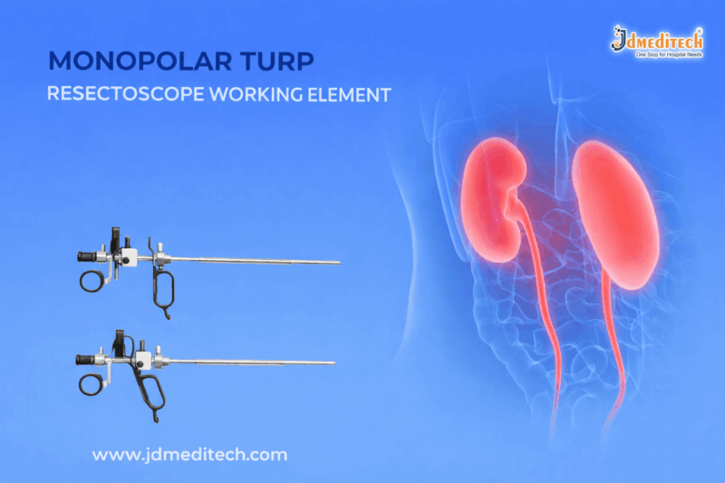
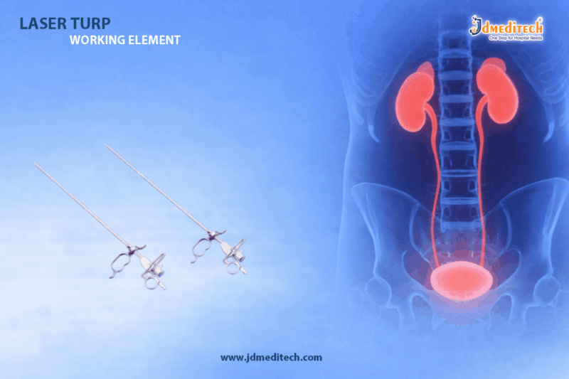
A Laser TURP Working Element is an integral part of a resectoscope system used during Transurethral Resection of the Prostate (TURP) procedures. Unlike conventional monopolar or bipolar systems, this working element is optimized for laser-based prostate surgery, offering enhanced control and safety.
It facilitates the movement and positioning of laser fibers, ensuring precise targeting of prostate tissue while minimizing damage to surrounding areas.
Key Features of Laser TURP Working Element
1. High Precision Control
The device provides exceptional control over laser fiber movement, allowing surgeons to perform accurate tissue removal.
2. Compatibility with Laser Systems
Designed to work seamlessly with modern holmium and other medical laser technologies, ensuring optimal performance.
3. Ergonomic Design
Its user-friendly structure reduces surgeon fatigue during long procedures and enhances handling efficiency.
4. Durable Medical-Grade Construction
Manufactured using high-quality stainless steel and advanced polymers, ensuring long-lasting durability and reliability.
5. Smooth Mechanism Operation
The working element offers a smooth sliding mechanism, allowing consistent and controlled surgical movements.
Benefits in Minimally Invasive TURP
Enhanced Surgical Accuracy
The Laser TURP Working Element ensures precise targeting, reducing the risk of unnecessary tissue damage.
Reduced Bleeding
Laser technology significantly minimizes bleeding compared to traditional TURP procedures.
Faster Recovery Time
Patients experience shorter hospital stays and quicker recovery, making it a preferred choice for modern urology.
Lower Risk of Complications
Improved control and precision help reduce complications such as TUR syndrome.
Applications in Urology
The Laser TURP Working Element is widely used in:
- Treatment of Benign Prostatic Hyperplasia (BPH)
- Laser prostatectomy procedures
- Minimally invasive urological surgeries
- Hospital and clinical urology setups
Why Choose a High-Quality Laser TURP Working Element?
Selecting a premium-quality Laser TURP Working Element ensures:
- Consistent surgical performance
- Enhanced patient safety
- Long-term cost efficiency
- Compatibility with advanced surgical systems
For healthcare providers, investing in reliable equipment directly impacts surgical outcomes and patient satisfaction.
Maintenance and Handling Tips
To ensure optimal performance:
- Clean and sterilize after every procedure
- Inspect for wear and tear regularly
- Use compatible laser fibers only
- Store in a dry and sterile environment
Proper maintenance extends the lifespan and ensures consistent functionality.
Conclusion
The Laser TURP Working Element represents a significant advancement in minimally invasive prostate surgery. With its precision, safety, and efficiency, it plays a vital role in improving surgical outcomes and patient recovery.
As urology continues to evolve, adopting advanced tools like the Laser TURP Working Element is essential for delivering high-quality, patient-centric care.
Get Connected:
+91 79909 93062 | +91 63513 72032 | exports@jdmeditech.com