Laparoscopic Adhesiolysis Surgery – Minimally Invasive Treatment for Abdominal Adhesions

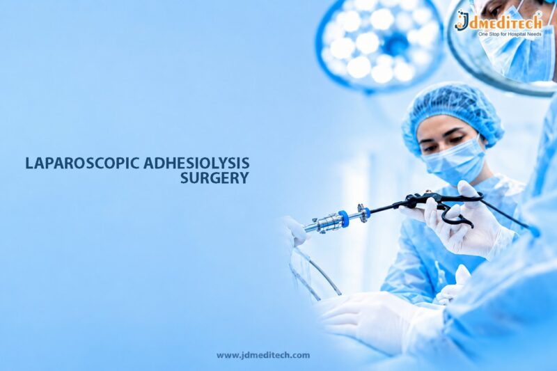
Laparoscopic adhesiolysis surgery is an advanced minimally invasive procedure performed to remove or separate abnormal scar tissue bands known as adhesions inside the abdomen or pelvis. Adhesions commonly develop after abdominal surgery, infections, inflammation, trauma, or radiation therapy. Although some adhesions remain asymptomatic, others may cause chronic abdominal pain, bowel obstruction, infertility, digestive problems, and restricted organ movement.

Traditionally, adhesion treatment often required open surgery with larger incisions and longer recovery periods. However, modern laparoscopic techniques now allow surgeons to perform adhesiolysis through small incisions using high-definition cameras and specialized endoscopic instruments. Consequently, patients benefit from reduced postoperative pain, shorter hospital stay, faster healing, and lower risk of additional adhesion formation.

Today, laparoscopic adhesiolysis has become a preferred surgical approach for managing many adhesion-related complications because of its precision, safety, and minimally invasive advantages.

What Is Laparoscopic Adhesiolysis Surgery?

Laparoscopic adhesiolysis surgery is a minimally invasive procedure used to carefully separate or remove internal scar tissue adhesions that abnormally connect organs, tissues, or abdominal structures.

Normally, abdominal organs move freely within the abdominal cavity. However, adhesions may form after tissue injury or inflammation, causing organs to stick together. As a result, patients may experience pain, bowel obstruction, or reproductive complications.

During the procedure, surgeons use a laparoscope, which is a thin camera-equipped instrument inserted through small abdominal incisions. Specialized laparoscopic instruments are then used to cut and release adhesions while minimizing trauma to surrounding tissues.

Ultimately, the primary goal of laparoscopic adhesiolysis is to restore normal organ movement, relieve symptoms, and improve abdominal function.

Why Is Laparoscopic Adhesiolysis Performed?

Laparoscopic adhesiolysis is recommended when adhesions cause persistent symptoms or serious complications.

Common Indications for Surgery Include:
  • Chronic abdominal pain
  • Intestinal obstruction
  • Pelvic pain
  • Female infertility caused by adhesions
  • Adhesion-related digestive symptoms
  • Bowel kinking or narrowing
  • Recurrent abdominal bloating
  • Complications after previous surgery

Additionally, timely surgical treatment may help prevent severe bowel complications and improve quality of life.

Understanding Abdominal Adhesions

Abdominal adhesions are bands of fibrous scar tissue that form between internal organs and surrounding tissues.

Common Causes of Adhesions Include:
  • Previous abdominal surgery
  • Pelvic surgery
  • Appendicitis
  • Endometriosis
  • Abdominal infections
  • Inflammatory bowel disease
  • Radiation therapy
  • Trauma or injury

In many cases, adhesions form naturally during the healing process after tissue inflammation or surgery.

Symptoms of Adhesion-Related Complications

Symptoms vary depending on the location and severity of adhesions.

Common Symptoms Include:
  • Chronic abdominal pain
  • Cramping
  • Bloating
  • Nausea and vomiting
  • Constipation
  • Difficulty passing gas
  • Pelvic pain
  • Infertility in women
  • Recurrent bowel obstruction

Therefore, persistent abdominal symptoms should be evaluated by an experienced surgeon.

Who Is a Suitable Candidate for Laparoscopic Adhesiolysis?

Generally, laparoscopic adhesiolysis may be recommended for:

  • Patients with symptomatic adhesions
  • Individuals with recurrent bowel obstruction
  • Women with infertility linked to pelvic adhesions
  • Patients with chronic unexplained abdominal pain
  • Individuals medically fit for minimally invasive surgery

Importantly, patient selection depends on symptom severity, previous surgical history, and overall health status.

Preoperative Evaluation Before Surgery

Before laparoscopic adhesiolysis, the surgeon performs comprehensive assessments to determine the extent of adhesions and plan treatment safely.

Medical History Review

Initially, the doctor reviews:

  • Previous surgeries
  • Abdominal pain history
  • Digestive symptoms
  • Fertility concerns
  • Prior infections
  • Medication history
Physical Examination

Furthermore, abdominal examination helps identify tenderness, distension, or signs of bowel obstruction.

Diagnostic Investigations

Additional tests may include:

  • CT scan
  • Abdominal ultrasound
  • MRI imaging
  • Blood investigations
  • X-rays
  • Colonoscopy in selected cases

Consequently, these evaluations help identify complications and guide surgical planning.

How Laparoscopic Adhesiolysis Surgery Is Performed

Typically, laparoscopic adhesiolysis is performed under general anesthesia using advanced minimally invasive surgical equipment.

Step-by-Step Surgical Procedure
1. Creation of Small Incisions

First, the surgeon creates several tiny abdominal incisions for insertion of the laparoscope and surgical instruments.

2. Inflation of the Abdomen

Next, carbon dioxide gas is introduced into the abdomen to create adequate working space.

3. Visualization of Adhesions

Afterward, the laparoscope provides magnified visualization of abdominal organs and adhesion bands.

4. Careful Separation of Adhesions

Then, specialized instruments are used to cut and release adhesions while preserving nearby tissues and organs.

5. Restoration of Organ Mobility

The surgeon carefully frees trapped organs and restores normal anatomical movement.

6. Inspection for Additional Problems

Furthermore, the abdominal cavity is examined for bowel injury, inflammation, or other abnormalities.

7. Closure of Incisions

Finally, the instruments are removed and the small incisions are closed with sutures or surgical glue.

Overall, the procedure usually takes between 1 and 3 hours depending on the severity of adhesions.

Benefits of Laparoscopic Adhesiolysis Surgery

Laparoscopic adhesiolysis offers several important advantages compared to traditional open surgery.

Minimally Invasive Technique

Because the procedure uses small incisions, tissue trauma and postoperative discomfort are significantly reduced.

Faster Recovery

Additionally, patients often recover more quickly and resume normal activities sooner.

Reduced Risk of New Adhesion Formation

Minimally invasive surgery may lower the risk of future adhesions compared to open surgery.

Improved Surgical Precision

Magnified visualization allows accurate identification and separation of adhesions.

Reduced Blood Loss

Furthermore, laparoscopic techniques often minimize intraoperative bleeding.

Better Cosmetic Results

Smaller incisions produce minimal visible scarring.

Recovery After Laparoscopic Adhesiolysis

Recovery after laparoscopic adhesiolysis is generally smoother compared to open abdominal surgery.

Common Postoperative Symptoms

Initially, patients may experience:

  • Mild abdominal discomfort
  • Temporary bloating
  • Fatigue
  • Mild nausea
  • Shoulder pain from surgical gas

However, these symptoms usually improve within several days.

Recovery Guidelines

To support proper healing, patients are advised to:

  • Walk regularly after surgery
  • Stay hydrated
  • Follow dietary instructions
  • Avoid heavy lifting temporarily
  • Maintain incision hygiene
  • Attend follow-up appointments

As a result, many individuals return to routine activities within 1 to 3 weeks.

Risks and Complications

Although laparoscopic adhesiolysis is considered safe, certain complications may occasionally occur.

Possible Complications Include:
  • Bleeding
  • Infection
  • Bowel injury
  • Organ perforation
  • Recurrence of adhesions
  • Blood clots
  • Anesthesia-related complications

Nevertheless, complication rates are generally lower when surgery is performed by experienced laparoscopic surgeons.

Adhesion Recurrence Prevention

Although adhesions may recur after surgery, several measures may help reduce recurrence risk.

Preventive Strategies Include:
  • Minimally invasive surgical techniques
  • Gentle tissue handling
  • Use of adhesion barriers
  • Early postoperative movement
  • Proper infection control

Consequently, these measures may improve long-term outcomes and reduce repeat surgery risk.

Success Rates of Laparoscopic Adhesiolysis

Laparoscopic adhesiolysis has favorable outcomes for carefully selected patients.

Factors Affecting Outcomes

Several factors influence surgical success, including:

  • Severity of adhesions
  • Presence of bowel obstruction
  • Number of previous surgeries
  • Patient health condition
  • Surgical expertise
General Outcomes

Modern laparoscopic adhesiolysis typically provides:

  • Improved pain relief
  • Better bowel function
  • Reduced obstruction episodes
  • Faster recovery
  • Enhanced quality of life

Therefore, early intervention often helps improve long-term results.

Lifestyle Tips After Surgery

Healthy lifestyle habits may support recovery and digestive health after surgery.

Recommended Lifestyle Measures

Patients are encouraged to:

  • Stay physically active
  • Maintain healthy bowel habits
  • Follow balanced nutrition
  • Stay hydrated
  • Avoid smoking
  • Follow postoperative instructions carefully

Consequently, these habits may improve healing and reduce complications.

Choosing the Right Surgeon

Successful laparoscopic adhesiolysis requires expertise in advanced minimally invasive abdominal surgery.

Patients Should Look For:
  • Experienced laparoscopic surgeons
  • Expertise in complex abdominal surgery
  • Advanced hospital facilities
  • Comprehensive postoperative care
  • Strong patient safety standards

Ultimately, experienced surgical teams improve both safety and long-term outcomes.

Conclusion

Laparoscopic adhesiolysis surgery is a modern minimally invasive solution for treating adhesion-related abdominal and pelvic complications. By combining advanced laparoscopic technology with precise surgical techniques, the procedure provides effective symptom relief, faster recovery, reduced pain, and improved quality of life.

Moreover, early diagnosis and timely surgical management can help prevent severe complications such as bowel obstruction and chronic pain. Therefore, patients experiencing persistent abdominal symptoms after previous surgery or infection should seek evaluation from an experienced laparoscopic surgeon.

With continuous advancements in minimally invasive surgery and postoperative care, laparoscopic adhesiolysis continues to remain an effective treatment option for adhesion-related disorders worldwide.


Explore More Product >>

Get Connected:
+91 79909 93062 | +91 63513 72032 | exports@jdmeditech.com 

Laparoscopic Fundoplication Surgery – Minimally Invasive GERD and Acid Reflux Treatment

Laparoscopic fundoplication surgery is an advanced minimally invasive procedure used to treat gastroesophageal reflux disease (GERD) and chronic acid reflux. GERD is a digestive disorder in which stomach acid repeatedly flows back into the esophagus, causing symptoms such as heartburn, chest discomfort, regurgitation, and difficulty swallowing. If left untreated, chronic acid reflux may lead to serious complications, including esophageal inflammation, ulcers, and Barrett’s esophagus.

Traditionally, severe reflux disease was managed mainly with medications and lifestyle modifications. However, when symptoms persist despite medical treatment, laparoscopic fundoplication offers an effective long-term surgical solution. Moreover, this minimally invasive procedure provides excellent symptom control with reduced postoperative pain, smaller incisions, and faster recovery.

Today, laparoscopic fundoplication is widely recognized as one of the most successful surgical treatments for GERD and acid reflux disease.

What Is Laparoscopic Fundoplication Surgery?

Laparoscopic fundoplication surgery is a minimally invasive procedure in which the upper part of the stomach, called the fundus, is wrapped around the lower esophagus to strengthen the lower esophageal sphincter (LES).

The lower esophageal sphincter normally acts as a valve between the esophagus and stomach. However, when this valve becomes weak, stomach acid can flow backward into the esophagus and cause reflux symptoms.

During laparoscopic fundoplication, surgeons use a laparoscope and specialized instruments inserted through small abdominal incisions. Consequently, the procedure restores the barrier between the stomach and esophagus, helping prevent acid reflux.

Ultimately, the surgery aims to reduce reflux symptoms, improve quality of life, and prevent long-term esophageal damage.

Understanding GERD and Acid Reflux

GERD occurs when stomach acid repeatedly enters the esophagus due to weakness of the lower esophageal sphincter.

Common Symptoms of GERD Include:
  • Frequent heartburn
  • Acid regurgitation
  • Chest discomfort
  • Difficulty swallowing
  • Chronic cough
  • Sore throat
  • Hoarseness
  • Bitter taste in the mouth

In severe cases, GERD may interfere with sleep, eating habits, and daily activities.

Why Is Laparoscopic Fundoplication Performed?

Laparoscopic fundoplication is recommended when acid reflux symptoms become chronic or fail to improve with conservative treatment.

Common Indications for Surgery Include:
  • Severe GERD
  • Chronic acid reflux
  • Inadequate response to medications
  • Hiatal hernia associated with reflux
  • Esophagitis
  • Barrett’s esophagus
  • Regurgitation symptoms
  • Long-term dependence on acid suppression medications

Additionally, surgery may help improve long-term symptom control and reduce medication dependence.

Who Is a Suitable Candidate for Laparoscopic Fundoplication?

Generally, laparoscopic fundoplication may be recommended for:

  • Patients with confirmed GERD
  • Individuals with persistent reflux symptoms
  • Patients with reflux-related complications
  • Individuals medically fit for surgery
  • Patients seeking long-term reflux control

Importantly, proper evaluation is necessary before deciding on surgical treatment.

Preoperative Evaluation Before Surgery

Before laparoscopic fundoplication, the surgeon performs detailed assessments to confirm GERD and evaluate esophageal function.

Medical History Review

Initially, the doctor reviews:

  • Frequency of heartburn
  • Severity of reflux symptoms
  • Medication history
  • Dietary habits
  • Previous abdominal surgeries
  • Associated respiratory symptoms
Physical Examination

Furthermore, physical examination helps assess general health and abdominal findings.

Diagnostic Investigations

Additional tests may include:

  • Upper GI endoscopy
  • Esophageal pH monitoring
  • Esophageal manometry
  • Barium swallow study
  • CT scan in selected cases

Consequently, these investigations help confirm reflux disease and guide surgical planning.

How Laparoscopic Fundoplication Surgery Is Performed

Typically, laparoscopic fundoplication is performed under general anesthesia using advanced minimally invasive surgical equipment.

Step-by-Step Surgical Procedure
1. Creation of Small Incisions

First, the surgeon creates several tiny abdominal incisions for insertion of the laparoscope and surgical instruments.

2. Inflation of the Abdomen

Next, carbon dioxide gas is introduced to create adequate working space inside the abdomen.

3. Visualization of the Esophagus and Stomach

Afterward, the laparoscope provides magnified views of the stomach, diaphragm, and lower esophagus.

4. Hiatal Hernia Repair if Present

If a hiatal hernia is present, the surgeon repairs the opening in the diaphragm.

5. Wrapping of the Stomach Fundus

Then, the upper part of the stomach is wrapped around the lower esophagus to strengthen the reflux barrier.

6. Securing the Wrap

The wrap is carefully secured using sutures to maintain proper positioning.

7. Closure of Incisions

Finally, the surgical instruments are removed and the small incisions are closed.

Overall, the surgery usually takes between 1 and 3 hours depending on complexity.

Types of Fundoplication Procedures

Several types of fundoplication may be performed based on patient condition and esophageal function.

Procedure Type Description Common Use
Nissen Fundoplication Complete 360-degree wrap Standard GERD treatment
Toupet Fundoplication Partial 270-degree wrap Reduced swallowing difficulty risk
Dor Fundoplication Anterior partial wrap Selected esophageal conditions
Hiatal Hernia Repair with Fundoplication Combined repair GERD with hiatal hernia

Ultimately, the surgeon selects the most appropriate technique for each patient.

Benefits of Laparoscopic Fundoplication Surgery

Laparoscopic fundoplication offers several important advantages over traditional open surgery and long-term medication use.

Minimally Invasive Technique

Because the procedure uses small incisions, postoperative pain and tissue trauma are significantly reduced.

Faster Recovery

Additionally, patients usually return to normal activities more quickly.

Reduced Acid Reflux Symptoms

Most patients experience significant improvement in heartburn and regurgitation.

Smaller Scars

Minimal incisions provide better cosmetic outcomes.

Reduced Dependence on Medication

Furthermore, many patients no longer require long-term acid reflux medications after surgery.

Improved Quality of Life

Consequently, surgery often improves sleep, eating comfort, and daily functioning.

Recovery After Laparoscopic Fundoplication

Recovery after laparoscopic fundoplication is generally smooth when postoperative instructions are followed properly.

Common Postoperative Symptoms

Initially, patients may experience:

  • Mild abdominal discomfort
  • Temporary bloating
  • Difficulty swallowing
  • Fatigue
  • Shoulder pain from surgical gas

However, these symptoms usually improve gradually during recovery.

Recovery Guidelines

To support proper healing, patients are advised to:

  • Follow a soft or liquid diet initially
  • Eat smaller meals
  • Avoid carbonated beverages temporarily
  • Walk regularly
  • Avoid heavy lifting
  • Attend follow-up appointments

As a result, most individuals return to normal activities within 2 to 4 weeks.

Diet After Fundoplication Surgery

Dietary management plays an important role during recovery after surgery.

Recommended Dietary Tips

Patients are encouraged to:

  • Eat soft foods initially
  • Chew food thoroughly
  • Avoid overeating
  • Eat slowly
  • Stay hydrated
  • Gradually reintroduce solid foods

Consequently, these measures help reduce swallowing discomfort and support healing.

Risks and Complications

Although laparoscopic fundoplication is considered safe, certain complications may occasionally occur.

Possible Complications Include:
  • Difficulty swallowing
  • Gas bloating
  • Bleeding
  • Infection
  • Wrap loosening
  • Recurrence of reflux
  • Injury to nearby organs
  • Anesthesia-related complications

Nevertheless, complication rates are generally low when surgery is performed by experienced surgeons.

Success Rates of Laparoscopic Fundoplication

Laparoscopic fundoplication has excellent long-term success rates for GERD management.

Factors Affecting Outcomes

Several factors influence surgical success, including:

  • Severity of reflux disease
  • Presence of hiatal hernia
  • Esophageal function
  • Patient compliance
  • Surgical expertise
General Outcomes

Modern laparoscopic fundoplication typically provides:

  • Significant symptom relief
  • Improved quality of life
  • Reduced acid exposure
  • Lower medication dependence
  • Long-term reflux control

Therefore, proper patient selection and timely treatment are extremely important.

Lifestyle Tips After Surgery

Healthy lifestyle habits may further improve long-term reflux control after surgery.

Recommended Lifestyle Measures

Patients are encouraged to:

  • Maintain healthy body weight
  • Avoid smoking
  • Limit alcohol intake
  • Avoid late-night meals
  • Exercise regularly
  • Follow balanced nutrition

Consequently, these habits may support long-term digestive health.

Choosing the Right Surgeon

Successful laparoscopic fundoplication requires expertise in minimally invasive upper gastrointestinal surgery.

Patients Should Look For:
  • Experienced laparoscopic surgeons
  • Expertise in GERD treatment
  • Advanced hospital facilities
  • Comprehensive postoperative care
  • Strong patient safety standards

Ultimately, experienced specialists improve surgical precision and long-term outcomes.

Conclusion

Laparoscopic fundoplication surgery is an advanced minimally invasive treatment for GERD and chronic acid reflux. By strengthening the lower esophageal sphincter through precise laparoscopic techniques, the procedure provides long-term symptom relief, reduced medication dependence, and improved quality of life.

Moreover, modern surgical advancements have made laparoscopic fundoplication safer, more effective, and associated with faster recovery. Therefore, patients with severe or persistent acid reflux symptoms should seek evaluation from an experienced gastrointestinal surgeon to determine the most appropriate treatment approach.

With expert surgical care and proper lifestyle management, laparoscopic fundoplication continues to remain one of the most reliable solutions for chronic reflux disease.


Explore More Product >>

Get Connected:
+91 79909 93062 | +91 63513 72032 | exports@jdmeditech.com 

Tintara Uterine Manipulator for Advanced Gynecologic Procedures

Modern gynecologic surgery increasingly depends on minimally invasive techniques that improve surgical precision, reduce patient trauma, and support faster recovery. To achieve successful outcomes in laparoscopic and advanced gynecological procedures, surgeons require dependable instruments that provide accurate uterine positioning and stable anatomical access. Among these instruments, the Tintara Uterine Manipulator plays a critical role.

The Tintara Uterine Manipulator is an advanced gynecological instrument designed to provide controlled uterine movement during minimally invasive procedures. Its precision-focused design supports better visualization, enhanced surgical access, and improved procedural efficiency. As a result, gynecologists can perform complex procedures with greater accuracy and confidence.

This blog explores the features, benefits, applications, and clinical importance of the Tintara Uterine Manipulator while maintaining strong SEO performance and excellent readability.

What is a Tintara Uterine Manipulator?
Understanding the Instrument

A Tintara Uterine Manipulator is a specialized medical device used to mobilize, position, and stabilize the uterus during gynecological and laparoscopic surgeries. The instrument helps surgeons achieve optimal pelvic access and improved visualization of reproductive anatomy.

The device typically includes:

  • Ergonomic handle
  • Manipulation shaft
  • Adjustable uterine positioning system
  • Cervical attachment components
  • Locking mechanism for stability

Its precision-control design allows multidirectional uterine movement during advanced surgical procedures.

Importance of Uterine Manipulators in Gynecologic Surgery
Supporting Better Surgical Access and Visualization

Accurate uterine positioning is essential during minimally invasive gynecologic procedures. A high-quality Tintara Uterine Manipulator helps surgeons improve exposure of pelvic structures while maintaining procedural safety.

Clinical benefits include:

  • Enhanced pelvic visualization
  • Improved surgical precision
  • Better access to reproductive anatomy
  • Controlled uterine movement
  • Reduced tissue trauma
  • Increased procedural efficiency

Reliable uterine manipulation significantly improves the success of laparoscopic surgeries.

Key Features of the Tintara Uterine Manipulator
Ergonomic and Surgeon-Friendly Design

The manipulator features an ergonomic handle that allows surgeons to control uterine movement comfortably during lengthy procedures.

Advantages of ergonomic handling include:

  • Reduced hand fatigue
  • Improved instrument control
  • Better maneuverability
  • Enhanced surgical comfort

A user-friendly design supports precision throughout the operation.

Adjustable Uterine Manipulation System
Supporting Multidirectional Uterine Movement

The adjustable manipulation mechanism enables surgeons to position the uterus in multiple directions for improved surgical access.

Benefits include:

  • Better anatomical exposure
  • Easier laparoscopic navigation
  • Improved operative visibility
  • Enhanced procedural accuracy

Precise uterine control is essential during complex gynecologic surgeries.

High-Quality Medical-Grade Construction
Durable and Sterilization-Compatible Materials

Premium Tintara Uterine Manipulators are manufactured using surgical-grade stainless steel and durable medical-grade materials for long-term reliability.

Material advantages include:

  • Corrosion resistance
  • Long instrument lifespan
  • Consistent mechanical performance
  • Compatibility with sterilization protocols

Durable construction ensures dependable performance during repeated surgical use.

Designed for Minimally Invasive Procedures
Supporting Advanced Laparoscopic Surgery

The slim-profile design allows surgeons to perform minimally invasive gynecological procedures efficiently and safely.

Clinical benefits include:

  • Reduced tissue damage
  • Smaller surgical incisions
  • Lower bleeding risk
  • Faster patient recovery
  • Reduced postoperative pain

Minimally invasive compatibility enhances both patient comfort and surgical efficiency.

Applications of Tintara Uterine Manipulators
Common Gynecological Procedures

The Tintara Uterine Manipulator supports a wide range of diagnostic and operative gynecologic surgeries.

Common applications include:

Total Laparoscopic Hysterectomy (TLH)

The manipulator helps position the uterus effectively during hysterectomy procedures.

Diagnostic Laparoscopy

Surgeons use the device to improve pelvic visualization and anatomical access.

Endometriosis Treatment

Controlled uterine movement supports accurate access to endometrial lesions.

Myomectomy Procedures

The instrument assists in exposing uterine fibroids for precise removal.

Fertility and Reproductive Surgeries

The manipulator supports accurate uterine positioning during reproductive procedures.

Advantages of Using a Tintara Uterine Manipulator
Improved Surgical Precision

Precise uterine positioning allows surgeons to perform delicate procedures more accurately.

Precision-related advantages include:

  • Better surgical control
  • Improved anatomical visualization
  • Accurate tissue dissection
  • Reduced procedural errors

High-precision instruments support improved clinical outcomes.

Enhanced Surgical Efficiency
Streamlining Operating Room Workflow

The manipulator simplifies uterine handling and improves procedural workflow.

Efficiency benefits include:

  • Reduced surgical time
  • Easier instrument navigation
  • Faster procedural setup
  • Improved operating room productivity

Efficient instrumentation contributes to smoother gynecological surgeries.

Increased Patient Safety
Supporting Safer Minimally Invasive Procedures

Controlled uterine manipulation reduces tissue trauma and improves surgical safety.

Safety advantages include:

  • Reduced organ injury risk
  • Controlled tissue handling
  • Better visualization of pelvic anatomy
  • Lower complication rates

Patient safety remains essential in modern gynecologic surgery.

Easy Cleaning and Sterilization
Supporting Hospital Infection Control Standards

Reusable Tintara Uterine Manipulators are designed for efficient cleaning and sterilization according to hospital protocols.

Reprocessing benefits include:

  • Smooth instrument surfaces
  • Sterilization compatibility
  • Reduced contamination risk
  • Long-term usability

Proper sterilization maintains patient safety and instrument durability.

Choosing the Right Tintara Uterine Manipulator
Important Factors to Consider

Selecting the right uterine manipulator improves procedural reliability and surgical performance.

Factors to evaluate include:

Material Quality

Choose surgical-grade materials for durability and safety.

Ergonomic Design

Comfortable handling improves precision and surgeon control.

Adjustable Functionality

Ensure the manipulator supports multidirectional uterine movement.

Sterilization Compatibility

Verify compatibility with standard hospital sterilization procedures.

Manufacturer Reputation

Select trusted manufacturers known for high-quality gynecological instruments.

Role of Advanced Uterine Manipulators in Modern Gynecology
Supporting Precision-Based Surgical Care

Modern gynecology increasingly relies on advanced instruments that improve surgical access, visualization, and patient outcomes. The Tintara Uterine Manipulator supports these goals through reliable uterine positioning and precision control.

Advanced manipulators help surgeons:

  • Improve surgical accuracy
  • Reduce operative time
  • Enhance patient comfort
  • Minimize tissue trauma
  • Improve procedural consistency

As minimally invasive gynecology continues to evolve, precision uterine manipulation remains essential.

Maintenance Tips for Tintara Uterine Manipulators
Ensuring Long-Term Instrument Performance

Routine maintenance helps preserve instrument quality and operational reliability.

Recommended maintenance practices include:

  • Clean immediately after use
  • Follow validated sterilization guidelines
  • Inspect moving components regularly
  • Store instruments safely
  • Check for wear before procedures

Proper maintenance supports consistent performance during critical surgeries.

Conclusion
Enhancing Surgical Precision in Modern Gynecology

The Tintara Uterine Manipulator is an essential instrument for advanced gynecologic and laparoscopic procedures. Its precision-control system, ergonomic design, and minimally invasive compatibility help surgeons achieve better uterine positioning, enhanced visualization, and safer surgical outcomes.

As healthcare facilities continue adopting minimally invasive surgical techniques, high-quality uterine manipulators remain critical for improving procedural efficiency and patient care. A premium Tintara Uterine Manipulator supports accurate uterine control, smoother workflows, and improved surgical confidence across hospitals and gynecology centers.


Explore More Product >>

Get Connected:
+91 79909 93062 | +91 63513 72032 | exports@jdmeditech.com 

V Type Uterine Manipulator for Advanced Gynecological Surgery

Minimally invasive gynecological procedures require precision, stability, and excellent uterine positioning to achieve successful surgical outcomes. Among the essential instruments used in laparoscopic and gynecological surgeries, the V Type Uterine Manipulator stands out as a reliable solution for controlled uterine movement and enhanced pelvic access.

A V Type Uterine Manipulator is a specialized gynecological instrument designed to provide accurate uterine manipulation during laparoscopic procedures. Its advanced design supports improved visualization, easier instrument navigation, and better access to pelvic anatomy. As a result, surgeons can perform procedures with greater confidence, precision, and efficiency.

This blog explores the features, benefits, applications, and importance of the V Type Uterine Manipulator in modern gynecological surgery while maintaining strong SEO and readability.

What is a V Type Uterine Manipulator?
Understanding the Instrument

A V Type Uterine Manipulator is a medical device used during gynecological and laparoscopic procedures to mobilize, position, and stabilize the uterus. The instrument allows surgeons to achieve optimal surgical access while improving visualization of pelvic structures.

The manipulator generally consists of:

  • Ergonomic handle
  • Manipulation shaft
  • Vaginal cup or cervical attachment
  • Adjustable positioning mechanism
  • Locking and stabilization components

The “V Type” configuration enhances maneuverability and supports multidirectional uterine movement during surgery.

Importance of Uterine Manipulators in Gynecological Surgery
Supporting Better Surgical Access and Visibility

Proper uterine manipulation is critical during minimally invasive gynecological procedures. A high-quality V Type Uterine Manipulator helps surgeons improve surgical exposure and access difficult anatomical areas safely.

Clinical advantages include:

  • Improved pelvic visualization
  • Enhanced surgical precision
  • Better uterine positioning
  • Reduced tissue handling
  • Increased procedural efficiency
  • Safer laparoscopic access

Efficient uterine manipulation contributes significantly to successful surgical outcomes in gynecology.

Key Features of a V Type Uterine Manipulator
Ergonomic and User-Friendly Design

Modern uterine manipulators feature ergonomic handles that allow smooth operation and precise movement control during surgery.

Benefits of ergonomic handling include:

  • Reduced surgeon fatigue
  • Better maneuverability
  • Improved comfort during procedures
  • Enhanced operational stability

An ergonomic structure ensures greater control during delicate laparoscopic surgeries.

Adjustable Uterine Positioning Mechanism
Supporting Multidirectional Manipulation

The adjustable mechanism allows surgeons to move the uterus in different directions for optimal surgical exposure.

Advantages include:

  • Improved anatomical access
  • Better instrument navigation
  • Enhanced laparoscopic visibility
  • Accurate uterine positioning

Controlled movement is essential during complex gynecological procedures.

High-Quality Medical-Grade Construction
Durable and Sterilization-Compatible Materials

Premium V Type Uterine Manipulators are manufactured using surgical-grade stainless steel and high-quality medical polymers for durability and repeated use.

Material benefits include:

  • Corrosion resistance
  • Long instrument lifespan
  • Reliable performance
  • Compatibility with sterilization protocols

Durable construction supports long-term clinical reliability.

Minimally Invasive Surgical Compatibility
Designed for Advanced Laparoscopic Procedures

The slim profile and precision design make the manipulator ideal for minimally invasive gynecological surgeries.

Clinical benefits include:

  • Reduced tissue trauma
  • Smaller incisions
  • Lower bleeding risk
  • Faster patient recovery
  • Reduced postoperative discomfort

Minimally invasive compatibility improves both patient experience and surgical efficiency.

Applications of V Type Uterine Manipulators
Common Gynecological Procedures

The V Type Uterine Manipulator is widely used in diagnostic and operative gynecological surgeries.

Common applications include:

Total Laparoscopic Hysterectomy (TLH)

The manipulator helps position the uterus for improved surgical access during hysterectomy procedures.

Diagnostic Laparoscopy

Surgeons use the device to improve visualization of pelvic anatomy.

Endometriosis Surgery

Controlled uterine movement helps surgeons access endometrial lesions effectively.

Fertility and Reproductive Procedures

The manipulator supports precise uterine positioning during reproductive surgeries.

Myomectomy Procedures

The device assists in exposing fibroids and improving operative access.

Advantages of Using a V Type Uterine Manipulator
Improved Surgical Precision

Precise uterine positioning allows surgeons to operate more accurately during minimally invasive procedures.

Precision-related benefits include:

  • Better surgical control
  • Enhanced visualization
  • Accurate tissue dissection
  • Reduced procedural complications

Precision instruments improve overall surgical outcomes.

Enhanced Procedural Efficiency
Streamlining Gynecological Workflows

The manipulator simplifies uterine handling and supports smoother surgical workflows.

Efficiency benefits include:

  • Reduced operating time
  • Easier instrument navigation
  • Faster surgical setup
  • Improved operating room productivity

Efficient instruments contribute to optimized procedural performance.

Increased Patient Safety
Supporting Safer Surgical Outcomes

Controlled uterine manipulation minimizes unnecessary tissue trauma and improves procedural safety.

Safety advantages include:

  • Reduced organ injury risk
  • Better anatomical exposure
  • Controlled tissue handling
  • Lower complication rates

Patient safety remains a primary focus in minimally invasive gynecology.

Easy Cleaning and Sterilization
Supporting Infection Control Standards

Reusable V Type Uterine Manipulators are designed for easy cleaning and sterilization in hospital environments.

Reprocessing benefits include:

  • Smooth instrument surfaces
  • Sterilization compatibility
  • Reduced contamination risk
  • Long-term usability

Proper sterilization ensures patient safety and instrument longevity.

Choosing the Right V Type Uterine Manipulator
Important Selection Factors

Selecting a high-quality uterine manipulator improves procedural efficiency and surgical accuracy.

Factors to consider include:

Material Quality

Choose instruments manufactured from premium surgical-grade materials.

Ergonomic Design

Comfortable handling improves control and reduces fatigue.

Adjustable Functionality

Ensure the manipulator supports multidirectional uterine movement.

Sterilization Compatibility

Verify compatibility with hospital sterilization protocols.

Manufacturer Reliability

Purchase from trusted surgical instrument manufacturers with proven quality standards.

Role of Advanced Manipulators in Modern Gynecology
Supporting Minimally Invasive Surgical Advancements

Modern gynecology increasingly relies on advanced instruments that improve surgical precision and patient outcomes. The V Type Uterine Manipulator supports these advancements by enhancing visibility, access, and control during laparoscopic procedures.

Advanced uterine manipulators help surgeons:

  • Improve procedural accuracy
  • Enhance patient safety
  • Reduce surgical time
  • Minimize tissue trauma
  • Achieve better clinical outcomes

As minimally invasive surgery continues to evolve, high-performance manipulation instruments remain essential.

Maintenance Tips for V Type Uterine Manipulators
Ensuring Long-Term Performance

Routine maintenance helps preserve instrument quality and surgical reliability.

Recommended maintenance practices include:

  • Clean immediately after use
  • Follow validated sterilization guidelines
  • Inspect moving components regularly
  • Store instruments safely
  • Check for wear before procedures

Proper maintenance ensures consistent and reliable performance during surgery.

Conclusion
Enhancing Gynecological Precision and Surgical Access

The V Type Uterine Manipulator is an essential instrument in modern minimally invasive gynecological surgery. Its advanced manipulation capabilities, ergonomic design, and precision control help surgeons achieve optimal uterine positioning and improved surgical visibility.

As laparoscopic procedures continue to advance, healthcare professionals increasingly depend on high-quality uterine manipulators that combine safety, efficiency, and durability. A premium V Type Uterine Manipulator supports better surgical workflows, enhanced patient outcomes, and improved procedural confidence across hospitals and gynecology centers.


Explore More Product >>

Get Connected:
+91 79909 93062 | +91 63513 72032 | exports@jdmeditech.com 


PCOD Needle for Safe and Precise PCOD Aspiration Procedures

Polycystic Ovarian Disease (PCOD) is one of the most common hormonal disorders affecting women of reproductive age. In many gynecological and fertility-related procedures, clinicians require precise and minimally invasive instruments to perform ovarian aspiration safely and effectively. A PCOD Needle plays a critical role in these procedures by providing controlled access for ovarian cyst aspiration and related interventions.

Designed with precision engineering and medical-grade materials, the PCOD aspiration needle helps gynecologists perform procedures with enhanced accuracy, patient comfort, and procedural efficiency. Its fine tip, optimal flexibility, and smooth penetration capability make it an essential instrument in fertility clinics, gynecology centers, and minimally invasive surgical setups.

This blog explores the features, applications, advantages, and clinical importance of PCOD needles while providing valuable SEO-focused insights for healthcare professionals and medical buyers.

What is a PCOD Needle?
Understanding the Role of PCOD Aspiration Needles

A PCOD Needle is a specialized gynecological instrument used for controlled aspiration procedures involving polycystic ovaries. Doctors commonly use it during ovarian drilling support procedures, cyst aspiration, and fertility-related interventions requiring precision access to ovarian follicles or cystic structures.

The needle is carefully designed to:

  • Ensure accurate puncture
  • Minimize tissue trauma
  • Support smooth fluid aspiration
  • Improve procedural control
  • Enhance patient safety

Because of its fine and sharp structure, the needle allows clinicians to perform delicate gynecological procedures with confidence and precision.

Key Features of PCOD Needle
Precision-Engineered Design for Better Clinical Performance

Modern PCOD aspiration needles include several advanced features that improve usability and surgical outcomes.

Fine Sharp Tip

The ultra-sharp tip enables smooth tissue penetration while reducing trauma and discomfort during aspiration procedures.

Medical-Grade Stainless Steel

Manufacturers use high-quality stainless steel to ensure durability, corrosion resistance, and reliable performance.

Controlled Aspiration Access

The needle design supports accurate placement and controlled fluid aspiration from ovarian cysts or follicles.

Ergonomic Hub Design

An easy-grip hub improves handling and provides better procedural stability for gynecologists.

Sterile Disposable Configuration

Many PCOD needles come in sterile disposable packaging to reduce cross-contamination risks and support infection control.

Compatible with Aspiration Systems

The needle easily integrates with syringes and aspiration devices commonly used in gynecology and fertility clinics.

Applications of PCOD Needle in Gynecology
Essential Instrument for Minimally Invasive Ovarian Procedures

The PCOD Needle supports multiple gynecological and fertility-related applications.

Ovarian Cyst Aspiration

Doctors use the needle to aspirate cystic fluid safely during minimally invasive ovarian procedures.

PCOD Management Procedures

In selected PCOD treatment approaches, aspiration techniques help reduce ovarian cyst volume and improve clinical evaluation.

Fertility and IVF Procedures

The needle assists in follicular access and aspiration during reproductive medicine interventions.

Diagnostic Aspiration

Clinicians may use the needle for collecting samples for diagnostic evaluation and laboratory analysis.

Ultrasound-Guided Procedures

The needle performs effectively in ultrasound-guided aspiration procedures requiring high precision.

Benefits of Using PCOD Needle
Improving Safety, Precision, and Procedural Efficiency

Using a high-quality PCOD aspiration needle offers several clinical and operational advantages.

Enhanced Precision

The fine needle structure allows accurate targeting of ovarian follicles and cysts.

Reduced Tissue Trauma

Sharp and smooth penetration minimizes tissue injury and procedural discomfort.

Better Procedural Control

The ergonomic design helps clinicians maintain stable handling throughout the procedure.

Improved Patient Comfort

Minimally invasive aspiration techniques support faster recovery and reduced post-procedure discomfort.

Reliable Fluid Aspiration

Optimized lumen design ensures efficient aspiration and fluid flow.

Infection Control Support

Sterile disposable variants reduce contamination risks and simplify workflow management.

Important Considerations While Choosing a PCOD Needle
Factors That Influence Performance and Safety

Healthcare providers should evaluate several factors before selecting a PCOD aspiration needle.

Needle Gauge and Length

Choosing the appropriate size ensures compatibility with the intended procedure and patient anatomy.

Sterility Standards

Single-use sterile packaging enhances procedural safety and compliance.

Material Quality

Medical-grade stainless steel improves durability and procedural reliability.

Ultrasound Visibility

Some needles include echogenic markings for improved visualization during ultrasound-guided procedures.

Manufacturer Reliability

Selecting products from trusted medical device manufacturers ensures quality consistency and regulatory compliance.

Role of PCOD Needle in Fertility Clinics
Supporting Modern Reproductive Healthcare

Fertility specialists rely on precision instruments for successful ovarian access procedures. The PCOD Needle supports modern reproductive medicine by improving aspiration accuracy and procedural efficiency.

In IVF and fertility treatment settings, precise aspiration tools help clinicians perform delicate interventions with greater confidence. Furthermore, minimally invasive procedures reduce recovery time and improve overall patient experience.

As fertility treatments continue to advance, the demand for high-quality gynecological aspiration instruments is steadily increasing worldwide.

Safety and Sterilization Standards
Ensuring Safe Gynecological Procedures

Patient safety remains a top priority during aspiration procedures. Therefore, manufacturers design PCOD needles according to strict medical standards.

Important safety features include:

  • Sterile packaging
  • Biocompatible materials
  • Smooth needle finish
  • Secure hub connection
  • Reliable aspiration performance

Additionally, disposable needles help healthcare facilities maintain high infection control standards while improving operational efficiency.

Why High-Quality PCOD Needles Matter
Precision Instruments Improve Clinical Outcomes

Using a well-designed PCOD Needle significantly impacts procedural success and patient safety. High-quality instruments help clinicians achieve:

  • Better procedural accuracy
  • Reduced complication risks
  • Smoother aspiration performance
  • Improved patient satisfaction
  • Greater procedural confidence

Moreover, reliable aspiration needles support standardized clinical workflows in busy gynecology and fertility centers.

Future Trends in PCOD Aspiration Instruments
Advancements in Gynecological Device Technology

The field of gynecological medical devices continues to evolve rapidly. Manufacturers are introducing advanced aspiration needles with improved ergonomics, enhanced ultrasound visibility, and optimized fluid control systems.

Future innovations may include:

  • Enhanced echogenic needle surfaces
  • Improved flexible needle designs
  • Advanced aspiration control systems
  • Better compatibility with imaging technologies
  • Environmentally conscious disposable materials

These advancements aim to improve procedural precision while supporting safer minimally invasive gynecology practices.

Conclusion
PCOD Needle as an Essential Tool in Modern Gynecology

The PCOD Needle is an important medical instrument that supports safe, accurate, and minimally invasive ovarian aspiration procedures. Its precision-engineered design, controlled access capability, and compatibility with modern aspiration systems make it highly valuable in gynecology and fertility care.

By combining safety, efficiency, and procedural accuracy, the PCOD aspiration needle helps healthcare professionals deliver better patient outcomes while maintaining high clinical standards.

As minimally invasive gynecological procedures continue to grow, reliable aspiration instruments will remain essential for modern reproductive healthcare practices.


Explore More Product >>

Get Connected:
+91 79909 93062 | +91 63513 72032 | exports@jdmeditech.com 


Gynecology Aspiration Kit – Complete Sterile Solution for Safe and Controlled Uterine Aspiration.

A Gynecology Aspiration Kit plays an important role in modern gynecological procedures. It supports safe, controlled, and efficient uterine aspiration in hospitals, fertility centers, and gynecology clinics. Medical professionals use this sterile kit during diagnostic and therapeutic procedures that require accurate suction and smooth instrument handling.

Healthcare providers demand instruments that improve procedural safety while maintaining patient comfort. Therefore, a high-quality aspiration kit offers precision components, sterile packaging, and dependable performance throughout the procedure.

In addition, modern gynecological practices focus heavily on infection control, procedural efficiency, and patient-centered care. A complete aspiration kit helps physicians achieve these goals while reducing preparation time in clinical settings.

What Is a Gynecology Aspiration Kit?
Understanding the Purpose of the Kit

A Gynecology Aspiration Kit is a sterile medical set designed for uterine aspiration procedures. The kit usually includes aspiration cannulas, tubing, syringes, connectors, collection components, and other essential accessories required for controlled aspiration.

Doctors commonly use these kits for:

  • Endometrial aspiration
  • Uterine evacuation procedures
  • Diagnostic sampling
  • Fertility-related procedures
  • Gynecological suction procedures
  • Early pregnancy management
  • Biopsy collection

Because the kit comes pre-packed and sterile, healthcare professionals can prepare quickly and maintain a hygienic surgical environment.

Key Components of a Gynecology Aspiration Kit
Sterile and Procedure-Ready Instruments

A complete Gynecology Aspiration Kit may include:

  • Aspiration cannulas in multiple sizes
  • Flexible connecting tubing
  • Manual vacuum aspiration syringe
  • Connectors and adapters
  • Collection chamber or specimen trap
  • Dilators (in selected kits)
  • Sterile drapes and accessories

Each component supports smooth aspiration control and helps physicians perform procedures with greater confidence.

Moreover, manufacturers use medical-grade materials to ensure safety, durability, and compatibility with gynecological applications.

Benefits of Using a Gynecology Aspiration Kit
Enhanced Safety and Procedural Efficiency

A well-designed aspiration kit offers several important advantages.

1. Sterile Single-Use Design

The sterile packaging reduces the risk of cross-contamination and infection. Since the kit is disposable, it supports better infection prevention protocols.

2. Controlled Aspiration Performance

The aspiration system allows smooth and controlled suction during uterine procedures. As a result, physicians can perform procedures with improved precision.

3. Reduced Procedure Preparation Time

A pre-assembled sterile kit minimizes setup time. Therefore, healthcare staff can improve workflow efficiency in busy clinics and operating rooms.

4. Improved Patient Comfort

Soft and smooth cannulas help reduce procedural discomfort. Additionally, controlled aspiration may support less traumatic tissue handling.

5. Convenient Clinical Use

Because all essential components come together in one kit, medical professionals can perform procedures without searching for separate instruments.

Applications of Gynecology Aspiration Kits
Common Gynecological and Diagnostic Uses

Gynecology aspiration kits support various clinical applications, including:

  • Endometrial biopsy procedures
  • Uterine aspiration
  • Diagnostic uterine sampling
  • Fertility investigations
  • Gynecological suction procedures
  • Early pregnancy care
  • Outpatient gynecology procedures

Furthermore, these kits help clinicians maintain procedural consistency across different healthcare settings.

Features of a High-Quality Gynecology Aspiration Kit
Important Factors to Consider

Healthcare facilities should evaluate several features before selecting an aspiration kit.

Sterility Assurance

A reliable kit must meet strict sterilization standards. This feature helps maintain patient safety during every procedure.

Medical-Grade Materials

High-quality tubing and cannulas improve durability and procedural reliability.

Multiple Cannula Sizes

Different patient requirements demand various cannula sizes. Therefore, versatile kits improve procedural flexibility.

Ergonomic Design

Comfortable handling supports better procedural control for physicians.

Secure Connections

Leak-resistant connectors help maintain stable aspiration pressure throughout the procedure.

Importance of Sterility in Gynecological Procedures
Supporting Infection Prevention Standards

Sterility remains one of the most critical factors in gynecological care. Contaminated instruments can increase procedural risks and complications.

A sterile Gynecology Aspiration Kit helps:

  • Minimize infection risks
  • Maintain operating room hygiene
  • Improve patient confidence
  • Support hospital sterilization protocols
  • Reduce instrument handling errors

Consequently, many healthcare providers prefer ready-to-use sterile kits for routine gynecological procedures.

Why Healthcare Professionals Prefer Complete Aspiration Kits
Simplifying Clinical Workflow

Complete aspiration kits improve efficiency in hospitals and clinics. Since all components are packaged together, clinicians can save valuable preparation time.

In addition, standardized kits help healthcare teams maintain consistency during procedures. This organized setup supports smoother clinical operations and improved procedural readiness.

Medical professionals also appreciate the convenience of disposable sterile systems because they reduce post-procedure cleaning requirements.

Choosing the Right Gynecology Aspiration Kit
Factors Healthcare Facilities Should Evaluate

When selecting a gynecology aspiration kit, healthcare providers should consider:

  • Sterility standards
  • Component quality
  • Cannula compatibility
  • Ease of use
  • Packaging integrity
  • Manufacturer reliability
  • Clinical application suitability

Choosing a trusted medical device manufacturer ensures consistent quality and dependable clinical performance.

Future Trends in Gynecological Aspiration Devices
Advancements in Safety and Comfort

Modern gynecology continues to evolve rapidly. Manufacturers now focus on:

  • Improved ergonomic designs
  • Enhanced aspiration control
  • Better patient comfort
  • Advanced sterile packaging
  • Procedure-specific kits
  • Eco-friendly medical packaging

As minimally invasive gynecological procedures become more common, aspiration kits will continue to support safer and more efficient treatment methods.

Conclusion

A Gynecology Aspiration Kit provides a complete sterile solution for safe and controlled uterine aspiration procedures. Its ready-to-use design improves procedural efficiency, supports infection prevention, and enhances clinician confidence during gynecological interventions.

Moreover, the combination of sterile components, controlled suction capability, and user-friendly design makes these kits essential in modern gynecology practice. Hospitals, fertility centers, and outpatient clinics rely on high-quality aspiration kits to maintain procedural safety and patient care standards.

By selecting a reliable and well-designed aspiration kit, healthcare providers can achieve smoother procedures, improved workflow, and better clinical outcomes.


Explore More Product >>

Get Connected:
+91 79909 93062 | +91 63513 72032 | exports@jdmeditech.com 


Hysteroscopy Diagnostic Sheath for Safe & Accurate Uterine Examination

Modern gynecological procedures demand precision, safety, and patient comfort. Among the essential instruments used in minimally invasive gynecology, the Hysteroscopy Diagnostic Sheath plays a crucial role in supporting smooth and controlled uterine access during diagnostic hysteroscopy procedures.

Designed to provide stable instrument guidance and effective fluid management, the hysteroscopy diagnostic sheath helps gynecologists perform accurate uterine examinations with enhanced visualization and minimal trauma. Its ergonomic construction and reliable compatibility with hysteroscopes make it an important component in outpatient and hospital-based hysteroscopy procedures.

This blog explores the features, benefits, applications, and importance of the hysteroscopy diagnostic sheath in modern gynecological practice.

What is a Hysteroscopy Diagnostic Sheath?

A Hysteroscopy Diagnostic Sheath is a specialized outer sheath designed to house and guide a hysteroscope during diagnostic hysteroscopy procedures. It facilitates smooth insertion into the uterine cavity while maintaining continuous fluid inflow and outflow for clear visualization.

The sheath creates a stable entry pathway for the hysteroscope and helps reduce tissue trauma during insertion. Additionally, it supports efficient uterine distension, allowing physicians to examine the cervical canal and uterine cavity with greater accuracy.

Diagnostic sheaths are commonly used in:

  • Office hysteroscopy
  • Fertility assessments
  • Evaluation of abnormal uterine bleeding
  • Endometrial examinations
  • Investigation of uterine abnormalities
Importance of Hysteroscopy Diagnostic Sheaths in Gynecology

Diagnostic hysteroscopy requires clear visibility and stable instrument control. A high-quality diagnostic sheath significantly improves procedural efficiency and patient safety.

Smooth and Controlled Entry

The sheath supports gentle insertion into the cervical canal, minimizing discomfort and reducing the risk of trauma during diagnostic procedures.

Enhanced Visualization

Efficient fluid management through inflow and outflow channels helps maintain a clear operative field, enabling accurate visualization of the uterine cavity.

Improved Instrument Stability

The sheath securely guides the hysteroscope, allowing better maneuverability and controlled navigation inside the uterus.

Reduced Procedure Time

A stable and ergonomic sheath system streamlines hysteroscopy procedures, helping clinicians perform examinations more efficiently.

Key Features of Hysteroscopy Diagnostic Sheath
Ergonomic Design

The sheath features a lightweight and user-friendly design that improves handling and procedural comfort for gynecologists.

Smooth Surface Finish

Its polished surface supports atraumatic insertion and minimizes friction during uterine access.

Continuous Flow System

Many diagnostic sheaths include dedicated inflow and outflow channels that maintain optimal uterine distension and clear visibility throughout the procedure.

Durable Medical-Grade Construction

Manufactured from high-quality medical-grade materials, the sheath offers reliability, durability, and resistance to repeated sterilization cycles.

Secure Scope Compatibility

The diagnostic sheath is designed to fit compatible hysteroscopes securely, ensuring stable instrument positioning during examinations.

Optimal Diameter Options

Available in various diameters, the sheath accommodates different patient anatomies and procedural requirements.

Applications of Hysteroscopy Diagnostic Sheath

The hysteroscopy diagnostic sheath supports a wide range of gynecological diagnostic procedures.

Evaluation of Abnormal Uterine Bleeding

Gynecologists use diagnostic hysteroscopy to identify causes of abnormal bleeding, including polyps, fibroids, or endometrial abnormalities.

Infertility Investigations

The sheath assists in fertility evaluations by enabling clear visualization of the uterine cavity and detection of structural abnormalities.

Assessment of Uterine Pathologies

Clinicians use the sheath during examinations for:

  • Endometrial polyps
  • Submucosal fibroids
  • Adhesions
  • Congenital uterine anomalies
  • Endometrial hyperplasia
Postmenopausal Bleeding Evaluation

Diagnostic hysteroscopy provides direct visualization of the endometrial lining in patients with postmenopausal bleeding.

Follow-Up Examinations

The sheath also supports follow-up hysteroscopic evaluations after surgical or therapeutic procedures.

Benefits of Using Hysteroscopy Diagnostic Sheath
Better Patient Comfort

The smooth entry design reduces cervical trauma and improves patient tolerance during office-based hysteroscopy.

Accurate Diagnosis

Clear visualization helps physicians identify abnormalities with greater precision and confidence.

Minimally Invasive Procedure Support

The sheath enables less invasive uterine examination compared to traditional exploratory methods.

Efficient Fluid Management

Continuous irrigation maintains a clean visual field and improves procedural accuracy.

Reliable Clinical Performance

Its stable design supports controlled scope movement and consistent performance throughout the procedure.

How Hysteroscopy Diagnostic Sheath Enhances Diagnostic Accuracy

Accurate diagnosis depends heavily on visibility and instrument stability. The hysteroscopy diagnostic sheath contributes to procedural precision in several ways:

Maintains Clear Uterine Distension

The sheath allows continuous fluid circulation, preventing cavity collapse and supporting uninterrupted examination.

Supports Stable Scope Navigation

Stable hysteroscope positioning improves examination accuracy and reduces unintended movement.

Minimizes Visual Obstruction

Efficient outflow channels help remove blood, mucus, and debris from the operative field.

Enables Detailed Cavity Examination

The sheath supports thorough inspection of the uterine walls, tubal ostia, and cervical canal.

Choosing the Right Hysteroscopy Diagnostic Sheath

Selecting an appropriate diagnostic sheath is important for procedural safety and effectiveness.

Compatibility

Ensure the sheath is compatible with the hysteroscope system used in the facility.

Diameter Selection

Choose the appropriate sheath diameter based on patient anatomy and clinical requirements.

Material Quality

High-quality medical-grade materials improve durability, sterility, and patient safety.

Fluid Flow Efficiency

A well-designed inflow and outflow system supports optimal visualization during hysteroscopy.

Ease of Sterilization

Reusable sheaths should withstand repeated sterilization without compromising performance.

Maintenance and Sterilization

Proper cleaning and sterilization are essential for maintaining the safety and longevity of hysteroscopy diagnostic sheaths.

Cleaning Guidelines
  • Rinse immediately after use
  • Remove biological debris carefully
  • Use approved medical cleaning solutions
  • Inspect channels for blockage
Sterilization Procedures

Follow manufacturer-recommended sterilization protocols to ensure effective infection control and device integrity.

Routine Inspection

Regularly inspect the sheath for:

  • Surface damage
  • Cracks
  • Distortion
  • Channel obstruction
Why Quality Matters in Diagnostic Hysteroscopy Instruments

High-quality hysteroscopy instruments directly influence procedural success and patient outcomes. A premium diagnostic sheath provides:

  • Smooth procedural workflow
  • Better visualization
  • Reduced patient discomfort
  • Enhanced diagnostic confidence
  • Long-term durability

Healthcare facilities should prioritize reliable and precision-engineered gynecological instruments to support safe and efficient hysteroscopy procedures.

Conclusion

The Hysteroscopy Diagnostic Sheath is an essential instrument for safe, accurate, and minimally invasive uterine examinations. Its stable design, smooth insertion capability, and efficient fluid management system help gynecologists achieve superior visualization and improved diagnostic accuracy.

From infertility evaluations to abnormal bleeding assessments, the diagnostic sheath plays a critical role in modern gynecological diagnostics. By supporting patient comfort and procedural precision, it continues to be an indispensable component in contemporary hysteroscopy practice.


Explore More Product >>

Get Connected:
+91 79909 93062 | +91 63513 72032 | exports@jdmeditech.com 

Hysteroscopy Operative Sheath for Safe Instrument Introduction

Minimally invasive gynecological procedures continue to transform women’s healthcare by improving surgical precision, reducing patient discomfort, and shortening recovery time. Among the essential instruments used in operative hysteroscopy, the Hysteroscopy Operative Sheath plays a vital role in supporting safe and controlled instrument access into the uterine cavity.

Designed to work seamlessly with hysteroscopes and surgical instruments, the operative sheath helps gynecologists perform diagnostic and operative procedures with greater efficiency and visibility. Whether used for polyp removal, fibroid treatment, biopsy collection, or adhesiolysis, a high-quality hysteroscopy operative sheath contributes significantly to procedural success.

What is a Hysteroscopy Operative Sheath?

A Hysteroscopy Operative Sheath is a specialized medical device used during hysteroscopic procedures to facilitate the insertion and operation of surgical instruments within the uterus. It acts as a protective and functional channel surrounding the hysteroscope, allowing controlled access for operative tools while maintaining continuous fluid management and visualization.

Components of an Operative Sheath
  • Instrument channels
  • Inflow and outflow ports
  • Locking mechanisms
  • Smooth atraumatic edges
  • Compatibility with operative hysteroscopes

By enabling stable instrument introduction and efficient fluid circulation, the operative sheath improves procedural safety and accuracy.

Importance of Hysteroscopy Operative Sheaths in Gynecological Procedures

In operative hysteroscopy, maintaining clear visualization and smooth instrument handling is essential. The operative sheath supports these goals by creating a controlled surgical environment.

Safe Instrument Introduction

The sheath provides a guided pathway for surgical instruments, reducing trauma to cervical and uterine tissues during insertion.

Improved Visualization

Integrated inflow and outflow channels help maintain continuous uterine distension and clear visibility throughout the procedure.

Enhanced Procedural Control

Surgeons can manipulate operative instruments more accurately through stable working channels.

Reduced Tissue Trauma

Smooth, rounded edges minimize patient discomfort and lower the risk of tissue injury.

Efficient Fluid Management

Balanced fluid flow helps remove blood and debris, ensuring uninterrupted visualization.

Key Features of a High-Quality Hysteroscopy Operative Sheath

Modern operative sheaths are engineered for performance, compatibility, and procedural safety.

Ergonomic Design

The sheath features a lightweight and surgeon-friendly design that supports comfortable handling during lengthy procedures.

Precision Instrument Channels

Dedicated working channels allow smooth insertion of forceps, scissors, graspers, electrodes, and other operative tools.

High-Quality Medical-Grade Material

Manufactured from durable medical-grade stainless steel or advanced biocompatible materials, the sheath ensures long-term reliability and sterilization compatibility.

Continuous Flow System

The continuous flow mechanism supports effective irrigation and suction for optimal intrauterine visualization.

Secure Locking Mechanism

Reliable locking systems stabilize the hysteroscope and attached instruments during surgery.

Multiple Size Options

Available in various diameters and configurations to meet different procedural requirements and patient anatomies.

Applications of Hysteroscopy Operative Sheath

The Hysteroscopy Operative Sheath is widely used in diagnostic and operative gynecological procedures.

Polypectomy

Supports safe removal of endometrial and cervical polyps with excellent visualization.

Myomectomy

Allows controlled access for the resection of submucosal fibroids.

Endometrial Biopsy

Facilitates accurate tissue sampling for diagnostic evaluation.

Adhesiolysis

Assists in the treatment of intrauterine adhesions and scar tissue.

Septum Resection

Enables precise correction of uterine septa in fertility-related procedures.

Foreign Body Removal

Helps retrieve retained intrauterine devices or other foreign objects safely.

Benefits of Using Hysteroscopy Operative Sheath

Healthcare professionals rely on advanced operative sheaths because they improve both surgical efficiency and patient outcomes.

Better Surgical Precision

Stable instrument access allows more accurate operative movements.

Reduced Procedure Time

Efficient instrument exchange and continuous visualization streamline surgical workflow.

Improved Patient Comfort

Minimally traumatic insertion contributes to a more comfortable patient experience.

Lower Risk of Complications

Controlled access and fluid management help reduce procedural risks.

Enhanced Workflow Efficiency

Compatible designs support smooth integration with standard hysteroscopy systems.

Factors to Consider When Choosing a Hysteroscopy Operative Sheath

Selecting the right operative sheath is important for ensuring procedural effectiveness and safety.

Compatibility

Choose a sheath compatible with the hysteroscope and operative instruments used in your facility.

Diameter and Length

Appropriate sizing improves patient comfort and procedural access.

Fluid Management Capability

Efficient inflow and outflow systems help maintain visibility during surgery.

Material Quality

High-quality medical-grade materials provide durability and sterilization reliability.

Ease of Sterilization

Reusable sheaths should support standard sterilization protocols for infection control.

Why Minimally Invasive Gynecology Depends on Advanced Operative Sheaths

Modern gynecological surgery increasingly emphasizes minimally invasive approaches because they offer faster recovery, less pain, and reduced hospital stays. The Hysteroscopy Operative Sheath plays a critical supporting role in achieving these benefits.

Supporting Safer Procedures

By allowing smooth instrument introduction, stable access, and continuous visualization, the sheath helps surgeons perform delicate intrauterine procedures with greater confidence and efficiency.

Improving Clinical Outcomes

Advanced sheath systems contribute to reduced complications, shorter procedure durations, and improved patient satisfaction.

Maintenance and Sterilization Guidelines

Proper cleaning and sterilization are essential for maintaining instrument performance and patient safety.

Recommended Practices
  • Clean immediately after use
  • Follow manufacturer sterilization instructions
  • Inspect channels and valves for blockage
  • Check locking mechanisms regularly
  • Store in a clean and dry environment

Routine maintenance helps extend instrument lifespan and ensures consistent surgical performance.

Conclusion

The Hysteroscopy Operative Sheath is an essential component of modern operative hysteroscopy. Its role in safe instrument introduction, fluid management, and enhanced visualization makes it indispensable for minimally invasive gynecological procedures.

Trusted Solution for Modern Gynecology

From polypectomy and myomectomy to biopsy and adhesiolysis, this advanced sheath system supports precision, safety, and procedural efficiency. Healthcare providers seeking reliable surgical performance should invest in high-quality hysteroscopy operative sheaths designed to meet the evolving demands of modern gynecology.


Explore More Product >>

Get Connected:
+91 79909 93062 | +91 63513 72032 | exports@jdmeditech.com 

Hysteroscopy Forceps for Precision Minimally Invasive Gynecology

Modern gynecology increasingly depends on minimally invasive techniques that improve patient comfort, reduce recovery time, and deliver highly accurate clinical outcomes. Among the essential instruments used in these procedures, Hysteroscopy Forceps play a critical role in ensuring precision and control during intrauterine interventions.

Designed specifically for hysteroscopic procedures, these forceps allow gynecologists to grasp, remove, manipulate, or biopsy tissue safely within the uterine cavity. Their slim profile, ergonomic handling, and compatibility with hysteroscopes make them indispensable tools in diagnostic and operative hysteroscopy.

This blog explores the importance, features, applications, and benefits of hysteroscopy forceps while highlighting why they remain a trusted choice in minimally invasive gynecology.

What Are Hysteroscopy Forceps?

Hysteroscopy Forceps are specialized gynecological instruments used during hysteroscopy procedures to access and manipulate tissue inside the uterus. Surgeons insert the forceps through the operating channel of a hysteroscope to perform targeted interventions with excellent visibility and control.

These forceps support a wide range of procedures, including:

  • Endometrial biopsy
  • Polyp removal
  • Foreign body retrieval
  • Tissue grasping
  • Adhesion management
  • Removal of retained products of conception
  • Diagnostic sampling

Because hysteroscopic procedures require exceptional precision within a confined space, the design and quality of the forceps directly influence procedural success and patient safety.

Importance of Hysteroscopy Forceps in Minimally Invasive Gynecology

Minimally invasive gynecology focuses on reducing surgical trauma while improving treatment accuracy. Hysteroscopy forceps contribute significantly to these goals.

Enhanced Precision

The slim and finely engineered jaws allow surgeons to target delicate uterine tissue accurately without damaging surrounding structures.

Better Visualization

Since the forceps operate through the hysteroscope under direct visualization, surgeons can perform interventions with greater confidence and accuracy.

Reduced Patient Trauma

Minimally invasive access decreases discomfort, bleeding, and recovery time compared to traditional open procedures.

Improved Procedure Efficiency

High-quality forceps enable smooth tissue handling and faster procedural workflow, reducing operating time.

Reliable Tissue Collection

For diagnostic procedures, hysteroscopy forceps help obtain adequate biopsy samples for accurate pathological evaluation.

Key Features of High-Performance Hysteroscopy Forceps

Modern hysteroscopy forceps incorporate advanced design features that improve usability, durability, and surgical outcomes.

Slim and Flexible Shaft

The narrow shaft design ensures smooth insertion through the hysteroscope working channel while maintaining maneuverability inside the uterine cavity.

Precision Jaws

Finely crafted jaws provide secure tissue grasping and controlled manipulation during delicate procedures.

Ergonomic Handle Design

Comfortable handles reduce hand fatigue and improve surgeon control during lengthy procedures.

High-Quality Stainless Steel Construction

Medical-grade stainless steel enhances corrosion resistance, durability, and long-term performance.

Smooth Opening and Closing Mechanism

Reliable mechanical action allows consistent performance and precise tissue handling.

Compatibility with Standard Hysteroscopes

Most hysteroscopy forceps are designed to fit standard operative hysteroscopy systems used in gynecology clinics and hospitals.

Types of Hysteroscopy Forceps

Different procedures require specific forceps designs to optimize surgical performance.

Grasping Forceps

Used for holding and removing tissue, polyps, or foreign objects from the uterine cavity.

Biopsy Forceps

Designed to collect tissue samples for histopathological examination.

Scissor Forceps

Allow precise cutting of adhesions, septa, or soft tissue structures.

Alligator Forceps

Feature serrated jaws for improved grip during tissue retrieval procedures.

Punch Forceps

Commonly used for targeted endometrial biopsy and tissue excision.

Common Applications of Hysteroscopy Forceps

Hysteroscopy forceps support a wide range of gynecological procedures in both outpatient and operating room settings.

Endometrial Biopsy

Doctors use biopsy forceps to collect uterine lining samples for diagnosing abnormal bleeding, hyperplasia, or malignancy.

Polypectomy

Forceps assist in grasping and removing endometrial or cervical polyps under direct visualization.

Adhesiolysis

Surgeons use specialized forceps to separate intrauterine adhesions and restore uterine anatomy.

Retrieval of Retained Tissue

The instrument helps remove retained products of conception or misplaced intrauterine devices safely.

Fertility Evaluation and Treatment

Hysteroscopy forceps support minimally invasive fertility procedures by enabling targeted treatment within the uterine cavity.

Benefits of Using High-Quality Hysteroscopy Forceps

Selecting premium-quality hysteroscopy instruments offers significant advantages for both clinicians and healthcare facilities.

Increased Surgical Confidence

Reliable instrument performance improves procedural accuracy and surgeon confidence.

Better Patient Outcomes

Precise tissue handling minimizes complications and supports faster recovery.

Reduced Procedure Time

Efficient instrument functionality contributes to smoother and quicker procedures.

Long-Term Durability

High-grade materials withstand repeated sterilization cycles and frequent clinical use.

Cost-Effective Performance

Durable reusable forceps provide excellent long-term value for hospitals and clinics.

Factors to Consider When Choosing Hysteroscopy Forceps

Healthcare providers should evaluate several important factors before selecting hysteroscopy instruments.

Instrument Compatibility

Ensure compatibility with existing hysteroscopy systems and working channels.

Intended Clinical Application

Choose the appropriate jaw type and size based on the procedures commonly performed.

Material Quality

Medical-grade stainless steel improves longevity and sterilization reliability.

Ease of Sterilization

Reusable instruments should support standard hospital sterilization protocols.

Manufacturer Reliability

Selecting trusted medical instrument manufacturers ensures consistent quality and regulatory compliance.

Sterilization and Maintenance Guidelines

Proper cleaning and sterilization are essential to maintain performance and patient safety.

Recommended Maintenance Practices
  • Clean instruments immediately after use
  • Use approved enzymatic cleaning solutions
  • Inspect jaws and moving parts regularly
  • Follow manufacturer sterilization guidelines
  • Store instruments in protective trays
  • Avoid excessive force during use

Routine maintenance extends instrument life and ensures dependable surgical performance.

The Growing Demand for Minimally Invasive Gynecology Instruments

As healthcare providers increasingly adopt minimally invasive gynecological techniques, the demand for advanced hysteroscopy instruments continues to rise.

Hospitals and fertility clinics prefer instruments that offer:

  • Greater procedural accuracy
  • Improved patient comfort
  • Reduced hospital stay
  • Faster recovery
  • Lower complication rates

Hysteroscopy forceps remain a key component of this transition toward safer and more efficient gynecological care.

Why Choose Premium Hysteroscopy Forceps?

Premium-quality hysteroscopy forceps provide the precision, control, and reliability required in modern operative hysteroscopy. Their advanced design helps clinicians perform delicate procedures confidently while maintaining excellent patient safety standards.

Healthcare facilities that invest in high-performance hysteroscopy instruments benefit from:

  • Enhanced surgical efficiency
  • Consistent clinical outcomes
  • Improved workflow
  • Reduced instrument replacement costs
  • Greater procedural versatility
Conclusion

Hysteroscopy Forceps are essential tools in modern minimally invasive gynecology, supporting accurate diagnosis and effective treatment within the uterine cavity. Their precision engineering, ergonomic handling, and versatile functionality make them indispensable for hysteroscopic procedures.

From biopsy collection to tissue removal and fertility-related interventions, these forceps enable clinicians to perform delicate procedures safely and efficiently. As minimally invasive gynecology continues to evolve, high-quality hysteroscopy forceps will remain critical in delivering superior patient care and surgical confidence.

For healthcare providers seeking reliable gynecological instruments, investing in premium hysteroscopy forceps ensures long-term performance, procedural accuracy, and improved clinical outcomes.

More Product >>

Get Connected:
+91 79909 93062 | +91 63513 72032 | exports@jdmeditech.com

Plastic Vaginal Speculums for Safe & Comfortable Exams

Plastic Vaginal Speculums play a vital role in modern gynecological examinations and diagnostic procedures. These medical devices help healthcare professionals gain clear visualization of the vaginal canal and cervix while ensuring patient comfort and procedural accuracy. Designed for single-use applications, disposable plastic vaginal speculums support hygienic practices and reduce the risk of cross-contamination in hospitals, clinics, fertility centers, and outpatient facilities.

Today, healthcare providers increasingly prefer plastic vaginal speculums because they offer convenience, smooth handling, and enhanced patient confidence during examinations. Their lightweight construction, sterile packaging, and transparent material improve visibility while simplifying clinical workflows.

This blog explains the features, benefits, applications, and importance of Plastic Vaginal Speculums in modern gynecological care.

What Are Plastic Vaginal Speculums?

Plastic Vaginal Speculums are disposable medical instruments used to gently open the vaginal walls during pelvic examinations and gynecological procedures. They allow doctors and clinicians to inspect the cervix, collect samples, perform screenings, and conduct minor procedures with improved visibility and access.

Manufacturers typically produce these speculums from high-quality medical-grade plastic that ensures smooth insertion and patient safety. Unlike reusable metal speculums, disposable plastic speculums eliminate the need for sterilization after every use.

Healthcare professionals commonly use them for:

  • Routine gynecological examinations
  • Pap smear testing
  • Cervical screening
  • STI testing
  • Fertility evaluations
  • Colposcopy procedures
  • Vaginal infection assessments
Key Features of Plastic Vaginal Speculums
Medical-Grade Plastic Construction

Manufacturers use durable, non-toxic medical-grade plastic to ensure safety, reliability, and patient comfort during examinations.

Smooth Rounded Edges

The smooth edges reduce discomfort during insertion and removal. This design helps improve the patient experience, especially during sensitive examinations.

Transparent Material for Better Visibility

Many plastic vaginal speculums feature transparent or semi-transparent construction, allowing clinicians to achieve improved visibility throughout the examination.

Disposable and Sterile

Each speculum comes individually packed and sterilized for single use. This feature significantly reduces contamination risks and enhances infection control.

Adjustable Locking Mechanism

The integrated locking system allows clinicians to maintain the desired opening position securely during procedures.

Multiple Size Options

Plastic vaginal speculums are available in different sizes to accommodate varying patient anatomies and clinical requirements.

Benefits of Using Plastic Vaginal Speculums
Enhanced Patient Comfort

Plastic vaginal speculums often feel less intimidating than traditional metal instruments. Their lightweight structure and smooth finish help minimize patient anxiety and discomfort.

Improved Hygiene Standards

Since these speculums are disposable, they eliminate the possibility of contamination caused by improper sterilization. Clinics can maintain higher hygiene standards with single-use devices.

Faster Clinical Workflow

Healthcare facilities save valuable time because staff members do not need to clean and sterilize the instrument after every procedure.

Reduced Risk of Cross-Contamination

Single-use plastic speculums support safer examinations by reducing the spread of bacteria, viruses, and other infectious agents.

Cost-Effective for High-Volume Practices

Many hospitals and outpatient clinics prefer disposable speculums because they reduce sterilization costs, labor requirements, and equipment maintenance expenses.

Quiet and Non-Cold Surface

Unlike metal instruments, plastic speculums do not produce cold sensations or metallic sounds, helping patients feel more relaxed during examinations.

Common Applications of Plastic Vaginal Speculums
Routine Pelvic Examinations

Gynecologists use plastic vaginal speculums for regular pelvic assessments and reproductive health evaluations.

Pap Smear and Cervical Cancer Screening

Clinicians rely on speculums to access the cervix for sample collection during Pap smear procedures.

Fertility and IVF Clinics

Fertility specialists use disposable speculums during embryo transfer preparation, fertility assessments, and reproductive examinations.

STI and Infection Testing

Doctors perform vaginal swabs and infection screenings more effectively using properly positioned speculums.

Colposcopy Procedures

Plastic vaginal speculums help provide clear cervical visibility during diagnostic colposcopy examinations.

Prenatal and Postnatal Care

Obstetricians may use these instruments during pregnancy-related examinations and postpartum assessments.

Why Disposable Plastic Vaginal Speculums Are Preferred
Better Infection Prevention

Modern healthcare settings prioritize infection control. Disposable instruments support safer patient care while meeting strict hygiene protocols.

Convenient for Busy Clinics

Single-use speculums simplify inventory management and improve procedural efficiency in high-patient-volume environments.

Reduced Maintenance Requirements

Clinics no longer need sterilization units, cleaning chemicals, or dedicated staff time for reusable instrument processing.

Consistent Quality

Every sterile pack provides a fresh, ready-to-use instrument with consistent performance and reliability.

Important Factors to Consider When Choosing Plastic Vaginal Speculums
Size Availability

Healthcare providers should choose the correct size to ensure patient comfort and optimal examination visibility.

Sterility Assurance

Clinics should select speculums that come individually packed and properly sterilized according to international medical standards.

Material Quality

High-quality medical-grade plastic improves durability and minimizes the risk of cracking or breakage during use.

Ergonomic Design

Comfortable grip handles and reliable locking mechanisms help clinicians perform examinations efficiently.

Regulatory Compliance

Always choose products manufactured according to recognized medical device regulations and quality certifications.

Plastic Vaginal Speculums vs Metal Speculums
Feature Plastic Vaginal Speculums Metal Speculums
Usage Disposable Reusable
Sterilization Not required Required
Patient Comfort Higher Moderate
Weight Lightweight Heavier
Infection Control Excellent Dependent on sterilization
Visibility Often transparent Opaque
Maintenance Minimal Higher
Role of Plastic Vaginal Speculums in Modern Gynecology

Plastic Vaginal Speculums continue to improve women’s healthcare by combining patient comfort with clinical precision. Their disposable nature supports modern infection control practices, while their ergonomic design enhances examination accuracy and workflow efficiency.

As healthcare facilities increasingly adopt single-use medical devices, plastic vaginal speculums remain an essential component of safe and effective gynecological care.

Conclusion

Plastic Vaginal Speculums offer a dependable solution for accurate, hygienic, and comfortable gynecological examinations. Their disposable design, smooth handling, and enhanced visibility make them an ideal choice for hospitals, clinics, diagnostic centers, and fertility facilities.

Healthcare providers benefit from improved workflow efficiency, better infection control, and consistent examination quality. At the same time, patients experience greater comfort and reduced anxiety during procedures.

Choosing high-quality Plastic Vaginal Speculums helps medical professionals deliver safer and more effective women’s healthcare services while maintaining modern clinical standards.


Explore More Product >>

Get Connected:
+91 79909 93062 | +91 63513 72032 | exports@jdmeditech.com