Stress urinary incontinence (SUI) affects millions of women worldwide and can significantly impact daily life, confidence, and overall comfort. Modern minimally invasive urogynecological procedures have improved treatment outcomes, and the TOT Needle plays a vital role in these advancements. Designed for precision and control, the TOT Needle helps surgeons achieve safe and accurate placement during transobturator tape (TOT) procedures.
This specialized surgical instrument supports effective mesh or sling placement while minimizing tissue trauma and improving procedural efficiency. As a result, healthcare professionals can perform transobturator surgeries with greater confidence and reliability.
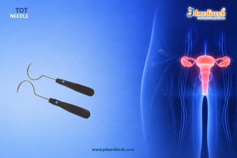
What is a TOT Needle?
A TOT Needle is a specialized surgical instrument used during Transobturator Tape (TOT) procedures for treating female stress urinary incontinence. Surgeons use this needle to guide the placement of a supportive sling beneath the urethra through the obturator foramen.
The needle features an ergonomic design and curved structure that allows smooth navigation through pelvic anatomy. Consequently, surgeons can achieve precise sling placement while reducing the risk of complications.
TOT procedures have become highly popular because they offer minimally invasive treatment with faster recovery times and reduced postoperative discomfort.
Importance of TOT Needle in Transobturator Procedures
The accuracy of sling placement directly affects the success of stress urinary incontinence treatment. Therefore, a high-quality TOT Needle is essential for achieving optimal surgical outcomes.
Key Advantages Include:
Accurate Sling Placement
The curved design ensures controlled passage through anatomical pathways, enabling precise positioning of the sling.
Reduced Tissue Trauma
The smooth surface and atraumatic tip minimize tissue damage during insertion and removal.
Enhanced Surgical Control
Ergonomic handling improves surgeon comfort and maneuverability throughout the procedure.
Faster Procedure Time
Efficient needle design simplifies sling placement and reduces overall operative duration.
Improved Patient Safety
Accurate placement lowers the chances of injury to surrounding tissues and organs.
Common Applications of TOT Needle
The TOT Needle is widely used in urogynecological and pelvic floor repair procedures.
Stress Urinary Incontinence (SUI) Treatment
The primary application involves the placement of transobturator slings for managing female stress urinary incontinence.
Minimally Invasive Pelvic Floor Procedures
Surgeons use the needle in modern minimally invasive techniques that require accurate mesh or tape placement.
Urogynecological Surgeries
The instrument supports procedures aimed at restoring pelvic floor stability and urinary function.
Key Features of a High-Quality TOT Needle
A premium TOT Needle offers superior precision, durability, and surgical efficiency.
Ergonomic Handle Design
The handle provides a secure grip and better control during delicate surgical maneuvers.
Curved Needle Configuration
The anatomically designed curve ensures smooth transobturator passage.
Atraumatic Tip
The rounded or specially designed tip reduces tissue trauma and enhances patient safety.
Medical-Grade Stainless Steel
High-quality stainless steel construction ensures corrosion resistance, durability, and sterilization compatibility.
Reusable or Disposable Options
Hospitals and clinics can choose reusable or single-use variants depending on surgical requirements.
Benefits of Using TOT Needle
Healthcare professionals prefer TOT Needles because they improve both procedural efficiency and patient outcomes.
Minimally Invasive Approach
The TOT technique requires smaller incisions and causes less tissue disruption compared to traditional procedures.
Reduced Recovery Time
Patients often recover faster and experience less postoperative discomfort.
Lower Risk of Complications
Accurate sling placement helps reduce bladder, bowel, and vascular injuries.
Better Surgical Precision
The needle allows controlled navigation through pelvic anatomy.
High Success Rates
TOT procedures performed with precision instruments often provide long-term symptom relief.
How TOT Needle Supports Safe Surgical Outcomes
Safety remains one of the most important factors in urogynecological surgery. The TOT Needle contributes to safer procedures by allowing controlled tissue passage and accurate sling positioning.
Additionally, modern TOT Needles are designed to reduce unnecessary force during insertion. This feature helps surgeons maintain anatomical accuracy while minimizing complications.
Because of these advantages, many hospitals and surgical centers rely on precision-engineered TOT Needles for routine incontinence surgeries.
Choosing the Right TOT Needle
Selecting the right surgical instrument can significantly improve procedural success.
Material Quality
Choose medical-grade stainless steel instruments for long-lasting performance and sterilization safety.
Needle Design
The curvature and tip design should support smooth transobturator access.
Surgeon Comfort
An ergonomic handle improves handling during lengthy procedures.
Sterilization Compatibility
Reusable instruments must withstand repeated sterilization cycles without compromising quality.
Manufacturer Reliability
Purchase from trusted medical device manufacturers known for consistent quality standards.
Maintenance and Sterilization
Proper instrument maintenance extends product life and ensures patient safety.
Cleaning
Clean the TOT Needle immediately after use to remove biological residues.
Sterilization
Follow standard hospital sterilization protocols using approved autoclave methods.
Inspection
Inspect the instrument regularly for wear, bending, or surface damage.
Proper Storage
Store in sterile trays to prevent contamination and maintain instrument integrity.
Why Healthcare Professionals Trust TOT Needles
Surgeons and healthcare facilities trust TOT Needles because they combine precision, safety, and reliability in one instrument. Their role in minimally invasive urogynecological surgery continues to grow as demand for effective stress urinary incontinence treatments increases.
Furthermore, advancements in instrument engineering have improved handling, patient comfort, and long-term procedural success.
Conclusion
The TOT Needle is an essential surgical instrument for safe and accurate transobturator procedures. Its precision design supports effective sling placement, reduces tissue trauma, and improves overall surgical outcomes in stress urinary incontinence treatment.
With ergonomic handling, durable construction, and enhanced procedural control, the TOT Needle remains a trusted solution in modern urogynecological surgery. Choosing a high-quality TOT Needle can help healthcare professionals achieve reliable performance, improved patient safety, and successful long-term results.