Introduction to Diagnostic Hysteroscopy Surgery
Diagnostic Hysteroscopy Surgery is a minimally invasive gynecological procedure that allows healthcare professionals to examine the inside of the uterus using a thin, illuminated instrument called a hysteroscope. Doctors commonly recommend this examination to investigate abnormal uterine bleeding, suspected endometrial polyps, and other conditions affecting the uterine cavity.
Unlike traditional abdominal surgery, diagnostic hysteroscopy usually requires no abdominal incision. Instead, the healthcare professional introduces the hysteroscope through the vagina and cervix. As a result, the procedure provides direct visualization of the uterine cavity without cutting through the abdominal wall.
Furthermore, modern hysteroscopy equipment supports clear visualization and targeted assessment of suspected abnormalities. When clinically appropriate, doctors may also collect a small tissue sample for laboratory examination.
In this comprehensive guide, JDMeditech explains diagnostic hysteroscopy surgery, its clinical applications, essential instruments, benefits, risks, patient preparation, recovery, and frequently asked questions.
What Is Diagnostic Hysteroscopy Surgery?
Diagnostic hysteroscopy is a procedure that helps gynecologists examine the uterine cavity directly. During the examination, a hysteroscope passes through the cervix into the uterus and provides images of the uterine lining.
Additionally, the healthcare professional introduces a suitable distension medium, commonly sterile saline, to expand the uterine cavity for better visualization. Consequently, the doctor can assess the endometrium and identify selected structural abnormalities.
For example, hysteroscopy may help detect endometrial polyps, submucosal fibroids, intrauterine adhesions, and certain uterine cavity abnormalities.
However, visual examination alone cannot diagnose every condition. Therefore, doctors may recommend endometrial biopsy or other investigations when necessary.
Understanding the Uterus and Endometrial Cavity
The uterus is a muscular reproductive organ located in the female pelvis. Its inner lining, called the endometrium, changes throughout the menstrual cycle.
During menstruation, the body sheds part of the endometrial lining. Meanwhile, hormonal changes prepare the lining for a possible pregnancy during each reproductive cycle.
However, structural abnormalities or changes in the endometrium may cause unusual bleeding or other symptoms. Therefore, direct examination of the uterine cavity can help doctors investigate selected conditions.
Why Is Diagnostic Hysteroscopy Performed?
Doctors recommend diagnostic hysteroscopy for several clinical reasons. In particular, it can help identify abnormalities that affect the uterine cavity.
1. Abnormal Uterine Bleeding
Abnormal uterine bleeding is a common reason for hysteroscopy. For example, patients may experience unusually heavy periods, irregular bleeding, or bleeding between menstrual cycles.
Furthermore, direct visualization may help doctors identify structural causes. However, the healthcare team may also recommend imaging, blood tests, or endometrial sampling.
2. Postmenopausal Bleeding
Bleeding after menopause requires appropriate medical assessment. In selected cases, diagnostic hysteroscopy helps doctors examine the uterine lining and investigate suspected abnormalities.
Importantly, doctors may need an endometrial tissue sample to assess for precancerous or cancerous changes.
3. Endometrial Polyps
Endometrial polyps are growths that develop from the uterine lining. Although many are benign, some require further investigation.
Consequently, diagnostic hysteroscopy may help identify their location and appearance. Additionally, doctors may recommend hysteroscopic removal when appropriate.
4. Submucosal Fibroids
Submucosal fibroids are growths that extend into the uterine cavity. As a result, they may contribute to heavy menstrual bleeding or reproductive concerns.
Diagnostic hysteroscopy allows direct assessment of selected fibroids. However, doctors may need ultrasound or other imaging to evaluate their size and relationship to the uterine wall.
5. Intrauterine Adhesions
Intrauterine adhesions are bands of scar tissue that develop inside the uterus. They may occur after certain uterine procedures or other conditions.
Furthermore, adhesions can affect menstrual bleeding and reproductive health. Therefore, hysteroscopy may help doctors confirm their presence and plan suitable treatment.
6. Recurrent Pregnancy Loss
Doctors may use hysteroscopy in selected patients with recurrent pregnancy loss when they suspect an abnormality inside the uterine cavity.
However, recurrent pregnancy loss can have several causes. Consequently, a broader clinical assessment may also be necessary.
7. Selected Infertility Investigations
Hysteroscopy may help assess suspected uterine cavity abnormalities in selected patients undergoing infertility evaluation.
Nevertheless, doctors do not recommend routine hysteroscopy for every patient with infertility. Instead, they consider symptoms, imaging findings, and previous investigations.
8. Missing or Malpositioned Intrauterine Device
When an intrauterine device cannot be located or removed using standard methods, hysteroscopy may help the healthcare professional identify its position.
Additionally, suitable instruments may assist with removal when clinically appropriate.
Common Symptoms That May Require Uterine Cavity Examination
Patients may require further gynecological evaluation when they experience certain symptoms. However, these symptoms do not automatically mean hysteroscopy is necessary.
- Heavy menstrual bleeding
- Irregular menstrual bleeding
- Bleeding between periods
- Postmenopausal bleeding
- Suspected uterine polyps
- Possible submucosal fibroids
- Selected fertility concerns
- Recurrent pregnancy loss with suspected uterine abnormalities
- Possible intrauterine adhesions
- Problems involving an intrauterine device
Therefore, a qualified gynecologist should determine whether hysteroscopy or another investigation is appropriate.
Types of Hysteroscopy Procedures
Healthcare professionals generally distinguish between diagnostic and operative hysteroscopy. Although both use a hysteroscope, their main purposes differ.
Diagnostic Hysteroscopy
Diagnostic hysteroscopy focuses on examining the uterine cavity and identifying abnormalities. Additionally, the healthcare professional may obtain a small biopsy when clinically indicated.
Operative Hysteroscopy
Operative hysteroscopy allows the surgeon to perform selected treatments using compatible instruments. For example, doctors may remove certain polyps, treat suitable fibroids, or divide intrauterine adhesions.
However, operative procedures may require additional equipment, pain management, and monitoring.
Office Hysteroscopy
Office hysteroscopy takes place in an appropriately equipped outpatient clinic. Furthermore, selected patients can undergo diagnostic examination without general anesthesia.
Nevertheless, the healthcare team must prioritize patient comfort and provide suitable pain relief options.
Diagnostic Hysteroscopy vs Operative Hysteroscopy
| Feature | Diagnostic Hysteroscopy | Operative Hysteroscopy |
|---|---|---|
| Main Purpose | Examine the uterine cavity | Treat selected uterine conditions |
| Typical Equipment | Hysteroscope and visualization system | Hysteroscope and treatment instruments |
| Tissue Removal | Biopsy when indicated | May involve polyp or fibroid removal |
| Anesthesia | Varies by patient and setting | Depends on procedure complexity |
| Procedure Duration | Often relatively short | May require additional time |
| Recovery | Often rapid after uncomplicated examination | Depends on the treatment performed |
How Should Patients Prepare for Diagnostic Hysteroscopy?
Preparation depends on the patient’s medical history and the planned examination. Therefore, healthcare professionals provide individualized instructions before the appointment.
First, the doctor reviews symptoms, previous investigations, and relevant medical conditions. Next, the team explains the procedure and discusses available pain relief options.
Importantly, pregnancy must be reasonably excluded before hysteroscopy. Additionally, clinicians may postpone the examination when active pelvic infection or another contraindication is present.
For patients with regular menstrual cycles, doctors may schedule the examination after menstruation when visualization is often easier.
Finally, patients should follow the healthcare team’s instructions about medicines, eating, drinking, and transportation, especially when sedation or general anesthesia is planned.
How Is Diagnostic Hysteroscopy Surgery Performed?
A qualified gynecologist or appropriately trained healthcare professional performs diagnostic hysteroscopy in a suitable clinical setting.
First, the healthcare team confirms the patient’s identity, clinical indication, and consent. Next, the clinician prepares the patient and provides the agreed pain management.
During the examination, the hysteroscope passes through the vagina and cervix into the uterine cavity. Depending on the approach, the clinician may use a vaginal speculum or perform a vaginoscopic examination without one.
Then, sterile saline or another suitable distension medium helps expand the cavity. As a result, the clinician can examine the uterine lining and identify visible abnormalities.
When appropriate, the healthcare professional may collect a targeted tissue sample. Afterward, the team completes the examination and explains the findings and next steps.
However, the patient may request that the procedure stop if the discomfort becomes unacceptable. Therefore, effective communication and pain management remain essential throughout the examination.
Essential Instruments Used in Diagnostic Hysteroscopy Surgery
Diagnostic hysteroscopy instruments support uterine cavity visualization, fluid management, examination, and selected tissue sampling.
Furthermore, hospitals should choose instruments according to their clinical requirements and existing equipment.
The following list describes common hysteroscopy equipment categories. It does not represent a confirmed JDMeditech instrument kit.
1. Diagnostic Hysteroscope Telescope
A diagnostic hysteroscope provides direct visualization of the uterine cavity. Additionally, its optical system allows the clinician to examine the endometrium and relevant anatomical structures.
2. Diagnostic Hysteroscopy Sheath
A compatible hysteroscopy sheath supports the telescope and may provide channels for fluid circulation. Therefore, the clinician must select a sheath that matches the hysteroscope.
3. Hysteroscopy Camera System
A camera system transfers hysteroscopic images to a monitor. As a result, the healthcare team can observe the examination clearly.
4. Medical Light Source
A medical light source illuminates the uterine cavity through a compatible optical system. Furthermore, suitable illumination supports detailed visualization.
5. Fiber-Optic Light Cable
A compatible light cable transfers illumination from the light source to the hysteroscope. Therefore, equipment compatibility is important.
6. Hysteroscopy Fluid Management System
A suitable fluid management system helps deliver and monitor the distension medium. Importantly, the healthcare team must follow appropriate fluid monitoring and safety practices.
7. Hysteroscopy Irrigation Pump
A compatible irrigation pump may support controlled fluid delivery during hysteroscopy. Additionally, the operator must follow the equipment’s approved intended use and operating instructions.
8. Sterile Saline Delivery Accessories
Compatible tubing and fluid accessories support uterine cavity distension. Consequently, the clinician can maintain suitable visualization during the examination.
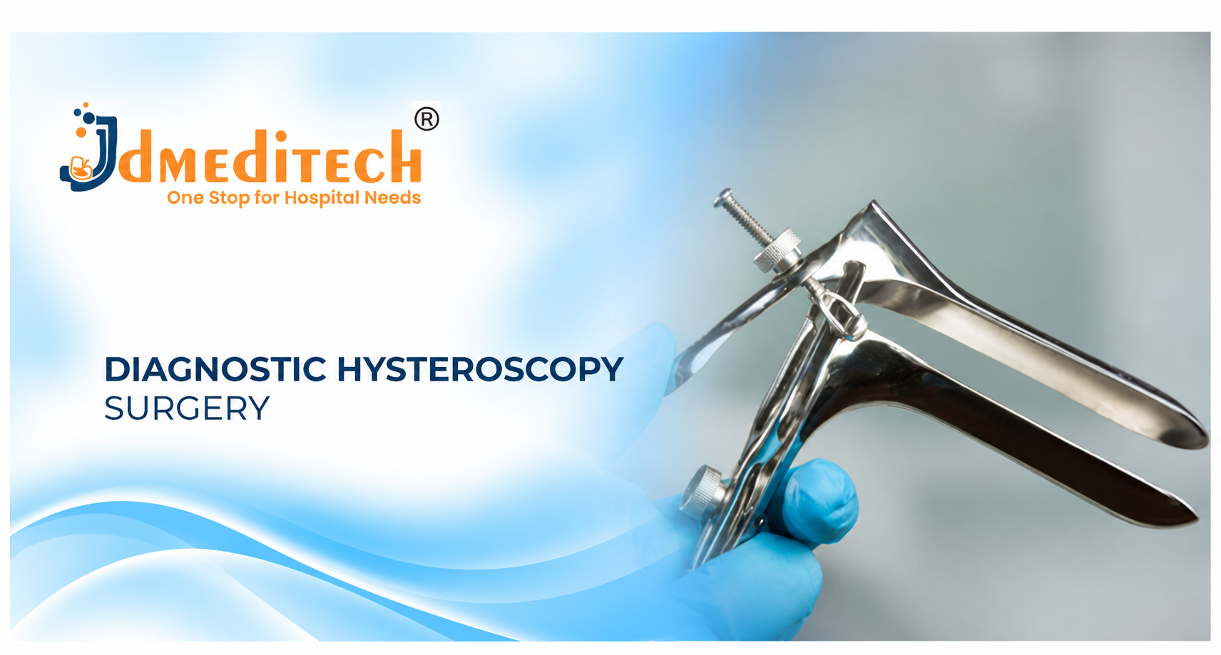
9. Vaginal Speculum
A vaginal speculum may help expose the cervix during conventional hysteroscopy. However, some clinicians use a vaginoscopic approach without a speculum.
10. Cervical Tenaculum
A cervical tenaculum may assist with cervical stabilization when necessary. Nevertheless, not every diagnostic hysteroscopy requires this instrument.
11. Cervical Dilators
Suitable cervical dilators may help prepare the cervical canal in selected cases. However, routine cervical dilation is not necessary for every diagnostic examination.
12. Hysteroscopic Biopsy Forceps
Compatible biopsy forceps support targeted endometrial tissue sampling when clinically indicated. Afterward, the healthcare team sends the specimen for pathological examination.
13. Hysteroscopic Grasping Forceps
Small hysteroscopic grasping forceps may support selected diagnostic or minor operative tasks. Additionally, the instrument must match the working channel and intended procedure.
14. Hysteroscopic Scissors
Compatible hysteroscopic scissors support selected operative tasks. However, they are not routinely required for a purely diagnostic examination.
15. Surgical Monitor
A medical-grade monitor displays images from the hysteroscopy camera system. Consequently, the healthcare team can observe the uterine cavity during the procedure.
16. Hysteroscopy Equipment Trolley
An equipment trolley supports organized placement of suitable hysteroscopy devices. Furthermore, proper arrangement helps the team manage the examination setup.
17. Sterile Examination Accessories
Suitable drapes, gloves, fluid tubing, and other accessories support the planned examination. Additionally, healthcare professionals follow appropriate infection-control practices.
Diagnostic Hysteroscopy Instruments and Their Functions
| Instrument | Primary Function |
|---|---|
| Diagnostic Hysteroscope | Provides uterine cavity visualization |
| Hysteroscopy Sheath | Supports the telescope and fluid channels |
| Camera System | Displays examination images |
| Light Source | Provides illumination |
| Light Cable | Transfers illumination |
| Fluid Management System | Supports distension fluid delivery and monitoring |
| Irrigation Pump | Supports compatible fluid delivery |
| Vaginal Speculum | Supports cervical exposure when used |
| Cervical Tenaculum | Supports selected cervical stabilization tasks |
| Cervical Dilators | Support cervical preparation when indicated |
| Biopsy Forceps | Support targeted tissue sampling |
| Grasping Forceps | Support selected instrument tasks |
| Hysteroscopic Scissors | Support selected operative procedures |
| Surgical Monitor | Displays hysteroscopic images |
Potential Benefits of Diagnostic Hysteroscopy Surgery
Diagnostic hysteroscopy provides several advantages when doctors select it for appropriate clinical indications. However, the benefits depend on the patient’s condition and examination findings.
1. Direct Visualization of the Uterine Cavity
Hysteroscopy allows the healthcare professional to examine the uterine lining directly. As a result, the clinician can identify certain abnormalities that may require further assessment.
2. No Abdominal Incision
Diagnostic hysteroscopy usually requires no abdominal incision. Consequently, suitable patients may recover quickly after an uncomplicated examination.
3. Targeted Endometrial Biopsy
When clinically appropriate, the clinician can collect a targeted tissue sample. Furthermore, pathological examination may provide important diagnostic information.
4. Outpatient Procedure Option
Many patients can undergo diagnostic hysteroscopy in an appropriately equipped outpatient clinic. Therefore, hospital admission and general anesthesia may not be necessary.
5. Assessment of Structural Abnormalities
Diagnostic hysteroscopy may help identify polyps, selected fibroids, and intrauterine adhesions. Additionally, the findings can support treatment planning.
6. Faster Recovery in Suitable Cases
Many patients return to normal activities soon after uncomplicated diagnostic hysteroscopy. However, recovery depends on pain, anesthesia, and any additional procedures.
7. Improved Treatment Planning
Direct examination may clarify the location and appearance of certain uterine abnormalities. Consequently, doctors can discuss appropriate follow-up investigations or treatment options.
Risks and Complications of Diagnostic Hysteroscopy
Diagnostic hysteroscopy is generally safe. Nevertheless, like other medical procedures, it carries potential risks.
- Temporary menstrual-like cramping
- Light vaginal bleeding or spotting
- Discomfort during the examination
- Vasovagal symptoms, such as dizziness or faintness
- Uterine or cervical injury
- Infection
- Excessive fluid absorption in rare circumstances
- Anesthesia-related complications when anesthesia is used
- Possible need to stop or repeat the examination
Furthermore, operative hysteroscopy generally carries additional risks compared with a simple diagnostic examination. Therefore, the healthcare professional should explain the planned procedure and obtain informed consent.
Pain Management During Diagnostic Hysteroscopy
Pain experiences vary considerably between patients. For example, some people experience mild menstrual-like cramping, while others find the examination significantly uncomfortable.
Therefore, clinicians should discuss pain relief before the appointment. Depending on the patient’s needs, options may include suitable oral medicines, local anesthesia, sedation, or general anesthesia.
Additionally, a vaginoscopic approach may reduce discomfort in selected patients because it can avoid the use of a speculum and tenaculum.
Importantly, patients should know that they can ask the healthcare professional to stop the procedure.
Recovery After Diagnostic Hysteroscopy Surgery
Recovery after uncomplicated diagnostic hysteroscopy is often relatively quick. However, the timeline varies according to the examination and the pain management used.
Initially, patients may experience mild cramping or light vaginal spotting. These symptoms often improve over the following days.
Furthermore, patients who receive sedation or general anesthesia may need additional monitoring and help with transportation home.
Most patients can gradually resume normal activities according to their healthcare professional’s advice. Nevertheless, they should follow any instructions about exercise, sexual activity, and vaginal products.
Postoperative Care After Diagnostic Hysteroscopy
Appropriate aftercare supports recovery and helps patients recognize complications. Therefore, the healthcare team should provide clear instructions before discharge.
- Follow the clinician’s pain relief instructions
- Use sanitary pads for spotting when advised
- Attend scheduled follow-up appointments
- Review biopsy results when applicable
- Follow instructions about sexual activity
- Follow advice about exercise and daily activities
- Report unusual bleeding or discharge
- Seek advice for persistent or worsening pain
Additionally, patients should follow any special instructions provided after sedation or general anesthesia.
Warning Signs After Diagnostic Hysteroscopy
Although serious complications are uncommon, certain symptoms require prompt medical assessment.
- Heavy or worsening vaginal bleeding
- Severe or increasing lower abdominal pain
- High or persistent fever
- Unusual or foul-smelling vaginal discharge
- Persistent dizziness or fainting
- Other unexpected or severe symptoms
Importantly, severe bleeding, collapse, or other emergency symptoms require immediate medical care.
Diagnostic Hysteroscopy vs Ultrasound
Ultrasound and hysteroscopy both help doctors investigate gynecological conditions. However, they provide different types of information.
| Feature | Diagnostic Hysteroscopy | Pelvic Ultrasound |
|---|---|---|
| Main Method | Direct camera examination | Sound-wave imaging |
| Uterine Cavity | Directly visualizes the cavity lining | Shows uterine structures through imaging |
| Biopsy | Can support targeted sampling | Does not directly obtain tissue |
| Instrument Entry | Hysteroscope passes through the cervix | Transabdominal or transvaginal probe |
| Typical Role | Selected direct cavity assessment | Common initial imaging investigation |
In many cases, ultrasound serves as an initial investigation. Subsequently, doctors may recommend hysteroscopy when direct visualization or tissue sampling would provide additional information.
How to Choose Diagnostic Hysteroscopy Instruments
Hospitals and gynecology departments should evaluate their clinical requirements before purchasing hysteroscopy equipment.
First, purchasing teams should identify whether they need diagnostic, office-based, or operative hysteroscopy instruments. Next, they should review hysteroscope dimensions, optical specifications, and compatible sheaths.
Furthermore, hospitals should confirm compatibility between camera systems, light sources, irrigation equipment, and fluid management accessories.
For reusable instruments, cleaning and sterilization instructions are especially important. Therefore, purchasing teams should review the manufacturer’s approved intended uses and technical documentation.
Important Hysteroscopy Equipment Selection Factors
- Approved intended use
- Diagnostic or operative application
- Hysteroscope diameter
- Viewing angle and optical specifications
- Compatible diagnostic sheath
- Camera system compatibility
- Medical light source compatibility
- Light cable compatibility
- Distension fluid delivery requirements
- Fluid management and monitoring capabilities
- Irrigation pump compatibility
- Working channel dimensions
- Biopsy forceps compatibility
- Reusable or single-use classification
- Cleaning and sterilization instructions
- Manufacturer technical documentation
- Applicable regulatory documentation
Diagnostic Hysteroscopy Instruments – JDMeditech
JDMeditech welcomes inquiries from hospitals, gynecology departments, hysteroscopy centers, medical institutions, and international distributors regarding hysteroscopy instruments and related accessories.
For example, customers can inquire about available diagnostic hysteroscopy sheaths, hysteroscopic telescopes, grasping instruments, scissors, irrigation equipment, and compatible accessories.
Furthermore, the JDMeditech team can discuss available product configurations, technical specifications, intended uses, and applicable documentation.
Therefore, hospitals and medical distributors can contact JDMeditech to discuss their specific diagnostic hysteroscopy equipment requirements.
For product information and international business inquiries, visit www.jdmeditech.com.
Frequently Asked Questions About Diagnostic Hysteroscopy Surgery
1. What is diagnostic hysteroscopy surgery?
Diagnostic hysteroscopy is a procedure that allows healthcare professionals to examine the inside of the uterus using a thin, illuminated hysteroscope.
2. Why do doctors recommend diagnostic hysteroscopy?
Doctors may recommend it to investigate abnormal uterine bleeding, suspected polyps, selected fibroids, adhesions, or other uterine cavity abnormalities.
3. Is diagnostic hysteroscopy a major surgery?
Usually not. Diagnostic hysteroscopy often takes place as a short outpatient procedure without an abdominal incision.
4. Does diagnostic hysteroscopy require general anesthesia?
Not always. Many patients undergo office hysteroscopy without general anesthesia. However, pain relief options depend on individual circumstances.
5. Is diagnostic hysteroscopy painful?
Discomfort varies. Some patients experience mild cramps, while others find the procedure painful. Therefore, doctors should discuss pain management in advance.
6. How long does diagnostic hysteroscopy take?
A simple diagnostic examination often takes approximately 10 to 15 minutes. However, additional sampling or treatment can increase the duration.
7. Does hysteroscopy require an abdominal incision?
No. The hysteroscope usually enters the uterus through the vagina and cervix.
8. Can hysteroscopy detect uterine polyps?
Yes. Direct visualization can help doctors identify endometrial polyps. Additionally, they may recommend biopsy or removal when appropriate.
9. Can diagnostic hysteroscopy detect fibroids?
It can identify selected fibroids that project into the uterine cavity. However, ultrasound or other imaging may be necessary for complete assessment.
10. Can hysteroscopy diagnose uterine cancer?
Hysteroscopy may reveal suspicious abnormalities and support tissue sampling. However, a pathological examination is generally necessary to confirm a cancer diagnosis.
11. Is hysteroscopy useful for infertility assessment?
It may help selected patients when doctors suspect uterine cavity abnormalities. Nevertheless, routine hysteroscopy is not necessary for every infertility evaluation.
12. Can hysteroscopy be performed during pregnancy?
Routine diagnostic hysteroscopy is generally avoided during pregnancy. Therefore, doctors should reasonably exclude pregnancy before the examination.
13. What instruments are used in diagnostic hysteroscopy?
Common instruments include a diagnostic hysteroscope, compatible sheath, camera system, light source, and suitable fluid management equipment.
14. What is the difference between diagnostic and operative hysteroscopy?
Diagnostic hysteroscopy examines the uterine cavity. In contrast, operative hysteroscopy allows surgeons to perform selected treatments.
15. What is office hysteroscopy?
Office hysteroscopy is a hysteroscopic examination performed in an appropriately equipped outpatient setting.
16. What are the main risks of diagnostic hysteroscopy?
Potential risks include pain, bleeding, infection, uterine injury, and rare fluid-related complications.
17. How long does recovery take?
Many patients resume normal activities the same day or the following day. However, recovery depends on symptoms, anesthesia, and additional procedures.
18. Is spotting normal after hysteroscopy?
Light spotting can occur after the examination. Nevertheless, patients should seek medical advice if bleeding becomes heavy or persistent.
19. Can patients go home after diagnostic hysteroscopy?
Yes. Many patients return home shortly after an uncomplicated outpatient examination. However, those receiving anesthesia may require additional observation.
20. Where can hospitals inquire about JDMeditech hysteroscopy instruments?
Hospitals and medical distributors can visit JDMeditech to inquire about available hysteroscopy instruments and related accessories.
Conclusion
Diagnostic Hysteroscopy Surgery provides a valuable method for examining the uterine cavity and investigating selected gynecological conditions. Furthermore, direct visualization may help healthcare professionals identify structural abnormalities and plan suitable treatment.
Unlike abdominal surgery, diagnostic hysteroscopy usually requires no abdominal incision. Consequently, many patients can undergo the examination in an outpatient setting and recover relatively quickly.
However, appropriate patient selection, pain management, equipment compatibility, and clinical expertise remain essential. Therefore, healthcare professionals must consider individual circumstances and follow current medical guidance.
For diagnostic hysteroscopy instrument inquiries and international business requirements, contact JDMeditech – One Stop for Hospital Needs.
Website: www.jdmeditech.com
Medical Disclaimer: This article provides general educational information only. It does not replace professional medical advice, diagnosis, or surgical training. Qualified healthcare professionals must make treatment and equipment decisions according to current clinical guidance, local regulations, and approved manufacturer instructions.