Laparoscopic Pyeloplasty Surgery: Complete Guide
Laparoscopic Pyeloplasty Surgery is a minimally invasive reconstructive urological procedure used to treat ureteropelvic junction obstruction. Doctors also call this condition UPJ obstruction or PUJ obstruction.
Normally, urine flows from the kidney’s renal pelvis into the ureter. However, a narrowed ureteropelvic junction can restrict this flow.
As a result, urine may accumulate inside the kidney and cause hydronephrosis. Furthermore, severe or persistent obstruction can affect kidney function.
Therefore, doctors evaluate symptoms, urine drainage, kidney health, and imaging results before recommending treatment.
In this guide, JDMeditech explains laparoscopic pyeloplasty, surgical techniques, instruments, benefits, risks, recovery, and frequently asked questions.
What Is Ureteropelvic Junction Obstruction?
Ureteropelvic junction obstruction is a narrowing or functional blockage where the renal pelvis connects to the ureter.
The renal pelvis collects urine produced by the kidney. Next, the ureter transports urine toward the bladder.
However, UPJ obstruction slows or restricts drainage at this connection.
Consequently, pressure may increase within the kidney’s collecting system.
Understanding the Kidney and Ureter
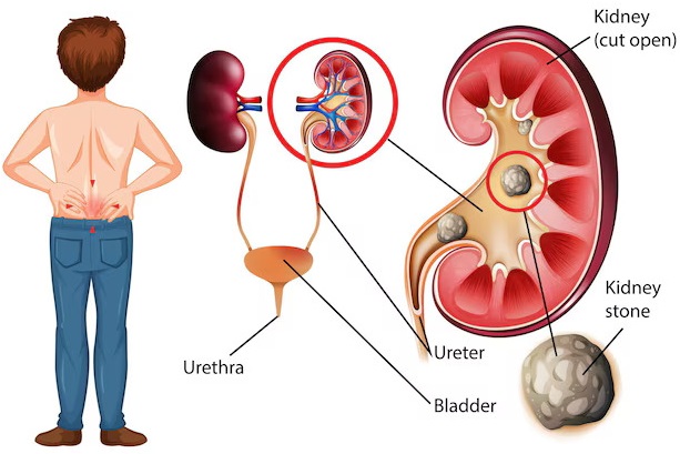
The urinary system includes the kidneys, ureters, bladder, and urethra.
First, the kidneys filter blood and produce urine. Next, urine collects in the renal pelvis.
Afterward, the ureters carry urine into the bladder.
Finally, urine leaves the body through the urethra.
In patients with UPJ obstruction, the narrowing occurs near the beginning of the ureter.
What Causes UPJ Obstruction?
UPJ obstruction may be congenital or acquired.
1. Congenital Narrowing
Some patients are born with an abnormally narrow ureteropelvic junction.
Furthermore, abnormal muscle development may interfere with normal urine movement.
2. Crossing Blood Vessel
A blood vessel passing near the UPJ may contribute to obstruction in selected patients.
Therefore, surgeons assess nearby vessels during reconstructive planning.
3. Scar Tissue
Previous surgery, inflammation, or injury may cause narrowing in some patients.
4. High Ureteral Insertion
An unusual connection between the ureter and renal pelvis may contribute to impaired drainage.
5. Other Acquired Conditions
Selected patients may develop obstruction related to stones, inflammatory changes, or other urinary tract abnormalities.
Common Symptoms of UPJ Obstruction
Symptoms depend on the severity of obstruction and the patient’s age.
For example, some infants have no noticeable symptoms, while older patients may experience pain or infection.
- Flank pain
- Abdominal discomfort
- Intermittent pain after increased fluid intake
- Nausea or vomiting
- Hydronephrosis
- Recurrent urinary tract infections
- Fever during urinary infection
- Blood in urine in selected cases
- Kidney stones in some patients
- Reduced kidney function
- Abnormal prenatal ultrasound findings
However, these symptoms can occur with other urinary conditions. Therefore, specialist evaluation is necessary.
UPJ Obstruction in Infants and Children
Some children receive a diagnosis before birth when prenatal ultrasound shows hydronephrosis.
However, urinary tract dilation does not always mean that surgery is required.
Furthermore, some cases improve with growth and careful monitoring.
Therefore, pediatric urologists use follow-up ultrasound and selected functional investigations to identify children who may benefit from surgery.
UPJ Obstruction in Adults
Adults may present with intermittent flank pain, recurrent infections, or unexplained hydronephrosis.
Additionally, a crossing blood vessel may contribute to symptoms in selected patients.
Therefore, imaging and functional kidney assessment help determine the most suitable treatment.
What Is Hydronephrosis?
Hydronephrosis is dilation of the kidney’s urine-collecting system.
It develops when urine drainage is impaired or another urinary condition causes collecting-system enlargement.
However, hydronephrosis does not always indicate clinically significant obstruction.
Therefore, doctors assess the degree of dilation together with symptoms and kidney function.
How Is UPJ Obstruction Diagnosed?
Diagnosis involves medical history, examination, imaging, and selected functional tests.
1. Renal Ultrasound
Ultrasound helps assess kidney structure and hydronephrosis.
2. Diuretic Renal Scan
A diuretic renogram, commonly using MAG3, helps evaluate urine drainage and relative kidney function.
3. CT Urography
Selected adults may undergo CT imaging to assess urinary anatomy and possible causes of obstruction.
4. MR Urography
MRI-based imaging may provide additional anatomical or functional information in selected cases.
5. Blood Tests
Serum creatinine and other investigations help assess overall kidney function.
6. Urine Tests
Urinalysis and urine culture help identify infection or other urinary abnormalities.
7. Additional Imaging
Doctors may recommend other investigations when reflux, stones, or anatomical abnormalities are suspected.
Comparison of UPJ Diagnostic Tests
| Test | Main Purpose | Clinical Use |
|---|---|---|
| Renal Ultrasound | Assess hydronephrosis | Initial and follow-up imaging |
| MAG3 Renogram | Evaluate drainage and relative kidney function | Selected functional assessment |
| CT Urography | Assess urinary tract anatomy | Selected adult cases |
| MR Urography | Provide additional anatomical information | Selected patients |
| Blood Tests | Assess kidney function | Clinical evaluation |
| Urine Culture | Detect urinary infection | When clinically indicated |
When Is Laparoscopic Pyeloplasty Surgery Recommended?
Not every patient with hydronephrosis or UPJ narrowing requires surgery.
However, doctors may recommend pyeloplasty when obstruction causes symptoms or threatens kidney health.
Possible indications include:
- Persistent or recurrent flank pain
- Confirmed clinically significant UPJ obstruction
- Progressive hydronephrosis
- Declining relative kidney function
- Recurrent infections associated with obstruction
- Selected patients with kidney stones
- Progressive thinning of kidney tissue
- Other clinically significant obstructive findings
Furthermore, the decision depends on patient age, imaging results, symptoms, and overall health.
What Is Laparoscopic Pyeloplasty?
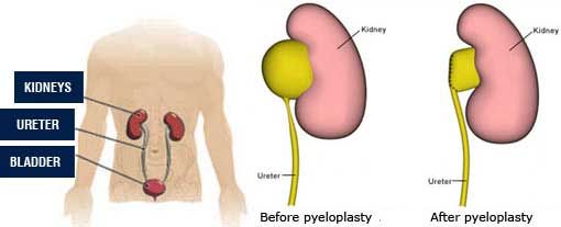
Laparoscopic Pyeloplasty is a minimally invasive operation that reconstructs the ureteropelvic junction to improve urine drainage.
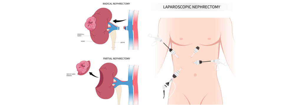
The surgeon uses a laparoscopic camera and compatible instruments through small abdominal or flank incisions.
Next, the narrowed junction is repaired or reconstructed.
Consequently, urine can move more freely from the renal pelvis into the ureter.
Types of Pyeloplasty Surgery
1. Open Pyeloplasty
Open pyeloplasty uses a conventional surgical incision to reconstruct the ureteropelvic junction.
Furthermore, it remains an established treatment, particularly in selected pediatric or complex cases.
2. Laparoscopic Pyeloplasty
Laparoscopic surgery uses a camera and long instruments introduced through small incisions.
As a result, suitable patients may experience smaller scars and faster recovery than with conventional open surgery.
3. Robotic-Assisted Pyeloplasty
Robotic-assisted pyeloplasty uses surgeon-controlled robotic instruments for reconstructive surgery.
However, equipment availability, cost, and surgeon expertise influence treatment selection.
4. Anderson-Hynes Dismembered Pyeloplasty
Anderson-Hynes pyeloplasty is an established reconstructive technique.
It removes the narrowed UPJ segment and reconnects the healthy ureter to the renal pelvis.
Furthermore, the surgeon can address selected crossing-vessel anatomy during reconstruction.
5. Non-Dismembered Pyeloplasty
Selected patients may undergo reconstruction without completely separating the ureter from the renal pelvis.
However, suitability depends on the specific anatomy.
6. Endopyelotomy
Endopyelotomy treats selected UPJ narrowings through an endoscopic incision.
However, it generally has a higher recurrence risk than standard pyeloplasty and is not suitable for every patient.
Comparison of Pyeloplasty Techniques
| Technique | Approach | Important Consideration |
|---|---|---|
| Open Pyeloplasty | Conventional incision | Established reconstructive treatment |
| Laparoscopic Pyeloplasty | Small incisions and camera | Minimally invasive reconstruction |
| Robotic Pyeloplasty | Robot-assisted reconstruction | Requires specialized equipment |
| Anderson-Hynes | Dismembered reconstruction | Established standard technique |
| Non-Dismembered Repair | Selected reconstructive techniques | Depends on anatomy |
| Endopyelotomy | Endoscopic incision | Selected cases; higher recurrence risk |
Transperitoneal and Retroperitoneal Laparoscopic Pyeloplasty
Surgeons may approach the kidney through the peritoneal cavity or directly through the retroperitoneal space.
The transperitoneal approach provides a familiar operative space for many surgeons.
In contrast, the retroperitoneal approach provides access without routinely entering the peritoneal cavity.
However, each technique has different advantages and limitations.
Therefore, surgeons choose the approach according to anatomy, experience, and patient needs.
Preparation Before Laparoscopic Pyeloplasty Surgery
Preparation begins with a detailed urological assessment.
First, the surgeon reviews symptoms, medical history, and imaging results.
Next, the healthcare team evaluates kidney function and possible infection.
Furthermore, the anesthesiologist assesses the patient’s readiness for surgery.
Preparation may include:
- Medical history review
- Physical examination
- Renal ultrasound
- Diuretic renogram when indicated
- Blood tests
- Urine testing
- Medication review
- Allergy assessment
- Anesthesia assessment
- Discussion of surgical alternatives
- Explanation of possible stent placement
- Informed consent
- Preoperative fasting instructions
How Is Laparoscopic Pyeloplasty Surgery Performed?
Laparoscopic pyeloplasty is generally performed under general anesthesia.
First, the surgical team positions the patient and prepares the operative field.
Next, the surgeon introduces a laparoscopic camera and suitable instruments through small incisions.
Afterward, the surgeon identifies the renal pelvis, ureter, and obstructed ureteropelvic junction.
In a dismembered repair, the narrowed segment is removed. The surgeon then reconnects the healthy ureter to the renal pelvis.
Furthermore, a temporary ureteral stent may be placed to support drainage during healing.
Finally, the surgeon checks the reconstruction and closes the incisions.
However, the exact technique depends on anatomy and the selected surgical approach.
How Long Does Laparoscopic Pyeloplasty Take?
Operating time varies according to the complexity of reconstruction and the surgical technique.
Many pyeloplasty procedures take approximately two to three hours. However, complex cases may require additional time.
Furthermore, anesthesia preparation and postoperative monitoring increase the total hospital time.
Essential Instruments Used in Laparoscopic Pyeloplasty Surgery
Laparoscopic Pyeloplasty Surgical Instruments support internal visualization, tissue dissection, precise suturing, and temporary urinary drainage.
However, the exact equipment depends on the surgeon’s technique, patient anatomy, and institutional protocol.
The following list describes general instrument categories and does not represent a verified JDMeditech pyeloplasty kit.
1. Laparoscopic Telescope
A laparoscopic telescope provides internal visualization during minimally invasive surgery.
2. Laparoscopic Camera System
The camera system displays the operative field on a monitor.
3. LED or Xenon Light Source
A compatible light source provides illumination during laparoscopy.
4. Fiber-Optic Light Cable
A suitable light cable transfers illumination to compatible endoscopic equipment.
5. Laparoscopic Trocar
A trocar provides access for compatible laparoscopic instruments.
6. Laparoscopic Cannula
A cannula maintains an access channel for selected laparoscopic equipment.
7. Veress Needle
A Veress needle may be used for pneumoperitoneum creation in selected laparoscopic access techniques.
8. CO2 Insufflator
A compatible insufflator helps maintain the operating space during laparoscopy.
9. Laparoscopic Atraumatic Grasper
An atraumatic grasper supports gentle handling of delicate tissues.
10. Laparoscopic Maryland Dissector
A Maryland dissector supports controlled tissue separation and manipulation.
11. Laparoscopic Right-Angle Dissector
A right-angle dissector supports selected dissection around anatomical structures.
12. Laparoscopic Metzenbaum Scissors
Metzenbaum-style laparoscopic scissors support delicate tissue cutting.
13. Laparoscopic Hook Electrode
A compatible hook electrode may support selected electrosurgical dissection.
14. Laparoscopic Bipolar Forceps
Bipolar forceps may support bleeding control during suitable procedures.
15. Electrosurgical Generator
A compatible generator provides energy to approved electrosurgical instruments.
16. Laparoscopic Needle Holder
A laparoscopic needle holder supports precise suturing during reconstruction.
17. Laparoscopic Needle Holder with Curved Jaw
A suitable curved-jaw needle holder may help manipulate needles during selected suturing tasks.
18. Laparoscopic Suction and Irrigation Instrument
This instrument helps remove fluid and maintain visibility.
19. Laparoscopic Clip Applicator
A compatible clip applicator may support selected vessel or tissue management tasks.
20. Laparoscopic Tissue Retractor
A suitable retractor helps maintain access to the operative field.
21. Laparoscopic Monopolar Scissors
Compatible monopolar scissors may support cutting and electrosurgical dissection.
22. Laparoscopic Knot Pusher
A knot pusher may support selected intracorporeal or extracorporeal suturing techniques.
23. Fine Absorbable Surgical Sutures
Suitable sutures support reconstruction of the ureteropelvic junction.
24. Double J Ureteral Stent
A Double J stent may provide temporary internal urinary drainage during healing.
25. Ureteral Guide Wire
A compatible guide wire may support selected ureteral stent-placement techniques.
26. Ureteral Catheter
A suitable ureteral catheter may support selected drainage or identification requirements.
27. Foley Urinary Catheter
A Foley catheter provides temporary bladder drainage when indicated.
28. Cystoscope
A compatible cystoscope may support retrograde ureteral stent placement or removal.
29. Cystoscope Sheath
A suitable sheath supports compatible cystoscopic instruments.
30. Endoscopic Stent Grasping Forceps
Compatible forceps may support retrieval of an internal ureteral stent.
31. Surgical Drain
A suitable drain may be used in selected cases to monitor postoperative fluid collection.
32. Sterile Surgical Drapes
Sterile drapes help establish the operative field.
33. Sterile Gauze
Sterile gauze supports wound care and bleeding management.
34. Surgical Dressing
A suitable dressing protects the incision sites.
35. Surgical Instrument Tray
A compatible tray supports instrument organization and reprocessing.
Laparoscopic Pyeloplasty Instruments and Their Functions
| Instrument | Primary Function | Typical Application |
|---|---|---|
| Laparoscopic Telescope | Internal visualization | Laparoscopic repair |
| Camera System | Displays operative field | Minimally invasive surgery |
| Trocar | Instrument access | Laparoscopic approach |
| Veress Needle | Selected access technique | Pneumoperitoneum creation |
| Maryland Dissector | Controlled dissection | UPJ exposure |
| Atraumatic Grasper | Gentle tissue handling | Reconstruction |
| Laparoscopic Scissors | Precise cutting | Tissue dissection |
| Needle Holder | Reconstructive suturing | Ureteropelvic anastomosis |
| Bipolar Forceps | Bleeding control | Selected procedures |
| Suction-Irrigation Instrument | Fluid management | Operative visibility |
| Double J Stent | Temporary internal drainage | Selected postoperative care |
| Guide Wire | Selected stent placement support | Ureteral access |
| Foley Catheter | Bladder drainage | Perioperative care |
| Cystoscope | Endoscopic urinary access | Selected stent procedures |
Why Is Instrument Selection Important?
Laparoscopic pyeloplasty requires delicate dissection and precise reconstructive suturing.
Therefore, surgeons need appropriately sized instruments with suitable handling characteristics.
Furthermore, endoscopic visualization, insufflation, and electrosurgical equipment must be compatible.
Consequently, hospitals should verify device specifications and manufacturer instructions before use.
Benefits of Laparoscopic Pyeloplasty Surgery
1. Improved Urine Drainage
Successful reconstruction aims to improve urine flow from the renal pelvis into the ureter.
2. Relief of Obstruction-Related Pain
Patients with symptoms caused by UPJ obstruction may experience pain relief after successful treatment.
3. Protection of Kidney Function
Appropriate surgery may help prevent further obstruction-related kidney injury.
4. Smaller Incisions
Laparoscopic techniques generally use smaller incisions than conventional open surgery.
5. Reduced Postoperative Discomfort
Suitable patients may experience less postoperative pain compared with open surgery.
6. Faster Recovery
Minimally invasive approaches may support earlier recovery and shorter hospital stays in selected patients.
7. Improved Cosmetic Appearance
Small laparoscopic incisions may leave less noticeable scars.
Risks and Complications of Laparoscopic Pyeloplasty
Although laparoscopic pyeloplasty is an established procedure, complications can occur.
- Bleeding
- Infection
- Postoperative pain
- Urine leakage
- Ureteral narrowing
- Persistent or recurrent UPJ obstruction
- Injury to nearby organs or blood vessels
- Temporary urinary symptoms
- Stent-related discomfort
- Catheter-related complications
- Need for repeat surgery
- Conversion to open surgery
- Anesthesia-related complications
Therefore, patients should discuss individual risks with the treating urologist.
Recovery After Laparoscopic Pyeloplasty Surgery
Recovery varies according to patient age, surgical complexity, and overall health.
Initially, the healthcare team monitors pain, urine output, and signs of complications.
Furthermore, selected patients require temporary bladder drainage or ureteral stenting.
Many patients begin recovering over one to two weeks. However, full return to strenuous activities may take several weeks.
Therefore, patients should follow the surgeon’s specific recovery instructions.
Postoperative Care After Pyeloplasty
- Take prescribed medicines as directed
- Follow wound-care instructions
- Follow catheter-care instructions when applicable
- Monitor urine output
- Follow hydration recommendations
- Avoid restricted physical activities
- Attend follow-up appointments
- Complete recommended imaging
- Arrange stent removal when required
- Report fever or worsening pain
How Long Does a Double J Stent Remain After Pyeloplasty?
A Double J stent may remain temporarily while the reconstruction heals.
However, the duration depends on the surgical technique, patient anatomy, and surgeon’s protocol.
Therefore, the healthcare team should provide a specific stent-removal plan.
Does Every Patient Need a Double J Stent?
No. Stent use varies among surgeons and techniques.
For example, some procedures use internal stents, while others use alternative drainage methods or selected stentless approaches.
Therefore, the decision should be individualized.
Can UPJ Obstruction Return After Pyeloplasty?
Yes. Although pyeloplasty has high success rates, recurrent narrowing can occur.
For example, scar tissue may develop near the reconstructed junction.
Therefore, persistent pain, infection, or worsening hydronephrosis may require further assessment.
Does Hydronephrosis Disappear After Surgery?
Hydronephrosis often improves gradually after successful reconstruction.
However, some collecting-system dilation may remain even when urine drainage has improved.
Therefore, doctors interpret follow-up imaging together with symptoms and kidney function.
Long-Term Follow-Up After Pyeloplasty
Follow-up helps determine whether urine drainage remains satisfactory.
For example, doctors may recommend renal ultrasound after surgery.
Furthermore, selected patients require a diuretic renal scan to assess drainage and relative kidney function.
Therefore, the follow-up schedule depends on patient age, symptoms, and clinical findings.
Warning Signs After Laparoscopic Pyeloplasty
Patients should contact the healthcare team if concerning symptoms develop.
- Fever or chills
- Severe or worsening flank pain
- Persistent vomiting
- Difficulty passing urine
- Little or no urine output
- Increasing abdominal swelling
- Wound redness or discharge
- Heavy bleeding in urine
- Catheter or stent problems
- Increasing weakness or signs of serious illness
Importantly, inability to pass urine, severe illness, or fever with significant flank pain requires urgent medical assessment.
How to Choose Laparoscopic Pyeloplasty Instruments
Hospitals and urology departments should evaluate their procedural requirements before purchasing equipment.
First, purchasing teams should confirm the selected laparoscopic approach and patient population.
Next, they should review telescope specifications, trocar dimensions, graspers, scissors, and needle holders.
Furthermore, hospitals should verify camera, light source, insufflation, and electrosurgical compatibility.
Finally, purchasing departments should review sterilization requirements, approved intended uses, and regulatory documentation.
Important Equipment Selection Factors
- Approved intended use
- Instrument diameter and working length
- Adult or pediatric compatibility
- Trocar dimensions
- Telescope viewing angle
- Camera system compatibility
- Light source compatibility
- CO2 insufflation requirements
- Grasper and dissector configuration
- Scissors specifications
- Needle holder jaw design
- Electrosurgical compatibility
- Ureteral stent specifications
- Guide wire compatibility
- Catheter dimensions
- Reusable or single-use classification
- Cleaning and sterilization instructions
- Manufacturer technical documentation
- Applicable regulatory requirements
Laparoscopic Pyeloplasty Surgical Instruments – JDMeditech
JDMeditech welcomes inquiries from hospitals, urology departments, surgical centers, healthcare institutions, and international medical distributors regarding urological surgical instruments and related medical equipment.
For example, customers can inquire about available laparoscopic instruments, trocars, graspers, needle holders, ureteral catheters, guide wires, and Double J stents.
Furthermore, healthcare facilities may discuss instrument configuration requirements with the JDMeditech team.
However, customers should confirm product availability, approved intended uses, dimensions, and applicable regulatory documentation before ordering.
For product information and international business inquiries, visit www.jdmeditech.com.
Frequently Asked Questions About Laparoscopic Pyeloplasty Surgery
1. What is laparoscopic pyeloplasty?
It is a minimally invasive operation that reconstructs the ureteropelvic junction to improve urine drainage from the kidney.
2. What is UPJ obstruction?
UPJ obstruction is narrowing or impaired drainage where the renal pelvis joins the ureter.
3. What causes UPJ obstruction?
Possible causes include congenital narrowing, abnormal muscle development, crossing vessels, or scar tissue.
4. What are common UPJ obstruction symptoms?
Symptoms may include flank pain, nausea, urinary infections, and hydronephrosis.
5. Can UPJ obstruction occur in children?
Yes. It is an important cause of hydronephrosis in infants and children.
6. Does every patient with hydronephrosis need surgery?
No. Some patients can be monitored without immediate surgery.
7. How is UPJ obstruction diagnosed?
Doctors use ultrasound, functional kidney imaging, and selected additional tests.
8. What is a MAG3 renal scan?
It is a nuclear medicine test used to assess kidney drainage and relative kidney function.
9. What is Anderson-Hynes pyeloplasty?
It is an established technique that removes the narrowed UPJ segment and reconnects the healthy ureter to the renal pelvis.
10. What is the difference between open and laparoscopic pyeloplasty?
Open surgery uses a conventional incision, while laparoscopic surgery uses smaller incisions and specialized instruments.
11. What is robotic-assisted pyeloplasty?
It is a reconstructive procedure performed using surgeon-controlled robotic instruments.
12. What instruments are used in laparoscopic pyeloplasty?
Common equipment includes laparoscopic telescopes, trocars, graspers, dissectors, scissors, needle holders, and urinary drainage accessories.
13. Is general anesthesia required?
Yes. Laparoscopic pyeloplasty is generally performed under general anesthesia.
14. How long does pyeloplasty take?
Many procedures take approximately two to three hours, although duration varies.
15. Is a Double J stent always required?
No. Stent use depends on the surgeon’s technique and the patient’s needs.
16. How long does recovery take?
Initial recovery often takes one to two weeks, while full recovery may require several additional weeks.
17. Can UPJ obstruction return after surgery?
Yes. Recurrent narrowing is possible, although established pyeloplasty techniques have high success rates.
18. Does hydronephrosis disappear immediately?
No. Kidney dilation may improve gradually and can persist despite successful drainage.
19. Is follow-up imaging necessary?
Yes. Doctors commonly use ultrasound and selected functional tests to monitor recovery.
20. Where can hospitals inquire about JDMeditech pyeloplasty instruments?
Hospitals and medical distributors can visit JDMeditech for information about available urological instruments and accessories.
Conclusion
Laparoscopic Pyeloplasty Surgery is an established minimally invasive treatment for selected patients with ureteropelvic junction obstruction.
Furthermore, the procedure aims to improve urine drainage, relieve obstruction-related symptoms, and protect kidney health.
However, not every patient requires surgery. Therefore, treatment should depend on symptoms, kidney function, imaging findings, and specialist assessment.
For urological surgical instrument inquiries and international business requirements, contact JDMeditech – One Stop for Hospital Needs.
Website: www.jdmeditech.com
Medical Disclaimer: This article provides general educational information only. It does not replace professional medical advice, diagnosis, informed consent, or surgical training. Qualified healthcare professionals must make treatment and equipment decisions according to current clinical guidance, applicable regulations, and approved manufacturer instructions.