Posterior Urethral Valve Ablation Surgery: Complete Guide
Posterior Urethral Valve Ablation Surgery is a pediatric urological procedure that treats an obstruction in the posterior urethra. The condition, known as posterior urethral valves (PUV), develops before birth and affects boys.
Normally, urine flows from the bladder through the urethra without significant obstruction. However, abnormal tissue folds can restrict urine flow.
As a result, pressure may increase inside the bladder and upper urinary tract. Furthermore, severe obstruction can damage the kidneys and affect bladder development.
Therefore, early diagnosis, appropriate urinary drainage, and specialist treatment are important.
In this guide, JDMeditech explains PUV symptoms, diagnosis, valve ablation techniques, surgical instruments, benefits, risks, recovery, and long-term follow-up.
What Are Posterior Urethral Valves?
Posterior urethral valves are congenital obstructing tissue membranes located within the posterior male urethra.
The posterior urethra passes through the prostate and nearby structures between the bladder and the external urethra.
However, abnormal valve tissue can partially block urine leaving the bladder.
Consequently, the bladder may become enlarged or thick-walled, while urine pressure affects the ureters and kidneys.
Understanding the Male Urinary System
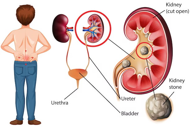
The urinary system includes the kidneys, ureters, bladder, and urethra.
First, the kidneys produce urine. Next, the ureters carry urine to the bladder.
Finally, urine passes through the urethra during urination.
In boys with PUV, obstructing tissue in the posterior urethra interferes with this process.
Therefore, the condition may affect several parts of the urinary system.
What Causes Posterior Urethral Valves?
PUV develops during fetal formation of the urinary tract.
However, the exact developmental mechanism is not fully understood.
Importantly, PUV is not caused by ordinary childhood activities, toilet training, or poor hygiene.
Furthermore, the condition is generally considered sporadic rather than a straightforward inherited disorder.
How Common Are Posterior Urethral Valves?
Posterior urethral valves are an uncommon congenital condition affecting boys.
Nevertheless, they are an important cause of severe urinary obstruction during infancy.
Furthermore, some children develop long-term kidney disease despite early treatment.
Therefore, PUV requires specialized pediatric urological care.
Types of Posterior Urethral Valve Obstruction
Historically, doctors classified posterior urethral valves into anatomical types.
However, modern clinical management focuses more on the presence and severity of obstruction than on older classifications.
Additionally, clinicians may use the term congenital obstructive posterior urethral membrane.
Symptoms of Posterior Urethral Valves
Symptoms depend on the severity of obstruction and the child’s age.
For example, severe cases may become apparent before birth, while milder cases may be detected later.
Common findings include:
- Weak urinary stream
- Difficulty passing urine
- Urinary retention
- Dribbling of urine
- Enlarged bladder
- Hydronephrosis
- Recurrent urinary tract infections
- Fever during urinary infection
- Poor feeding in infants
- Vomiting
- Poor weight gain
- Abnormal kidney function tests
- Urinary incontinence in older children
- Incomplete bladder emptying
However, these symptoms can occur with other conditions. Therefore, medical assessment is necessary.
Posterior Urethral Valves Before Birth
Some cases are detected during prenatal ultrasound.
For example, imaging may show a large bladder, urinary tract dilation, or reduced amniotic fluid.
Furthermore, severe fetal urinary obstruction may affect kidney and lung development.
Therefore, prenatal findings require evaluation by an experienced fetal medicine and pediatric urology team.
Posterior Urethral Valves in Newborn Babies
Newborns with severe obstruction may have difficulty passing urine.
Additionally, doctors may identify a distended bladder, hydronephrosis, or abnormal kidney function.
Consequently, urgent bladder drainage and stabilization may be necessary.
However, definitive valve ablation is generally performed after the baby becomes clinically stable.
Posterior Urethral Valves in Older Children
Some boys receive a diagnosis later in childhood.
For example, they may experience a weak urinary stream, recurrent infections, or urinary incontinence.
Furthermore, poor bladder emptying may cause persistent urinary symptoms.
Therefore, older children with unexplained urinary problems may require evaluation for obstruction.
Complications of Untreated Posterior Urethral Valves
Severe or prolonged urinary obstruction can cause significant complications.
- Hydronephrosis
- Hydroureter
- Bladder wall thickening
- Urinary retention
- Vesicoureteral reflux
- Recurrent urinary infections
- Kidney injury
- Chronic kidney disease
- Electrolyte disturbances
- Bladder dysfunction
- Growth-related problems in severe cases
- Kidney failure in some patients
Importantly, some kidney abnormalities develop before birth. Therefore, surgery may relieve obstruction without reversing existing damage.
How Are Posterior Urethral Valves Diagnosed?
Diagnosis involves clinical examination, laboratory testing, and urinary tract imaging.
1. Prenatal Ultrasound
Prenatal ultrasound may identify urinary tract dilation, bladder enlargement, or reduced amniotic fluid.
2. Renal and Bladder Ultrasound
Ultrasound helps assess kidney swelling, ureteral dilation, bladder wall appearance, and urinary tract anatomy.
3. Voiding Cystourethrogram (VCUG)
A VCUG is an important investigation for confirming suspected PUV.
Furthermore, it may demonstrate a dilated posterior urethra and associated vesicoureteral reflux.
4. Blood Tests
Serum creatinine and electrolyte tests help assess kidney function and metabolic disturbances.
5. Urine Tests
Urinalysis and urine culture may help detect urinary tract infection.
6. Cystoscopy
A suitable pediatric cystoscope allows direct visualization of the posterior urethra.
Additionally, the surgeon may perform endoscopic valve ablation during the procedure.
7. Functional Kidney Assessment
Selected patients require renal scans or other investigations to evaluate kidney function.
Comparison of PUV Diagnostic Tests
| Investigation | Main Purpose | Clinical Consideration |
|---|---|---|
| Ultrasound | Assess kidneys and bladder | Common initial investigation |
| VCUG | Confirm urethral obstruction and assess reflux | Important diagnostic examination |
| Serum Creatinine | Assess kidney function | Important for monitoring and prognosis |
| Urine Culture | Detect infection | Used when clinically indicated |
| Cystoscopy | Direct urethral visualization | May permit valve ablation |
| Renal Scan | Assess selected aspects of kidney function | Used in selected patients |
What Is Posterior Urethral Valve Ablation Surgery?
Posterior Urethral Valve Ablation Surgery is an endoscopic procedure that relieves obstruction caused by abnormal valve tissue.
The surgeon uses a suitable pediatric endoscope to visualize the urethra.
Next, the obstructing tissue is divided or ablated using an appropriate instrument.
Consequently, urine can pass more freely from the bladder through the urethra.
However, successful valve ablation does not guarantee normal bladder or kidney function.
When Is PUV Ablation Surgery Recommended?
Endoscopic valve ablation is a standard definitive treatment when the diagnosis is confirmed and the child is clinically stable.
However, immediate priorities in a sick newborn include urinary drainage and medical stabilization.
Therefore, the pediatric urology team determines the safest timing.
Treatment Options for Posterior Urethral Valves
1. Initial Bladder Drainage
Doctors usually drain the bladder when a newborn has suspected lower urinary tract obstruction.
Furthermore, the healthcare team monitors kidney function and electrolytes.
2. Endoscopic Valve Ablation
Once the child is stable, the surgeon may perform endoscopic ablation to relieve the obstruction.
3. Temporary Vesicostomy
A vesicostomy creates an opening for urine drainage through the lower abdominal wall.
For example, this approach may be necessary when the urethra is too small for safe endoscopic treatment.
4. Other Urinary Diversion Procedures
Selected complex cases may require additional urinary diversion when drainage remains inadequate.
However, these procedures are not routinely necessary for every child.
5. Long-Term Bladder Management
Some children need additional treatment for bladder storage or emptying problems.
Therefore, medication, catheterization, or other specialist management may be required.
Types of Posterior Urethral Valve Ablation
1. Endoscopic Cold-Knife Incision
A compatible endoscopic cutting instrument can divide obstructing valve tissue without using electrosurgical energy.
However, suitability depends on the instrument and patient anatomy.
2. Endoscopic Electrosurgical Ablation
Selected surgeons use compatible electrosurgical instruments to treat valve tissue.
However, excessive thermal injury can increase the risk of urethral scarring.
3. Laser-Assisted Valve Ablation
Selected specialist centers use suitable laser systems for endoscopic valve treatment.
However, laser equipment is not required for every PUV procedure.
Comparison of PUV Ablation Techniques
| Technique | Method | Important Consideration |
|---|---|---|
| Cold-Knife Incision | Mechanical division of valve tissue | Avoids thermal cutting injury |
| Electrosurgical Ablation | Compatible electrical cutting or coagulation | Requires careful control of thermal effects |
| Laser Ablation | Laser-based treatment | Requires specialized compatible equipment |
Preparation Before Posterior Urethral Valve Ablation Surgery
Preparation depends on the child’s condition and the severity of obstruction.
First, the medical team ensures appropriate urinary drainage.
Next, doctors evaluate kidney function, electrolytes, and possible infection.
Furthermore, the pediatric anesthesiologist assesses the child’s readiness for surgery.
Preparation may include:
- Medical history review
- Physical examination
- Urinary drainage assessment
- Renal and bladder ultrasound review
- VCUG review
- Serum creatinine measurement
- Electrolyte assessment
- Urine testing
- Infection management
- Anesthesia evaluation
- Equipment size assessment
- Discussion of possible complications
- Informed consent
How Is Posterior Urethral Valve Ablation Surgery Performed?
PUV ablation is generally performed under suitable anesthesia by a pediatric urology specialist.
First, the surgical team confirms that the child is stable enough for the procedure.
Next, the surgeon uses an appropriately sized pediatric endoscope to examine the urethra and identify obstructing valve tissue.
Afterward, the surgeon divides or ablates the valve tissue using the selected compatible instrument.
Furthermore, the surgical team assesses whether urine can drain adequately.
Finally, temporary catheter drainage may be used according to the child’s needs.
However, the precise technique and equipment depend on the patient’s anatomy and clinical condition.
How Long Does PUV Ablation Surgery Take?
The duration depends on the child’s size, anatomy, medical condition, and equipment requirements.
Furthermore, anesthesia preparation and postoperative observation increase the total hospital time.
Therefore, caregivers should ask the treating team for an individualized estimate.
Essential Instruments Used in Posterior Urethral Valve Ablation Surgery
Posterior Urethral Valve Ablation Instruments support pediatric urethral visualization, treatment of obstructing tissue, irrigation, and urinary drainage.
However, the exact instrument selection depends on the child’s size and surgical technique.
The following list describes general instrument categories rather than a verified JDMeditech PUV surgical kit.
1. Pediatric Cystoscope
A pediatric cystoscope provides direct visualization of the urethra and bladder.
2. Pediatric Cystoscope Telescope
A compatible telescope supports visualization of small urinary structures.
3. Pediatric Cystoscope Sheath
A suitable sheath supports compatible endoscopic equipment and irrigation.
4. Pediatric Resectoscope
A compatible pediatric resectoscope may support selected endoscopic valve procedures.
5. Resectoscope Working Element
A suitable working element controls compatible endoscopic cutting or treatment accessories.
6. Endoscopic Cold Knife
A compatible cold knife supports mechanical incision of obstructing tissue.
7. Endoscopic Electrode
A suitable electrode may support electrosurgical treatment when clinically indicated.
8. Electrosurgical Generator
A compatible generator provides energy to approved electrosurgical instruments.
9. Endoscopic Laser Fiber
A suitable laser fiber may support treatment in selected laser-assisted procedures.
10. Laser Generator
A compatible laser generator supplies energy for approved laser-based treatment.
11. Endoscopic Camera System
The camera system displays the internal operative field on a monitor.
12. Endoscopic Light Source
A compatible light source illuminates the urethra during endoscopy.
13. Fiber-Optic Light Cable
A suitable light cable transfers illumination to compatible endoscopic equipment.
14. Irrigation Tubing
Irrigation tubing supports appropriate fluid flow during endoscopic visualization.
15. Irrigation Fluid Delivery System
A compatible system helps maintain suitable visualization during the procedure.
16. Endoscopic Instrument Bridge
A compatible bridge supports selected accessories used with suitable endoscopes.
17. Urethral Catheter
An appropriately sized urethral catheter supports bladder drainage before or after treatment.
18. Pediatric Foley Catheter
A suitable pediatric Foley catheter may provide temporary bladder drainage when indicated.
19. Suprapubic Catheter
A suprapubic catheter may provide alternative urinary drainage in selected patients.
20. Urethral Guide Wire
A compatible guide wire may support selected catheter-placement procedures.
21. Ureteral Catheter
A suitable ureteral catheter may be required for selected additional urinary tract procedures, but it is not a routine component of every PUV ablation.
22. Surgical Scalpel
A surgical scalpel supports incision during selected urinary diversion procedures.
23. Metzenbaum Scissors
Metzenbaum scissors support soft-tissue dissection during selected open procedures.
24. Atraumatic Tissue Forceps
Atraumatic forceps support gentle tissue handling when open surgical access is required.
25. Fine Needle Holder
A fine needle holder supports suturing during selected reconstructive or diversion procedures.
26. Fine Hemostatic Forceps
Fine hemostatic forceps support bleeding control during selected open procedures.
27. Surgical Retractor
A suitable retractor supports visibility during open urinary diversion procedures.
28. Surgical Sutures
Appropriate sutures support wound closure during selected procedures.
29. Sterile Surgical Drapes
Sterile drapes help establish the operative field.
30. Sterile Gauze
Sterile gauze supports wound care and bleeding management.
31. Surgical Dressing
A suitable dressing protects surgical wounds when required.
32. Surgical Instrument Tray
A compatible tray supports instrument organization and reprocessing.
PUV Ablation Surgical Instruments and Their Functions
| Instrument | Primary Function | Typical Application |
|---|---|---|
| Pediatric Cystoscope | Urethral visualization | Endoscopic PUV treatment |
| Pediatric Telescope | Internal imaging | Cystoscopy |
| Pediatric Resectoscope | Supports compatible treatment instruments | Selected valve ablation |
| Cold Knife | Mechanical tissue incision | Selected valve incision |
| Endoscopic Electrode | Electrosurgical treatment | Selected ablation techniques |
| Laser Fiber | Laser energy delivery | Selected laser-assisted procedures |
| Camera System | Visualization on monitor | Endoscopic surgery |
| Light Source | Internal illumination | Endoscopy |
| Irrigation Tubing | Fluid management | Endoscopic visualization |
| Pediatric Foley Catheter | Bladder drainage | Perioperative care |
| Suprapubic Catheter | Alternative urinary drainage | Selected complex cases |
| Fine Needle Holder | Wound closure | Selected open diversion procedures |
Why Is Instrument Selection Important?
Posterior urethral valve surgery involves the delicate urethra of an infant or child.
Therefore, appropriately sized instruments are essential for safe access and visualization.
Furthermore, equipment must be compatible with the selected surgical technique.
Consequently, hospitals should verify device dimensions, intended uses, and manufacturer instructions before treatment.
Potential Benefits of PUV Ablation Surgery
1. Relief of Urethral Obstruction
Successful valve ablation reduces the blockage caused by abnormal urethral tissue.
2. Improved Urine Flow
Relieving obstruction may allow urine to pass more freely through the urethra.
3. Reduced Urinary Tract Pressure
Improved drainage may reduce pressure affecting the bladder and upper urinary tract.
4. Protection of Remaining Kidney Function
Timely treatment may help limit additional obstruction-related kidney injury.
5. Improved Urinary Drainage Management
Definitive treatment can reduce dependence on temporary drainage in suitable patients.
Risks and Complications of Posterior Urethral Valve Ablation
Although valve ablation is an established treatment, complications can occur.
For example, endoscopic treatment may cause temporary bleeding or urinary discomfort.
Possible complications include:
- Postoperative pain
- Bleeding
- Urinary tract infection
- Urethral injury
- Urethral stricture
- Incomplete valve ablation
- Residual obstruction
- Temporary urinary retention
- Catheter-related complications
- Need for repeat cystoscopy or ablation
- Anesthesia-related complications
Furthermore, persistent bladder dysfunction and chronic kidney disease may develop despite technically successful surgery.
Therefore, continued specialist monitoring is essential.
What Is a Urethral Stricture?
A urethral stricture is a narrowing caused by scar tissue inside the urethra.
Consequently, the urinary stream may become weak or obstructed.
However, the risk depends on the procedure, instrument size, and other factors.
Therefore, persistent urinary symptoms require specialist assessment.
What Is Residual Posterior Urethral Valve Tissue?
Residual valve tissue is obstructing tissue that remains after the initial procedure.
As a result, the child may continue to experience poor urine flow or abnormal bladder findings.
Therefore, the surgeon may recommend additional imaging or repeat endoscopic assessment.
Recovery After Posterior Urethral Valve Ablation Surgery
Recovery depends on the child’s age, kidney health, and severity of urinary obstruction.
Initially, the healthcare team monitors urine output and kidney function.
Furthermore, some children need temporary catheter drainage after surgery.
However, hospital stay and catheter duration vary according to clinical needs.
Therefore, caregivers should follow the individualized postoperative plan.
Postoperative Care After PUV Ablation
- Follow prescribed medication instructions
- Monitor urine output
- Follow catheter-care instructions
- Attend follow-up appointments
- Complete recommended kidney function tests
- Complete follow-up ultrasound examinations
- Monitor for fever or urinary infection
- Report poor urine flow
- Follow bladder-management recommendations
- Discuss growth and nutrition concerns with the healthcare team
How Long Does Recovery Take?
Initial recovery from the endoscopic procedure may be relatively short.
However, infants with severe kidney dysfunction may require prolonged medical care.
Furthermore, long-term treatment may continue after the surgical wounds have healed.
Therefore, recovery and ongoing disease management should be considered separately.
Does Valve Ablation Completely Cure PUV?
Valve ablation can relieve the anatomical obstruction.
However, some children continue to have abnormal bladder function or kidney disease.
For example, a child may experience urinary incontinence, incomplete bladder emptying, or persistent kidney impairment.
Therefore, successful surgery does not eliminate the need for follow-up.
What Is Valve Bladder Syndrome?
Valve bladder syndrome refers to bladder dysfunction that can persist or develop after relief of posterior urethral obstruction.
For example, the bladder may have difficulty storing urine at safe pressures or emptying completely.
Furthermore, these problems may change as the child grows.
Therefore, long-term bladder assessment is an important part of PUV care.
Can Posterior Urethral Valves Cause Kidney Failure?
Yes. Severe PUV can cause lasting kidney injury.
Furthermore, some children develop chronic kidney disease despite appropriate treatment.
In advanced cases, dialysis or kidney transplantation may eventually be required.
Therefore, early treatment and long-term kidney monitoring are essential.
Why Is Lifelong Follow-Up Important?
Posterior urethral valves can affect bladder and kidney function for many years.
Therefore, ongoing monitoring helps identify complications early.
Follow-up may include:
- Blood pressure assessment
- Kidney function blood tests
- Electrolyte monitoring
- Renal and bladder ultrasound
- Urinary infection assessment
- Urine flow testing when appropriate
- Post-void residual urine assessment
- Bladder function testing when indicated
- Growth and nutritional assessment
- Transition to adult urology care
Warning Signs After PUV Ablation Surgery
Caregivers should contact the healthcare team if concerning symptoms develop.
- Fever or chills
- Little or no urine output
- Catheter blockage
- Difficulty passing urine
- Severe abdominal discomfort
- Persistent vomiting
- Heavy bleeding in urine
- Increasing urinary symptoms
- Signs of dehydration
- Unusual lethargy or poor feeding
Importantly, inability to pass urine, serious illness, or signs of urinary infection in a young infant require urgent medical assessment.
How to Choose PUV Ablation Surgical Instruments
Hospitals and pediatric urology departments should evaluate their equipment requirements before purchasing instruments.
First, purchasing teams should confirm the patient’s age range and required pediatric dimensions.
Next, they should review cystoscope compatibility, telescope size, working elements, and treatment accessories.
Furthermore, hospitals should confirm camera, light source, and irrigation compatibility.
Finally, purchasing departments should verify intended use, cleaning instructions, and applicable regulatory documentation.
Important Equipment Selection Factors
- Approved intended use
- Pediatric instrument diameter
- Cystoscope compatibility
- Telescope specifications
- Resectoscope working element compatibility
- Cold-knife accessory compatibility
- Electrode specifications
- Electrosurgical generator compatibility
- Laser fiber compatibility when applicable
- Camera and light source compatibility
- Irrigation system requirements
- Pediatric catheter dimensions
- Sterile packaging
- Reusable or single-use classification
- Cleaning and sterilization instructions
- Manufacturer technical documentation
- Applicable regulatory documentation
Posterior Urethral Valve Ablation Instruments – JDMeditech
JDMeditech welcomes inquiries from hospitals, pediatric urology departments, surgical centers, healthcare institutions, and international medical distributors regarding urological surgical instruments and related medical equipment.
For example, customers can inquire about available cystoscopy instruments, pediatric endoscopic accessories, resectoscope components, urethral catheters, and compatible equipment.
Furthermore, healthcare facilities may discuss instrument configuration requirements with the JDMeditech team.
However, customers should confirm product availability, approved intended uses, technical specifications, and applicable regulatory documentation before ordering.
For product information and international business inquiries, visit www.jdmeditech.com.
Frequently Asked Questions About Posterior Urethral Valve Ablation Surgery
1. What are posterior urethral valves?
Posterior urethral valves are congenital tissue membranes that obstruct urine flow through the posterior male urethra.
2. What does PUV stand for?
PUV stands for posterior urethral valves.
3. Who can develop PUV?
PUV is a congenital condition affecting boys.
4. What causes posterior urethral valves?
The condition develops before birth. However, the exact developmental cause is not fully understood.
5. What are common PUV symptoms?
Symptoms may include weak urinary stream, urinary retention, infections, hydronephrosis, and abnormal kidney function.
6. Can PUV be detected before birth?
Yes. Prenatal ultrasound may identify findings that suggest fetal urinary obstruction.
7. How is PUV diagnosed?
Doctors use ultrasound, VCUG, laboratory tests, and selected cystoscopic examinations.
8. What is a VCUG?
A voiding cystourethrogram is an imaging test that helps confirm posterior urethral obstruction and assess urinary reflux.
9. What is PUV ablation surgery?
It is an endoscopic procedure that divides or ablates obstructing valve tissue.
10. Is bladder drainage needed before surgery?
Yes. Newborns with suspected significant lower urinary tract obstruction generally need prompt bladder drainage and stabilization.
11. What instruments are used in PUV surgery?
Common equipment includes pediatric cystoscopes, compatible cutting accessories, camera systems, light sources, and urinary drainage catheters.
12. What is a pediatric resectoscope?
It is a small endoscopic instrument system used for selected pediatric urinary tract procedures.
13. What is cold-knife valve incision?
It is a technique that mechanically divides obstructing valve tissue using a compatible endoscopic instrument.
14. Is laser treatment always required?
No. Laser-assisted ablation is one of several possible approaches and is not necessary for every patient.
15. Does PUV surgery require anesthesia?
Yes. Endoscopic valve ablation is generally performed under suitable anesthesia.
16. How long does surgery take?
Operating time varies according to the child’s size, medical condition, and selected equipment.
17. Can PUV return after surgery?
PUV is congenital, but residual obstructing tissue or postoperative urethral narrowing may require further treatment.
18. Does surgery restore normal kidney function?
Relieving obstruction may protect remaining kidney function. However, existing kidney damage may not fully reverse.
19. Is lifelong follow-up necessary?
Yes. Children with PUV require ongoing kidney and bladder monitoring into adulthood.
20. Where can hospitals inquire about JDMeditech PUV instruments?
Hospitals and medical distributors can visit JDMeditech to inquire about available urological surgical instruments and accessories.
Conclusion
Posterior Urethral Valve Ablation Surgery is an important treatment for congenital urinary obstruction in boys.
Furthermore, endoscopic valve ablation aims to restore urine flow and reduce ongoing obstruction-related damage.
However, some children continue to experience bladder dysfunction or chronic kidney disease after treatment.
Therefore, timely specialist care and lifelong follow-up are essential.
For urological surgical instrument inquiries and international business requirements, contact JDMeditech – One Stop for Hospital Needs.
Website: www.jdmeditech.com
Medical Disclaimer: This article provides general educational information only. It does not replace professional medical advice, diagnosis, informed consent, or surgical training. Qualified healthcare professionals must make treatment and equipment decisions according to current clinical guidance, applicable regulations, and approved manufacturer instructions.