PTFE Guide Wire for Urology – Smooth & Reliable Access

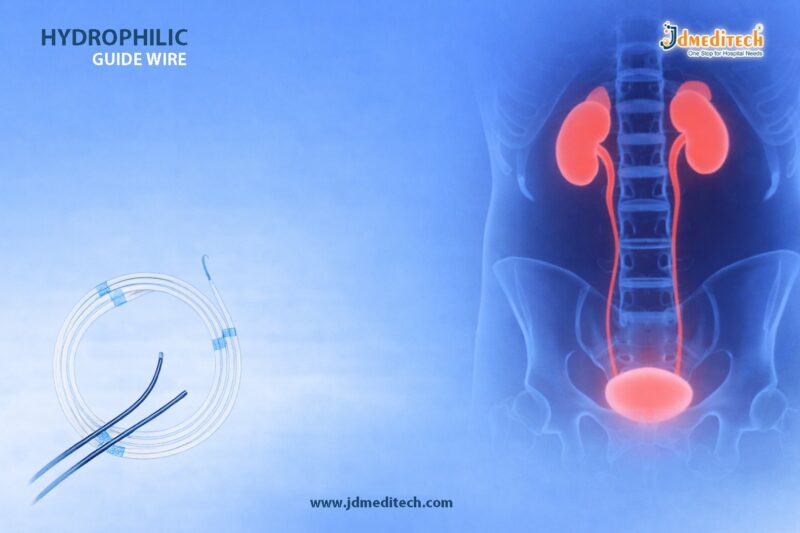
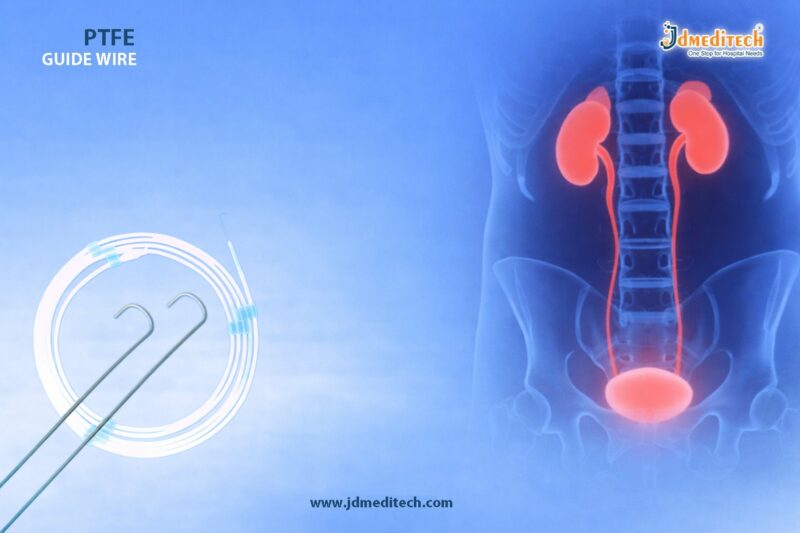
The PTFE Guide Wire plays a crucial role in modern urological procedures. It ensures smooth navigation and reliable access through the urinary tract. Doctors depend on the PTFE Guide Wire for precision, flexibility, and patient safety.

In minimally invasive urology, the right guide wire improves procedural success. Therefore, the PTFE Guide Wire has become a preferred choice for many specialists worldwide.

What is a PTFE Guide Wire?

A PTFE Guide Wire is a medical device coated with Polytetrafluoroethylene (PTFE). This coating reduces friction and allows the wire to glide smoothly through anatomical pathways.

Because of its unique properties, the PTFE Guide Wire offers excellent control during procedures such as:

  • Ureteroscopy
  • Catheter placement
  • Stone removal procedures
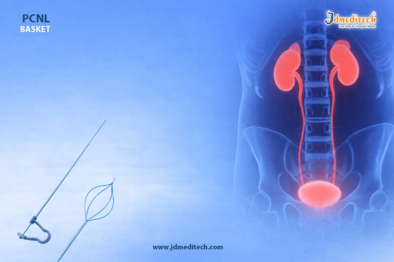
  • Percutaneous nephrolithotomy (PCNL)
Key Features of PTFE Guide Wire
1. Smooth Surface for Easy Navigation

The PTFE coating ensures minimal resistance. As a result, the PTFE Guide Wire moves easily through narrow and complex pathways.

2. Excellent Flexibility

Flexibility allows the PTFE Guide Wire to adapt to anatomical curves. This reduces trauma during insertion.

3. High Strength and Durability

Despite its flexibility, the PTFE Guide Wire maintains strong structural integrity.

4. Hydrophobic Coating

Unlike hydrophilic wires, the PTFE Guide Wire provides controlled movement without excessive slipperiness.

5. Kink Resistance

The design prevents bending or kinking, ensuring consistent performance.

Applications of PTFE Guide Wire in Urology

The PTFE Guide Wire is widely used in various urological procedures. Its versatility makes it an essential tool.

Common Uses Include:
  • Assisting catheter insertion
  • Guiding ureteral stents
  • Supporting endoscopic procedures
  • Facilitating access during kidney stone removal

Because of its reliability, the PTFE Guide Wire improves procedural efficiency.

Advantages of Using PTFE Guide Wire
1. Enhanced Procedural Control

The PTFE Guide Wire provides better tactile feedback. Surgeons can guide it accurately.

2. Reduced Tissue Trauma

Its smooth surface minimizes friction. Therefore, it reduces irritation and injury.

3. Cost-Effective Solution

Compared to advanced coated wires, the PTFE Guide Wire offers a balance between performance and cost.

4. Reliable Performance

The consistent behavior of the PTFE Guide Wire makes it suitable for routine procedures.

PTFE Guide Wire vs Hydrophilic Guide Wire
Feature PTFE Guide Wire Hydrophilic Guide Wire
Surface Smooth (low friction) Extremely slippery when wet
Control High control Less tactile feedback
Use Case Routine procedures Complex or tight pathways
Cost More economical Higher cost

While hydrophilic wires excel in complex cases, the PTFE Guide Wire remains ideal for standard procedures.

How to Choose the Right PTFE Guide Wire

When selecting a PTFE Guide Wire, consider the following factors:

  • Diameter and length based on procedure
  • Tip design (straight or J-tip)
  • Stiffness level required
  • Compatibility with other instruments

Choosing the right PTFE Guide Wire ensures better outcomes and safety.

Safety and Handling Tips

Proper handling of the PTFE Guide Wire is essential. Follow these guidelines:

  • Always maintain sterility
  • Avoid excessive force during insertion
  • Use imaging guidance when necessary
  • Inspect for damage before use

These practices enhance patient safety and device performance.

Why Choose PTFE Guide Wire from JDMeditech?

At JDMeditech, we offer high-quality PTFE Guide Wire solutions designed for precision and reliability.

Our Key Benefits:
  • Premium-grade materials
  • Strict quality control
  • Designed for smooth navigation
  • Suitable for various urological procedures
Conclusion

The PTFE Guide Wire is an essential tool in urological procedures. It provides smooth access, reliable control, and enhanced safety. Because of its performance and affordability, the PTFE Guide Wire remains a trusted choice among medical professionals.

If you aim for precision and efficiency in urology, the PTFE Guide Wire is the right solution.


Explore More Product >>

Get Connected:

+91 79909 93062 | +91 63513 72032 | exports@jdmeditech.com

Pancreatic Drainage Procedure for Fluid Build-Up Relief

Introduction to Ross ProcedureThe Pancreatic Drainage Procedure is a specialized surgical technique used to treat pancreatic fluid accumulation caused by conditions such as pancreatitis or pancreatic cysts. This Pancreatic Drainage Procedure helps relieve pressure, reduce pain, and prevent serious complications.

When pancreatic fluid builds up in or around the pancreas, it can lead to infection, inflammation, and organ dysfunction. Therefore, doctors recommend the Pancreatic Drainage Procedure to restore normal pancreatic function and improve patient outcomes.

What is a Pancreatic Drainage Procedure?

A Pancreatic Drainage Procedure involves creating a pathway to drain excess pancreatic fluid from cysts, pseudocysts, or blocked ducts. Surgeons perform this Pancreatic Drainage Procedure using minimally invasive or open surgical techniques.

In many cases, the Pancreatic Drainage Procedure connects the fluid collection to the stomach or small intestine, allowing safe internal drainage.

Why is Pancreatic Drainage Procedure Needed?

Doctors recommend a Pancreatic Drainage Procedure when fluid collections cause symptoms or complications. Common indications include:

  • Pancreatic pseudocysts
  • Chronic pancreatitis
  • Pancreatic duct obstruction
  • Infected fluid collections
  • Persistent abdominal pain

Without timely treatment, these conditions may worsen. Therefore, the Pancreatic Drainage Procedure plays a crucial role in preventing further damage.

Types of Pancreatic Drainage Procedure
1. Endoscopic Pancreatic Drainage Procedure

This Pancreatic Drainage Procedure uses an endoscope to access the pancreas through the digestive tract. It is minimally invasive and offers faster recovery.

2. Percutaneous Pancreatic Drainage Procedure

In this Pancreatic Drainage Procedure, a catheter is inserted through the skin under imaging guidance to drain fluid externally.

3. Surgical Pancreatic Drainage Procedure

Surgeons perform this Pancreatic Drainage Procedure when other methods are not effective. It includes procedures like cystogastrostomy or pancreaticojejunostomy.

Procedure Steps of Pancreatic Drainage Procedure

The Pancreatic Drainage Procedure typically follows these steps:

  1. Patient evaluation and imaging (CT scan or MRI)
  2. Administration of anesthesia
  3. Accessing the fluid collection
  4. Creating a drainage pathway
  5. Placement of stents or drains
  6. Monitoring and post-procedure care

Each Pancreatic Drainage Procedure is tailored to the patient’s condition and severity.

Benefits of Pancreatic Drainage Procedure

The Pancreatic Drainage Procedure offers several advantages:

  • Relieves abdominal pain and pressure
  • Prevents infection and complications
  • Improves pancreatic function
  • Minimally invasive options available
  • Faster recovery time

Because of these benefits, the Pancreatic Drainage Procedure is widely used in modern gastroenterology.

Risks and Complications

Although the Pancreatic Drainage Procedure is generally safe, some risks may include:

  • Bleeding
  • Infection
  • Leakage of pancreatic fluid
  • Damage to surrounding organs

However, experienced surgeons minimize these risks during the Pancreatic Drainage Procedure.

Recovery After Pancreatic Drainage Procedure

Recovery from a Pancreatic Drainage Procedure depends on the technique used. Most patients experience:

  • Short hospital stay
  • Gradual return to normal diet
  • Pain relief within days
  • Regular follow-up imaging

Proper post-operative care ensures successful outcomes after the Pancreatic Drainage Procedure.

Advanced Technologies in Pancreatic Drainage Procedure

Modern advancements have improved the success of the Pancreatic Drainage Procedure:

  • Endoscopic ultrasound (EUS)-guided drainage
  • Biodegradable stents
  • High-resolution imaging systems
  • Minimally invasive surgical tools

These innovations make the Pancreatic Drainage Procedure safer and more effective.

Conclusion

The Pancreatic Drainage Procedure is a highly effective solution for managing pancreatic fluid build-up. It relieves symptoms, prevents complications, and restores normal pancreatic function.

With advanced techniques and improved safety, the Pancreatic Drainage Procedure continues to play a vital role in modern surgical care. Early diagnosis and timely intervention can significantly improve patient outcomes.


Explore More Product >>

Get Connected:

+91 79909 93062 | +91 63513 72032 | exports@jdmeditech.com 

Hydrophilic Guide wire for Urology – Smooth & Precise Navigation

In modern urology, precision and control play a critical role in achieving successful outcomes. The Hydrophilic Guide wire stands out as an essential tool that enables smooth and efficient navigation through complex anatomical pathways. Due to its advanced coating and flexibility, this guide wire reduces friction and enhances access during minimally invasive procedures.

In this blog, we explore the features, benefits, applications, and importance of the Hydrophilic Guide wire in urological interventions.

What is a Hydrophilic Guide wire?

A Hydrophilic Guide wire is a specialized medical device designed to assist physicians in navigating through the urinary tract during diagnostic and therapeutic procedures. It features a hydrophilic (water-attracting) coating that becomes extremely slippery when activated by fluids.

As a result, the Hydrophilic Guide wire easily glides through narrow, tortuous, or obstructed pathways, making it ideal for challenging cases.

Key Features of Hydrophilic Guide wire
1. Hydrophilic Coating for Smooth Glide

The defining feature of a Hydrophilic Guide wire is its lubricious coating. When exposed to saline or bodily fluids, it significantly reduces friction, allowing effortless insertion and movement.

2.Excellent Flexibility and Control

The Hydrophilic Guide wire offers excellent flexibility while maintaining sufficient stiffness for control. This balance ensures precise navigation without compromising safety.

3. Atraumatic Tip Design

Most Hydrophilic Guide wire models come with a soft, atraumatic tip. Therefore, they minimize the risk of tissue injury during insertion.

4. Radiopacity for Visibility

The Hydrophilic Guide wire is visible under fluoroscopy, allowing clinicians to monitor its position in real-time for enhanced accuracy.

5. Kink Resistance

High-quality materials provide durability and kink resistance, ensuring consistent performance throughout the procedure.

Applications of Hydrophilic Guide wire in Urology

The Hydrophilic Guide wire plays a vital role in various urological procedures. Its smooth navigation capabilities make it indispensable in complex cases.

1. Ureteroscopy

During ureteroscopy, the Hydrophilic Guide wire helps access the ureter and kidney with ease, especially in tight or obstructed passages.

2. PCNL (Percutaneous Nephrolithotomy)

The Hydrophilic Guide wire assists in establishing access to the renal collecting system, ensuring safe and efficient tract creation.

3. Stone Management Procedures

In stone removal procedures, the Hydrophilic Guide wire facilitates device placement and improves procedural success rates.

4. Stent Placement

The Hydrophilic Guide wire is widely used for guiding ureteral stents into the correct position.

5. Difficult Anatomy Navigation

In cases involving strictures or anatomical variations, the Hydrophilic Guide wire provides reliable access where standard guide wires may fail.

Benefits of Using Hydrophilic Guide wire
Enhanced Maneuverability

The Hydrophilic Guide wire allows smooth passage through complex pathways, reducing procedural difficulty.

Reduced Trauma

Its lubricious surface and soft tip minimize tissue damage, ensuring patient safety.

Improved Procedural Efficiency

By enabling faster and easier navigation, the Hydrophilic Guide wire reduces procedure time.

Higher Success Rates

Clinicians achieve better outcomes in challenging cases with the help of a Hydrophilic Guide wire.

Versatility

The Hydrophilic Guide wire adapts to multiple urological applications, making it a valuable addition to any surgical setup.

How to Choose the Right Hydrophilic Guide wire

Selecting the appropriate Hydrophilic Guide wire depends on several factors:

  • Length and Diameter : Choose based on procedural requirements
  • Tip Type : Straight or angled depending on anatomy
  • Stiffness Level : Balance between flexibility and support
  • Coating Quality : Ensure consistent hydrophilic performance
Why Hydrophilic Guide wire is Essential in Modern Urology

The growing demand for minimally invasive procedures has increased the importance of advanced tools like the Hydrophilic Guide wire. Its ability to provide smooth, controlled, and safe navigation makes it indispensable for urologists.

Moreover, as procedural complexity increases, reliance on high-performance devices like the Hydrophilic Guide wire continues to grow.

Conclusion

The Hydrophilic Guide wire has transformed urological procedures by offering unmatched smoothness, flexibility, and control. Whether dealing with complex anatomy or routine interventions, this device ensures safe and efficient access.

For healthcare providers aiming to enhance precision and patient outcomes, investing in a high-quality Hydrophilic Guide wire is a smart and necessary choice.


Explore More Product >>

Get Connected:

+91 79909 93062 | +91 63513 72032 | exports@jdmeditech.com

Ross Procedure: Natural Valve Replacement for Heart Function

The Ross Procedure is a highly advanced cardiac surgery used to treat aortic valve disease. In this technique, surgeons replace the damaged valve with the patient’s own pulmonary valve. As a result, the Ross Procedure restores natural heart function more effectively than artificial alternatives.

Moreover, the Ross Procedure reduces dependency on synthetic valves. Because it uses living tissue, it improves long-term performance. Therefore, many specialists consider the Ross Procedure a superior option for selected patients.

What is Ross Procedure?

The Ross Procedure is a surgical method that replaces the diseased aortic valve using the patient’s pulmonary valve. Then, surgeons insert a donor valve into the pulmonary position. This unique approach allows the Ross Procedure to maintain natural valve dynamics.

In addition, the Ross Procedure ensures better blood flow and pressure control. Consequently, patients experience improved cardiovascular efficiency.

Why Ross Procedure is Performed

Doctors recommend the Ross Procedure for several conditions:

  • Aortic stenosis
  • Aortic regurgitation
  • Congenital valve defects
  • Pediatric valve disorders

Furthermore, the Ross Procedure is ideal for patients who want to avoid long-term anticoagulants. Because of this advantage, the Ross Procedure is widely used in younger patients.

Key Benefits of Ross Procedure

The Ross Procedure provides multiple clinical benefits.

Natural Valve Function

The Ross Procedure uses a living valve, which adapts naturally inside the body.

No Lifelong Blood Thinners

Unlike mechanical valves, the Ross Procedure does not require permanent anticoagulation.

Growth Compatibility

Importantly, the Ross Procedure supports growth in children and young adults.

Long-Term Performance

With proper care, the Ross Procedure delivers excellent durability.

Improved Lifestyle

Patients undergoing the Ross Procedure often return to active lifestyles quickly.

Step-by-Step Ross Procedure

The Ross Procedure involves precise surgical steps:

  1. Surgeons access the heart through open surgery
  2. They remove the damaged aortic valve
  3. They transfer the pulmonary valve
  4. They implant it in the aortic position
  5. They place a donor valve in the pulmonary area
  6. They restore and test heart function

Each stage of the Ross Procedure requires expert surgical skill.

Risks of Ross Procedure

Although the Ross Procedure is effective, some risks exist:

  • Infection
  • Bleeding
  • Valve degeneration
  • Reoperation in rare cases

However, with experienced surgeons, the success rate of the Ross Procedure remains high.

Recovery After Ross Procedure

Recovery after the Ross Procedure is generally smooth.

Initially, patients stay in the hospital for about a week. Then, they gradually resume daily activities. In addition, regular follow-ups help monitor the success of the Ross Procedure.

Most importantly, patients report better heart function after the Ross Procedure.

Ross Procedure vs Other Valve Surgeries

The Ross Procedure differs significantly from other options:

Feature Ross Procedure Mechanical Valve Tissue Valve
Natural Function Yes No Partial
Anticoagulants Not required Required Not required
Durability High Very high Moderate
Growth Potential Yes No No

Clearly, the Ross Procedure offers a balanced and natural solution.

Who Should Consider Ross Procedure

The Ross Procedure is best suited for:

  • Children and young adults
  • Active individuals
  • Patients avoiding anticoagulants
  • Those seeking natural valve replacement

Before choosing the Ross Procedure, doctors perform detailed evaluations.

Future of Ross Procedure

Modern advancements continue to improve the Ross Procedure. Surgeons now use refined techniques for better outcomes. As a result, the Ross Procedure is gaining popularity worldwide.

Conclusion

The Ross Procedure stands as one of the most advanced solutions for aortic valve replacement. By using the patient’s own valve, the Ross Procedure restores natural heart function effectively.

Ultimately, the Ross Procedure offers durability, safety, and improved quality of life. Therefore, it remains a preferred choice for long-term cardiac care.


Explore More Product >>

Get Connected:

+91 79909 93062 | +91 63513 72032 | exports@jdmeditech.com

Hepaticojejunostomy Surgery for Biliary Flow Restoration

Hepaticojejunostomy surgery plays a vital role in restoring normal bile flow when the bile ducts become blocked or damaged. Surgeons perform hepaticojejunostomy surgery to create a direct connection between the hepatic duct and the jejunum. As a result, bile bypasses the obstruction and flows smoothly into the intestine.

This procedure is widely used in hepatobiliary surgery because it offers a reliable and long-term solution for bile duct injuries and strictures.

What is Hepaticojejunostomy Surgery?

Hepaticojejunostomy surgery is a reconstructive surgical procedure that connects the hepatic duct to a segment of the small intestine known as the jejunum. Doctors recommend hepaticojejunostomy surgery when bile cannot flow naturally from the liver to the intestine.

In many cases, hepaticojejunostomy surgery becomes necessary after bile duct injury, tumor removal, or chronic inflammation. Therefore, it ensures proper digestion and prevents complications such as jaundice and infection.

Why is Hepaticojejunostomy Surgery Performed?

Doctors perform hepaticojejunostomy surgery to treat various hepatobiliary conditions. Some of the most common indications include:

  • Bile duct injury during gallbladder surgery
  • Biliary strictures or narrowing
  • Cholangiocarcinoma (bile duct cancer)
  • Congenital bile duct abnormalities
  • Chronic pancreatitis affecting bile flow

Because these conditions disrupt bile drainage, hepaticojejunostomy surgery provides an effective bypass solution.

Hepaticojejunostomy Surgery Procedure

Hepaticojejunostomy surgery involves precise surgical steps to ensure long-term success.

1. Preparation

Surgeons evaluate the patient using imaging tests such as MRI, CT scans, or ERCP. Proper planning helps achieve accurate reconstruction.

2. Surgical Approach

Surgeons may perform hepaticojejunostomy surgery using:

  • Open surgery
  • Laparoscopic techniques
  • Robotic-assisted surgery

Minimally invasive methods often reduce recovery time.

3. Creating the Connection

The surgeon connects the hepatic duct to a loop of the jejunum. This connection, known as an anastomosis, allows bile to flow directly into the intestine.

4. Ensuring Bile Flow

Surgeons check the connection carefully to prevent leakage or blockage. Proper alignment is essential for long-term success.

Benefits of Hepaticojejunostomy Surgery

Hepaticojejunostomy surgery offers several advantages:

  • Restores normal bile flow
  • Prevents bile leakage and infections
  • Reduces jaundice and liver damage
  • Provides a long-term solution for bile duct obstruction
  • Improves digestion and nutrient absorption

Because of these benefits, hepaticojejunostomy surgery remains a preferred treatment option.

Risks and Complications

Although hepaticojejunostomy surgery is generally safe, some risks may occur:

  • Infection at the surgical site
  • Bile leakage
  • Anastomotic stricture (narrowing of the connection)
  • Bleeding
  • Delayed gastric emptying

However, experienced surgeons minimize these risks with proper technique and postoperative care.

Recovery After Hepaticojejunostomy Surgery

Recovery after hepaticojejunostomy surgery depends on the surgical approach and patient condition.

  • Hospital stay usually lasts 5–10 days
  • Patients gradually resume normal diet
  • Regular follow-up ensures proper bile flow
  • Full recovery may take several weeks

Following medical advice plays a key role in successful healing.

Hepaticojejunostomy Surgery in Modern Healthcare

With advancements in surgical technology, hepaticojejunostomy surgery has become safer and more effective. Laparoscopic and robotic techniques improve precision and reduce complications.

Medical device innovations also support better outcomes. High-quality surgical instruments ensure accurate anastomosis and reduce procedural risks.

Conclusion

Hepaticojejunostomy surgery stands as a highly effective solution for restoring biliary flow in patients with bile duct obstruction. By creating a direct connection between the liver and intestine, hepaticojejunostomy surgery ensures proper digestion and prevents serious complications.

With modern surgical precision and advanced medical tools, hepaticojejunostomy surgery continues to improve patient outcomes and quality of life.


Explore More Product >>

Get Connected:

+91 79909 93062 | +91 63513 72032 | exports@jdmeditech.com

ICD Implantation – Lifesaving Heart Procedure for Arrhythmia

ICD implantation is a critical medical procedure designed to prevent sudden cardiac death in patients with life-threatening heart rhythm disorders. An Implantable Cardioverter Defibrillator (ICD) continuously monitors heart rhythms and delivers electrical therapy when dangerous arrhythmias occur.

Cardiovascular diseases remain a leading cause of mortality worldwide. Therefore, ICD implantation has become a trusted and effective solution for patients at high risk of sudden cardiac arrest.

What is ICD Implantation?

ICD implantation refers to the surgical placement of a small electronic device inside the chest. This device monitors the heart’s electrical activity and responds instantly to abnormal rhythms such as ventricular tachycardia or ventricular fibrillation.

The ICD system consists of:

  • A pulse generator (battery and circuitry)
  • Leads (thin wires placed in the heart)
  • Electrodes to deliver therapy

When the device detects an irregular heartbeat, it delivers a shock to restore normal rhythm.

Why is ICD Implantation Necessary?

Doctors recommend ICD implantation for patients who have a high risk of sudden cardiac arrest or severe arrhythmias.

Common Indications Include:
  • History of cardiac arrest
  • Ventricular tachycardia or fibrillation
  • Heart failure with reduced ejection fraction
  • Cardiomyopathy (dilated or hypertrophic)
  • Genetic heart rhythm disorders

By performing ICD implantation, healthcare providers significantly reduce mortality rates and improve patient survival.

How ICD Implantation Works

The primary function of ICD implantation is to detect and correct abnormal heart rhythms automatically.

Device Functions:
  • Continuous heart rhythm monitoring
  • Anti-tachycardia pacing (ATP)
  • Cardioversion (low-energy shock)
  • Defibrillation (high-energy shock)

The device acts within seconds, making ICD implantation one of the most reliable emergency interventions inside the body.

Procedure of ICD Implantation

ICD implantation is a minimally invasive procedure usually performed under local anesthesia with sedation.

Step-by-Step Process:
  1. A small incision is made below the collarbone
  2. Leads are guided through a vein into the heart
  3. The ICD device is positioned under the skin
  4. Leads are connected to the device
  5. Device testing ensures proper functioning

The entire ICD implantation procedure typically takes 1–2 hours.

Benefits of ICD Implantation

There are numerous advantages of ICD implantation, especially for high-risk cardiac patients.

Key Benefits:
  • Prevents sudden cardiac death
  • Provides continuous monitoring
  • Immediate response to arrhythmias
  • Improves long-term survival
  • Enhances quality of life

Because of these benefits, ICD implantation is widely recommended in modern cardiology.

Risks and Complications of ICD Implantation

Although ICD implantation is generally safe, some risks may occur.

Possible Complications:
  • Infection at the implant site
  • Bleeding or swelling
  • Lead displacement
  • Device malfunction
  • Rare allergic reactions

Proper post-operative care reduces the likelihood of complications after ICD implantation.

Recovery After ICD Implantation

Recovery following ICD implantation is usually quick and smooth.

Post-Procedure Care:
  • Avoid heavy lifting for a few weeks
  • Keep the incision area clean and dry
  • Attend regular follow-up visits
  • Avoid strong electromagnetic fields

Most patients return to normal activities within a short period after ICD implantation.

ICD Implantation vs Pacemaker

While both devices regulate heart rhythm, ICD implantation serves a more advanced purpose.

Feature ICD Implantation Pacemaker
Function Corrects life-threatening arrhythmias Controls slow heart rate
Shock Delivery Yes No
Emergency Response Immediate Limited

Thus, ICD implantation is essential for patients at risk of sudden cardiac arrest.

Latest Advancements in ICD Implantation

Modern ICD implantation technology has evolved significantly.

Innovations Include:
  • Subcutaneous ICDs (no leads in the heart)
  • MRI-compatible devices
  • Remote monitoring systems
  • Longer battery life

These advancements have made ICD implantation safer, more efficient, and patient-friendly.

Conclusion

ICD implantation is a lifesaving heart procedure that plays a crucial role in preventing sudden cardiac death. With advanced technology and proven effectiveness, it offers patients a reliable safeguard against dangerous arrhythmias.

If you or your patients are at risk of severe heart rhythm disorders, ICD implantation could be the key to long-term cardiac safety and improved quality of life.


Explore More Product >>

Get Connected:

+91 79909 93062 | +91 63513 72032 | exports@jdmeditech.com

PCNL Basket – Advanced Tool for Kidney Stone Extraction

Kidney stones can cause severe discomfort and complications if not treated effectively. Modern urology offers advanced minimally invasive solutions like Percutaneous Nephrolithotomy (PCNL). One of the most essential tools used during this procedure is the PCNL Basket.

A PCNL Basket plays a critical role in ensuring safe, precise, and efficient stone extraction. In this blog, we will explore how the PCNL Basket enhances nephrolithotomy surgery and why it remains a preferred choice among urologists.

What is a PCNL Basket?

A PCNL Basket is a specialized urological instrument designed to capture, hold, and remove kidney stones during PCNL procedures. It is typically introduced through a nephroscope and allows surgeons to retrieve stone fragments with high accuracy.

The design of a PCNL Basket ensures flexibility, durability, and controlled manipulation inside the kidney.

Role of PCNL Basket in Nephrolithotomy Surgery

During Percutaneous Nephrolithotomy, surgeons create a small tract to access the kidney. Once the stones are fragmented, the PCNL Basket is used to retrieve them.

Key Functions:
  • Captures fragmented stones effectively
  • Prevents stone migration
  • Ensures complete clearance
  • Minimizes procedure time

The use of a PCNL Basket significantly improves surgical outcomes and reduces the risk of residual stones.

Types of PCNL Basket

Different types of PCNL Basket devices are available to suit various clinical needs:

1. Nitinol PCNL Basket
  • Highly flexible
  • Shape memory for better control
  • Ideal for complex anatomies
2. Stainless Steel PCNL Basket
  • Strong and durable
  • Suitable for larger stones
  • Cost-effective option
3. Tipless PCNL Basket
  • Reduces trauma risk
  • Allows safer navigation
  • Preferred for delicate procedures

Each PCNL Basket type offers specific advantages depending on the surgical requirement.

Key Features of PCNL Basket

A high-quality PCNL Basket includes several advanced features:

  • Excellent flexibility for smooth navigation
  • Strong grip for secure stone capture
  • Corrosion-resistant materials
  • Ergonomic handle for better control
  • Radiopaque design for visibility

These features make the PCNL Basket an indispensable tool in modern urology.

Advantages of Using PCNL Basket

The PCNL Basket offers multiple benefits during nephrolithotomy surgery:

1. High Precision

It allows accurate stone capture, reducing the chances of incomplete removal.

2. Improved Efficiency

Surgeons can quickly retrieve stone fragments, shortening procedure time.

3. Minimally Invasive Support

It complements the minimally invasive nature of PCNL.

4. Reduced Complications

Using a PCNL Basket lowers the risk of residual stones and repeat procedures.

How to Choose the Right PCNL Basket

Selecting the right PCNL Basket depends on several factors:

  • Size and type of kidney stones
  • Patient anatomy
  • Surgeon preference
  • Material (Nitinol vs Stainless Steel)
  • Basket design (tipless or standard)

Choosing the appropriate PCNL Basket ensures better surgical outcomes and patient safety.

Applications of PCNL Basket

The PCNL Basket is widely used in:

  • Kidney stone removal procedures
  • Percutaneous nephrolithotomy surgeries
  • Retrieval of stone fragments
  • Complex urological interventions

Its versatility makes the PCNL Basket a standard instrument in urology setups.

Best Practices for Using PCNL Basket

To achieve optimal results, surgeons should:

  • Use gentle manipulation techniques
  • Avoid excessive force
  • Select the correct basket size
  • Ensure proper visualization during use

Following these practices enhances the performance of the PCNL Basket.

Why Choose JDMeditech PCNL Basket

At JDMeditech, we provide high-quality PCNL Basket solutions designed for precision and reliability.

Our Key Highlights:
  • Premium-grade materials
  • Advanced ergonomic design
  • High flexibility and durability
  • Strict quality standards
  • Competitive pricing

Our PCNL Basket products are trusted by healthcare professionals for delivering consistent performance.

Conclusion

The PCNL Basket is a crucial instrument in nephrolithotomy surgery, enabling efficient and safe stone extraction. With advancements in urological technology, the demand for high-quality PCNL Basket devices continues to grow.

By choosing the right PCNL Basket, surgeons can improve procedural success, reduce complications, and ensure better patient outcomes.


Explore More Product >>

Get Connected:

+91 79909 93062 | +91 63513 72032 | exports@jdmeditech.com

Arterial Switch Operation for TGA – Best Surgical Solution for TGA

The Arterial Switch Operation for TGA is a life-saving cardiac surgery performed to correct Transposition of the Great Arteries (TGA), a serious congenital heart defect. In this condition, the positions of the main arteries are reversed, which disrupts normal blood circulation.

Without timely intervention, TGA can lead to severe oxygen deficiency in the body. However, the Arterial Switch Operation for TGA restores proper blood flow and significantly improves survival rates in newborns.

What is Transposition of the Great Arteries (TGA)?

Transposition of the Great Arteries is a condition where:

  • The aorta arises from the right ventricle
  • The pulmonary artery arises from the left ventricle

As a result:

  • Oxygen-poor blood circulates through the body
  • Oxygen-rich blood cycles back to the lungs

This abnormal circulation makes early surgical correction essential. The Arterial Switch Operation for TGA corrects this issue effectively.

Why is Arterial Switch Operation for TGA Necessary?

Doctors recommend the Arterial Switch Operation for TGA because it:

  • Restores normal blood circulation
  • Ensures proper oxygen supply to vital organs
  • Prevents life-threatening complications
  • Offers long-term survival and improved quality of life

Early surgery, usually within the first few weeks of life, delivers the best outcomes.

Procedure of Arterial Switch Operation for TGA

The Arterial Switch Operation for TGA involves precise surgical steps:

1. Open Heart Access

Surgeons open the chest and connect the baby to a heart-lung machine.

2. Artery Repositioning

They detach and switch the positions of the aorta and pulmonary artery.

3. Coronary Artery Transfer

Surgeons carefully reattach the coronary arteries to the new aorta.

4. Restoration of Blood Flow

They reconstruct the vessels to establish normal blood circulation.

This procedure ensures that oxygen-rich blood reaches the body efficiently.

Benefits of Arterial Switch Operation for TGA

The Arterial Switch Operation for TGA offers several advantages:

  •  Restores normal heart anatomy
  •  Provides near-normal life expectancy
  •  Improves oxygen levels instantly
  •  Reduces long-term cardiac complications
  •  Supports healthy growth and development
Risks and Complications

Although the Arterial Switch Operation for TGA is highly successful, some risks may include:

  • Bleeding or infection
  • Coronary artery complications
  • Arrhythmias
  • Narrowing of arteries (stenosis)

However, with modern surgical techniques, complications are minimal and manageable.

Recovery After Arterial Switch Operation for TGA

Recovery after the Arterial Switch Operation for TGA requires careful monitoring:

  • Initial stay in the neonatal intensive care unit (NICU)
  • Gradual return to normal feeding and activity
  • Regular follow-ups with a pediatric cardiologist

Most infants recover well and lead active, healthy lives.

Long-Term Outcomes

The long-term success of the Arterial Switch Operation for TGA is excellent:

  • High survival rates
  • Normal physical development
  • Minimal restrictions in daily activities

Regular check-ups ensure ongoing heart health.

Medical Equipment Used in Arterial Switch Operation for TGA

High-quality medical devices play a crucial role in successful outcomes. These include:

  • Cardiac surgical instruments
  • Heart-lung machines
  • Vascular clamps and forceps
  • Surgical sutures and monitoring systems

At JDMeditech, we provide advanced medical solutions designed to support complex cardiac procedures like the Arterial Switch Operation for TGA.

Conclusion

The Arterial Switch Operation for TGA remains the gold standard for correcting Transposition of the Great Arteries. It restores normal heart function and offers newborns a chance at a healthy life.

With timely diagnosis, skilled surgeons, and advanced medical equipment, this procedure delivers outstanding results and long-term success.


Explore More Product >>

Get Connected:

+91 79909 93062 | +91 63513 72032 | exports@jdmeditech.com

Cardiac Resynchronization Therapy – CRT Treatment Guide

Heart failure affects millions of people worldwide, significantly reducing quality of life and increasing mortality risk. Fortunately, Cardiac Resynchronization Therapy has emerged as a revolutionary solution. This advanced treatment improves heart function, reduces symptoms, and helps patients live longer and healthier lives.

In this detailed guide, we will explore how Cardiac Resynchronization Therapy works, its benefits, procedure, and why it is considered a breakthrough in modern cardiology.

What is Cardiac Resynchronization Therapy?

Cardiac Resynchronization Therapy (CRT) is a specialized treatment designed to improve the coordination of heart contractions. In many heart failure patients, the heart’s ventricles do not beat in sync, which reduces pumping efficiency.

CRT uses a small implantable device, similar to a pacemaker, to restore synchronized heart rhythms. As a result, Cardiac Resynchronization Therapy enhances cardiac performance and reduces complications.

Why is Cardiac Resynchronization Therapy Needed?

Heart failure often leads to electrical conduction delays in the heart. This causes inefficient pumping and worsens symptoms like fatigue and shortness of breath.

Key reasons to consider Cardiac Resynchronization Therapy:
  • Improves irregular heart rhythms
  • Enhances blood circulation
  • Reduces hospitalization rates
  • Improves survival outcomes
  • Boosts overall quality of life

Therefore, Cardiac Resynchronization Therapy plays a crucial role in managing advanced heart failure.

How Does Cardiac Resynchronization Therapy Work?

Cardiac Resynchronization Therapy works by delivering electrical impulses to both ventricles, ensuring they contract simultaneously.

The system includes:
  • A pulse generator (battery-powered device)
  • Leads (wires) placed in the heart chambers
  • A programming system to control timing

Because of this synchronized action, Cardiac Resynchronization Therapy restores efficient heart pumping and reduces strain on the heart.

Types of Cardiac Resynchronization Therapy Devices

There are two main types of Cardiac Resynchronization Therapy devices:

1. CRT-P (Pacemaker)

This device helps regulate heart rhythm without defibrillation.

2. CRT-D (Defibrillator)

This advanced device includes a defibrillator that can prevent sudden cardiac arrest.

Choosing the right Cardiac Resynchronization Therapy device depends on the patient’s condition and risk factors.

Procedure of Cardiac Resynchronization Therapy

The Cardiac Resynchronization Therapy procedure is minimally invasive and usually completed within a few hours.

Step-by-step process:
  1. Local anesthesia is administered
  2. Leads are inserted through veins into the heart
  3. The device is implanted under the skin near the chest
  4. The system is tested and programmed

After implantation, Cardiac Resynchronization Therapy begins improving heart function almost immediately.

Benefits of Cardiac Resynchronization Therapy

Patients receiving Cardiac Resynchronization Therapy often experience significant improvements.

Major benefits include:
  • Improved heart efficiency
  • Reduced symptoms of heart failure
  • Better exercise tolerance
  • Lower risk of hospitalization
  • Enhanced survival rates

Because of these advantages, Cardiac Resynchronization Therapy is widely recommended by cardiologists.

Risks and Considerations

Although Cardiac Resynchronization Therapy is generally safe, some risks may occur.

Possible risks:
  • Infection at the implant site
  • Lead displacement
  • Device malfunction
  • Bleeding or swelling

However, with proper medical care, complications related to Cardiac Resynchronization Therapy remain minimal.

Who is an Ideal Candidate for Cardiac Resynchronization Therapy?

Not every patient with heart failure qualifies for Cardiac Resynchronization Therapy.

Ideal candidates include:
  • Patients with moderate to severe heart failure
  • Individuals with delayed ventricular conduction
  • Patients not responding well to medication
  • Those with reduced ejection fraction

A cardiologist evaluates whether Cardiac Resynchronization Therapy is suitable based on diagnostic tests.

Recovery and Lifestyle After CRT

Recovery after Cardiac Resynchronization Therapy is usually quick. Most patients resume normal activities within a few weeks.

Post-procedure care tips:
  • Avoid heavy lifting initially
  • Attend regular follow-ups
  • Monitor device function
  • Maintain a heart-healthy lifestyle

By following these guidelines, patients can maximize the benefits of Cardiac Resynchronization Therapy.

Future of Cardiac Resynchronization Therapy

Advancements in technology continue to improve Cardiac Resynchronization Therapy devices. Innovations like wireless pacing and AI-driven monitoring are shaping the future of heart failure treatment.

As research progresses, Cardiac Resynchronization Therapy will become even more effective and accessible.

Conclusion

Cardiac Resynchronization Therapy has transformed the management of heart failure. By restoring synchronized heart function, it significantly improves patient outcomes and quality of life.

If you or your patients are dealing with advanced heart failure, Cardiac Resynchronization Therapy offers a reliable and life-enhancing solution.


Explore More Product >>

+91 79909 93062 | +91 63513 72032 | exports@jdmeditech.com

Laparoscopic Choledocholithotomy – Bile Duct Stone Removal

Choledocholithotomy is a highly effective surgical procedure used to remove stones from the common bile duct. These stones, also known as choledocholithiasis, can cause severe pain, infection, and life-threatening complications if left untreated.

Today, advancements in medical technology have made choledocholithotomy a minimally invasive and safer solution. As a result, patients experience faster recovery, reduced hospital stays, and fewer complications.

What is Choledocholithotomy?

Choledocholithotomy refers to the surgical removal of stones from the bile duct. Surgeons perform this procedure when stones block bile flow and lead to symptoms such as jaundice, abdominal pain, and infection.

In many cases, doctors prefer minimally invasive approaches such as laparoscopic or endoscopic techniques. These methods improve patient outcomes and reduce surgical risks.

Why is Choledocholithotomy Needed?

Doctors recommend choledocholithotomy when bile duct stones cannot pass naturally or be removed through less invasive methods like ERCP.

Common Indications Include:
  • Obstructive jaundice
  • Severe abdominal pain
  • Cholangitis (bile duct infection)
  • Pancreatitis caused by bile duct stones
  • Failed ERCP stone removal

Without timely choledocholithotomy, these conditions can worsen quickly and lead to serious complications.

Types of Choledocholithotomy Procedures
1. Open Choledocholithotomy

Surgeons make a larger incision to access the bile duct directly. Although effective, this method is less common today due to longer recovery times.

2. Laparoscopic Choledocholithotomy

This minimally invasive approach uses small incisions and specialized instruments. It offers quicker recovery, minimal pain, and reduced scarring.

3. Endoscopic Approach (ERCP-Assisted)

In some cases, surgeons combine choledocholithotomy with ERCP techniques to remove stones without external incisions.

Step-by-Step Procedure

Understanding the steps of choledocholithotomy helps patients feel more confident before surgery.

  1. Anesthesia Administration – The patient receives general anesthesia
  2. Access to Bile Duct – Surgeons use laparoscopic or open techniques
  3. Stone Identification – Imaging tools locate the stones
  4. Stone Removal – Specialized instruments extract the stones
  5. Drain Placement (if needed) – A T-tube may be inserted
  6. Closure – The incision is closed carefully

Each step in choledocholithotomy focuses on safety and complete stone removal.

Advantages of Minimally Invasive Choledocholithotomy

Modern choledocholithotomy techniques offer several benefits:

  • Smaller incisions and less scarring
  • Reduced postoperative pain
  • Faster recovery time
  • Shorter hospital stay
  • Lower risk of infection

Because of these advantages, laparoscopic choledocholithotomy has become the preferred choice in many hospitals.

Risks and Complications

Although choledocholithotomy is generally safe, some risks may occur:

  • Infection
  • Bleeding
  • Bile leakage
  • Injury to surrounding structures
  • Recurrence of stones

However, experienced surgeons significantly reduce these risks during choledocholithotomy.

Recovery After Choledocholithotomy

Recovery depends on the type of procedure performed.

  • Laparoscopic surgery : 1–2 weeks recovery
  • Open surgery : 3–6 weeks recovery

Patients usually resume normal activities quickly after choledocholithotomy, especially with minimally invasive techniques.

Postoperative Care Tips:
  • Follow dietary recommendations
  • Avoid heavy lifting initially
  • Take prescribed medications
  • Attend follow-up visits

Proper care ensures a smooth recovery after choledocholithotomy.

Role of Advanced Medical Devices

High-quality surgical instruments play a critical role in successful choledocholithotomy procedures. Precision tools ensure accurate stone removal and improve patient safety.

At JDMeditech, we provide advanced medical devices designed for minimally invasive gastrointestinal and surgical procedures, ensuring reliability and superior performance.

Conclusion

Choledocholithotomy remains a trusted and effective solution for bile duct stone removal. With the rise of minimally invasive techniques, the procedure has become safer, faster, and more patient-friendly.

Early diagnosis and timely choledocholithotomy can prevent serious complications and restore normal bile flow effectively.


Explore More Product >>

Get Connected:

+91 79909 93062 | +91 63513 72032 | exports@jdmeditech.com