
Heart Valve Repair Surgery – Precision Cardiac Treatment

Heart valve repair surgery is an advanced cardiac procedure that restores the normal function of damaged heart valves. Unlike replacement, this approach preserves the patient’s natural valve. As a result, patients often experience better long-term outcomes.

The heart contains four valves—mitral, aortic, tricuspid, and pulmonary—that regulate blood flow. However, when any of these valves become damaged, blood flow becomes inefficient. Consequently, the heart must work harder, which may lead to serious complications.

Therefore, heart valve repair surgery plays a critical role in modern cardiac care. In addition, it helps reduce the need for artificial implants and long-term medication.

Why Heart Valve Repair Surgery is Needed

Heart valve disorders can significantly affect overall heart function. For instance, when a valve fails to open or close properly, blood flow becomes disrupted. In such cases, doctors often recommend heart valve repair surgery.

Common Conditions That Require Repair
  • Valve Regurgitation : Blood flows backward due to improper closure
  • Valve Stenosis : Narrowed valves restrict blood flow
  • Congenital Defects : Structural abnormalities present from birth
  • Infective Endocarditis : Infection damages valve tissues

Moreover, early diagnosis ensures timely heart valve repair surgery. As a result, patients can avoid severe complications and improve their quality of life.

Types of Heart Valve Repair Techniques

Heart valve repair surgery includes several techniques. However, the choice depends on the severity and type of valve damage.

1. Annuloplasty

In this technique, surgeons place a ring around the valve. Consequently, the valve regains its proper shape and closes effectively.

2. Leaflet Repair

Surgeons reshape or remove damaged parts of the valve leaflets. As a result, blood flow improves significantly.

3. Chordal Repair

Doctors repair or replace the supporting chordae tendineae. Therefore, the valve opens and closes smoothly.

4. Patch Repair

A patch is used to repair holes or tears. In turn, this restores the valve’s structural integrity.

5. Minimally Invasive Repair

This approach uses smaller incisions. As a result, patients experience less pain and faster recovery. Furthermore, it reduces surgical risks.

Procedure of Heart Valve Repair Surgery

Heart valve repair surgery follows a precise and step-by-step process. First, doctors conduct a detailed evaluation. Then, they proceed with the surgery.

Step-by-Step Process
  1. Preoperative Evaluation : Doctors perform imaging tests such as echocardiography
  2. Anesthesia : The patient is placed under general anesthesia
  3. Surgical Access : Surgeons choose open-heart or minimally invasive methods
  4. Valve Repair : The damaged valve is repaired using appropriate techniques
  5. Closure and Monitoring : Finally, the incision is closed, and the patient is monitored

Furthermore, modern surgical tools enhance the accuracy of heart valve repair surgery. As a result, outcomes continue to improve.

Benefits of Heart Valve Repair Surgery

Heart valve repair surgery offers several advantages. Therefore, it is often preferred over valve replacement.

  • Preserves the natural valve
  • Reduces the risk of infection
  • Eliminates the need for lifelong blood thinners (in most cases)
  • Improves heart efficiency
  • Enhances overall quality of life

In addition, patients typically recover faster compared to replacement procedures.

Risks and Complications

Although heart valve repair surgery is generally safe, certain risks may occur. However, these risks are relatively low.

  • Bleeding
  • Infection
  • Blood clots
  • Irregular heartbeat
  • Rare need for reoperation

Nevertheless, advancements in technology have significantly reduced these complications. Therefore, heart valve repair surgery remains a reliable option.

Recovery After Heart Valve Repair Surgery

Recovery is an essential phase of heart valve repair surgery. After the procedure, patients require proper care and monitoring.

Recovery Timeline
  • Hospital Stay : 5 to 7 days
  • Initial Recovery : 2 to 4 weeks
  • Full Recovery : 6 to 12 weeks
Post-Surgery Care

To ensure a smooth recovery, patients should:

  • Attend regular follow-up visits
  • Take medications as prescribed
  • Maintain a heart-healthy diet
  • Gradually resume physical activity

Additionally, cardiac rehabilitation programs provide structured recovery support. As a result, patients regain strength more efficiently.

Who is a Suitable Candidate?

Heart valve repair surgery is suitable for patients with specific conditions. However, doctors carefully evaluate each case.

  • Moderate to severe valve damage
  • Repairable valve structure
  • Good overall health

Therefore, a detailed medical assessment is essential before proceeding with heart valve repair surgery.

Heart Valve Repair vs Replacement

Understanding the difference between these options is important. While both treatments are effective, they serve different needs.

Feature Repair Surgery Replacement Surgery
Valve Type Natural Valve Artificial Valve
Medication Usually Not Required Often Required
Infection Risk Lower Higher
Outcome More Natural Depends on Valve Type

Thus, heart valve repair surgery is often the preferred choice when feasible.

Latest Innovations in Heart Valve Repair Surgery

Technological advancements continue to improve heart valve repair surgery. As a result, procedures are now safer and more efficient.

  • Robotic-assisted surgery for enhanced precision
  • 3D imaging for accurate diagnosis
  • Minimally invasive techniques for faster recovery
  • Transcatheter procedures like MitraClip for high-risk patients

Consequently, these innovations have transformed cardiac care.

Conclusion

Heart valve repair surgery is a precise and effective solution for treating valve disorders. By preserving the natural valve, it improves heart function and reduces long-term risks.

Most importantly, early diagnosis and timely treatment lead to better outcomes. Therefore, patients should consult a cardiac specialist as soon as symptoms appear.


Explore More Product >>

Get Connected:

+91 79909 93062 | +91 63513 72032 | exports@jdmeditech.com
 

Heart Valve Replacement Surgery – Benefits & Recovery

Heart valve replacement surgery is a life-saving procedure that restores proper blood flow in patients with severe valve disease. When heart valves fail to open or close correctly, the heart struggles to pump blood efficiently. As a result, patients experience fatigue, breathlessness, and serious complications.

However, heart valve replacement surgery provides an effective and reliable solution. Moreover, modern advancements have made heart valve replacement surgery safer and more successful than ever before.

What is Heart Valve Replacement Surgery?

Heart valve replacement surgery is a procedure in which a damaged heart valve is removed and replaced with a new valve. This new valve can be mechanical or biological.

In addition, heart valve replacement surgery helps restore normal circulation and reduces strain on the heart. Doctors usually recommend heart valve replacement surgery when the valve condition becomes severe and symptoms worsen.

Types of Valves Used in Heart Valve Replacement Surgery
1. Mechanical Valves

Mechanical valves are strong and long-lasting. Therefore, they are ideal for younger patients. However, patients need lifelong blood-thinning medication after heart valve replacement surgery.

2. Biological Valves

Biological valves come from animal or human tissue. In contrast, they offer more natural function. Although they may wear out over time, they usually do not require long-term medication after heart valve replacement surgery.

When Do You Need Heart Valve Replacement Surgery?

Doctors recommend heart valve replacement surgery when valve damage becomes severe. For example, common conditions include:

  • Aortic stenosis
  • Mitral regurgitation
  • Congenital valve defects
  • Endocarditis

Furthermore, symptoms that indicate the need for heart valve replacement surgery include:

  • Shortness of breath
  • Chest pain
  • Fatigue
  • Irregular heartbeat

Therefore, early diagnosis plays a crucial role in successful heart valve replacement surgery.

Procedure of Heart Valve Replacement Surgery
Preoperative Preparation

Before heart valve replacement surgery, doctors perform tests such as echocardiography, blood tests, and imaging scans. These tests help plan the procedure effectively.

Surgical Process

During heart valve replacement surgery, the surgeon removes the damaged valve and replaces it with a new one. The procedure may involve open-heart surgery or minimally invasive techniques.

Advanced Techniques

Today, techniques like TAVR (Transcatheter Aortic Valve Replacement) make heart valve replacement surgery less invasive. As a result, patients recover faster and face fewer complications.

Benefits of Heart Valve Replacement Surgery

Heart valve replacement surgery offers several important benefits. For instance:

  • Improves blood flow
  • Reduces symptoms quickly
  • Enhances quality of life
  • Increases survival rate

In addition, heart valve replacement surgery allows patients to return to normal daily activities with improved energy levels.

Risks of Heart Valve Replacement Surgery

Although heart valve replacement surgery is safe, some risks may occur. These include:

  • Infection
  • Bleeding
  • Blood clots
  • Valve-related complications

However, experienced surgeons and advanced technology significantly reduce these risks during heart valve replacement surgery.

Recovery After Heart Valve Replacement Surgery

Recovery after heart valve replacement surgery is gradual but steady.

Hospital Stay

Patients usually stay in the hospital for 5–10 days after heart valve replacement surgery.

Rehabilitation

Cardiac rehabilitation helps patients regain strength. Moreover, it improves heart health after heart valve replacement surgery.

Lifestyle Changes

Patients should follow a healthy lifestyle after heart valve replacement surgery, including:

  • Balanced diet
  • Regular exercise
  • Avoiding smoking

Consequently, these steps ensure long-term success.

Latest Innovations in Heart Valve Replacement Surgery

Modern medicine continues to improve heart valve replacement surgery. For example:

  • Minimally invasive procedures
  • TAVR technology
  • Advanced imaging systems
  • Durable valve materials

Therefore, heart valve replacement surgery has become safer, faster, and more effective.

Conclusion

Heart valve replacement surgery is a proven and life-saving solution for patients with severe heart valve disease. It restores normal heart function and improves quality of life.

In conclusion, heart valve replacement surgery offers excellent outcomes when performed at the right time. With proper care and follow-up, patients can live longer, healthier lives.


Explore More Product >>

Get Connected:

+91 79909 93062 | +91 63513 72032 | exports@jdmeditech.com
Angioplasty (PCI) for Heart Artery Blockages

CABG surgery is one of the most trusted treatments for severe coronary artery disease. It helps restore healthy blood flow to the heart when the coronary arteries become blocked or narrow. Because of that, CABG surgery often reduces chest pain, improves daily function, and lowers the risk of serious cardiac events.

Doctors usually suggest CABG surgery when medicines, lifestyle changes, or angioplasty cannot provide enough relief. In many cases, this procedure offers better long-term outcomes for patients with multiple arterial blockages. Therefore, understanding CABG surgery can help patients and families feel more prepared and confident.

What Is CABG Surgery?

CABG surgery, or coronary artery bypass grafting, is a heart operation that creates a new path for blood to flow around blocked coronary arteries. During CABG surgery, the surgeon uses a healthy blood vessel from another part of the body and attaches it to bypass the blocked section.

These grafts may come from:

  • the leg vein
  • the internal mammary artery in the chest
  • the radial artery in the arm

As a result, the heart muscle receives better oxygen-rich blood. This improved circulation helps reduce symptoms and supports better heart function after CABG surgery.

Why Is CABG Surgery Needed?

Doctors recommend CABG surgery for patients with severe coronary artery disease. This condition develops when fatty deposits build up inside the coronary arteries and restrict blood flow.

Common reasons for CABG surgery include:
  • severe chest pain or angina
  • multiple blocked coronary arteries
  • left main coronary artery disease
  • failed stenting or angioplasty
  • high risk of heart attack
  • reduced heart function due to poor blood flow

In these situations, CABG surgery can become the most reliable treatment option. Moreover, it may improve survival in selected high-risk patients.

How CABG Surgery Works

The main goal of CABG surgery is to bypass blocked arteries and restore blood flow to the heart muscle. Instead of removing the blockage directly, the surgeon creates a new route around it.

For example, when one artery is blocked, a single graft may be enough. However, if several arteries are blocked, the surgeon may perform double, triple, or even quadruple bypass CABG surgery.

Because every patient has a different condition, the exact surgical plan may vary. Still, the basic purpose of CABG surgery remains the same: improve circulation and protect the heart.

Types of CABG Surgery

There are several approaches to CABG surgery, and the doctor chooses the best one based on the patient’s health and heart condition.

1. Traditional On-Pump CABG Surgery

In this method, the surgeon temporarily stops the heart and uses a heart-lung machine during the procedure.

2. Off-Pump CABG Surgery

In off-pump CABG surgery, the heart continues beating while the surgeon performs the bypass. This method may reduce certain complications in selected patients.

3. Minimally Invasive CABG Surgery

This approach uses smaller incisions instead of opening the full chest. Therefore, recovery may be faster for suitable patients.

Step-by-Step CABG Surgery Procedure

Understanding the CABG surgery process can reduce fear and improve patient awareness.

The usual CABG surgery steps are:
  1. The patient receives general anesthesia.
  2. The surgeon makes an incision in the chest.
  3. A healthy blood vessel is collected from the leg, chest, or arm.
  4. The surgeon attaches the graft above and below the blocked artery.
  5. Blood flow is redirected through the new pathway.
  6. The chest is closed carefully after the bypass is completed.

Most CABG surgery procedures take between 3 and 6 hours. However, the total time depends on the number of grafts and the complexity of the case.

Benefits of CABG Surgery

CABG surgery offers many important short-term and long-term benefits. That is why it remains a widely used cardiac procedure across the world.

Major benefits of CABG surgery:
  • improved blood flow to the heart
  • reduced chest pain
  • lower risk of future heart attack
  • better exercise tolerance
  • improved quality of life
  • better long-term results in complex coronary artery disease

In many patients, CABG surgery allows a return to more active and comfortable daily living.

Risks of CABG Surgery

Like any major operation, CABG surgery has some risks. However, experienced cardiac teams manage these risks carefully.

Possible risks of CABG surgery include:
  • infection
  • bleeding
  • irregular heartbeat
  • stroke
  • kidney problems
  • temporary memory or concentration issues

Even so, many patients complete CABG surgery safely and recover well with proper medical care.

Recovery After CABG Surgery

Recovery after CABG surgery takes time, but most patients improve steadily with the right support and follow-up care.

Typical CABG surgery recovery timeline:
  • hospital stay: 5 to 7 days
  • early recovery: 6 to 8 weeks
  • fuller recovery: around 2 to 3 months

After CABG surgery, patients should follow their doctor’s instructions closely. In addition, family support and cardiac rehabilitation can make recovery easier.

Helpful recovery tips after CABG surgery:
  • take medicines on time
  • walk regularly as advised
  • eat a heart-friendly diet
  • avoid smoking
  • get enough rest
  • attend follow-up appointments
  • join a cardiac rehabilitation program

These steps help patients heal better after CABG surgery and reduce future heart problems.

Lifestyle Changes After CABG Surgery

Although CABG surgery improves blood flow, patients must still protect their heart health after the operation. Long-term results depend greatly on healthy daily habits.

Important lifestyle changes after CABG surgery:
  • eat low-fat and low-salt foods
  • control blood pressure and cholesterol
  • exercise regularly
  • maintain a healthy weight
  • manage diabetes carefully
  • reduce stress
  • avoid tobacco completely

Therefore, lifestyle improvement remains essential even after successful CABG surgery.

Conclusion

CABG surgery is a proven and highly effective treatment for severe coronary artery disease. It improves blood flow, relieves symptoms, and supports better heart health in patients with serious arterial blockages.

Because heart disease can become life-threatening, early diagnosis and timely treatment matter a lot. CABG surgery continues to save lives and improve long-term outcomes for patients around the world. With expert care and healthy lifestyle changes, patients can enjoy a stronger recovery and a better quality of life after CABG surgery.

Explore More Product >>

Get Connected:

+91 79909 93062 | +91 63513 72032 | exports@jdmeditech.com
Meatal Dilator for Urethral Meatus Dilation | JDMeditech

The Meatal Dilator is an essential urological instrument designed to gently widen the urethral meatus. This tool plays a critical role in managing urethral narrowing (meatal stenosis) and improving urinary flow.

Moreover, healthcare professionals widely use the Meatal Dilator in both diagnostic and therapeutic procedures. Because of its simple design and reliable performance, it ensures safe and controlled dilation.

What is a Meatal Dilator?

A Meatal Dilator is a specialized medical device used to dilate the external opening of the urethra. Typically, it features a smooth, tapered structure that allows gradual expansion without causing trauma.

In addition, manufacturers produce these dilators in various sizes to accommodate different patient needs. As a result, clinicians can perform progressive dilation with precision and comfort.

Why is Meatal Dilation Necessary?

Meatal dilation becomes necessary when the urethral opening narrows, which can lead to discomfort and urinary complications. Therefore, the Meatal Dilator helps restore normal function efficiently.

Common Indications
  • Meatal stenosis
  • Difficulty in urination
  • Weak urinary stream
  • Post-surgical urethral narrowing
  • Pediatric and adult urological conditions

Furthermore, early intervention with a Meatal Dilator prevents complications and improves patient quality of life.

Key Features of Meatal Dilator

The Meatal Dilator offers several features that enhance safety and effectiveness:

1. Smooth Tapered Design

First, the rounded and polished tip ensures gentle insertion. Consequently, it minimizes discomfort and reduces the risk of injury.

2. High-Quality Material

Most Meatal Dilators are made from medical-grade stainless steel or biocompatible plastic. Therefore, they provide durability and easy sterilization.

3. Multiple Size Options

Additionally, the availability of graduated sizes allows step-by-step dilation. This feature ensures controlled expansion.

4. Ergonomic Handling

The instrument provides a firm grip. Hence, clinicians can perform procedures with better control and accuracy.

Benefits of Using a Meatal Dilator

Using a Meatal Dilator offers numerous advantages in urological care.

  • Ensures safe and gradual dilation
  • Improves urinary flow effectively
  • Reduces patient discomfort during procedures
  • Minimizes the risk of urethral trauma
  • Supports both diagnostic and therapeutic applications

As a result, the Meatal Dilator becomes a reliable tool for urologists worldwide.

Procedure: How Meatal Dilation is Performed

The procedure using a Meatal Dilator is simple yet requires clinical expertise.

  1. First, the clinician applies a sterile lubricant.
  2. Then, they gently insert the smallest size dilator.
  3. After that, they gradually increase the size step by step.
  4. Finally, they achieve the desired dilation safely.

Because of this controlled approach, the Meatal Dilator ensures minimal discomfort and effective results.

Applications in Urology

The Meatal Dilator finds use in various urological settings.

  • Hospitals and surgical centers
  • Urology clinics
  • Pediatric urology procedures
  • Post-operative care

In addition, it supports routine examinations and specialized interventions.

Safety and Sterilization

Proper handling of the Meatal Dilator is essential for patient safety.

  • Always use sterile equipment
  • Follow hospital sterilization protocols
  • Inspect for damage before use
  • Use appropriate lubrication

Therefore, maintaining hygiene ensures optimal performance and reduces infection risks.

Choosing the Right Meatal Dilator

Selecting the appropriate Meatal Dilator is crucial for effective treatment.

  • Consider material quality
  • Choose the correct size range
  • Ensure compliance with medical standards
  • Prefer trusted manufacturers like JDMeditech

As a result, the right choice improves procedural outcomes and patient safety.

Conclusion

The Meatal Dilator is a vital instrument for managing urethral meatus narrowing. Not only does it provide safe and controlled dilation, but it also improves patient comfort and urinary function.

Furthermore, its versatile design and ease of use make it an indispensable tool in modern urology. Therefore, healthcare providers rely on the Meatal Dilator for effective and reliable results.


Explore More Product >>

Get Connected:

+91 79909 93062 | +91 63513 72032 | exports@jdmeditech.com
Endovascular Aneurysm Repair Procedure – Complete Lifesaving Guide

The aneurysm repair procedure is a critical intervention in modern cardiology. It is specifically designed to treat weakened and bulging blood vessels, known as aneurysms. If left untreated, aneurysms can rupture and cause life-threatening complications. Therefore, timely diagnosis and treatment are essential.

In this blog, we will explain the aneurysm repair procedure, its types, benefits, risks, and recovery process. Moreover, we will highlight why this procedure plays a vital role in saving lives.

What is an Aneurysm?

An aneurysm occurs when a section of a blood vessel wall weakens and bulges outward. Most commonly, aneurysms develop in:

  • The aorta (aortic aneurysm)
  • Brain (cerebral aneurysm)
  • Peripheral arteries

However, aortic aneurysms are the most common and dangerous type. As a result, they often require an aneurysm repair procedure to prevent rupture.

What is an Aneurysm Repair Procedure?

The aneurysm repair procedure is a medical treatment used to fix or reinforce a weakened artery. It aims to prevent rupture and restore normal blood flow.

There are two primary types of aneurysm repair:

1. Open Surgical Repair

In this traditional approach, surgeons make an incision to access the affected artery. Then, they replace the weakened section with a synthetic graft.

Key advantages:

  • Durable long-term results
  • Suitable for complex aneurysms
2. Endovascular Aneurysm Repair (EVAR)

This is a minimally invasive aneurysm repair procedure. Doctors insert a catheter through a small incision and place a stent graft inside the artery.

Key advantages:

  • Faster recovery
  • Less pain
  • Reduced hospital stay
When is an Aneurysm Repair Procedure Needed?

Doctors recommend an aneurysm repair procedure when:

  • The aneurysm is large or growing rapidly
  • There is a high risk of rupture
  • The patient experiences symptoms like pain
  • Imaging tests show structural weakness

Therefore, early intervention significantly improves patient outcomes.

Benefits of Aneurysm Repair Procedure

The aneurysm repair procedure offers several life-saving benefits:

  • Prevents rupture and internal bleeding
  • Restores proper blood circulation
  • Reduces risk of sudden death
  • Improves long-term survival

Additionally, minimally invasive methods like EVAR enhance patient comfort and recovery.

Risks and Complications

Although the aneurysm repair procedure is generally safe, some risks may occur:

  • Infection
  • Bleeding
  • Blood clots
  • Graft leakage (in EVAR)
  • Heart or lung complications

However, with advanced technology and skilled surgeons, these risks are minimized.

Recovery After Aneurysm Repair Procedure

Recovery depends on the type of aneurysm repair procedure performed.

After Open Surgery:
  • Hospital stay: 5–10 days
  • Full recovery: 6–12 weeks
After EVAR:
  • Hospital stay: 1–3 days
  • Recovery: 2–4 weeks

Furthermore, patients should follow medical advice, maintain a healthy lifestyle, and attend regular follow-ups.

Technological Advancements in Aneurysm Repair

Modern cardiology has significantly improved the aneurysm repair procedure. Innovations include:

  • Advanced stent grafts
  • Imaging-guided precision
  • Minimally invasive techniques

As a result, outcomes are safer and more effective than ever before.

Why Choose High-Quality Medical Devices?

The success of an aneurysm repair procedure depends on high-quality medical equipment. Reliable devices ensure:

  • Accurate placement
  • Better durability
  • Reduced complications

At JDMeditech, we provide advanced cardiology devices designed to support safe and effective procedures.

Conclusion

The aneurysm repair procedure is a vital, life-saving treatment in cardiology. It not only prevents fatal complications but also improves patient quality of life. While both open surgery and EVAR have their benefits, the choice depends on the patient’s condition.

In conclusion, early diagnosis, timely intervention, and high-quality medical devices are key to successful outcomes.


Explore More Product >>

Get Connected:

+91 79909 93062 | +91 63513 72032 | exports@jdmeditech.com
Savary Gilliard Guide Wire – Reliable Navigation in Endoscopy

In modern gastrointestinal endoscopy, precision and control are critical for successful outcomes. Among the essential tools that enable smooth and safe procedures, the Savary Gilliard Guide Wire stands out as a trusted solution. It plays a vital role in guiding dilators and other devices through complex anatomical pathways with accuracy.

Because of its excellent flexibility and strength, the Savary Gilliard Guide Wire has become a preferred choice for gastroenterologists worldwide. Whether used in esophageal dilation or advanced therapeutic procedures, this guide wire ensures reliable navigation and improved patient safety.

What is a Savary Gilliard Guide Wire?

The Savary Gilliard Guide Wire is a specialized medical device used in endoscopic procedures to guide dilation instruments safely through the gastrointestinal tract. It is commonly used during esophageal dilation to treat strictures and obstructions.

Designed with a flexible yet durable structure, this guide wire allows clinicians to navigate challenging anatomies without causing trauma.

Key Features of Savary Gilliard Guide Wire
1. High Flexibility for Smooth Navigation

The wire offers excellent flexibility, allowing it to pass easily through curved and narrow pathways.

2. Optimal Stiffness for Support

While flexible, the Savary Gilliard Guide Wire maintains sufficient stiffness to support dilators during procedures.

3. Atraumatic Tip Design

Its soft, atraumatic tip minimizes the risk of mucosal injury, ensuring patient safety.

4. Radiopaque Markings

Radiopaque properties enable clear visualization under fluoroscopy, improving procedural accuracy.

5. Durable Construction

Manufactured using high-quality materials, the guide wire resists kinking and ensures long-lasting performance.

Clinical Applications in Endoscopy

The Savary Gilliard Guide Wire is widely used in various gastrointestinal procedures, including:

  • Esophageal dilationfor strictures
  • Treatment of dysphagia
  • Guiding bougies and dilators
  • Support in complex GI interventions

Because of its versatility, it is an indispensable tool in both diagnostic and therapeutic endoscopy.

Advantages of Using Savary Gilliard Guide Wire
Enhanced Procedural Safety

The atraumatic tip and controlled navigation reduce the risk of complications.

Improved Accuracy

Radiopaque visibility ensures precise placement during fluoroscopic guidance.

Time Efficiency

Smooth passage and reliable support help reduce procedure time.

Versatility

Compatible with a wide range of endoscopic devices and dilation systems.

Why Choose Savary Gilliard Guide Wire from JDMeditech?

At JDMeditech, we prioritize quality, precision, and reliability. Our Savary Gilliard Guide Wire is designed to meet international standards and deliver consistent performance in demanding clinical environments.

Our Key Benefits:
  • Strict quality control and sterile packaging
  • Designed for optimal flexibility and strength
  • Competitive pricing without compromising quality
  • Trusted by healthcare professionals
Best Practices for Safe Usage

To maximize the performance of the Savary Gilliard Guide Wire, clinicians should:

  • Ensure proper lubrication before insertion
  • Use fluoroscopic guidance when necessary
  • Handle the wire gently to avoid damage
  • Follow standard sterilization and handling protocols
Conclusion

The Savary Gilliard Guide Wire plays a crucial role in ensuring safe, efficient, and precise navigation during endoscopic procedures. Its combination of flexibility, strength, and atraumatic design makes it an essential tool for gastroenterologists.

As endoscopic techniques continue to evolve, reliable accessories like the Savary Gilliard Guide Wire remain fundamental to achieving successful clinical outcomes.


Explore More Product >>

Get Connected:

+91 79909 93062 | +91 63513 72032 | exports@jdmeditech.com
H. pylori Test Kit – Rapid & Reliable Gastric Infection Detection

The H. pylori Rapid Test Kit plays a vital role in modern diagnostics. It helps healthcare professionals detect Helicobacter pylori infections quickly and accurately. Since H. pylori infection is a leading cause of gastritis, peptic ulcers, and even gastric cancer, early detection is essential.

Moreover, traditional diagnostic methods often take longer and require complex laboratory setups. In contrast, a rapid H. pylori test kit provides results within minutes. Therefore, it improves clinical decision-making and patient outcomes significantly.

What is an H. pylori Rapid Test Kit?

An H. pylori Rapid Test Kit is a diagnostic device designed to identify the presence of Helicobacter pylori antigens or antibodies in a patient sample. Typically, it uses stool, blood, or serum samples.

Additionally, the test works on immunochromatographic principles. This ensures fast, reliable, and easy-to-interpret results without the need for advanced equipment.

Why is H. pylori Detection Important?

Detecting H. pylori infection at an early stage is critical. This bacterium can silently damage the stomach lining over time.

Key reasons include:
  • Prevents progression to peptic ulcers
  • Reduces the risk of gastric cancer
  • Helps manage chronic gastritis
  • Enables targeted antibiotic therapy
  • Improves overall digestive health

Therefore, using a rapid H. pylori test kit ensures timely diagnosis and treatment.

How Does the H. pylori Rapid Test Kit Work?

The H. pylori Rapid Test Kit operates using a simple and efficient process:

  1. Collect the sample (stool, blood, or serum)
  2. Apply it to the test cassette
  3. Add buffer solution
  4. Wait for the reaction (usually 5–15 minutes)
  5. Read the results (visible colored lines)

Because of this straightforward method, healthcare providers can perform testing even in low-resource settings.

Key Features of H. pylori Rapid Test Kit
1. Quick Results

The test delivers results within minutes. This allows immediate clinical decisions.

2. High Accuracy

It provides reliable sensitivity and specificity, ensuring trustworthy outcomes.

3. Easy to Use

The kit requires minimal training. Therefore, it is suitable for clinics, hospitals, and laboratories.

4. Non-Invasive Options

Stool-based tests reduce patient discomfort compared to invasive procedures.

5. Portable and Convenient

Compact design makes it ideal for point-of-care testing.

Applications in Medical Practice

The H. pylori Rapid Test Kit is widely used across various healthcare settings:

  • Hospitals for quick diagnosis
  • Clinics for routine screening
  • Diagnostic laboratories
  • Emergency care units
  • Rural and remote healthcare centers

Furthermore, it plays a crucial role in mass screening programs.

Advantages Over Traditional Methods

Traditional diagnostic methods such as endoscopy and biopsy are effective but invasive and time-consuming.

In comparison, rapid test kits offer:
  • Faster turnaround time
  • Cost-effectiveness
  • Minimal infrastructure requirement
  • Immediate treatment planning

As a result, many healthcare providers prefer the H. pylori rapid detection test.

Limitations to Consider

Although the H. pylori Rapid Test Kit is highly efficient, it has some limitations:

  • May require confirmation with advanced tests in certain cases
  • Accuracy can vary depending on sample type
  • Early infections may sometimes show false negatives

Therefore, clinicians should interpret results alongside clinical findings.

Best Practices for Accurate Results

To achieve reliable results, follow these guidelines:

  • Use fresh and properly collected samples
  • Follow manufacturer instructions strictly
  • Store the kit under recommended conditions
  • Avoid contamination during testing

By maintaining these practices, the H. pylori test kit delivers optimal performance.

Why Choose JDMeditech for H. pylori Rapid Test Kits?

At JDMeditech, we provide high-quality H. pylori Rapid Test Kits designed for accuracy, reliability, and ease of use.

Our advantages include:
  • Strict quality standards
  • Competitive pricing
  • Consistent performance
  • Bulk supply capability
  • Trusted by healthcare professionals
Conclusion

The H. pylori Rapid Test Kit has transformed the way gastric infections are diagnosed. With fast results, high accuracy, and ease of use, it is an essential tool in modern healthcare.

Early detection of H. pylori infection not only improves treatment outcomes but also prevents serious complications. Therefore, adopting rapid testing solutions is a smart choice for healthcare providers.


Explore More Product >>

Get Connected:

+91 79909 93062 | +91 63513 72032 | exports@jdmeditech.com

Endoscopy Trolley – Organized Mobility for Precision Procedures

An Endoscopy Trolley plays a vital role in modern healthcare settings by ensuring smooth workflow, organized equipment handling, and safe mobility during endoscopic procedures. In busy operation theaters and endoscopy suites, efficiency and precision are critical. Therefore, having a well-designed endoscopy trolley significantly improves procedural outcomes and staff productivity.

In this blog, we will explore the features, benefits, applications, and importance of using an endoscopy trolley in medical environments.

What is an Endoscopy Trolley?

An endoscopy trolley is a specially designed mobile unit used to hold, transport, and organize endoscopy equipment such as monitors, light sources, processors, and accessories. It ensures that all essential devices remain accessible and securely placed during procedures.

Moreover, the trolley enhances workflow by reducing clutter and enabling easy movement between different areas of the hospital.

Key Features of Endoscopy Trolley
1. Robust and Durable Structure

The endoscopy trolley is made from high-quality materials such as stainless steel or powder-coated metal. As a result, it offers long-lasting durability and resistance to corrosion.

2. Multi-Level Shelving System

It includes multiple shelves that allow organized placement of:

  • Endoscopy monitors
  • Light source units
  • Camera control units
  • Irrigation pumps

Thus, it ensures easy access during procedures.

3. Smooth Mobility with Lockable Wheels

The trolley is equipped with 360-degree rotating wheels. Additionally, lockable casters provide stability during procedures.

4. Cable Management System

Modern endoscopy trolleys feature cable organizers to prevent tangling. This improves safety and maintains a clean working environment.

5. Compact and Ergonomic Design

Its compact design allows easy movement in confined spaces. Furthermore, ergonomic handling improves user convenience.

Benefits of Using an Endoscopy Trolley
Enhanced Workflow Efficiency

An organized endoscopy trolley reduces setup time and allows healthcare professionals to focus on the procedure.

Improved Patient Safety

By securely holding equipment, the trolley minimizes the risk of accidental falls or disruptions.

Better Equipment Management

All devices remain properly arranged, which reduces damage and increases equipment lifespan.

Easy Mobility Across Departments

The endoscopy trolley allows seamless transport of equipment between OT, ICU, and endoscopy rooms.

Applications of Endoscopy Trolley

The endoscopy trolley is widely used in:

  • Gastroenterology procedures (EGD, colonoscopy)
  • Urology endoscopy
  • Bronchoscopy procedures
  • ENT endoscopic surgeries
  • Operation theaters and diagnostic centers

Hence, it is an essential component in any facility performing minimally invasive procedures.

Why Choose a High-Quality Endoscopy Trolley?

Choosing a premium endoscopy trolley ensures reliability and efficiency. Poor-quality trolleys may lead to instability, equipment damage, and workflow interruptions.

At JDMeditech, we focus on providing:

  • High-quality construction
  • Smooth mobility
  • Customizable configurations
  • Reliable performance

Therefore, our products support healthcare professionals in delivering precise and safe procedures.

Maintenance Tips for Endoscopy Trolley

To ensure long-term performance:

  • Clean and disinfect regularly
  • Check wheel locks periodically
  • Avoid overloading shelves
  • Inspect for loose fittings

Proper maintenance enhances durability and ensures safety.

Conclusion

An endoscopy trolley is more than just a storage unit—it is a crucial tool for improving efficiency, safety, and organization in medical procedures. With its structured design and mobility features, it supports healthcare professionals in delivering precise and effective care.

If you are looking for a reliable and durable endoscopy trolley, JDMeditech offers high-quality solutions tailored to modern healthcare needs.

Nephrostomy Balloon Dilator for PCNL | Precision Tract Dilation

The Nephrostomy Balloon Dilator plays a crucial role in modern urological procedures, especially during Percutaneous Nephrolithotomy (PCNL). As kidney stone cases continue to rise, urologists increasingly rely on advanced tools that ensure safe, controlled, and precise tract dilation.

Unlike traditional dilators, the Nephrostomy Balloon Dilator offers uniform radial expansion, minimizing trauma and improving procedural efficiency. As a result, it has become an essential device in urology setups worldwide.

What is a Nephrostomy Balloon Dilator?

A Nephrostomy Balloon Dilator is a specialized medical device designed to dilate the nephrostomy tract during PCNL procedures. It uses a high-pressure balloon mechanism to expand the tract smoothly and evenly.

Because of its design, it reduces the risk of complications associated with sequential dilation methods.

Why is Nephrostomy Balloon Dilation Important in PCNL?

During PCNL procedures, creating a safe and effective access tract to the kidney is critical. Therefore, controlled dilation becomes essential.

Key reasons include:

  • Ensures precise tract formation
  • Reduces bleeding and tissue trauma
  • Improves procedure speed
  • Enhances surgeon control and visibility

Consequently, the Nephrostomy Balloon Dilator significantly improves overall surgical outcomes.

Key Features of Nephrostomy Balloon Dilator
1. High-Pressure Balloon Technology

The device uses a durable balloon capable of handling high pressure, ensuring consistent dilation.

2. Uniform Radial Expansion

Unlike metal dilators, it expands evenly in all directions, reducing tissue injury.

3. Radiopaque Markers

These markers improve fluoroscopic visibility, allowing precise placement.

4. Easy Handling and Control

The ergonomic design allows surgeons to perform procedures efficiently.

5. Sterile and Single-Use Design

Ensures patient safety and eliminates cross-contamination risks.

Advantages Over Conventional Dilators

The Nephrostomy Balloon Dilator offers multiple advantages over traditional dilation techniques:

  • Minimized trauma to renal tissue
  • Reduced procedural time
  • Lower risk of complications
  • Improved success rate in PCNL procedures
  • Better patient recovery outcomes

In addition, balloon dilation eliminates the need for multiple instrument exchanges.

Clinical Applications

The Nephrostomy Balloon Dilator is widely used in:

  • Percutaneous Nephrolithotomy (PCNL)
  • Kidney stone removal procedures
  • Urological tract dilation
  • Access creation for nephrostomy placement

Thus, it serves as a versatile tool in endourology.

Step-by-Step Use in PCNL Procedures
  1. Guidewire Placement – Inserted into the renal collecting system
  2. Initial Access Creation – Needle puncture performed
  3. Balloon Advancement – Dilator advanced over the guidewire
  4. Balloon Inflation – Controlled expansion to desired size
  5. Sheath Placement – Access sheath introduced post dilation

This method ensures a safe and efficient tract formation.

Safety and Performance Considerations

Although highly effective, proper usage is essential:

  • Use under fluoroscopic or ultrasound guidance
  • Monitor balloon pressure carefully
  • Ensure correct size selection
  • Maintain strict sterile technique

Following these practices helps avoid complications.

Why Choose High-Quality Nephrostomy Balloon Dilators?

Quality plays a vital role in medical procedures. Therefore, selecting a reliable product ensures:

  • Consistent performance
  • Enhanced patient safety
  • Reduced intraoperative risks
  • Better long-term outcomes
Conclusion

The Nephrostomy Balloon Dilator has transformed PCNL procedures by offering precision, safety, and efficiency. With its advanced balloon technology and user-friendly design, it ensures optimal tract dilation with minimal complications.

As urology continues to evolve, adopting innovative tools like the Nephrostomy Balloon Dilator is essential for delivering superior patient care.


Explore More Product >>

Get Connected:

+91 79909 93062 | +91 63513 72032 | exports@jdmeditech.com

Safe Retrieval Made Simple – Foreign Body Hood in Endoscopy

Endoscopic procedures play a vital role in diagnosing and treating gastrointestinal (GI) conditions. However, foreign body retrieval remains one of the most delicate and risk-prone tasks in endoscopy. This is where the Foreign Body Hood in Endoscopic Procedures becomes an essential accessory, ensuring safe, controlled, and efficient removal of ingested or impacted objects.

In modern GI practice, a foreign body hood enhances visualization, protects mucosal surfaces, and significantly reduces procedural complications. As a result, it has become a must-have tool for gastroenterologists performing therapeutic endoscopy.

What is a Foreign Body Hood in Endoscopic Procedures?

A Foreign Body Hood in Endoscopic Procedures is a transparent distal attachment (cap) fitted onto the tip of an endoscope. It is specifically designed to facilitate the safe retrieval of foreign objects such as coins, bones, food boluses, dentures, or sharp items from the gastrointestinal tract.

Because of its clear and ergonomic design, the hood improves visibility while creating a protective barrier between the foreign object and the mucosal lining.

Why is a Foreign Body Hood Important?

Foreign body removal can lead to complications like mucosal tears, bleeding, or perforation if not handled properly. Therefore, using a foreign body hood in endoscopic procedures offers several clinical advantages:

  • Enhanced Safety : Protects the GI mucosa during extraction
  • Improved Visualization : Transparent design allows clear endoscopic view
  • Better Control : Helps stabilize and guide the foreign object
  • Reduced Trauma : Minimizes injury caused by sharp or irregular objects
  • Efficient Retrieval : Speeds up the procedure and improves success rates
Key Features of Foreign Body Hood in Endoscopic Procedures
1. Transparent Medical-Grade Material

The hood is made from high-quality, clear material that ensures unobstructed visualization during the procedure.

2. Smooth Edges for Safety

Rounded and polished edges help prevent mucosal injury, even during complex retrieval cases.

3. Secure Fit on Endoscope Tip

Designed to fit snugly on various endoscopes, ensuring stable attachment without slipping.

4. Multiple Size Options

Available in different diameters to accommodate various endoscope models and clinical needs.

5. Disposable or Reusable Variants

Depending on the clinical setup, both single-use and reusable options are available.

Applications in Endoscopic Procedures

The foreign body hood in endoscopic procedures is widely used across multiple clinical scenarios:

  • Esophageal Foreign Body Removal
  • Gastric Object Retrieval
  • Food Bolus Impaction Management
  • Sharp Object Extraction (bones, pins, needles)
  • Pediatric Endoscopy Cases

Because of its versatility, it is commonly used in emergency endoscopy as well as planned therapeutic procedures.

How Does a Foreign Body Hood Work?

The working mechanism of a foreign body hood in endoscopic procedures is simple yet highly effective:

  1. The hood is attached to the distal end of the endoscope.
  2. The endoscope is inserted into the GI tract to locate the foreign object.
  3. The hood helps enclose or stabilize the object.
  4. Retrieval tools such as forceps, baskets, or snares are used.
  5. The hood protects surrounding tissues during extraction.

As a result, the procedure becomes safer, smoother, and more controlled.

Benefits of Using Foreign Body Hood in Endoscopic Procedures

Using a foreign body hood in endoscopic procedures provides multiple advantages:

  •  Minimizes risk of perforation
  •  Reduces procedure time
  •  Enhances endoscopic precision
  •  Improves patient safety and comfort
  •  Supports successful retrieval in difficult cases

Moreover, it significantly improves outcomes in complex and high-risk retrieval procedures.

Choosing the Right Foreign Body Hood

When selecting a foreign body hood in endoscopic procedures, consider the following factors:

  • Compatibility with endoscope size
  • Type of foreign body (sharp, blunt, large)
  • Procedure type (emergency or elective)
  • Material quality and transparency
  • Sterility and disposability

Choosing the right hood ensures optimal performance and safety.

Best Practices for Safe Use

To maximize the effectiveness of a foreign body hood in endoscopic procedures, follow these best practices:

  • Always ensure proper attachment before insertion
  • Select the appropriate hood size
  • Combine with suitable retrieval devices
  • Maintain clear visualization throughout the procedure
  • Follow sterilization protocols (if reusable)

These steps help achieve safe and complication-free outcomes.

Conclusion

The Foreign Body Hood in Endoscopic Procedures is a simple yet powerful tool that transforms the safety and efficiency of foreign body retrieval. By improving visualization, reducing trauma, and enhancing control, it plays a crucial role in modern GI endoscopy.

For healthcare providers aiming to deliver high-quality patient care, incorporating a reliable foreign body hood is essential. It not only ensures safer procedures but also boosts clinical confidence and success rates.


Explore More Product >>

Get Connected:

+91 79909 93062 | +91 63513 72032 | exports@jdmeditech.com
 

Ureteral Balloon Dilator for Precise Ureteral Stricture Treatment

Ureteral strictures can significantly impact urinary flow and kidney function if left untreated. Therefore, achieving safe and controlled dilation is essential in urological procedures. The ureteral balloon dilator has emerged as a reliable and minimally invasive solution for treating ureteral narrowing with precision.

In modern urology, surgeons prefer tools that provide accurate dilation, minimal trauma, and predictable outcomes. As a result, the ureteral balloon dilator plays a crucial role in endourological interventions.

What is a Ureteral Balloon Dilator?

A ureteral balloon dilator is a specialized medical device designed to dilate narrowed segments of the ureter. It consists of a flexible catheter with an inflatable balloon at its distal end. Once positioned at the stricture site, the balloon inflates to widen the ureter safely.

Because of its controlled expansion, the ureteral balloon dilator ensures uniform dilation without excessive force, reducing the risk of complications.

Key Features of Ureteral Balloon Dilator
1. Controlled and Uniform Dilation

The device allows precise balloon inflation, ensuring consistent pressure distribution across the stricture.

2. High-Pressure Capability

Most ureteral balloon dilators are designed to withstand high الضغط levels, enabling effective dilation of tough strictures.

3. Flexible and Kink-Resistant Shaft

The flexible catheter ensures smooth navigation through the ureter, even in complex anatomies.

4. Radiopaque Markers

Radiopaque bands help clinicians accurately position the balloon under fluoroscopic guidance.

5. Minimally Invasive Design

Since the procedure uses an endoscopic approach, it reduces tissue trauma and speeds up patient recovery.

Applications in Urology

The ureteral balloon dilator is widely used in various urological procedures, including:

  • Treatment of ureteral strictures
  • Preparation for ureteroscopy
  • Dilation prior to stent placement
  • Management of ureteropelvic junction (UPJ) obstruction
  • Access creation for endourological procedures

Because of its versatility, it has become a standard tool in both diagnostic and therapeutic interventions.

Procedure Overview

During the procedure, the surgeon inserts the ureteral balloon dilator over a guidewire. Then, they position it precisely at the stricture site using imaging guidance.

Once in place:

  1. The balloon inflates gradually to a predefined pressure
  2. The narrowed segment expands uniformly
  3. The balloon deflates and is removed carefully

This controlled approach ensures effective dilation with minimal risk of ureteral injury.

Advantages of Ureteral Balloon Dilator
1. High Precision

The ureteral balloon dilator provides targeted dilation without damaging surrounding tissues.

2. Reduced Trauma

Unlike mechanical dilators, it minimizes shearing forces on the ureter.

3. Faster Recovery

Patients often experience shorter recovery times due to the minimally invasive nature.

4. Improved Success Rates

Uniform dilation enhances procedural success and reduces recurrence.

5. Better Patient Comfort

The technique results in less postoperative discomfort compared to traditional methods.

Why Choose Ureteral Balloon Dilator from JDMeditech?

At JDMeditech, we focus on delivering high-quality urology devices that meet global standards. Our ureteral balloon dilators are designed for precision, durability, and ease of use.

Key Benefits:
  • High-quality medical-grade materials
  • Excellent pressure tolerance
  • Smooth insertion and navigation
  • Sterile and ready-to-use packaging
  • Cost-effective solutions for hospitals and clinics
Best Practices for Safe Use

To achieve optimal outcomes, clinicians should follow these best practices:

  • Always use imaging guidance for accurate placement
  • Inflate the balloon gradually to recommended pressure levels
  • Avoid over-dilation to prevent ureteral damage
  • Ensure proper guidewire positioning before dilation

Following these steps enhances both safety and effectiveness.

Conclusion

The ureteral balloon dilator has transformed the management of ureteral strictures by offering a precise, safe, and minimally invasive solution. With its ability to deliver controlled dilation, it improves procedural outcomes and patient comfort.

As urological techniques continue to advance, the ureteral balloon dilator remains an essential tool for modern endourology.


Explore More Product >>

Get Connected:

+91 79909 93062 | +91 63513 72032 | exports@jdmeditech.com
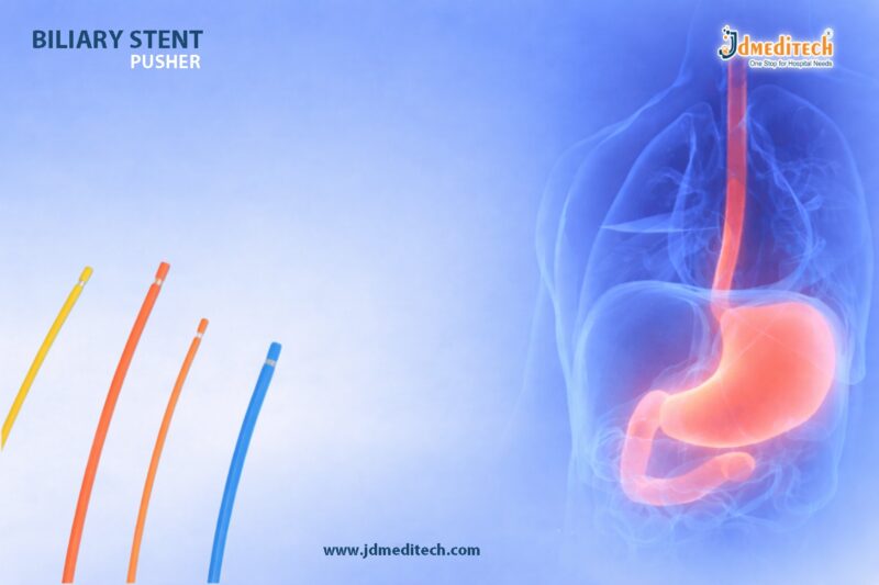
Accurate Stent Delivery in ERCP with Biliary Stent Pusher

In advanced gastrointestinal procedures, accurate stent delivery in ERCP plays a crucial role in achieving successful outcomes. One of the most important tools that ensures precision and control during stent placement is the biliary stent pusher. Designed to provide smooth deployment and optimal positioning, this device enhances the efficiency of biliary interventions.

During Endoscopic Retrograde Cholangiopancreatography (ERCP), clinicians rely on high-quality accessories to navigate complex biliary anatomy. A biliary stent pusher supports precise stent placement, reducing complications and improving patient outcomes.

What is a Biliary Stent Pusher?

A biliary stent pusher is a specialized endoscopic accessory used during ERCP procedures to facilitate the accurate placement of biliary stents within the bile duct. It is designed to push the stent through the endoscope channel and position it precisely at the targeted location.

This tool ensures accurate stent delivery in ERCP, especially in cases involving strictures, obstructions, or tumors within the biliary system.

Importance of Accurate Stent Delivery in ERCP

Achieving accurate stent delivery in ERCP is essential for several reasons:

1. Improved Patient Outcomes

Precise placement ensures effective bile drainage, reducing symptoms like jaundice and infection.

2. Reduced Procedure Time

A reliable biliary stent pusher allows faster deployment, improving procedural efficiency.

3. Minimization of Complications

Accurate positioning reduces risks such as stent migration or improper drainage.

4. Enhanced Clinical Confidence

Doctors can perform procedures with better control and precision.

Key Features of Biliary Stent Pusher

A high-quality biliary stent pusher is engineered for performance and reliability:

  • High Pushability – Ensures smooth advancement of the stent
  • Optimal Flexibility – Navigates complex biliary anatomy with ease
  • Radiopaque Markers – Allows clear visualization under fluoroscopy
  • Kink Resistance – Maintains structural integrity during insertion
  • Ergonomic Design – Provides better handling and control

These features collectively support accurate stent delivery in ERCP, making the procedure safer and more efficient.

Applications in ERCP Procedures

The biliary stent pusher is widely used in various clinical scenarios:

  • Treatment of biliary strictures
  • Management of bile duct obstructions
  • Drainage in malignant conditions
  • Support in post-operative bile leaks
  • Palliative care in advanced GI diseases

In each application, achieving accurate stent delivery in ERCP is critical for therapeutic success.

Advantages of Using a Biliary Stent Pusher
1. Precision Placement

Ensures the stent is delivered exactly where required.

2. Smooth Deployment

Reduces resistance during insertion.

3. Better Control

Allows clinicians to manage stent positioning effectively.

4. Time Efficiency

Speeds up ERCP procedures.

5. Reduced Risk of Re-intervention

Accurate placement minimizes the need for repeat procedures.

Why Quality Matters in ERCP Accessories

Using a high-quality biliary stent pusher significantly impacts the success of ERCP procedures. Poor-quality devices may lead to:

  • Difficult navigation
  • Increased procedural time
  • Higher complication rates
  • Inaccurate stent placement

Therefore, selecting reliable and well-designed accessories is essential for achieving accurate stent delivery in ERCP.

Tips for Optimal Stent Placement

To ensure accurate stent delivery in ERCP, clinicians should:

  • Choose the appropriate stent size and type
  • Use compatible accessories
  • Maintain clear fluoroscopic guidance
  • Advance the stent gently and steadily
  • Confirm final positioning before deployment
Conclusion

The biliary stent pusher is an indispensable tool in modern ERCP procedures. It plays a vital role in ensuring accurate stent delivery in ERCP, improving both procedural efficiency and patient outcomes.

With advancements in endoscopic technology, the demand for high-quality accessories continues to grow. Investing in reliable devices enhances precision, reduces complications, and supports better clinical results.


Explore More Product >>

Get Connected:

+91 79909 93062 | +91 63513 72032 | exports@jdmeditech.com
Ureteral Access Sheath for Safe and Smooth Urology Procedures

The ureteral access sheath is a crucial tool in modern urology. It provides smooth, repeated access to the urinary tract during procedures like ureteroscopy and stone management. Because of its efficiency and safety, the ureteral access sheath has become an essential device for urologists worldwide.

In this blog, we will explore how a ureteral access sheath improves procedural outcomes, enhances safety, and supports advanced urological interventions.

What is a Ureteral Access Sheath?

A ureteral access sheath is a flexible, hollow tube inserted into the ureter. It creates a continuous working channel that allows easy passage of instruments such as ureteroscopes, laser fibers, and retrieval devices.

As a result, the sheath minimizes repeated trauma to the ureter and ensures smoother instrument exchange.

Why is Ureteral Access Sheath Important?

The ureteral access sheath plays a vital role in improving both efficiency and patient safety.

Key Advantages:
  • Reduces ureteral trauma during repeated instrument insertion
  • Maintains low intrarenal pressure
  • Improves visualization during procedures
  • Facilitates faster and smoother device exchange
  • Enhances overall surgical efficiency

Therefore, many urologists prefer using a ureteral access sheath in complex procedures.

Key Features of Ureteral Access Sheath
1. Smooth Hydrophilic Coating

The hydrophilic coating reduces friction. Consequently, it allows easy insertion and removal without damaging the ureter.

2. Flexible Yet Durable Design

The sheath offers excellent flexibility. At the same time, it maintains enough strength to prevent kinking.

3. Radiopaque Markers

Radiopaque markers ensure accurate placement under fluoroscopy. This improves procedural safety.

4. Dual-Layer Construction

Many ureteral access sheaths feature a dual-layer design. This combination provides both flexibility and structural support.

5. Wide Range of Sizes

Available in different diameters and lengths, the sheath suits various patient anatomies and procedural needs.

Applications of Ureteral Access Sheath

The ureteral access sheath is widely used in:

  • Ureteroscopy (URS)
  • Kidney stone removal procedures
  • Laser lithotripsy
  • Retrograde intrarenal surgery (RIRS)
  • Diagnostic urological procedures

Because of its versatility, it is a standard tool in modern urology.

Benefits of Using Ureteral Access Sheath
Improved Procedure Efficiency

The sheath allows quick instrument exchanges. Therefore, it significantly reduces operation time.

Enhanced Patient Safety

By minimizing ureteral trauma and maintaining low pressure, the sheath reduces complications.

Better Visualization

Continuous irrigation flow improves visibility, which helps surgeons perform precise procedures.

Reduced Infection Risk

Lower intrarenal pressure helps prevent bacterial migration, thereby reducing infection risk.

How to Use a Ureteral Access Sheath?
  1. Insert a guidewire into the ureter
  2. Advance the ureteral access sheath over the guidewire
  3. Position it correctly under imaging guidance
  4. Introduce the ureteroscope and other instruments
  5. Perform the procedure efficiently

Proper technique ensures safe and effective outcomes.

Choosing the Right Ureteral Access Sheath

When selecting a ureteral access sheath, consider:

  • Size and length compatibility
  • Coating quality for smooth insertion
  • Flexibility and strength
  • Compatibility with instruments
  • Patient anatomy

Choosing the right sheath improves both performance and safety.

Why Choose JDMeditech Ureteral Access Sheath?

At JDMeditech, we provide high-quality ureteral access sheaths designed for reliability and precision.

Our Advantages:
  • Premium-grade materials
  • Smooth hydrophilic coating
  • Excellent flexibility and durability
  • Sterile and ready-to-use packaging
  • Competitive pricing with superior quality

Explore more products at: https://www.jdmeditech.com

Conclusion

The ureteral access sheath is an indispensable device in urological procedures. It ensures smooth access, improved safety, and better surgical outcomes. As urology continues to advance, the importance of reliable tools like the ureteral access sheath will only grow.


Explore More Product >>

Get Connected:

+91 79909 93062 | +91 63513 72032 | exports@jdmeditech.com
Endoscopy LED Light Source for Precision Imaging

In modern endoscopic procedures, clear visualization is critical for accurate diagnosis and treatment. The Endoscopy LED Light Source plays a vital role by delivering bright, consistent, and energy-efficient illumination. As a result, it enhances image clarity and supports precision during minimally invasive procedures.

Today, healthcare professionals prefer LED technology over traditional light sources because it offers longer lifespan, lower heat generation, and superior illumination quality. Therefore, investing in a high-quality Endoscopy LED Light Source ensures better clinical outcomes and improved patient safety.

What is an Endoscopy LED Light Source?

An Endoscopy LED Light Source is a medical device that provides high-intensity light to illuminate internal body structures during endoscopic procedures. It connects to an endoscope via a fiber optic cable, ensuring optimal visibility inside cavities such as the gastrointestinal tract, urinary system, and respiratory pathways.

Unlike conventional halogen or xenon light sources, LED technology offers stable brightness, reduced maintenance, and energy efficiency.

Key Features of Endoscopy LED Light Source
1. High-Intensity Illumination

The Endoscopy LED Light Source produces bright and uniform light, which improves visualization during procedures. Consequently, clinicians can identify even minor abnormalities with ease.

2. Energy Efficiency

LED technology consumes less power compared to traditional systems. Therefore, hospitals can reduce operational costs while maintaining high performance.

3. Long Lifespan

One of the biggest advantages of an Endoscopy LED Light Source is its durability. It offers thousands of hours of operation, reducing the need for frequent replacements.

4. Low Heat Generation

Unlike xenon light sources, LED systems generate minimal heat. As a result, they improve patient safety and reduce the risk of tissue damage.

5. Compact and User-Friendly Design

Modern systems are designed for easy integration with endoscopy units. Additionally, they feature intuitive controls for brightness adjustment.

Applications of Endoscopy LED Light Source

The Endoscopy LED Light Source is widely used across various medical specialties, including:

  • Gastroenterology – for procedures like gastroscopy, colonoscopy, and ERCP
  • Urology – for cystoscopy and ureteroscopy
  • Pulmonology – for bronchoscopy
  • Gynecology – for hysteroscopy
  • ENT procedures – for nasal and throat examinations

Because of its versatility, it has become an essential component in modern endoscopy systems.

Benefits of Using Endoscopy LED Light Source
Improved Visualization

The Endoscopy LED Light Source delivers consistent brightness, which helps in accurate diagnosis and treatment.

Enhanced Procedure Efficiency

Better illumination allows clinicians to perform procedures faster and with greater confidence.

Cost-Effective Solution

Since LED lights last longer and consume less energy, healthcare facilities can save significantly on maintenance and electricity.

Increased Patient Safety

Lower heat emission reduces the risk of burns or discomfort during procedures.

Why Choose JDMeditech Endoscopy LED Light Source?

At JDMeditech, we provide high-quality Endoscopy LED Light Source solutions designed for precision and reliability. Our products are manufactured using advanced technology to meet international medical standards.

Key Advantages:
  • Superior brightness and clarity
  • Durable and long-lasting performance
  • Energy-efficient design
  • Easy compatibility with various endoscopy systems
  • Competitive pricing with assured quality
Maintenance Tips for Endoscopy LED Light Source

To ensure optimal performance of your Endoscopy LED Light Source, follow these simple tips:

  • Regularly clean the external surfaces
  • Check fiber optic cables for damage
  • Avoid overloading the system
  • Ensure proper ventilation during operation
  • Schedule routine servicing

Proper maintenance extends the life of the device and ensures consistent performance.

Future of LED Technology in Endoscopy

The future of Endoscopy LED Light Source technology looks promising. With continuous advancements, newer models are becoming more efficient, compact, and intelligent. Additionally, integration with digital imaging systems will further enhance diagnostic accuracy.

Conclusion

The Endoscopy LED Light Source is an essential component in modern medical procedures. Its ability to provide bright, efficient, and reliable illumination significantly improves the accuracy and safety of endoscopic interventions.

By choosing a high-quality solution from JDMeditech, healthcare providers can ensure superior performance, long-term reliability, and better patient outcomes.


Explore More Product >>

Get Connected:

+91 79909 93062 | +91 63513 72032 | exports@jdmeditech.com

Double J Stent Set for Ureteral Drainage | Reliable Urology Solution

The Double J Stent Set plays a vital role in modern urology by ensuring smooth urine flow from the kidney to the bladder. Urologists rely on this device to manage ureteral obstructions, support post-surgical healing, and prevent complications.

Because of its unique design and dependable performance, the Double J Stent Set has become a standard solution for ureteral drainage and support across hospitals and surgical centers.

What is a Double J Stent Set?

A Double J Stent Set is a medical device used to maintain ureteral patency. It consists of a flexible stent with curls at both ends, forming a “J” shape on each side.

These curls help:

  • Secure the stent in the kidney and bladder
  • Prevent migration
  • Ensure continuous drainage

The set typically includes:

  • Double J stent
  • Pusher
  • Guidewire
  • Clamps or accessories
Why is the Double J Stent Set Important?

Ureteral blockage can lead to severe complications such as kidney damage or infection. Therefore, maintaining urine flow is critical.

The Double J Stent Set helps by:

  • Relieving ureteral obstruction
  • Supporting healing after urological procedures
  • Preventing urine backflow
  • Reducing pressure in the kidney

As a result, it improves patient outcomes and reduces recovery time.

Key Features of Double J Stent Set
1. High-Quality Biocompatible Material

Manufacturers use medical-grade materials to ensure safety and compatibility within the body.

2. Smooth Surface for Easy Insertion

The stent features a polished surface, allowing effortless placement and minimizing trauma.

3. Radiopaque Markings

Radiopaque lines enhance visibility under imaging, enabling precise positioning.

4. Optimal Flexibility and Strength

The stent adapts to the natural anatomy while maintaining structural integrity.

5. Anti-Migration Design

The double “J” curls secure the stent in place, preventing displacement.

Applications of Double J Stent Set

Healthcare professionals use the Double J Stent Set in various clinical scenarios:

  • Kidney stones (urolithiasis)
  • Ureteral strictures
  • Post-ureteroscopy procedures
  • Tumor-related obstructions
  • Post-surgical drainage support

Additionally, it serves both temporary and long-term drainage needs.

Benefits of Using Double J Stent Set
Ensures Continuous Urine Flow

The stent maintains an open ureter, allowing uninterrupted drainage.

Reduces Risk of Complications

It helps prevent hydronephrosis, infections, and kidney damage.

Enhances Patient Comfort

Its flexible design reduces irritation and discomfort.

Supports Faster Recovery

By stabilizing the ureter, it promotes quicker healing.

Easy Placement and Removal

The set enables efficient procedures for healthcare professionals.

Procedure Overview

Doctors typically place the Double J Stent Set using endoscopic techniques:

  1. Insert a guidewire into the ureter
  2. Advance the stent over the guidewire
  3. Position curls in the kidney and bladder
  4. Confirm placement using imaging

The procedure is minimally invasive and usually performed under anesthesia.

Safety and Considerations

Although the Double J Stent Set is highly effective, proper management is essential.

Key considerations include:
  • Timely removal or replacement
  • Monitoring for infection
  • Managing mild discomfort or urinary symptoms

Patients should always follow medical advice and attend follow-up visits.

Why Choose JDMeditech?

At JDMeditech, we focus on delivering high-quality urology solutions that meet global standards.

Our Double J Stent Set offers:

  • Precision-engineered design
  • Reliable performance
  • Superior material quality
  • Compliance with medical standards

We aim to support healthcare professionals with products that enhance patient care.

Conclusion

The Double J Stent Set stands as a trusted and effective solution for ureteral drainage and support. Its advanced design, ease of use, and clinical reliability make it indispensable in modern urology.

Whether managing obstructions or aiding recovery, this device ensures optimal outcomes for both patients and healthcare providers.


Explore More Product >>

Get Connected:

+91 79909 93062 | +91 63513 72032 | exports@jdmeditech.com

Hemorrhoid Band Ligator for Internal Hemorrhoids

The Hemorrhoid Band Ligator offers a quick and effective way to treat internal hemorrhoids. It helps doctors perform rubber band ligation with speed and accuracy. As a result, patients experience less discomfort and faster recovery.

Moreover, the hemorrhoid band ligator simplifies the procedure. Therefore, many clinics and hospitals prefer this method for non-surgical treatment.

What is a Hemorrhoid Band Ligator?

A Hemorrhoid Band Ligator is a medical device that places a small rubber band around the base of an internal hemorrhoid. This action stops blood flow and causes the tissue to shrink.

As a result, the hemorrhoid dries out and falls off within a few days. In most cases, doctors perform the procedure without anesthesia. Therefore, it suits outpatient settings perfectly.

How Does the Hemorrhoid Band Ligator Work?

The hemorrhoid band ligator follows a clear and simple process:

  • First, the doctor inserts a proctoscope to view the hemorrhoid
  • Next, the doctor gently grasps or suctions the tissue
  • Then, the ligator places a rubber band at the base
  • As a result, blood flow stops immediately
  • Finally, the hemorrhoid shrinks and falls off within 5–7 days

This method, known as rubber band ligation, delivers reliable results. In addition, it reduces the need for surgery.

Key Features of Hemorrhoid Band Ligator

The hemorrhoid band ligator provides several useful features:

  • Minimally invasive design
  • Easy handling and quick application
  • Accurate band placement
  • Reduced patient discomfort
  • Disposable and reusable options
  • Compatibility with standard proctoscopes

Because of these features, the hemorrhoid band ligator ensures consistent performance.

Benefits of Using a Hemorrhoid Band Ligator
Fast Procedure

The doctor completes the procedure within minutes. Therefore, it saves time for both patients and clinicians.

Effective Results

The hemorrhoid band ligator removes hemorrhoids at the source. As a result, it lowers the risk of recurrence.

Minimal Pain

Internal hemorrhoids contain fewer pain-sensitive nerves. Therefore, patients tolerate the procedure well.

Quick Recovery

Patients return to normal activities quickly. In most cases, they resume daily routines the same day.

Cost-Effective Solution

The hemorrhoid band ligator reduces treatment costs. Compared to surgery, it offers a more affordable option.

When Should You Use a Hemorrhoid Band Ligator?

Doctors commonly use the Hemorrhoid Band Ligator for:

  • Grade I internal hemorrhoids
  • Grade II internal hemorrhoids
  • Selected Grade III cases
  • Patients who want to avoid surgery

Therefore, it serves as a first-line treatment in many cases.

Procedure Overview

The procedure using a hemorrhoid band ligator follows these steps:

  1. First, the doctor positions the patient comfortably
  2. Next, the doctor inserts a proctoscope
  3. Then, the doctor identifies the hemorrhoid
  4. After that, the doctor applies the rubber band
  5. Finally, the doctor observes the patient briefly

Overall, the process remains simple, safe, and effective.

Why Choose a Hemorrhoid Band Ligator?

The hemorrhoid band ligator offers several advantages. First, it delivers proven clinical results. In addition, it allows easy handling for doctors.

Moreover, it reduces complications and speeds up recovery. As a result, patients report high satisfaction.

Safety and Precautions

Doctors must follow proper technique while using the hemorrhoid band ligator. For example:

  • Place the band above the dentate line
  • Capture only the required tissue
  • Monitor the patient after the procedure

When doctors follow these steps, they minimize risks. Therefore, complications rarely occur.

Use in Modern Healthcare

Today, many healthcare facilities use the Hemorrhoid Band Ligator. For instance:

  • Gastroenterology clinics
  • Proctology centers
  • Daycare units
  • Hospitals

Because of its efficiency, it has become a standard treatment tool.

Conclusion

The Hemorrhoid Band Ligator provides a fast, safe, and effective solution for internal hemorrhoids. It simplifies the procedure while improving patient comfort.

In conclusion, the hemorrhoid band ligator remains a reliable choice for modern medical practice.


Explore More Product >>

Get Connected:

+91 79909 93062 | +91 63513 72032 | exports@jdmeditech.com



Gastrostomy Replacement Tubes for Safe Enteral Nutrition

Gastrostomy replacement tubes play a critical role in modern enteral nutrition care. These tubes provide a reliable and safe route for delivering nutrition directly into the stomach when oral intake is not possible. Healthcare professionals widely use gastrostomy replacement tubes in both hospital and home care settings.

As patient care evolves, the demand for high-quality gastrostomy replacement tubes continues to grow. These devices improve patient comfort, reduce complications, and ensure consistent nutritional support.

What Are Gastrostomy Replacement Tubes?

Gastrostomy replacement tubes are medical devices designed to replace an existing gastrostomy tube without requiring a new surgical procedure. Doctors typically insert these tubes into a mature stoma tract to maintain enteral feeding access.

Unlike initial PEG tubes, replacement tubes allow quick and minimally invasive changes, which improves patient convenience and reduces healthcare costs.

Types of Gastrostomy Replacement Tubes
1. Balloon Gastrostomy Tubes

Balloon gastrostomy replacement tubes feature an inflatable balloon that secures the tube inside the stomach. Clinicians prefer them because they are easy to insert and remove.

2. Non-Balloon (Mushroom/Retention Dome) Tubes

These gastrostomy replacement tubes use a fixed internal retention dome. They provide long-term durability and reduce accidental dislodgement.

3. Low-Profile (Button) Tubes

Low-profile gastrostomy replacement tubes sit flush with the skin. They enhance patient comfort, improve mobility, and offer a discreet feeding option.

Key Features of Gastrostomy Replacement Tubes
  • Medical-grade, biocompatible materials
  • Smooth surface for patient comfort
  • Secure retention mechanisms
  • Anti-reflux valve systems
  • Radiopaque lines for imaging visibility
  • Multiple size options for customized care

These features ensure that gastrostomy replacement tubes deliver safe and effective enteral nutrition.

Clinical Applications

Gastrostomy replacement tubes are widely used in:

  • Neurological disorders (stroke, ALS, cerebral palsy)
  • Oncology patients requiring nutritional support
  • Pediatric feeding difficulties
  • Long-term critical care patients
  • Dysphagia management

Because of their versatility, gastrostomy replacement tubes remain essential in modern healthcare.

Benefits of Gastrostomy Replacement Tubes
1. Minimally Invasive Replacement

Clinicians can replace gastrostomy tubes quickly without surgery.

2. Improved Patient Comfort

Modern designs reduce irritation and enhance usability.

3. Reduced Risk of Complications

High-quality gastrostomy replacement tubes help prevent leakage and infection.

4. Cost-Effective Solution

Replacement procedures reduce hospital stays and overall treatment costs.

5. Enhanced Mobility

Low-profile options allow patients to maintain an active lifestyle.

Best Practices for Use

To maximize the effectiveness of gastrostomy replacement tubes:

  • Ensure proper tube size selection
  • Maintain strict hygiene during handling
  • Regularly check balloon integrity (if applicable)
  • Monitor for signs of infection or leakage
  • Replace tubes as per clinical guidelines

Following these practices ensures long-term success in enteral nutrition care.

Why Choose High-Quality Gastrostomy Replacement Tubes?

Choosing premium gastrostomy replacement tubes ensures better clinical outcomes. High-quality tubes offer superior durability, patient comfort, and safety. They also reduce the need for frequent replacements, which improves both patient satisfaction and healthcare efficiency.

Conclusion

Gastrostomy replacement tubes are essential tools in modern enteral nutrition care. They provide a safe, efficient, and patient-friendly solution for long-term feeding needs. With advancements in design and materials, these tubes continue to enhance clinical outcomes and improve quality of life for patients worldwide.

Healthcare providers should always prioritize high-quality gastrostomy replacement tubes to ensure optimal performance and patient safety.


Explore More Product >>

Get Connected:

+91 79909 93062 | +91 63513 72032 | exports@jdmeditech.com
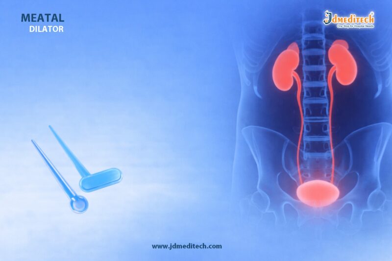
 

Urethroplasty Instrument Set for Precise Stricture Repair

The Urethroplasty Instrument Set plays a vital role in modern urology. Surgeons use the Urethroplasty Instrument Set to perform accurate urethral reconstruction. This procedure requires precision and control. Therefore, a complete Urethroplasty Instrument Set improves surgical outcomes.

Moreover, the Urethroplasty Instrument Set helps reduce complications and supports faster recovery. As a result, it has become essential in stricture repair procedures.

What is a Urethroplasty Instrument Set?

A Urethroplasty Instrument Set includes specialized tools for urethral reconstruction. Surgeons use the Urethroplasty Instrument Set to remove scar tissue and restore urine flow.

In addition, the Urethroplasty Instrument Set supports both simple and complex surgical techniques. It ensures consistency and control throughout the procedure.

Key Components of Urethroplasty Instrument Set

The Urethroplasty Instrument Set contains all essential tools. Each instrument supports a specific surgical step.

  • Fine scissors for precise cutting
  • Tissue forceps for gentle handling
  • Needle holders for suturing control
  • Dilators for urethral calibration
  • Retractors for better visibility
  • Hemostats for bleeding control
  • Scalpels for clean incisions

Furthermore, every Urethroplasty Instrument Set ensures smooth workflow in the operating room.

Features of Urethroplasty Instrument Set
High-Quality Material

The Urethroplasty Instrument Set uses premium stainless steel. It resists corrosion and ensures long-term use.

Precision Design

Each tool in the Urethroplasty Instrument Set delivers accurate performance. Surgeons achieve better control during procedures.

Ergonomic Handling

The Urethroplasty Instrument Set offers a comfortable grip. It reduces fatigue during long surgeries.

Complete Surgical Kit

The Urethroplasty Instrument Set includes all required instruments. Therefore, surgeons do not need extra tools.

Easy Sterilization

The Urethroplasty Instrument Set supports standard sterilization methods. This ensures safety and hygiene.

Applications of Urethroplasty Instrument Set

The Urethroplasty Instrument Set is widely used in:

  • Urethral stricture repair
  • Reconstructive urology procedures
  • Trauma-related urethral surgery
  • Congenital defect correction

Additionally, hospitals and urology centers rely on the Urethroplasty Instrument Set for consistent results.

Benefits of Urethroplasty Instrument Set

The Urethroplasty Instrument Set offers several advantages:

  • Improves surgical precision
  • Reduces procedure time
  • Enhances patient safety
  • Ensures reliable performance
  • Supports better recovery

As a result, the Urethroplasty Instrument Set improves overall surgical success.

Why Choose JDMeditech Urethroplasty Instrument Set?

The Urethroplasty Instrument Set from JDMeditech meets high-quality standards. It delivers consistent performance in critical procedures.

  • Advanced manufacturing process
  • Strict quality checks
  • Durable and reliable instruments
  • Designed for surgical efficiency

Therefore, many professionals trust the Urethroplasty Instrument Set from JD Meditech.

Best Practices for Using Urethroplasty Instrument Set

Follow these steps to use the Urethroplasty Instrument Set effectively:

  • Sterilize all instruments before use
  • Inspect each tool carefully
  • Handle instruments with care
  • Follow standard surgical protocols

These steps ensure safe use of the Urethroplasty Instrument Set.

Conclusion

The Urethroplasty Instrument Set is essential for successful stricture repair. It provides precision, control, and reliability. Moreover, the Urethroplasty Instrument Set supports better outcomes and faster recovery.

Therefore, choosing a high-quality Urethroplasty Instrument Set improves both surgical efficiency and patient care.


Explore More Product >>

Get Connected:

+91 79909 93062 | +91 63513 72032 | exports@jdmeditech.com

 

Male Urethral Dilator Set – Safe Stricture Treatment

The Male Urethral Dilator Set plays a vital role in urological procedures. It helps doctors manage urethral strictures safely and effectively. Urethral strictures can block urine flow and cause discomfort. Therefore, proper dilation becomes essential for restoring normal urinary function.

Moreover, a high-quality Male Urethral Dilator Set ensures smooth insertion and minimizes trauma. As a result, it improves patient outcomes and procedural efficiency.

What is a Male Urethral Dilator Set?

A Male Urethral Dilator Set is a collection of precisely designed instruments. These instruments gradually widen the urethra. Doctors use them to treat narrowing caused by strictures, injury, or infection.

Typically, the set includes dilators of increasing sizes. This allows controlled and progressive dilation. In addition, each dilator features a smooth surface to reduce friction and discomfort.

Importance in Urology Procedures

The Male Urethral Dilator Set is essential in modern urology. It supports both diagnosis and treatment.

Key Benefits:
  • Ensures gradual and controlled dilation
  • Reduces the risk of urethral injury
  • Improves urinary flow quickly
  • Supports minimally invasive treatment
  • Enhances procedural precision

Because of these benefits, urologists widely prefer a reliable Male Urethral Dilator Set for stricture management.

Key Features of a High-Quality Male Urethral Dilator Set

When selecting a Male Urethral Dilator Set, quality matters. The right design ensures safety and performance.

1. Smooth Surface Finish

Each dilator has a polished surface. This reduces friction during insertion.

2. Graduated Sizes

The set includes multiple sizes. Therefore, doctors can perform gradual dilation with precision.

3. Ergonomic Design

The instruments offer a comfortable grip. This improves control during procedures.

4. High-Quality Material

Most dilators are made from medical-grade materials. As a result, they ensure durability and patient safety.

5. Sterility and Hygiene

Many sets come sterile or are easy to sterilize. This reduces infection risks.

Applications of Male Urethral Dilator Set

The Male Urethral Dilator Set is used in various clinical situations.

  • Treatment of urethral strictures
  • Pre-operative urethral preparation
  • Post-surgical urethral maintenance
  • Diagnostic evaluation of urethral narrowing

Furthermore, it helps in both routine and complex urological cases.

How the Procedure Works

The use of a Male Urethral Dilator Set follows a systematic approach.

  1. The urethra is lubricated properly
  2. The smallest dilator is inserted gently
  3. Gradually larger dilators are used
  4. The process continues until the desired dilation is achieved

Because of this step-by-step method, the risk of complications remains low.

Safety Considerations

Although the Male Urethral Dilator Set is safe, proper technique is essential.

  • Always use adequate lubrication
  • Maintain sterile conditions
  • Avoid forceful insertion
  • Monitor patient discomfort
  • Follow medical guidelines strictly

Therefore, trained professionals should perform the procedure.

Why Choose JDMeditech?

At JDMeditech, we focus on delivering precision-engineered urology instruments. Our Male Urethral Dilator Set meets high standards of quality and performance.

Our Advantages:
  • Consistent product quality
  • Reliable performance in clinical settings
  • Designed for patient comfort
  • Competitive pricing
  • Trusted by healthcare professionals
Conclusion

The Male Urethral Dilator Set is a reliable and essential tool for managing urethral strictures. It ensures safe, controlled, and effective dilation. As a result, it improves patient comfort and treatment success.

Choosing a high-quality Male Urethral Dilator Set enhances both precision and safety. Therefore, it remains a cornerstone instrument in modern urology practice.


Explore More Product >>

Get Connected:

+91 79909 93062 | +91 63513 72032 | exports@jdmeditech.com
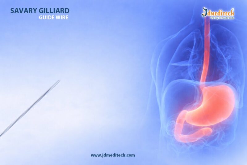


Sclerotherapy Needle for GI Bleeding

A sclerotherapy needle is a vital device in endoscopic procedures, especially for managing gastrointestinal (GI) bleeding. It allows clinicians to inject sclerosing agents directly into or around bleeding vessels, ensuring rapid hemostasis.

Because GI bleeding can become life-threatening, the sclerotherapy needle plays a critical role in emergency and therapeutic gastroenterology. Its precision and reliability make it a preferred choice for endoscopists worldwide.

What is a Sclerotherapy Needle?

A sclerotherapy needle is a specialized endoscopic instrument designed to deliver therapeutic agents into targeted tissues within the gastrointestinal tract.

It typically consists of:

  • A flexible catheter compatible with endoscopes
  • A sharp, retractable needle tip
  • An ergonomic handle for controlled injection

The design ensures safe, accurate, and efficient delivery of sclerosants during procedures.

Importance of Sclerotherapy Needle in GI Bleeding Management

The sclerotherapy needle is widely used to control bleeding from:

  • Esophageal varices
  • Gastric varices
  • Peptic ulcers
  • Dieulafoy lesions
Why it is essential:
  • Enables targeted injection therapy
  • Provides rapid bleeding control
  • Reduces need for surgical intervention
  • Supports minimally invasive treatment

As a result, the sclerotherapy needle significantly improves patient outcomes in critical situations.

Key Features of a Sclerotherapy Needle

Modern sclerotherapy needles are engineered for precision and safety. Key features include:

1. High-Precision Needle Tip

Ensures accurate penetration and controlled delivery of sclerosants.

2. Flexible Catheter Design

Allows smooth navigation through the endoscope channel.

3. Adjustable Needle Length

Provides versatility for different clinical requirements.

4. Secure Locking Mechanism

Prevents accidental needle exposure.

5. Radiopaque Markers

Enhance visibility under imaging guidance.

Clinical Applications of Sclerotherapy Needle

The sclerotherapy needle is used in various therapeutic endoscopy procedures:

  • Injection of sclerosants for variceal bleeding
  • Treatment of non-variceal GI bleeding
  • Endoscopic mucosal therapy
  • Hemostasis in emergency situations

Because of its versatility, the sclerotherapy needle remains a cornerstone tool in gastroenterology.

Advantages of Using a Sclerotherapy Needle

Using a sclerotherapy needle offers several benefits:

  • Minimally invasive approach
  • Quick and effective hemostasis
  • Reduced patient recovery time
  • Improved procedural control
  • Cost-effective treatment option

Therefore, it is widely adopted in hospitals and endoscopy centers.

How to Choose the Right Sclerotherapy Needle

Selecting the right sclerotherapy needle depends on several factors:

  • Needle gauge and length
  • Compatibility with endoscopes
  • Quality of materials
  • Sterility and disposability
  • Manufacturer reliability

Choosing a high-quality sclerotherapy needle ensures better performance and patient safety.

Safety Considerations

While the sclerotherapy needle is highly effective, proper handling is essential:

  • Use sterile, single-use devices
  • Follow correct injection techniques
  • Avoid excessive tissue penetration
  • Monitor patient response during the procedure

Adhering to these guidelines ensures optimal outcomes.

Why Choose JDMeditech Sclerotherapy Needle?

At JDMeditech, we provide high-quality sclerotherapy needles designed for precision and safety.

Key Highlights:
  • Premium-grade materials
  • Excellent flexibility and control
  • Reliable performance in critical procedures
  • Strict quality standards
Conclusion

The sclerotherapy needle is an indispensable tool in the management of gastrointestinal bleeding. Its precision, safety, and effectiveness make it a preferred choice for endoscopic therapy.

As healthcare continues to advance, the role of the sclerotherapy needle will remain crucial in delivering life-saving treatments with minimal invasiveness.


Explore More Product >>

Get Connected:

+91 79909 93062 | +91 63513 72032 | exports@jdmeditech.com
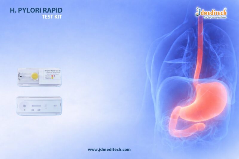
 

PCNL Forceps for Stone Grasping in Percutaneous Procedures

PCNL Forceps play a vital role in modern urology, especially during Percutaneous Nephrolithotomy (PCNL) procedures. These specialized instruments are designed to ensure safe, precise, and efficient stone grasping and extraction from the kidney.

As kidney stone cases continue to rise globally, surgeons rely on high-quality PCNL Forceps to improve procedural accuracy and patient outcomes. Therefore, selecting the right instrument becomes critical for both efficiency and safety.

What Are PCNL Forceps?

PCNL Forceps are precision surgical tools used during PCNL procedures to grasp, hold, and remove kidney stones through a nephroscope.

Unlike standard forceps, these instruments are specifically engineered to:

  • Navigate narrow percutaneous tracts
  • Provide firm grip on calculi
  • Minimize trauma to surrounding tissues

As a result, they significantly enhance surgical control during stone retrieval.

Role of PCNL Forceps in PCNL Procedures

During a PCNL procedure, the surgeon creates a small tract into the kidney to remove stones. At this stage, PCNL Forceps become essential.

Key Functions
  • Grasping fragmented or intact stones
  • Assisting in stone extraction
  • Improving procedural efficiency
  • Reducing operative time

Moreover, these forceps allow surgeons to handle stones of different sizes and shapes with ease.

Key Features of PCNL Forceps

High-quality PCNL Forceps are designed with advanced features to meet surgical demands.

1. Precision Tip Design

Ensures accurate stone gripping without slippage.

2. Ergonomic Handle

Provides better control and reduces hand fatigue during long procedures.

3. Durable Construction

Made from medical-grade stainless steel for longevity and repeated sterilization.

4. Variety of Jaw Types

Available in different designs such as:

  • Alligator jaws
  • Grasping jaws
  • Serrated tips

This variety allows surgeons to choose the most suitable option.

5. Compatibility

Designed to work seamlessly with nephroscopes and other PCNL instruments.

Benefits of Using PCNL Forceps

Using advanced PCNL Forceps offers multiple clinical advantages:

  • Improved Stone Retrieval Efficiency
  • Reduced Procedure Time
  • Enhanced Surgical Precision
  • Lower Risk of Tissue Damage
  • Better Patient Outcomes

Additionally, these instruments contribute to smoother workflow in operating rooms.

Applications of PCNL Forceps
PCNL Forceps are widely used in:
  • Percutaneous Nephrolithotomy (PCNL)
  • Kidney stone removal procedures
  • Urological surgeries involving calculi extraction

Furthermore, they are suitable for both fragmented and intact stone removal.

How to Choose the Right PCNL Forceps

Selecting the ideal PCNL Forceps depends on several factors:

Consider These Points
  • Type and size of kidney stones
  • Compatibility with existing instruments
  • Jaw design and grip strength
  • Surgeon preference
  • Sterilization and durability

Therefore, choosing the right forceps can directly impact procedural success.

Maintenance and Sterilization

Proper care of PCNL Forceps ensures long-term performance:

  • Clean immediately after use
  • Use standard sterilization protocols (autoclaving)
  • Inspect for wear and tear
  • Store in a dry, sterile environment

Consistent maintenance helps maintain precision and reliability.

Why Choose High-Quality PCNL Forceps?

Investing in premium PCNL Forceps ensures:

  • Consistent performance
  • Enhanced safety
  • Cost-effectiveness over time
  • Reliable surgical outcomes

In addition, high-quality instruments reduce complications and improve surgeon confidence.

Conclusion

PCNL Forceps are indispensable tools in percutaneous kidney stone procedures. Their ability to provide secure stone grasping, precision handling, and efficient extraction makes them a cornerstone of modern urology.

As surgical techniques evolve, the demand for reliable and high-performance PCNL Forceps continues to grow. Therefore, choosing the right instrument is essential for achieving optimal clinical results.


Explore More Product >>

Get Connected:

+91 79909 93062 | +91 63513 72032 | exports@jdmeditech.com


SPC Trocar: Safe Tool for Suprapubic Catheterization

The SPC Trocar is an essential instrument in urology. It enables safe and efficient suprapubic catheterization. Doctors use the SPC Trocar when urethral catheterization is not possible. Therefore, it provides a reliable alternative for bladder drainage.

Moreover, the SPC Trocar improves procedural accuracy and patient comfort. Because of its design, clinicians can perform the procedure quickly and safely.

What is an SPC Trocar?

An SPC Trocar is a specialized surgical instrument. It helps create a controlled entry into the bladder through the abdominal wall. As a result, it allows easy placement of a suprapubic catheter.

Typically, an SPC Trocar includes:

  • A sharp obturator
  • A hollow cannula
  • An ergonomic handle

Together, these components ensure smooth insertion and secure catheter placement.

Why SPC Trocar is Important

The SPC Trocar plays a key role in modern urology. First, it provides direct bladder access. Second, it reduces the risk of urethral injury. In addition, it supports fast intervention in emergency cases.

Furthermore, the SPC Trocar enhances patient comfort during long-term catheterization. Because of these benefits, it is widely used in hospitals and clinics.

Key Features of SPC Trocar

A high-quality SPC Trocar offers several important features:

1. Sharp Precision Tip

The SPC Trocar ensures smooth penetration with minimal tissue damage.

2. Durable Medical Material

Most SPC Trocar devices use stainless steel or medical-grade polymers. Therefore, they ensure strength and sterility.

3. Ergonomic Grip

The SPC Trocar provides better handling and control during procedures.

4. Secure Cannula Design

It allows stable and accurate catheter placement.

5. Sterile Packaging

Many SPC Trocar options come in sterile, single-use formats. As a result, infection risk decreases.

Clinical Applications of SPC Trocar

The SPC Trocar is widely used in several medical conditions:

  • Urinary retention
  • Neurogenic bladder
  • Urethral obstruction
  • Post-surgical bladder drainage
  • Long-term catheterization

Because of its versatility, the SPC Trocar is a must-have tool in urology.

Procedure Using SPC Trocar

Doctors follow a structured process when using an SPC Trocar:

  1. First, prepare the patient and apply local anesthesia.
  2. Next, locate the bladder, often using ultrasound.
  3. Then, insert the SPC Trocar above the pubic bone.
  4. After that, create a tract into the bladder.
  5. Insert the catheter through the cannula.
  6. Finally, remove the SPC Trocar and secure the catheter.

Thus, the SPC Trocar ensures a safe and controlled procedure.

Benefits of SPC Trocar

The SPC Trocar provides multiple advantages:

  • Minimally invasive approach
  • Reduced urethral complications
  • Improved patient comfort
  • Quick bladder decompression
  • Lower infection risk

Therefore, the SPC Trocar improves both safety and efficiency.

Safety Considerations

Although the SPC Trocar is effective, proper precautions are necessary.

  • Ensure bladder is full before insertion
  • Use ultrasound guidance when needed
  • Maintain sterile technique
  • Avoid incorrect placement
  • Monitor patients carefully

By following these steps, clinicians can safely use the SPC Trocar.

How to Choose the Right SPC Trocar

Choosing the right SPC Trocar is important. Consider the following:

  • Size compatibility
  • Material quality
  • Ease of use
  • Sterility options
  • Compliance with standards

A well-designed SPC Trocar ensures better outcomes.

Conclusion

The SPC Trocar is a vital tool for suprapubic catheterization. It provides safe, precise, and efficient bladder access. In addition, the SPC Trocar improves patient comfort and reduces complications.

Because of its reliability, the SPC Trocar remains essential in modern urology.


Explore More Product >>

Get Connected:

+91 79909 93062 | +91 63513 72032 | exports@jdmeditech.com
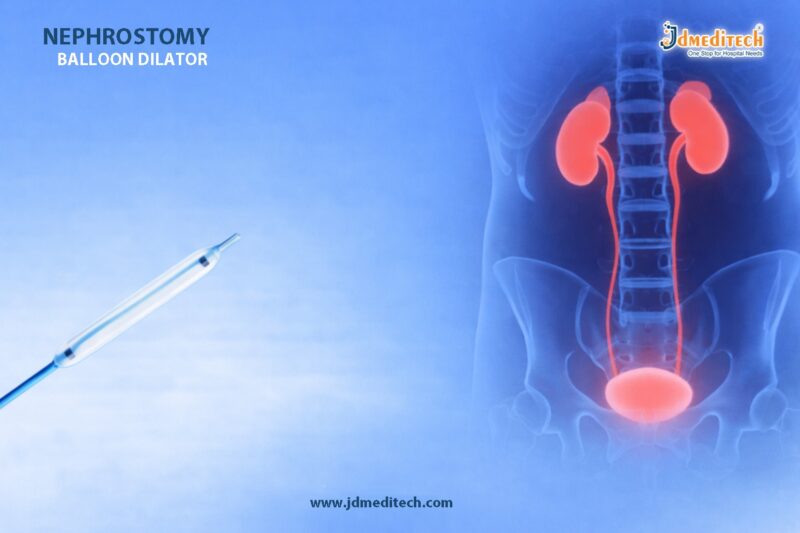
 

Biliary Stents: Advanced Solution for Bile Duct Obstruction

Biliary stents play a vital role in modern gastroenterology by providing effective solutions for bile duct obstruction. These small yet highly functional medical devices help restore bile flow, relieve symptoms, and improve patient outcomes. As minimally invasive treatment options gain popularity, biliary stents have become an indispensable tool in both diagnostic and therapeutic procedures.

What Are Biliary Stents?

Biliary stents are small tubular devices inserted into the bile duct to maintain or restore proper bile flow from the liver to the small intestine. They are typically placed during endoscopic procedures such as ERCP (Endoscopic Retrograde Cholangiopancreatography).

There are two main types of biliary stents:

  • Plastic Biliary Stents– Cost-effective and suitable for short-term use
  • Metal Biliary Stents (Self-Expandable Metal Stents – SEMS) – Durable and ideal for long-term placement
Applications of Biliary Stents in Gastroenterology
1. Management of Biliary Obstruction

Biliary stents are widely used to relieve obstruction caused by gallstones, tumors, or strictures. They help restore normal bile flow and prevent complications.

2. Treatment of Malignant Conditions

In patients with pancreatic cancer or cholangiocarcinoma, biliary stents provide palliative care by reducing jaundice and improving quality of life.

3. Benign Biliary Strictures

Biliary stents are effective in managing benign strictures resulting from surgery, inflammation, or injury.

4. Post-Surgical Support

After hepatobiliary surgeries, biliary stents help maintain duct patency and support healing.

5. Management of Bile Leaks

Biliary stents assist in diverting bile flow, allowing leaks to heal naturally.

Benefits of Biliary Stents
1. Minimally Invasive Treatment

Biliary stents can be placed endoscopically, reducing the need for open surgery and lowering patient risk.

2. Immediate Symptom Relief

Patients experience rapid improvement in symptoms such as jaundice, itching, and abdominal discomfort.

3. Improved Quality of Life

Especially in cancer patients, biliary stents significantly enhance comfort and daily functioning.

4. Cost-Effective Solution

Plastic stents offer an affordable option, while metal stents reduce the need for frequent replacements.

5. Reduced Complications

By restoring bile flow, biliary stents help prevent infections like cholangitis and liver damage.

Procedure Overview

The placement of biliary stents is commonly performed using ERCP:

  1. Endoscope insertion through the mouth to the duodenum
  2. Identification of bile duct obstruction
  3. Guidewire placement across the blockage
  4. Deployment of the biliary stent

This procedure is generally safe and performed under sedation.

Potential Risks and Considerations

While biliary stents are highly effective, some risks may include:

  • Stent occlusion or blockage
  • Infection (cholangitis)
  • Migration of the stent
  • Need for replacement (especially plastic stents)

Proper follow-up and timely replacement are essential for optimal outcomes.

Choosing the Right Biliary Stent

The selection of biliary stents depends on:

  • Nature of the obstruction (benign or malignant)
  • Duration of required stenting
  • Patient condition and prognosis
  • Cost considerations

Healthcare providers carefully evaluate these factors to ensure the best treatment approach.

Conclusion

Biliary stents have revolutionized the management of biliary disorders in gastroenterology. Their ability to provide quick, minimally invasive relief makes them a preferred choice for clinicians worldwide. From managing obstructions to improving patient quality of life, biliary stents continue to play a critical role in modern digestive healthcare.


Explore More Product >>

Get Connected:

+91 79909 93062 | +91 63513 72032 | exports@jdmeditech.com
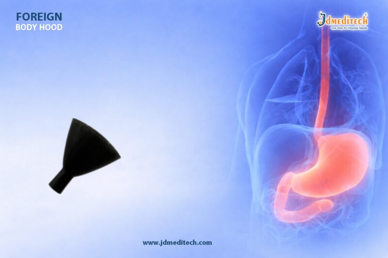


Alkaline Telescopic Dilator Set – Precision Tract Dilation Tool

The Alkaline Telescopic Dilator Set plays a critical role in modern urological procedures. It allows surgeons to achieve accurate and controlled tract dilation, especially in procedures like percutaneous nephrolithotomy (PCNL).

Because precision is essential in urology, the use of a reliable dilator system can significantly improve outcomes. The Alkaline Telescopic Dilator Set is designed to offer smooth, stepwise dilation while minimizing tissue trauma.

What is an Alkaline Telescopic Dilator Set?

An Alkaline Telescopic Dilator Set is a specialized surgical instrument used to gradually enlarge a tract during minimally invasive procedures.

It consists of multiple dilators of increasing diameters that are passed sequentially over a guidewire. This telescopic design ensures:

  • Controlled dilation
  • Reduced risk of injury
  • Consistent tract formation
Key Features of Alkaline Telescopic Dilator Set
1. Sequential Telescopic Design

The set includes multiple dilators that allow step-by-step tract dilation, ensuring maximum control.

2. High-Quality Material

Manufactured using medical-grade materials, the Alkaline Telescopic Dilator Set ensures durability and safety.

3. Smooth Surface Finish

The polished surface reduces friction, which helps in atraumatic insertion.

4. Precision Engineering

Each component is designed for accurate alignment, ensuring seamless procedural flow.

5. Ergonomic Handling

The set allows surgeons to maintain a firm and comfortable grip, improving control during procedures.

Applications in Urology

The Alkaline Telescopic Dilator Set is widely used in:

  • Percutaneous Nephrolithotomy (PCNL)
  • Renal access procedures
  • Urinary tract dilation
  • Stone removal surgeries

Because of its controlled expansion capability, it is essential for creating a safe access tract to the kidney.

Benefits of Using Alkaline Telescopic Dilator Set
1. Controlled Dilation

The telescopic mechanism ensures gradual expansion, reducing complications.

2. Reduced Tissue Trauma

Smooth transitions between dilators help minimize damage to surrounding tissues.

3. Improved Procedural Accuracy

The design supports precise tract creation, which is crucial in urology.

4. Enhanced Safety

Using a guidewire-based system improves procedural safety and consistency.

5. Time Efficiency

The sequential system allows faster dilation compared to traditional methods.

Why Choose Alkaline Telescopic Dilator Set?

Choosing the Alkaline Telescopic Dilator Set ensures reliability and performance. It is engineered to meet the demands of modern surgical environments.

Healthcare professionals prefer it because:

  • It provides consistent results
  • It supports minimally invasive techniques
  • It enhances patient safety
Comparison with Other Dilator Systems
Feature Telescopic Dilator Set Balloon Dilator Metal Dilators
Control High Moderate Low
Cost Cost-effective Expensive Moderate
Reusability Yes No Yes
Precision Excellent Good Moderate

The Alkaline Telescopic Dilator Set stands out due to its balance of precision, control, and affordability.

Best Practices for Use

To maximize performance of the Alkaline Telescopic Dilator Set, follow these guidelines:

  • Always use with a guidewire
  • Ensure proper lubrication
  • Insert dilators gradually and steadily
  • Maintain sterile conditions
  • Inspect instruments before use
Conclusion

The Alkaline Telescopic Dilator Set is an essential tool for achieving accurate and controlled tract dilation in urology. Its telescopic design, precision engineering, and safety features make it a preferred choice among surgeons.

As minimally invasive procedures continue to evolve, the importance of reliable tools like the Alkaline Telescopic Dilator Set will only grow.


Explore More Product >>

+91 79909 93062 | +91 63513 72032 | exports@jdmeditech.com
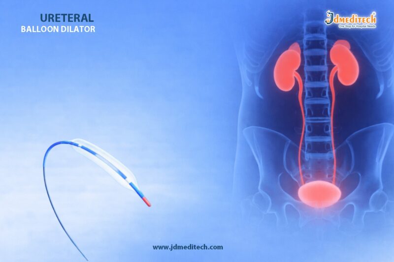


Dialysis ON OFF Kit for Safe Blood Flow Control

The Dialysis ON OFF Kit plays a critical role in modern hemodialysis by ensuring safe, controlled, and efficient blood flow management. In dialysis procedures, maintaining precise control over blood circulation is essential to prevent complications and ensure optimal treatment outcomes.

Designed for reliability and ease of use, the Dialysis ON OFF Kit supports clinicians in managing blood flow transitions smoothly during dialysis sessions. Its integration into dialysis systems enhances both patient safety and procedural efficiency.

What is a Dialysis ON OFF Kit?

A Dialysis ON OFF Kit is a specialized medical device used to regulate and control the flow of blood during dialysis procedures. It allows healthcare professionals to safely start (ON) and stop (OFF) blood flow between the patient and the dialysis machine.

This kit is essential in ensuring:

  • Controlled blood circulation
  • Prevention of blood loss or leakage
  • Smooth transition during dialysis setup and completion
Key Components of Dialysis ON OFF Kit

The Dialysis ON OFF Kit typically includes:

  • Cotton Balls
  • Gauze Swabs
  • Wrapping Sheet / Sterile Drape
  • Disposable Tray
  • Absorbent Towel
Applications of Dialysis ON OFF Kit

The Dialysis ON OFF Kit is widely used in:

  • Hemodialysis procedures
  • Dialysis centers and hospitals
  • Critical care units
  • Emergency dialysis setups

Its versatility makes it an essential component in both routine and advanced dialysis treatments.

Benefits of Dialysis ON OFF Kit
1. Enhanced Patient Safety

The Dialysis ON OFF Kit ensures controlled blood flow, reducing the risk of complications such as air embolism or blood loss.

2. Improved Clinical Efficiency

Quick ON/OFF functionality saves time and simplifies workflow for healthcare professionals.

3. Leak-Proof and Secure Design

High-quality connectors prevent leakage and ensure reliable performance.

4. Easy Handling and Operation

User-friendly design allows smooth handling during critical procedures.

5. Sterile and Disposable Options

Reduces the risk of infection and cross-contamination.

Why Choose a High-Quality Dialysis ON OFF Kit?

Selecting a reliable Dialysis ON OFF Kit is crucial for ensuring patient safety and achieving consistent dialysis outcomes. High-quality kits offer:

  • Durable and medical-grade materials
  • Accurate flow control
  • Compatibility with standard dialysis machines
  • Compliance with international healthcare standards
Best Practices for Using Dialysis ON OFF Kit

To maximize performance and safety:

  • Always check connections before initiating dialysis
  • Maintain sterile handling procedures
  • Ensure proper ON/OFF positioning during transitions
  • Dispose of single-use kits responsibly
Conclusion

The Dialysis ON OFF Kit is an indispensable tool in dialysis procedures, offering safe, efficient, and controlled blood flow management. Its role in enhancing patient safety and simplifying clinical workflow makes it a vital component in modern healthcare settings.

By choosing a high-quality Dialysis ON OFF Kit, healthcare providers can ensure reliable performance and better outcomes for patients undergoing dialysis.


Explore More Product >>

Get Connected:

+91 79909 93062 | +91 63513 72032 | exports@jdmeditech.com