Laparoscopic Orchidopexy Surgery – Advanced Treatment for Undescended Testicles

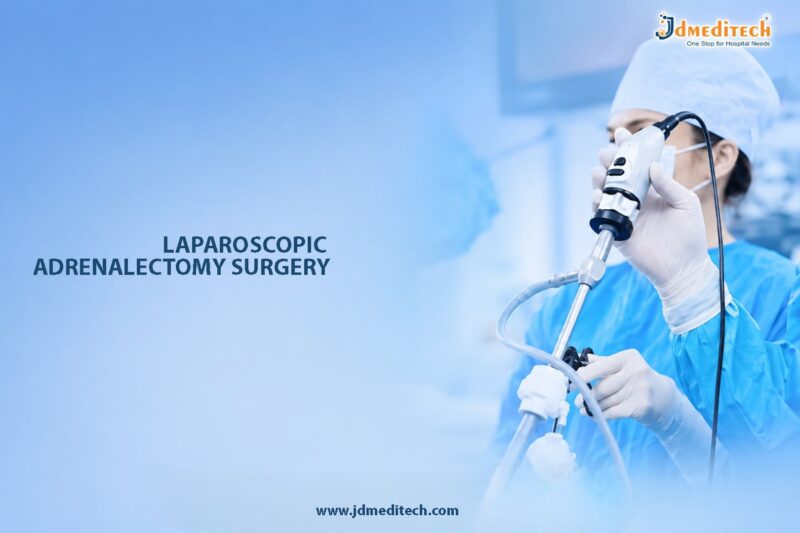
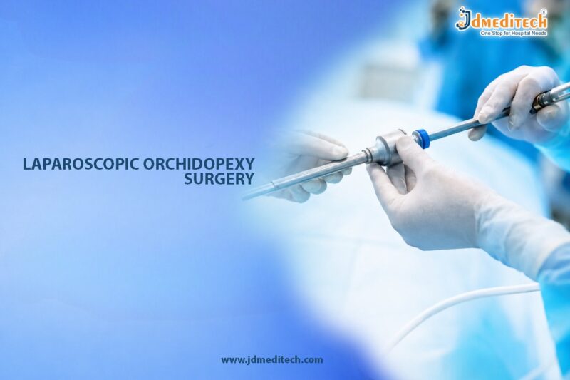
Laparoscopic orchidopexy surgery is a modern minimally invasive procedure performed to treat undescended testes, also known as cryptorchidism. Normally, the testicles descend into the scrotum before birth or during early infancy. However, in some children, one or both testes fail to move into the correct position. Consequently, surgical correction becomes necessary to protect fertility, hormonal function, and long-term testicular health.

Today, laparoscopic orchidopexy is widely preferred because it offers excellent visualization, precise surgical correction, smaller incisions, and faster recovery. Moreover, this advanced endoscopic technique allows surgeons to accurately locate and reposition the undescended testis while minimizing surgical trauma.

What Is Laparoscopic Orchidopexy Surgery?

Laparoscopic orchidopexy surgery is a minimally invasive urologic procedure used to locate, mobilize, and fix an undescended testicle inside the scrotum. The surgery is performed using a laparoscope, which is a thin camera-equipped instrument inserted through small abdominal incisions.

During the procedure, specialized surgical instruments are used to carefully free the testis and bring it down into the scrotum. Afterward, the testicle is securely fixed in place to prevent future retraction.

As a result, laparoscopic orchidopexy helps restore normal anatomical positioning and supports proper testicular development.

Why Is Laparoscopic Orchidopexy Performed?

Undescended testes may lead to several complications if left untreated. Therefore, early surgical correction is strongly recommended.

Common Reasons for Surgery Include:
  • Undescended testicle in infants or children
  • Intra-abdominal testis
  • Non-palpable testis
  • Prevention of infertility
  • Reduction of testicular cancer risk
  • Prevention of testicular torsion
  • Cosmetic and psychological concerns

In addition, timely surgery can improve long-term reproductive and hormonal outcomes.

Understanding Undescended Testicles

An undescended testicle occurs when the testis does not properly move from the abdomen into the scrotum during fetal development.

Types of Undescended Testes
Palpable Undescended Testis

In this condition, the testicle can be felt in the groin during physical examination.

Non-Palpable Testis

Here, the testicle cannot be felt because it may remain inside the abdomen or may be absent altogether.

Intra-Abdominal Testis

This type refers to a testicle located within the abdominal cavity, often requiring laparoscopic management.

Therefore, laparoscopy plays an important role in both diagnosis and treatment.

Who Is a Suitable Candidate for Laparoscopic Orchidopexy?

Generally, laparoscopic orchidopexy may be recommended for:

  • Infants older than 6 months with undescended testes
  • Children with non-palpable testes
  • Patients with intra-abdominal testes
  • Individuals with failed previous orchidopexy
  • Adolescents or adults with selected undescended testicular conditions

Importantly, early intervention usually provides better fertility preservation and improved testicular development.

Preoperative Evaluation Before Surgery

Before the procedure, the urologist performs a thorough clinical assessment to determine the exact location and condition of the testis.

Medical History Review

Initially, the surgeon evaluates:

  • Birth history
  • Developmental milestones
  • Previous surgeries
  • Family medical history
  • Associated congenital conditions
Physical Examination

Furthermore, careful examination helps determine whether the testicle is palpable or non-palpable.

Imaging Studies

In selected cases, imaging tests may include:

  • Ultrasound
  • MRI scan
  • Diagnostic laparoscopy

Consequently, these investigations help guide surgical planning.

How Laparoscopic Orchidopexy Surgery Is Performed

Typically, laparoscopic orchidopexy is performed under general anesthesia using advanced minimally invasive surgical equipment.

Step-by-Step Surgical Procedure
1. Creation of Small Incisions

First, the surgeon creates tiny incisions in the abdomen for insertion of the laparoscope and surgical instruments.

2. Identification of the Testicle

Next, the laparoscope provides magnified visualization to accurately locate the undescended testis.

3. Mobilization of the Testis

Afterward, surrounding tissues are carefully released to gain sufficient length for testicular descent.

4. Testicular Descent

Then, the testis is gently brought down into the scrotum without compromising blood supply.

5. Testicular Fixation

Finally, the testicle is securely fixed inside the scrotum to maintain proper positioning.

Overall, the surgery usually takes between 1 and 2 hours depending on surgical complexity.

Single-Stage vs Two-Stage Orchidopexy

In some patients, especially those with high intra-abdominal testes, a staged approach may be necessary.

Feature Single-Stage Orchidopexy Two-Stage Orchidopexy
Procedure Timing One operation Two separate operations
Complexity Lower Higher
Use Low-lying testes High intra-abdominal testes
Recovery Faster Gradual correction

Ultimately, the surgeon selects the most suitable approach based on testicular position and vascular length.

Benefits of Laparoscopic Orchidopexy Surgery

Laparoscopic orchidopexy offers several important advantages over traditional open surgery.

Minimally Invasive Technique

Because the procedure uses small incisions, postoperative pain and scarring are significantly reduced.

Better Visualization

Additionally, laparoscopy provides magnified internal views, allowing precise identification of the testis and surrounding structures.

Faster Recovery

Most patients recover quickly and resume normal activities sooner compared to open surgery.

Improved Cosmetic Results

Smaller incisions produce minimal visible scarring.

Fertility Preservation

Early correction supports normal testicular development and may improve future fertility potential.

Reduced Cancer Risk

Furthermore, placing the testicle in the scrotum allows easier monitoring for future abnormalities.

Recovery After Laparoscopic Orchidopexy

In most cases, recovery after laparoscopic orchidopexy is smooth and uncomplicated.

Immediate Postoperative Recovery

Initially, patients may experience:

  • Mild abdominal discomfort
  • Temporary swelling
  • Minor bruising
  • Mild fatigue

However, symptoms generally improve within a few days.

Recovery Guidelines

To ensure proper healing, patients are advised to:

  • Avoid strenuous activities temporarily
  • Maintain incision hygiene
  • Attend follow-up appointments
  • Wear comfortable clothing
  • Monitor for signs of infection

As a result, most children return to routine activities within 1 to 2 weeks.

Success Rates of Laparoscopic Orchidopexy

Laparoscopic orchidopexy has high success rates when performed by experienced pediatric urologists or laparoscopic surgeons.

Factors Influencing Success

Several factors affect outcomes, including:

  • Testicular location
  • Patient age
  • Blood supply to the testis
  • Surgical expertise
  • Presence of associated abnormalities
General Outcomes

Modern laparoscopic orchidopexy typically achieves:

  • Excellent testicular positioning
  • High long-term success rates
  • Improved fertility preservation
  • Reduced complication rates

Therefore, early diagnosis and treatment are strongly encouraged.

Risks and Complications

Although laparoscopic orchidopexy is considered safe, certain complications may still occur in rare cases.

Possible Complications Include:
  • Bleeding
  • Infection
  • Testicular atrophy
  • Recurrence of undescended testis
  • Injury to surrounding structures
  • Anesthesia-related risks

Nevertheless, complications are uncommon when surgery is performed by experienced specialists.

Long-Term Follow-Up After Orchidopexy

Long-term monitoring remains important even after successful surgery.

Follow-Up May Include:
  • Physical examination
  • Monitoring testicular growth
  • Assessment of testicular position
  • Evaluation during puberty
  • Fertility assessment in adulthood

Consequently, regular follow-up helps ensure proper long-term outcomes.

Choosing the Right Surgeon for Laparoscopic Orchidopexy

Selecting an experienced surgeon plays a major role in achieving successful surgical and functional results.

Patients Should Look For:
  • Expertise in pediatric urology
  • Advanced laparoscopic training
  • Experience with minimally invasive surgery
  • Modern surgical facilities
  • Comprehensive postoperative care

Ultimately, specialized surgical expertise improves both safety and treatment success.

Conclusion

Laparoscopic orchidopexy surgery is an advanced minimally invasive solution for correcting undescended testes and restoring proper testicular positioning. By combining precise endoscopic visualization with modern surgical techniques, the procedure offers excellent outcomes with minimal discomfort and faster recovery.

Moreover, early treatment helps preserve fertility potential, supports healthy testicular development, and reduces future complications. Therefore, timely consultation with an experienced urologist or pediatric surgeon is essential for achieving the best long-term results.


Explore More Product >>

Get Connected:
+91 79909 93062 | +91 63513 72032 | exports@jdmeditech.com 

Testicular Sperm Extraction Surgery – Advanced TESE Procedure for Male Infertility

Testicular Sperm Extraction (TESE) surgery is a specialized male infertility procedure performed to retrieve sperm directly from the testicular tissue in men with azoospermia. Azoospermia is a condition in which no sperm are present in the semen, making natural conception difficult or impossible. However, with advancements in reproductive medicine and microsurgical techniques, TESE surgery has become an effective fertility solution for many couples.

Today, TESE is widely used in men with both obstructive azoospermia and non-obstructive azoospermia. Moreover, the procedure is often combined with assisted reproductive technologies such as Intracytoplasmic Sperm Injection (ICSI) to improve the chances of successful fertilization and pregnancy.

Because TESE allows direct sperm retrieval from the testes, it offers hope to men who previously had limited fertility treatment options.

What Is Testicular Sperm Extraction Surgery?

Testicular Sperm Extraction surgery is a minor surgical procedure in which small samples of testicular tissue are removed to identify and retrieve viable sperm cells. Subsequently, the retrieved sperm can be used for fertility treatments such as IVF and ICSI.

Unlike natural ejaculation, TESE bypasses potential blockages or sperm production limitations by obtaining sperm directly from the testicular tissue. Therefore, the procedure is considered one of the most important surgical options in modern male infertility management.

In addition, TESE may be performed as a standalone procedure or combined with microscopic sperm retrieval techniques depending on the underlying condition.

Understanding Azoospermia

Azoospermia refers to the complete absence of sperm in the ejaculate. Generally, the condition is classified into two major categories.

Obstructive Azoospermia

In obstructive azoospermia, sperm production inside the testes is usually normal. However, a blockage in the reproductive tract prevents sperm from entering the semen.

Common Causes Include:
  • Previous vasectomy
  • Congenital absence of the vas deferens
  • Infection-related blockage
  • Ejaculatory duct obstruction
  • Prior pelvic surgery
Non-Obstructive Azoospermia

In non-obstructive azoospermia, sperm production is severely impaired or absent due to testicular dysfunction.

Common Causes Include:
  • Genetic abnormalities
  • Hormonal disorders
  • Testicular failure
  • Varicocele-related damage
  • Chemotherapy or radiation exposure

Therefore, TESE plays a critical role in evaluating and managing both conditions.

Why Is TESE Surgery Performed?

TESE surgery is recommended when sperm cannot be obtained through normal ejaculation. Consequently, direct sperm retrieval becomes necessary for fertility treatment.

Common Indications for TESE Include:
  • Obstructive azoospermia
  • Non-obstructive azoospermia
  • Failed vasectomy reversal
  • Ejaculatory disorders
  • Prior unsuccessful sperm retrieval procedures
  • Fertility preservation before cancer treatment

Additionally, TESE may help couples achieve biological parenthood using assisted reproductive technologies.

Who Is a Suitable Candidate for TESE Surgery?

Generally, TESE surgery may be recommended for:

  • Men diagnosed with azoospermia
  • Patients with blocked reproductive ducts
  • Individuals with impaired sperm production
  • Men undergoing IVF-ICSI treatment
  • Patients unable to produce ejaculated sperm

Importantly, a complete fertility evaluation is necessary before deciding on surgical sperm retrieval.

Preoperative Evaluation Before TESE

Before surgery, fertility specialists and urologists perform detailed assessments to identify the cause of azoospermia and estimate sperm retrieval success.

Medical History Review

Initially, the doctor evaluates:

  • Fertility history
  • Prior surgeries
  • Hormonal disorders
  • Childhood testicular conditions
  • Medication history
  • Lifestyle factors
Physical Examination

Furthermore, examination helps assess:

  • Testicular size
  • Presence of varicocele
  • Epididymal abnormalities
  • Secondary sexual characteristics
Laboratory Investigations

Diagnostic tests may include:

  • Semen analysis
  • Hormonal profile
  • Genetic testing
  • Ultrasound imaging

Consequently, these evaluations guide treatment planning and patient counseling.

Types of Testicular Sperm Retrieval Procedures

Several sperm retrieval techniques are available depending on the patient’s condition and fertility goals.

Conventional TESE

In this standard technique, small testicular tissue samples are surgically removed to search for sperm.

Micro-TESE

Microdissection TESE uses an operating microscope to identify seminiferous tubules more likely to contain sperm. Therefore, this technique improves sperm retrieval rates in men with non-obstructive azoospermia.

TESA (Testicular Sperm Aspiration)

TESA involves needle aspiration of testicular tissue and is less invasive than open surgical TESE.

PESA (Percutaneous Epididymal Sperm Aspiration)

PESA retrieves sperm from the epididymis and is commonly used in obstructive azoospermia.

Ultimately, the fertility specialist selects the most suitable procedure based on the underlying diagnosis.

How Testicular Sperm Extraction Surgery Is Performed

Typically, TESE surgery is performed under local anesthesia, sedation, or general anesthesia.

Step-by-Step Surgical Procedure
1. Surgical Preparation

First, the scrotal area is cleaned and prepared under sterile conditions.

2. Small Incision

Next, a small incision is made in the scrotum to access the testicular tissue.

3. Tissue Extraction

Afterward, tiny samples of testicular tissue are carefully removed.

4. Laboratory Examination

The tissue samples are immediately examined under a microscope to identify viable sperm cells.

5. Sperm Preservation

If sperm are successfully retrieved, they may be used immediately for IVF-ICSI or frozen for future use.

6. Closure

Finally, the incision is closed with absorbable sutures.

Overall, the procedure usually takes between 30 minutes and 1 hour.

Benefits of Testicular Sperm Extraction Surgery

TESE surgery offers several important benefits for infertile couples.

Direct Sperm Retrieval

Most importantly, TESE allows sperm retrieval even when no sperm are present in the ejaculate.

Opportunity for Biological Parenthood

Additionally, TESE enables many couples to achieve pregnancy using their own genetic material.

Minimally Invasive Options

Modern sperm retrieval techniques minimize tissue damage and improve recovery.

High Success with Assisted Reproduction

When combined with ICSI, TESE significantly improves fertilization opportunities.

Fertility Preservation

Furthermore, TESE allows sperm freezing before treatments such as chemotherapy.

Recovery After TESE Surgery

Recovery after TESE is generally quick and uncomplicated.

Common Postoperative Symptoms

Patients may temporarily experience:

  • Mild scrotal swelling
  • Minor discomfort
  • Bruising
  • Temporary tenderness

However, symptoms usually improve within a few days.

Recovery Guidelines

To promote proper healing, patients are advised to:

  • Wear scrotal support garments
  • Avoid heavy lifting
  • Limit strenuous activity temporarily
  • Maintain proper hygiene
  • Follow prescribed medications

As a result, most men resume normal activities within several days.

Success Rates of TESE Surgery

Success rates depend on multiple factors, including the underlying cause of azoospermia and the surgical technique used.

Factors Affecting Success

Several important factors include:

  • Type of azoospermia
  • Testicular function
  • Hormonal balance
  • Genetic conditions
  • Surgeon expertise
General Success Outcomes

In obstructive azoospermia, sperm retrieval rates are usually very high. Meanwhile, retrieval rates in non-obstructive azoospermia vary depending on sperm production capacity.

Nevertheless, micro-TESE has significantly improved outcomes in complex infertility cases.

Risks and Complications of TESE Surgery

Although TESE is considered safe, certain complications may occur in rare cases.

Possible Risks Include:
  • Bleeding
  • Infection
  • Scrotal hematoma
  • Temporary discomfort
  • Testicular injury
  • Reduced testosterone production in rare situations

Therefore, choosing an experienced fertility surgeon is essential for minimizing risks.

TESE vs Other Sperm Retrieval Techniques
Procedure Source of Sperm Common Use
TESE Testicular tissue Obstructive and non-obstructive azoospermia
Micro-TESE Microscopic testicular extraction Severe non-obstructive azoospermia
TESA Needle aspiration from testis Obstructive azoospermia
PESA Epididymal aspiration Ductal obstruction

Ultimately, treatment selection depends on the patient’s fertility diagnosis and reproductive goals.

Emotional and Psychological Aspects of Male Infertility

Male infertility can significantly affect emotional well-being and self-confidence. Therefore, psychological support and counseling may be beneficial throughout fertility treatment.

Helpful Support Measures Include:
  • Fertility counseling
  • Partner communication
  • Stress management
  • Support groups
  • Psychological therapy when needed

Consequently, emotional care plays an important role in overall fertility treatment success.

Choosing the Right Fertility Specialist

Successful TESE outcomes require specialized expertise in male infertility and microsurgery.

Patients Should Look For:
  • Experienced reproductive urologists
  • Advanced fertility centers
  • Access to IVF-ICSI facilities
  • Expertise in micro-TESE
  • Comprehensive fertility counseling

Ultimately, specialized care improves both sperm retrieval success and reproductive outcomes.

Conclusion

Testicular Sperm Extraction surgery is an advanced fertility procedure that offers hope to men with obstructive and non-obstructive azoospermia. By retrieving sperm directly from the testes, TESE enables many couples to pursue biological parenthood through assisted reproductive technologies.

Moreover, modern microsurgical advancements have improved sperm retrieval rates, reduced complications, and enhanced fertility outcomes. Therefore, early consultation with an experienced reproductive urologist is essential for selecting the most appropriate fertility treatment strategy.

With proper evaluation, expert surgical care, and advanced reproductive support, TESE surgery continues to play a major role in modern male infertility management.


Explore More Product >>

Get Connected:
+91 79909 93062 | +91 63513 72032 | exports@jdmeditech.com

Vasovasostomy Surgery – Advanced Vasectomy Reversal for Male Fertility Restoration

Vasovasostomy surgery is a highly specialized microsurgical procedure designed to restore fertility in men who have previously undergone a vasectomy. Over time, however, personal circumstances and family planning goals may change. As a result, many men seek effective options to regain their reproductive potential. Fortunately, vasovasostomy offers a reliable solution by reconnecting the vas deferens, thereby allowing sperm to once again travel naturally into the semen.

Moreover, advances in modern microsurgical techniques have significantly improved success rates, reduced recovery time, and enhanced the chances of natural conception. Therefore, this procedure is now considered one of the most effective fertility restoration treatments in modern urology.

What Is Vasovasostomy Surgery?

Vasovasostomy surgery, commonly known as vasectomy reversal surgery, is a microsurgical procedure used to reconnect the severed ends of the vas deferens after a vasectomy. Essentially, the vas deferens are the tubes responsible for transporting sperm from the testicles into the semen.

During a vasectomy, these tubes are intentionally cut or sealed to prevent sperm from reaching the ejaculate. Subsequently, vasovasostomy reverses this process by carefully rejoining the separated ends using advanced microscopic surgical techniques.

As a result, sperm can once again flow naturally through the reproductive tract. Ultimately, the primary goal of the procedure is to restore fertility and improve the possibility of achieving natural pregnancy.

Why Is Vasovasostomy Surgery Performed?

Men may choose vasovasostomy surgery for several personal and medical reasons. In many cases, life circumstances evolve after vasectomy. Therefore, fertility restoration becomes an important consideration.

Common Reasons Include:
  • Desire to have additional children
  • Remarriage or new family planning goals
  • Loss of a child
  • Change in personal lifestyle preferences
  • Chronic post-vasectomy pain in selected patients

In addition, vasovasostomy offers the opportunity for natural conception without relying entirely on assisted reproductive techniques.

Who Is a Suitable Candidate for Vasovasostomy?

Generally, vasovasostomy surgery may be recommended for men who:

  • Previously underwent vasectomy
  • Wish to restore fertility
  • Have healthy sperm production
  • Have a partner with acceptable fertility potential
  • Are medically fit for surgery

Importantly, success rates are often higher when the reversal is performed within 10 years of vasectomy. Nevertheless, many men still achieve favorable outcomes even after longer periods.

Preoperative Evaluation Before Vasovasostomy

Before surgery, the urologist performs a detailed assessment to determine whether the procedure is likely to succeed. Additionally, preoperative evaluation helps identify any factors that may influence fertility outcomes.

Medical History Review

Initially, the surgeon reviews:

  • Time since vasectomy
  • Previous fertility history
  • Prior scrotal surgeries
  • Existing medical conditions
  • Current medications and lifestyle habits
Physical Examination

Furthermore, a careful genital examination helps assess:

  • Testicular size and consistency
  • Presence of scar tissue
  • Condition of the vas deferens
Fertility Assessment

In some situations, the female partner may also undergo fertility evaluation. Consequently, this allows the couple to better understand their overall chances of conception.

How Vasovasostomy Surgery Is Performed

Typically, vasovasostomy is performed under general or regional anesthesia using an operating microscope for enhanced precision.

Step-by-Step Surgical Procedure
1. Surgical Exposure

First, the surgeon makes small incisions in the scrotum to access the previously divided vas deferens.

2. Identification of Vasal Ends

Next, both ends of the vas deferens are carefully identified and prepared for reconnection.

3. Fluid Examination

Afterward, fluid from the testicular side is examined under magnification for the presence of sperm. Based on these findings, the surgeon determines the most appropriate reconstruction technique.

4. Microsurgical Reconnection

Then, using ultra-fine sutures and microscopic magnification, the two ends of the vas deferens are meticulously reconnected.

5. Closure

Finally, the incisions are closed with absorbable sutures to promote proper healing.

Overall, the procedure usually takes between 2 and 4 hours depending on surgical complexity.

Vasovasostomy vs Vasoepididymostomy

Sometimes, long-standing vasectomy can lead to blockage closer to the epididymis. In such cases, sperm may not be found in the vasal fluid. Therefore, a more advanced procedure called vasoepididymostomy may be necessary.

Feature Vasovasostomy Vasoepididymostomy
Surgical Site Vas deferens Epididymis to vas deferens
Complexity Moderate Highly complex
Success Rate Higher Slightly lower
Common Use Standard vasectomy reversal Long-term obstruction

Ultimately, the final decision is often made during surgery based on microscopic findings.

Benefits of Vasovasostomy Surgery

Vasovasostomy provides several important advantages for couples seeking fertility restoration.

Restoration of Natural Fertility

Most importantly, the surgery allows sperm to return naturally to the semen, thereby enabling natural conception.

High Success Rates

Additionally, modern microsurgical techniques significantly improve sperm return rates and fertility outcomes.

Minimally Invasive Procedure

Because the surgery involves small incisions and precise techniques, tissue damage is minimized and recovery is generally faster.

Long-Term Fertility Potential

Unlike temporary fertility treatments, vasovasostomy may provide ongoing reproductive potential for future pregnancies.

Emotional and Psychological Benefits

Furthermore, restoring fertility can improve emotional well-being and strengthen long-term family planning confidence.

Recovery After Vasovasostomy Surgery

In most cases, recovery is smooth when postoperative instructions are followed carefully.

Immediate Postoperative Recovery

Initially, patients may experience:

  • Mild scrotal swelling
  • Minor discomfort
  • Temporary bruising

However, these symptoms usually improve within a few days.

Recovery Guidelines

To ensure proper healing, patients are generally advised to:

  • Wear scrotal support garments
  • Avoid strenuous physical activity
  • Refrain from heavy lifting
  • Maintain proper wound hygiene
  • Avoid sexual activity temporarily

As a result, most men can return to office-based work within several days.

When Does Fertility Return After Surgery?

Following surgery, sperm may reappear in the semen within several weeks to several months. However, recovery timelines can vary from patient to patient.

Therefore, regular semen analysis is performed to monitor:

  • Sperm count
  • Sperm motility
  • Semen quality

Once sperm flow is adequately restored, natural pregnancy may occur over time.

Success Rates of Vasovasostomy Surgery

Several important factors influence the success of vasovasostomy surgery.

Factors Affecting Success Include:
  • Time since vasectomy
  • Surgical technique
  • Surgeon expertise
  • Presence of secondary obstruction
  • Female partner fertility
General Outcome Statistics

Thanks to advanced microsurgical methods, modern vasovasostomy may achieve:

  • Sperm return rates exceeding 85%
  • Pregnancy rates ranging from 40% to 70%

Therefore, early consultation with a fertility-focused urologist is highly beneficial.

Possible Risks and Complications

Although vasovasostomy is generally considered safe, some risks may still occur.

Potential Complications Include:
  • Bleeding
  • Infection
  • Scrotal hematoma
  • Persistent discomfort
  • Scar tissue formation
  • Recurrent blockage

Nevertheless, choosing an experienced microsurgical surgeon can significantly reduce complication risks and improve overall outcomes.

Lifestyle Tips to Improve Fertility After Vasovasostomy

Healthy lifestyle habits can further support fertility restoration and sperm quality after surgery.

Recommended Lifestyle Measures

Patients are encouraged to:

  • Maintain a healthy body weight
  • Avoid smoking and excessive alcohol
  • Follow a balanced diet rich in antioxidants
  • Reduce stress levels
  • Avoid excessive heat exposure to the testes

Consequently, these measures may positively influence reproductive health and postoperative fertility outcomes.

Alternatives to Vasovasostomy Surgery

In certain situations, assisted reproductive techniques may also be considered.

Alternative Fertility Options
  • Sperm retrieval procedures
  • In vitro fertilization (IVF)
  • Intracytoplasmic sperm injection (ICSI)

However, vasovasostomy often remains the preferred option for couples seeking natural conception whenever feasible.

Choosing the Right Urologic Surgeon

Successful vasovasostomy requires advanced microsurgical expertise. Therefore, selecting the right surgeon is extremely important.

Patients Should Look For:
  • Fellowship-trained urologists
  • Experience in male infertility surgery
  • Specialized microsurgical expertise
  • Advanced surgical facilities
  • Comprehensive fertility counseling

Ultimately, experienced surgeons can significantly improve both surgical precision and fertility outcomes.

Conclusion

Vasovasostomy surgery is an advanced microsurgical solution for men seeking fertility restoration after vasectomy. By precisely reconnecting the vas deferens, the procedure helps restore natural sperm flow and improves the chances of conception.

Moreover, modern surgical advancements have made vasovasostomy safer, more effective, and increasingly successful. With proper patient selection, expert surgical care, and appropriate follow-up, many couples can successfully achieve pregnancy naturally.

Therefore, men considering fertility restoration should consult an experienced urologist to explore the most suitable treatment approach and maximize reproductive success.


Explore More Product >>

Get Connected:
+91 79909 93062 | +91 63513 72032 | exports@jdmeditech.com 

Vasoepididymostomy Surgery for Male Fertility Restoration

Vasoepididymostomy Surgery is a highly specialized microsurgical procedure performed to restore male fertility in men with epididymal blockage or obstruction. The surgery reconnects the vas deferens directly to the epididymis, allowing sperm to travel normally from the testicles into the semen.

This advanced fertility-restoration procedure is commonly recommended when sperm blockage cannot be corrected through standard vasectomy reversal techniques. Fortunately, modern microsurgical methods provide excellent precision, improved sperm flow, and higher chances of natural conception.

Moreover, vasoepididymostomy is considered one of the most delicate and technically advanced procedures in male reproductive microsurgery.

What Is Vasoepididymostomy Surgery?

Vasoepididymostomy is a microsurgical reconstructive procedure designed to bypass an obstruction in the epididymis. During surgery, the surgeon connects the vas deferens directly to a tiny epididymal tubule carrying sperm.

As a result, sperm can once again enter the semen naturally.

Unlike standard vasovasostomy, which reconnects the two ends of the vas deferens, vasoepididymostomy is performed when blockage exists closer to the epididymis.

Why Is Vasoepididymostomy Performed?

Doctors recommend this procedure when sperm transport is blocked due to epididymal obstruction.

Common Reasons for Epididymal Obstruction
  • Failed vasectomy reversal
  • Long-standing vasectomy
  • Infection-related scarring
  • Congenital reproductive tract blockage
  • Trauma or injury
  • Previous scrotal surgery
  • Inflammation of the epididymis

Therefore, accurate diagnosis is essential before planning surgery.

Symptoms and Signs of Obstructive Male Infertility

Many men with epididymal obstruction have no noticeable symptoms other than infertility.

Common Clinical Findings Include
  • Absence of sperm in semen (azoospermia)
  • Normal testosterone levels
  • Normal sexual function
  • History of vasectomy or infection
  • Testicular fullness in some cases

Since symptoms may be minimal, fertility evaluation plays a crucial role in diagnosis.

Diagnostic Evaluation Before Surgery

Before surgery, a detailed fertility assessment is performed to confirm obstruction and evaluate reproductive health.

Common Diagnostic Tests
Semen Analysis

Semen testing helps detect:

  • Absence of sperm
  • Semen volume abnormalities
  • Signs of obstruction
Hormone Testing

Blood tests evaluate:

  • Testosterone levels
  • Follicle-stimulating hormone (FSH)
  • Luteinizing hormone (LH)
Scrotal Ultrasound

Ultrasound helps assess:

  • Epididymal enlargement
  • Testicular structure
  • Scarring or obstruction
Physical Examination

The surgeon evaluates the reproductive anatomy and identifies possible sites of blockage.

Together, these investigations help determine whether vasoepididymostomy is the appropriate treatment option.

How Vasoepididymostomy Surgery Is Performed

The procedure is performed under general or regional anesthesia using advanced microsurgical techniques.

Step-by-Step Surgical Procedure
Step 1: Administration of Anesthesia

First, anesthesia is provided to ensure patient comfort throughout the surgery.

Step 2: Surgical Exposure

Next, a small incision is made in the scrotum to expose the vas deferens and epididymis.

Step 3: Identification of Healthy Epididymal Tubule

Using a high-powered operating microscope, the surgeon carefully identifies a healthy epididymal tubule containing sperm.

Step 4: Microsurgical Connection

Then, the vas deferens is meticulously connected to the epididymal tubule using extremely fine sutures.

Step 5: Confirmation of Proper Alignment

After that, the surgeon confirms accurate alignment to allow proper sperm flow.

Step 6: Closure of the Incision

Finally, the incision is closed carefully after completing the reconstruction.

Because of the complexity of the procedure, microsurgical expertise is essential for optimal success.

Benefits of Vasoepididymostomy Surgery

This advanced fertility-restoration surgery offers several important advantages.

Key Benefits
  • Restoration of natural fertility
  • Improved sperm flow into semen
  • Potential for natural conception
  • Minimally invasive microsurgical approach
  • High surgical precision
  • Preservation of reproductive function
  • Alternative to assisted reproductive techniques

Additionally, successful surgery may reduce the need for repeated fertility treatments.

Recovery After Vasoepididymostomy Surgery

Recovery is generally smooth, although proper postoperative care remains important.

What to Expect After Surgery
  • Mild scrotal discomfort
  • Temporary swelling or bruising
  • Gradual return to daily activities
  • Improvement in semen quality over time

Most patients resume non-strenuous activities within several days. However, heavy lifting and sexual activity should be avoided temporarily as advised by the surgeon.

Follow-Up and Fertility Monitoring

Regular follow-up is essential after surgery.

Postoperative Monitoring May Include
  • Semen analysis
  • Fertility evaluation
  • Physical examination
  • Assessment of sperm count and motility

Importantly, sperm may take several months to reappear in the semen after successful reconstruction.

Possible Risks and Complications

Although vasoepididymostomy is considered safe, rare complications may occur.

Potential Risks Include
  • Bleeding
  • Infection
  • Persistent obstruction
  • Scarring
  • Fluid collection
  • Failure to restore sperm flow
  • Chronic discomfort in rare cases

Nevertheless, experienced microsurgeons significantly improve the chances of successful outcomes.

Success Rates of Vasoepididymostomy

Success depends on:

  • Duration of obstruction
  • Surgical expertise
  • Overall reproductive health
  • Presence of healthy sperm production

In many cases, modern microsurgical techniques achieve favorable fertility restoration and improved pregnancy rates.

Furthermore, earlier treatment may increase the likelihood of successful sperm return.

Long-Term Outlook After Surgery

Many patients experience restoration of sperm flow and improved fertility potential after successful surgery.

Long-term benefits may include:

  • Natural conception opportunities
  • Improved reproductive confidence
  • Avoidance of complex fertility procedures
  • Enhanced quality of life

Therefore, vasoepididymostomy remains an important option for selected men with obstructive infertility.

When to Consult a Fertility Specialist

Medical evaluation is recommended if:

  • Pregnancy does not occur despite regular attempts
  • Semen analysis shows azoospermia
  • There is a history of vasectomy or scrotal surgery
  • Previous vasectomy reversal has failed
  • Male infertility concerns persist

Early fertility assessment can help identify the most appropriate treatment approach.

Conclusion

Vasoepididymostomy Surgery is a highly advanced microsurgical procedure designed to restore fertility in men with epididymal obstruction. By reconnecting the vas deferens directly to the epididymis, the surgery enables natural sperm transport and improves the possibility of conception.

Moreover, modern microsurgical techniques provide exceptional precision, improved reproductive outcomes, and long-term fertility benefits. Therefore, men experiencing obstructive infertility should consult an experienced male fertility specialist to explore whether vasoepididymostomy is the right treatment option.


Explore More Product >>

Get Connected:
+91 79909 93062 | +91 63513 72032 | exports@jdmeditech.com 

Vasectomy Surgery for Reliable Permanent Male Contraception

Vasectomy Surgery is a safe, effective, and minimally invasive male sterilization procedure designed to provide permanent contraception. The surgery prevents sperm from mixing with semen by blocking or cutting the vas deferens, the tubes responsible for carrying sperm from the testicles.

As a highly reliable birth control method, vasectomy is widely chosen by men seeking a long-term contraceptive solution. Moreover, the procedure is quick, usually performed on an outpatient basis, and associated with minimal recovery time.

Compared to temporary contraceptive methods, vasectomy offers long-lasting protection with a very high success rate.

What Is Vasectomy Surgery?

Vasectomy is a minor surgical procedure that permanently interrupts the transport of sperm. During ejaculation, semen is still produced normally; however, it no longer contains sperm capable of fertilization.

Importantly, vasectomy does not affect testosterone production, sexual performance, libido, or the ability to achieve erections and orgasm.

How Vasectomy Prevents Pregnancy

The procedure works by sealing, cutting, or blocking the vas deferens on both sides. Consequently, sperm cannot travel into the semen released during ejaculation.

Over time, the body naturally absorbs the unused sperm without causing harm.

Who Should Consider Vasectomy Surgery?

Vasectomy is recommended for men who are certain they do not want biological children in the future.

Common Reasons for Choosing Vasectomy
  • Desire for permanent contraception
  • Completion of family planning
  • Avoidance of female sterilization procedures
  • Preference for a highly effective birth control method
  • Medical reasons preventing pregnancy in a partner

Therefore, careful consideration and counseling are essential before undergoing the procedure.

Types of Vasectomy Procedures

Several surgical techniques may be used depending on the surgeon’s preference and the patient’s condition.

Conventional Vasectomy

In this method, small incisions are made in the scrotum to access and seal the vas deferens.

No-Scalpel Vasectomy

This modern technique uses a tiny puncture instead of surgical cuts. As a result, patients often experience:

  • Less bleeding
  • Reduced discomfort
  • Faster healing
  • Smaller scars

Today, no-scalpel vasectomy is one of the most commonly performed techniques worldwide.

Diagnostic Evaluation Before Surgery

Before surgery, the doctor performs a detailed consultation and physical examination.

Preoperative Assessment May Include
  • Medical history review
  • Physical examination
  • Discussion of permanent contraception
  • Review of medications
  • Counseling regarding postoperative expectations

Additionally, patients receive instructions regarding preparation and recovery.

How Vasectomy Surgery Is Performed

The procedure is generally completed within 20 to 30 minutes under local anesthesia.

Step-by-Step Surgical Procedure
Step 1: Administration of Local Anesthesia

First, local anesthesia is injected into the scrotal area to numb the surgical site.

Step 2: Accessing the Vas Deferens

Next, the surgeon identifies and gently brings the vas deferens to the surface.

Step 3: Cutting or Blocking the Vas Deferens

Then, the vas deferens is cut, sealed, clipped, or cauterized to stop sperm transport.

Step 4: Completion of the Procedure

Finally, the incision or puncture site is closed, and the patient is monitored briefly before discharge.

Since the procedure is minimally invasive, most patients return home the same day.

Benefits of Vasectomy Surgery

Vasectomy offers numerous advantages as a long-term contraceptive solution.

Key Benefits
  • Highly effective permanent contraception
  • Minimally invasive procedure
  • Quick surgical time
  • Outpatient treatment
  • Minimal recovery period
  • No effect on sexual performance
  • Cost-effective long-term birth control
  • Lower complication rates compared to female sterilization

Furthermore, vasectomy eliminates the need for ongoing contraceptive management.

Recovery After Vasectomy Surgery

Recovery is typically smooth and uncomplicated.

What to Expect After Surgery
  • Mild swelling or bruising
  • Minor discomfort for a few days
  • Temporary scrotal soreness
  • Ability to walk shortly after surgery

Most patients resume light activities within 24 to 48 hours. However, strenuous exercise should be avoided for about one week.

Postoperative Care Tips
  • Apply ice packs to reduce swelling
  • Wear supportive underwear
  • Avoid heavy lifting temporarily
  • Follow prescribed medications carefully

Additionally, proper rest supports faster healing.

Important Note About Fertility After Vasectomy

Vasectomy does not provide immediate sterility. Sperm may remain in the reproductive tract for several weeks or months after surgery.

Therefore, alternative contraception should be used until semen analysis confirms the absence of sperm.

Semen Analysis After Surgery

Doctors usually recommend semen testing several weeks after the procedure to confirm successful sterilization.

Only after a clear semen analysis can vasectomy be considered fully effective.

Possible Risks and Complications

Although vasectomy is considered very safe, rare complications may occur.

Potential Risks Include
  • Mild bleeding
  • Infection
  • Swelling or bruising
  • Temporary discomfort
  • Sperm granuloma
  • Chronic scrotal pain in rare cases
  • Vasectomy failure or reconnection

Nevertheless, serious complications are uncommon when performed by experienced surgeons.

Long-Term Outlook After Vasectomy

Vasectomy provides excellent long-term contraceptive success with very high reliability.

Most men experience:

  • Permanent pregnancy prevention
  • Normal sexual function
  • Unchanged hormone levels
  • Improved peace of mind regarding contraception

Importantly, the procedure does not increase the risk of prostate cancer, sexual dysfunction, or reduced masculinity.

When to Consult a Doctor

Medical attention may be necessary if you experience:

  • Severe pain
  • Persistent swelling
  • Fever
  • Excessive bleeding
  • Signs of infection
  • Continued fertility concerns after surgery

Early evaluation helps ensure proper recovery and treatment if complications occur.

Conclusion

Vasectomy Surgery is a highly effective and minimally invasive procedure that offers reliable permanent male contraception. By safely blocking sperm transport, the surgery provides long-term pregnancy prevention without affecting sexual performance or hormone production.

Moreover, quick recovery, high success rates, and minimal surgical risks make vasectomy one of the most trusted contraceptive options for men worldwide. Therefore, men seeking permanent birth control should consult an experienced urologist to determine whether vasectomy is the right choice for their family planning goals.


Explore More Product >>

Get Connected:
+91 79909 93062 | +91 63513 72032 | exports@jdmeditech.com 

Laparoscopic Adrenalectomy Surgery for Safe Adrenal Gland Removal

Laparoscopic Adrenalectomy Surgery is an advanced minimally invasive surgical procedure performed to remove one or both adrenal glands safely and effectively. The adrenal glands are small hormone-producing glands located above the kidneys and play a vital role in regulating blood pressure, metabolism, stress response, and hormone balance.

When adrenal tumors, hormonal disorders, or abnormal gland enlargement occur, surgical removal may become necessary. Fortunately, laparoscopic adrenalectomy allows surgeons to remove the adrenal gland through small incisions while minimizing pain, blood loss, and recovery time.

Compared to traditional open surgery, this minimally invasive approach offers faster healing, shorter hospital stays, and improved cosmetic results.

What Is Laparoscopic Adrenalectomy Surgery?

Laparoscopic adrenalectomy is a keyhole surgical technique used to remove diseased or overactive adrenal glands using specialized laparoscopic instruments and a high-definition camera.

During the procedure, surgeons create small abdominal incisions to access the adrenal gland. Subsequently, the affected gland is carefully separated and removed without the need for a large open incision.

As a result, patients experience less postoperative discomfort and quicker recovery.

Why Is Adrenal Gland Removal Needed?

Doctors may recommend adrenalectomy for several adrenal gland conditions, especially when tumors produce excessive hormones or show suspicious growth.

Common Conditions Treated With Laparoscopic Adrenalectomy
  • Adrenal adenoma
  • Pheochromocytoma
  • Cushing’s syndrome
  • Conn’s syndrome (primary aldosteronism)
  • Adrenal cysts
  • Adrenal gland enlargement
  • Functional adrenal tumors
  • Suspicious or cancerous adrenal masses

In many cases, early surgical treatment helps prevent serious hormonal complications and protects overall health.

Symptoms of Adrenal Gland Disorders

Symptoms vary depending on the type of adrenal disease and hormone imbalance.

Common Symptoms Include
  • High blood pressure
  • Unexplained weight gain
  • Muscle weakness
  • Excessive sweating
  • Anxiety or rapid heartbeat
  • Persistent fatigue
  • Headaches
  • Elevated blood sugar
  • Abdominal discomfort

Therefore, proper evaluation is essential when these symptoms persist.

Diagnostic Evaluation Before Surgery

Before surgery, doctors perform detailed investigations to confirm the diagnosis and determine the safest treatment plan.

Common Diagnostic Tests
Blood Hormone Tests

These tests evaluate:

  • Cortisol levels
  • Aldosterone production
  • Adrenal hormone activity
  • Catecholamine levels
Urine Tests

Urine analysis helps detect abnormal hormone secretion over a 24-hour period.

CT Scan or MRI

Imaging studies help determine:

  • Tumor size
  • Tumor location
  • Adrenal gland anatomy
  • Possible cancer spread
Nuclear Medicine Scans

In selected cases, specialized scans assess adrenal gland activity and tumor function.

Together, these investigations help surgeons plan the procedure accurately.

How Laparoscopic Adrenalectomy Surgery Is Performed

The surgery is performed under general anesthesia by an experienced laparoscopic or urologic surgeon.

Step-by-Step Surgical Procedure
Step 1: Administration of Anesthesia

First, the patient is placed under general anesthesia to ensure complete comfort during surgery.

Step 2: Creation of Small Incisions

Next, several tiny incisions are made in the abdomen to insert laparoscopic instruments and a camera.

Step 3: Visualization of the Adrenal Gland

A high-definition laparoscope provides magnified visualization of the adrenal gland and surrounding structures.

Step 4: Careful Dissection

Then, the surgeon carefully separates the adrenal gland from nearby tissues and blood vessels.

Step 5: Removal of the Adrenal Gland

After that, the diseased adrenal gland is safely removed through one of the small incisions.

Step 6: Closure of Incisions

Finally, the instruments are removed, and the small incisions are closed carefully.

Benefits of Laparoscopic Adrenalectomy Surgery

Laparoscopic adrenalectomy offers several advantages over conventional open adrenal surgery.

Key Benefits
  • Minimally invasive approach
  • Smaller surgical scars
  • Reduced postoperative pain
  • Less blood loss
  • Faster recovery
  • Shorter hospital stay
  • Lower risk of infection
  • Quicker return to normal activities
  • Better cosmetic appearance

Additionally, laparoscopic magnification improves surgical precision and safety.

Recovery After Laparoscopic Adrenalectomy

Recovery is usually smooth, although careful monitoring remains important after surgery.

What to Expect After Surgery
  • Mild abdominal discomfort
  • Temporary fatigue
  • Early walking within 24 hours
  • Gradual return to normal diet
  • Short hospital stay in most cases

Generally, most patients resume daily activities within a few weeks.

Hormone Monitoring After Surgery

Following adrenal gland removal, hormone levels may require monitoring. In some patients, temporary hormone replacement therapy may also be necessary.

Therefore, regular follow-up with the healthcare team is essential.

Possible Risks and Complications

Although laparoscopic adrenalectomy is considered safe, some rare complications may occur.

Potential Risks Include
  • Bleeding
  • Infection
  • Injury to nearby organs
  • Blood pressure fluctuations
  • Hormonal imbalance
  • Blood clots
  • Adverse anesthesia reactions

Nevertheless, experienced surgeons take extensive precautions to minimize surgical risks.

Long-Term Outlook After Surgery

Most patients experience significant improvement in symptoms after successful adrenal gland removal.

Long-term benefits may include:

  • Better blood pressure control
  • Improved hormone balance
  • Reduced headaches and sweating
  • Improved energy levels
  • Better quality of life

Furthermore, minimally invasive surgery allows faster recovery with excellent long-term outcomes.

When to Consult a Surgeon

Medical consultation is important if you experience:

  • Persistent high blood pressure
  • Unexplained hormonal symptoms
  • Adrenal tumors detected on imaging
  • Severe headaches with sweating
  • Rapid heartbeat episodes
  • Unexplained weight changes

Early diagnosis and treatment help prevent complications related to adrenal gland disorders.

Conclusion

Laparoscopic Adrenalectomy Surgery is a highly effective minimally invasive procedure used for the safe removal of diseased adrenal glands. By utilizing advanced laparoscopic techniques, surgeons can treat adrenal tumors and hormone-related disorders with exceptional precision and reduced surgical trauma.

Moreover, smaller incisions, quicker recovery, less pain, and excellent long-term outcomes make laparoscopic adrenalectomy a preferred treatment option for many adrenal gland conditions. Therefore, timely diagnosis and expert surgical care remain essential for achieving the best possible results.


Explore More Product >>

Get Connected:
+91 79909 93062 | +91 63513 72032 | exports@jdmeditech.com 

Laparoscopic Pyeloplasty Surgery for Safe Renal Reconstruction

Laparoscopic Pyeloplasty Surgery is an advanced minimally invasive urologic procedure used to treat ureteropelvic junction (UPJ) obstruction, a condition that blocks the normal flow of urine from the kidney to the ureter. As a result, this obstruction can lead to kidney swelling, pain, urinary infections, and gradual loss of kidney function if not treated promptly.

Fortunately, modern laparoscopic techniques allow surgeons to reconstruct the narrowed area with exceptional precision while minimizing surgical trauma. Compared to traditional open surgery, laparoscopic pyeloplasty offers faster recovery, reduced pain, smaller incisions, and excellent long-term success rates.

What Is Ureteropelvic Junction (UPJ) Obstruction?

UPJ obstruction occurs when the connection between the renal pelvis and the ureter becomes narrowed or blocked. Consequently, urine cannot drain properly from the kidney.

The condition may be congenital or acquired later in life due to scarring, kidney stones, infections, or previous surgeries. Therefore, timely diagnosis is essential to avoid long-term complications.

Common Symptoms of UPJ Obstruction

Symptoms can vary depending on the severity of obstruction and the patient’s age.

In Adults
  • Flank or abdominal pain
  • Pain after drinking large amounts of fluid
  • Recurrent urinary tract infections
  • Blood in urine
  • Kidney stones
  • Nausea or vomiting
In Children
  • Abdominal swelling
  • Urinary tract infections
  • Poor growth in severe cases
  • Pain or discomfort
  • Prenatal detection of hydronephrosis

Importantly, early diagnosis helps prevent permanent kidney damage and preserves renal function.

What Is Laparoscopic Pyeloplasty Surgery?

Laparoscopic pyeloplasty is a reconstructive surgery performed to remove the blocked segment at the ureteropelvic junction and reconnect the healthy portions of the kidney pelvis and ureter.

During the procedure, surgeons use small keyhole incisions and specialized laparoscopic instruments. In addition, a high-definition camera provides magnified visualization, allowing delicate reconstruction with high accuracy.

Why Is Laparoscopic Pyeloplasty Performed?

Doctors recommend laparoscopic pyeloplasty when UPJ obstruction causes symptoms, kidney swelling, or declining renal function.

Main Objectives of the Procedure
  • Restore normal urine drainage
  • Relieve kidney obstruction
  • Preserve kidney function
  • Reduce pain and infections
  • Prevent progressive kidney damage
  • Improve long-term urinary health

Without proper treatment, UPJ obstruction may eventually lead to serious renal complications. Therefore, surgical correction is often strongly recommended.

Diagnostic Evaluation Before Surgery

A detailed diagnostic assessment is necessary to confirm the obstruction and evaluate kidney function.

Common Diagnostic Tests
Ultrasound Scan

Ultrasound helps detect:

  • Hydronephrosis
  • Kidney swelling
  • Urinary tract abnormalities
CT Scan or MRI

Advanced imaging provides detailed anatomical information regarding:

  • Site of obstruction
  • Crossing blood vessels
  • Kidney structure
Renal Nuclear Scan

This test evaluates:

  • Kidney drainage
  • Differential kidney function
  • Severity of obstruction
Urine and Blood Tests

These tests assess:

  • Infection
  • Kidney function
  • Overall health status

Together, these investigations help surgeons plan the most effective treatment strategy.

How Laparoscopic Pyeloplasty Surgery Is Performed

The procedure is carried out under general anesthesia by an experienced urologic surgeon.

Step-by-Step Surgical Procedure
Step 1: Administration of Anesthesia

First, the patient is placed under general anesthesia to ensure complete comfort during surgery.

Step 2: Small Laparoscopic Incisions

Next, several tiny incisions are created in the abdomen to insert laparoscopic instruments and a camera.

Step 3: Identification of the Obstructed Area

After that, the surgeon carefully identifies the narrowed ureteropelvic junction.

Step 4: Removal of the Narrowed Segment

Then, the blocked or scarred portion is excised precisely.

Step 5: Renal Reconstruction

Subsequently, the healthy ureter is reconnected to the renal pelvis using fine sutures to create a wide drainage channel.

Step 6: Placement of Ureteral Stent

In many cases, a temporary ureteral stent is placed to support healing and maintain urine flow.

Step 7: Completion of Surgery

Finally, the instruments are removed, and the small incisions are closed carefully.

Benefits of Laparoscopic Pyeloplasty Surgery

Laparoscopic pyeloplasty offers several advantages over conventional open surgery.

Key Benefits
  • Minimally invasive technique
  • Smaller surgical incisions
  • Reduced postoperative pain
  • Minimal blood loss
  • Faster recovery
  • Shorter hospital stay
  • Lower risk of infection
  • Excellent cosmetic results
  • High long-term success rate

Moreover, magnified laparoscopic visualization improves surgical precision during reconstruction.

Recovery After Laparoscopic Pyeloplasty

Most patients recover smoothly following surgery. However, proper postoperative care remains important for optimal healing.

What to Expect After Surgery
  • Mild abdominal discomfort
  • Temporary urinary catheter in some cases
  • Early mobilization within 24 hours
  • Gradual return to normal diet

Generally, most patients can resume routine activities within a few weeks.

Ureteral Stent Removal

If a stent is placed, it is usually removed after a few weeks during a simple outpatient procedure. As a result, normal urinary drainage continues without obstruction.

Follow-Up Care

Regular follow-up may include:

  • Ultrasound imaging
  • Renal scans
  • Urine tests
  • Kidney function monitoring

These evaluations help ensure successful drainage and proper kidney recovery over time.

Possible Risks and Complications

Although laparoscopic pyeloplasty is considered safe, rare complications may still occur.

Potential Risks Include
  • Bleeding
  • Infection
  • Urine leakage
  • Stent discomfort
  • Recurrent obstruction
  • Injury to nearby structures
  • Delayed healing

Nevertheless, choosing an experienced urologic surgeon significantly reduces surgical risks.

Long-Term Outlook After Surgery

Laparoscopic pyeloplasty has an excellent long-term success rate, often exceeding 90%.

Following successful surgery, most patients experience:

  • Improved kidney drainage
  • Relief from pain
  • Reduced infection risk
  • Preservation of kidney function
  • Better quality of life

Furthermore, early treatment generally leads to superior outcomes and minimizes long-term renal complications.

When to Consult a Urologist

Medical evaluation is important if you experience:

  • Persistent flank pain
  • Recurrent urinary tract infections
  • Kidney swelling
  • Blood in urine
  • Kidney stones
  • Difficulty with urine drainage

In such cases, prompt medical attention can help prevent worsening kidney damage.

Conclusion

Laparoscopic Pyeloplasty Surgery is a highly advanced minimally invasive procedure designed to correct ureteropelvic junction obstruction safely and effectively. By performing precise renal reconstruction, the surgery restores normal urine flow, relieves symptoms, and preserves kidney function.

Additionally, smaller incisions, faster recovery, and outstanding long-term results make laparoscopic pyeloplasty a preferred treatment option for both adults and children with UPJ obstruction. Therefore, early intervention and expert surgical care play a vital role in achieving the best possible outcomes.


Explore More Product >>

Get Connected:
+91 79909 93062 | +91 63513 72032 | exports@jdmeditech.com 

Posterior Urethral Valve Ablation Surgery for Bladder Outlet Obstruction

Posterior Urethral Valve (PUV) Ablation Surgery is a specialized endoscopic urologic procedure performed to treat posterior urethral valves, a congenital condition that causes blockage in the urinary tract of male infants and children. This obstruction affects the normal flow of urine from the bladder and may lead to bladder dysfunction, recurrent urinary tract infections, kidney damage, and growth-related complications if left untreated.

With advances in pediatric urology, posterior urethral valve ablation has become a safe and highly effective minimally invasive surgery. The procedure removes the obstructing valve tissue using an endoscopic instrument, helping restore normal urine flow and protect long-term kidney function.

What Are Posterior Urethral Valves?

Posterior urethral valves are abnormal folds of tissue located in the posterior urethra, the part of the urethra closest to the bladder. These valves partially or completely block urine outflow and are present from birth.

This condition occurs only in boys and is considered one of the most common causes of urinary tract obstruction in male infants.

Common Symptoms of Posterior Urethral Valves

Symptoms may vary depending on the severity of obstruction and the child’s age.

In Newborns and Infants
  • Poor urinary stream
  • Difficulty passing urine
  • Swollen abdomen due to enlarged bladder
  • Failure to thrive
  • Fever due to urinary infection
  • Breathing difficulty caused by low amniotic fluid during pregnancy
In Older Children
  • Recurrent urinary tract infections
  • Daytime wetting or bedwetting
  • Weak urine flow
  • Frequent urination
  • Kidney function abnormalities
  • Delayed growth

Early diagnosis and timely surgery are essential to prevent permanent kidney damage.

What Is Posterior Urethral Valve Ablation Surgery?

Posterior urethral valve ablation surgery is a minimally invasive endoscopic procedure designed to remove or destroy the obstructing valve tissue inside the urethra.

During the surgery, a pediatric cystoscope is inserted through the urethral opening without making external cuts. The surgeon identifies the abnormal valve tissue and carefully ablates it using specialized instruments or energy sources.

As a result, urine can flow normally from the bladder, reducing pressure on the urinary system and kidneys.

Why Is Posterior Urethral Valve Ablation Needed?

Doctors recommend this surgery to relieve urinary obstruction and prevent complications associated with long-standing blockage.

Main Goals of the Procedure
  • Improve urine flow
  • Reduce bladder pressure
  • Protect kidney function
  • Prevent recurrent urinary infections
  • Improve bladder emptying
  • Minimize long-term urinary complications

Without treatment, posterior urethral valves can lead to chronic kidney disease and severe bladder dysfunction.

Diagnostic Evaluation Before Surgery

A detailed evaluation helps confirm the diagnosis and assess the severity of urinary obstruction.

Common Diagnostic Tests
Ultrasound Examination

Ultrasound helps evaluate:

  • Enlarged bladder
  • Thickened bladder wall
  • Kidney swelling (hydronephrosis)
  • Dilated ureters
Voiding Cystourethrogram (VCUG)

VCUG is the most important imaging test for diagnosing posterior urethral valves. It shows:

  • Dilated posterior urethra
  • Bladder abnormalities
  • Vesicoureteral reflux
Blood and Urine Tests

These tests assess:

  • Kidney function
  • Presence of infection
  • Electrolyte imbalance
Cystoscopy

Direct visualization using a cystoscope confirms the diagnosis during surgery.

How Posterior Urethral Valve Ablation Surgery Is Performed

The procedure is performed under general anesthesia by a pediatric urologist.

Step-by-Step Surgical Procedure
Step 1: Administration of Anesthesia

The child is placed under general anesthesia to ensure comfort and safety during surgery.

Step 2: Introduction of the Cystoscope

A small pediatric cystoscope is gently inserted through the urethra into the bladder.

Step 3: Identification of Valve Tissue

The surgeon carefully identifies the obstructing posterior urethral valve.

Step 4: Valve Ablation

The valve tissue is incised or ablated using:

  • Electrocautery
  • Cold knife
  • Laser energy in selected cases

This creates a wider passage for urine flow.

Step 5: Bladder Drainage

A urinary catheter may be temporarily placed to help bladder drainage after surgery.

Benefits of Posterior Urethral Valve Ablation Surgery

This minimally invasive surgery offers several important advantages.

Key Benefits
  • No external surgical incision
  • Improved urinary flow
  • Reduced bladder outlet obstruction
  • Preservation of kidney function
  • Short hospital stay
  • Faster recovery
  • Reduced risk of recurrent infections
  • Better long-term urinary health

Additionally, early intervention significantly improves long-term outcomes.

Recovery After Posterior Urethral Valve Ablation

Recovery is generally smooth, although regular follow-up remains important.

What to Expect After Surgery
  • Mild discomfort during urination for a short period
  • Temporary urinary catheter in some cases
  • Observation for urine output and kidney function
  • Antibiotics if infection is present

Most children can return home within a few days after surgery.

Follow-Up Care

Regular follow-up may include:

  • Ultrasound scans
  • Urine tests
  • Kidney function monitoring
  • Bladder function assessment

Long-term monitoring is essential because some children may continue to have bladder dysfunction even after successful valve removal.

Possible Risks and Complications

Although posterior urethral valve ablation is considered safe, certain complications may occur in rare cases.

Potential Risks Include
  • Bleeding
  • Urinary tract infection
  • Residual valve tissue
  • Urethral stricture
  • Persistent bladder dysfunction
  • Vesicoureteral reflux
  • Kidney impairment in severe cases

Experienced pediatric urologists take extensive precautions to minimize these risks.

Long-Term Outlook After Surgery

The long-term prognosis depends on:

  • Severity of obstruction
  • Degree of kidney damage before treatment
  • Age at diagnosis
  • Bladder function

Children diagnosed and treated early usually experience excellent outcomes with improved urinary function and better kidney preservation.

However, lifelong monitoring may still be necessary in some patients.

When to Consult a Pediatric Urologist

Parents should seek immediate medical attention if a child experiences:

  • Weak urinary stream
  • Difficulty urinating
  • Recurrent urinary infections
  • Swollen abdomen
  • Poor growth
  • Urinary dribbling or wetting problems

Prompt evaluation can help prevent serious complications.

Conclusion

Posterior Urethral Valve Ablation Surgery is an advanced endoscopic procedure that effectively relieves bladder outlet obstruction caused by posterior urethral valves. By restoring normal urine flow, this minimally invasive surgery helps protect kidney function, improve bladder health, and enhance the child’s overall quality of life.

Early diagnosis, expert surgical care, and regular follow-up play a crucial role in achieving successful long-term outcomes. With modern pediatric urologic techniques, most children can lead healthy and active lives after treatment.


Explore More Product >>

Get Connected:
+91 79909 93062 | +91 63513 72032 | exports@jdmeditech.com 

Vesicoureteral Reflux Surgery – Advanced Surgical Treatment for Urinary Reflux

Vesicoureteral Reflux Surgery is a specialized urologic procedure performed to correct abnormal backward flow of urine from the bladder into the ureters and kidneys. This condition, known as vesicoureteral reflux (VUR), commonly affects infants and children, although adults may also develop it in certain situations.

Normally, urine flows in one direction from the kidneys to the bladder. However, in vesicoureteral reflux, urine moves backward due to an abnormal valve mechanism at the junction of the ureter and bladder. As a result, repeated urinary tract infections, kidney damage, and long-term urinary complications may occur.

Today, modern surgical techniques provide safe and highly effective correction of urinary reflux with excellent long-term outcomes. Moreover, early intervention helps preserve kidney function and improves overall urinary health.

What is Vesicoureteral Reflux?

Vesicoureteral reflux is a condition in which urine flows backward from the bladder into one or both ureters and sometimes into the kidneys. Normally, a one-way valve at the ureterovesical junction prevents urine from returning upward.

However, when this valve mechanism becomes weak or malformed, urine reflux occurs. Consequently, bacteria from the bladder may travel toward the kidneys and cause recurrent infections.

Types of Vesicoureteral Reflux
Primary Vesicoureteral Reflux

This form is caused by congenital abnormal development of the ureter-bladder junction. In most cases, children are born with this condition.

Secondary Vesicoureteral Reflux

This type develops due to increased bladder pressure or urinary obstruction. Therefore, it may occur because of bladder dysfunction, nerve disorders, or urinary blockage.

Grades of Vesicoureteral Reflux

VUR is classified into different grades depending on severity.

Grade I

Urine refluxes only into the ureter without kidney involvement.

Grade II

Urine reaches the kidney without causing dilation.

Grade III

Mild swelling of the ureter and kidney occurs.

Grade IV

Moderate dilation and twisting of the ureter develop.

Grade V

Severe kidney and ureter enlargement with significant urinary reflux is present.

Higher grades usually carry a greater risk of kidney damage and often require surgical treatment.

Causes of Vesicoureteral Reflux

Several factors may contribute to the development of vesicoureteral reflux.

Common Causes
  • Congenital valve abnormality
  • Short ureteral tunnel into the bladder
  • Bladder outlet obstruction
  • Neurogenic bladder
  • Recurrent urinary tract infections
  • Posterior urethral valves
  • Dysfunctional voiding

Additionally, family history may increase the risk of VUR in children.

Symptoms of Vesicoureteral Reflux

Some children with VUR may not show symptoms initially. However, recurrent urinary infections are often the first warning sign.

Common Symptoms
  • Recurrent urinary tract infections
  • Fever without clear cause
  • Painful urination
  • Frequent urination
  • Bedwetting
  • Abdominal or flank pain
  • Poor growth in children
  • High blood pressure in severe cases

In infants, symptoms may sometimes be vague. Therefore, repeated unexplained fever should always be evaluated carefully.

Why is Vesicoureteral Reflux Dangerous?

Untreated reflux may lead to repeated kidney infections and permanent renal scarring.

Possible Complications
  • Kidney scarring
  • Chronic kidney disease
  • High blood pressure
  • Recurrent pyelonephritis
  • Reduced kidney growth
  • Kidney failure in severe cases

Therefore, early diagnosis and proper management are extremely important.

Diagnosis of Vesicoureteral Reflux

Accurate diagnosis helps determine the severity of reflux and the need for surgery.

Clinical Evaluation

The urologist evaluates:

  • History of urinary infections
  • Fever episodes
  • Urinary symptoms
  • Family history of reflux
Imaging and Diagnostic Tests
Ultrasound

Detects kidney swelling and structural abnormalities.

Voiding Cystourethrogram (VCUG)

This is the primary test used to diagnose VUR and determine its grade.

Nuclear Renal Scan

Evaluates kidney function and detects renal scarring.

Urine Tests

Help identify infection and assess urinary abnormalities.

Blood Tests

Used to evaluate kidney function in severe cases.

When is Vesicoureteral Reflux Surgery Needed?

Not all patients with VUR require surgery. Mild reflux may improve naturally with growth. However, surgery becomes necessary in persistent or severe cases.

Common Indications for Surgery
  • Recurrent urinary tract infections
  • High-grade reflux
  • Kidney scarring
  • Failure of medical management
  • Persistent reflux despite observation
  • Declining kidney function
  • Bilateral severe reflux

Consequently, surgical treatment helps prevent long-term kidney damage.

Types of Vesicoureteral Reflux Surgery

Several surgical options are available depending on the severity and patient condition.

Endoscopic Injection Therapy

This minimally invasive procedure involves injection of a bulking agent near the ureter opening.

Advantages
  • No large incision
  • Short recovery time
  • Day-care procedure
  • Minimal discomfort

However, some patients may require repeat treatment for complete correction.

Open Ureteral Reimplantation Surgery

This is considered the gold standard treatment for severe reflux.

Procedure Overview
  • The ureter is detached from the bladder
  • A new tunnel is created inside the bladder wall
  • The ureter is reimplanted to create a proper anti-reflux valve

As a result, urine flow becomes one-directional again.

Laparoscopic Reimplantation Surgery

This minimally invasive approach uses small incisions and specialized instruments.

Benefits
  • Reduced pain
  • Faster recovery
  • Smaller scars
  • Shorter hospital stay
Robotic-Assisted Reimplantation

Robotic surgery provides enhanced precision and improved visualization.

Advantages
  • Better surgical accuracy
  • Minimal blood loss
  • Faster healing
  • Excellent cosmetic results
Procedure of Vesicoureteral Reflux Surgery

The procedure is performed under general anesthesia by a pediatric or adult urologist.

Preoperative Preparation

Before surgery, patients may undergo:

  • Blood investigations
  • Urine culture
  • Imaging studies
  • Anesthesia evaluation

Additionally, active urinary infections are treated before surgery.

Surgical Steps
Step 1: Administration of Anesthesia

General anesthesia ensures patient comfort during the procedure.

Step 2: Surgical Access

The surgeon accesses the bladder through open, laparoscopic, or robotic techniques.

Step 3: Ureteral Reconstruction

The ureter is repositioned to create a stronger anti-reflux mechanism.

Step 4: Closure and Drainage

The surgical area is closed carefully, and temporary drainage tubes may be placed if necessary.

Recovery After Vesicoureteral Reflux Surgery

Recovery depends on the type of surgery performed.

Hospital Stay
  • Same-day discharge after endoscopic treatment
  • 1–3 days after minimally invasive surgery
  • Slightly longer after open surgery
Recovery Timeline
First Few Days

Patients may experience:

  • Mild pain
  • Temporary urinary discomfort
  • Blood-tinged urine
Within 1–2 Weeks

Most children and adults gradually resume normal activities.

Full Recovery

Complete healing usually occurs within several weeks.

Postoperative Care After VUR Surgery

Proper postoperative care supports successful recovery and long-term outcomes.

Important Care Instructions
  • Maintain proper hydration
  • Complete prescribed antibiotics if advised
  • Attend follow-up appointments
  • Monitor urinary symptoms
  • Avoid strenuous activity during recovery

Additionally, follow-up imaging may be recommended to confirm successful correction.

Benefits of Vesicoureteral Reflux Surgery

Surgical treatment offers significant long-term urinary protection.

Major Benefits
  • Prevention of recurrent infections
  • Protection of kidney function
  • Reduction of kidney scarring
  • Improved urinary drainage
  • Better quality of life
  • Reduced need for long-term antibiotics

Consequently, timely surgery can prevent serious renal complications later in life.

Risks and Complications

Although VUR surgery is generally safe, certain complications may occasionally occur.

Possible Complications
  • Bleeding
  • Infection
  • Persistent reflux
  • Urinary obstruction
  • Bladder spasms
  • Ureteral narrowing

Fortunately, most complications are uncommon and manageable with proper medical care.

Success Rate of Vesicoureteral Reflux Surgery

Modern surgical techniques provide excellent outcomes in most patients.

Expected Outcomes
  • High correction success rates
  • Reduced urinary infections
  • Improved kidney protection
  • Long-term reflux resolution

Open ureteral reimplantation surgery has success rates exceeding 95% in many cases.

Long-Term Follow-Up After Surgery

Regular follow-up helps ensure continued kidney health and proper urinary function.

Follow-Up Benefits
  • Monitoring kidney growth
  • Evaluating urinary function
  • Detecting recurrent reflux
  • Preventing future complications

In some patients, periodic imaging studies may be advised for long-term assessment.

Conclusion

Vesicoureteral Reflux Surgery is a highly effective urologic intervention designed to correct persistent urinary reflux and protect long-term kidney health. Today, advanced endoscopic, laparoscopic, and robotic surgical techniques provide excellent outcomes with minimal complications and faster recovery.

Most importantly, early diagnosis and timely surgical treatment help prevent recurrent infections, kidney scarring, and chronic renal damage. With expert urologic care and regular follow-up, most patients achieve lasting urinary health and improved quality of life.


Explore More Product >>

Get Connected:
+91 79909 93062 | +91 63513 72032 | exports@jdmeditech.com 

Undescended Testis Surgery – Advanced Orchiopexy Procedure for Cryptorchidism

Undescended Testis Surgery is a specialized pediatric urologic procedure performed to relocate a testicle that has not moved properly into the scrotum. The condition, medically known as cryptorchidism, is one of the most common congenital abnormalities seen in male infants. If left untreated, it may affect fertility, hormone production, and long-term testicular health.

Today, modern surgical techniques allow safe and effective repositioning of the testis into the scrotum with excellent success rates. Moreover, early treatment significantly reduces the risk of complications and supports normal reproductive development.

What is an Undescended Testis?

An undescended testis occurs when one or both testicles fail to descend into the scrotum before birth. Normally, the testes develop inside the abdomen during fetal growth and gradually move down into the scrotum before delivery.

However, in some infants, this process remains incomplete. As a result, the testis may remain inside the abdomen, groin, or upper scrotal region. Consequently, the affected testis may not develop normally if treatment is delayed.

Types of Undescended Testis
Unilateral Undescended Testis

Only one testicle fails to descend. In fact, this is the most common form of cryptorchidism.

Bilateral Undescended Testis

Both testicles remain undescended. Therefore, the risk of fertility problems may become higher in the future.

Palpable Undescended Testis

The testis can be felt during physical examination, usually in the groin area. In such cases, surgery is generally more straightforward.

Non-Palpable Undescended Testis

The testis cannot be felt and may be located inside the abdomen or may sometimes be absent. Consequently, additional imaging or laparoscopy may be required.

Causes of Undescended Testis

The exact cause of cryptorchidism is not always known. Nevertheless, several developmental, hormonal, and genetic factors may contribute to the condition.

Possible Risk Factors
  • Premature birth
  • Low birth weight
  • Family history of undescended testis
  • Hormonal imbalance during pregnancy
  • Genetic abnormalities
  • Maternal smoking or alcohol exposure

Additionally, certain syndromes and developmental disorders may further increase the risk of undescended testes.

Symptoms of Undescended Testis

The primary sign is the absence of one or both testicles from the scrotum. In most cases, the condition is painless and noticed soon after birth.

Common Symptoms and Signs
  • Empty scrotum on one or both sides
  • Smaller scrotal sac
  • Testis felt in the groin area
  • Asymmetrical scrotal appearance

Sometimes, parents may notice the abnormality during bathing or diaper changes. Therefore, early pediatric evaluation is important.

Why is Undescended Testis Surgery Important?

Timely surgical correction is extremely important because untreated cryptorchidism may lead to several long-term complications.

Potential Complications Without Treatment
  • Reduced fertility
  • Increased risk of testicular cancer
  • Testicular torsion
  • Inguinal hernia
  • Trauma to the undescended testis
  • Psychological concerns related to appearance

Therefore, pediatric urologists strongly recommend early diagnosis and treatment. In addition, proper testicular positioning helps support normal hormonal and reproductive function.

Diagnosis of Undescended Testis

Diagnosis is usually made through physical examination by a pediatric urologist or pediatric surgeon. Furthermore, early assessment allows timely treatment planning.

Clinical Examination

The doctor evaluates:

  • Position of the testis
  • Scrotal development
  • Presence of associated hernia
  • Whether the testis is palpable
Imaging Tests

In selected cases, additional investigations may include:

  • Ultrasound
  • MRI scan
  • Diagnostic laparoscopy

These tests help locate non-palpable testes and guide treatment planning more accurately. As a result, surgeons can select the most appropriate surgical technique.

What is Undescended Testis Surgery?

Undescended Testis Surgery, also called orchiopexy, is a procedure performed to move the testicle into the scrotum and secure it in the correct position.

The surgery helps preserve testicular function and supports normal growth and development. Furthermore, it improves cosmetic appearance and reduces future health risks.

Best Age for Undescended Testis Surgery

Early treatment is highly important for achieving the best outcomes.

Recommended Age
  • Between 6 months and 18 months of age

Performing surgery at an early age helps improve fertility potential and reduces future complications. However, older children and adults may still benefit from corrective surgery when diagnosis is delayed.

Types of Undescended Testis Surgery

The surgical approach depends on the location and condition of the testis. Therefore, every patient requires individualized evaluation.

Open Orchiopexy

This procedure is commonly performed for palpable testes located in the groin.

Procedure Overview
  • A small groin incision is made
  • The testis is carefully mobilized
  • The testis is placed inside the scrotum
  • The testis is secured in position

As a result, the testicle remains stable inside the scrotum.

Laparoscopic Orchiopexy

This minimally invasive technique is used for non-palpable or abdominal testes.

Advantages
  • Smaller incisions
  • Better visualization
  • Faster recovery
  • Reduced postoperative discomfort

Moreover, laparoscopic surgery allows accurate identification of abdominal testes.

Two-Stage Fowler-Stephens Procedure

In complex abdominal cases, a staged approach may be required to safely relocate the testis. Consequently, this technique improves the chances of successful preservation.

Procedure of Undescended Testis Surgery

The surgery is performed under general anesthesia and is usually completed within one to two hours.

Preoperative Preparation

Before surgery, the child may undergo:

  • Physical examination
  • Blood investigations
  • Anesthesia assessment

Additionally, parents receive detailed instructions regarding fasting and postoperative care.

Surgical Steps
Step 1: Administration of Anesthesia

General anesthesia ensures the child remains comfortable throughout the procedure.

Step 2: Identification of the Testis

The surgeon locates the undescended testis through open or laparoscopic techniques.

Step 3: Mobilization of the Testis

The testis and surrounding structures are carefully freed to allow proper positioning.

Step 4: Relocation into the Scrotum

The testis is gently moved into the scrotum. Subsequently, its blood supply is checked carefully.

Step 5: Fixation of the Testis

The testis is secured inside the scrotum to prevent retraction in the future.

Recovery After Undescended Testis Surgery

Most children recover quickly after orchiopexy. In fact, recovery is generally smooth with proper postoperative care.

Hospital Stay
  • Many children are discharged the same day
  • Some may require short observation
Recovery Timeline
First Few Days

Mild swelling and discomfort are common. However, symptoms usually improve rapidly within a few days.

Within 1–2 Weeks

Most children gradually return to normal activities. Nevertheless, strenuous activity should still be avoided.

Complete Healing

Full healing generally occurs within several weeks depending on the surgical technique used.

Parents are advised to avoid activities that may place pressure on the surgical area during recovery.

Postoperative Care After Orchiopexy

Proper care supports successful healing and minimizes complications.

Important Care Instructions
  • Keep the surgical area clean
  • Give prescribed medications regularly
  • Avoid strenuous physical activity
  • Attend follow-up appointments
  • Monitor swelling or redness

Parents should contact the doctor immediately if fever, severe pain, or wound discharge develops.

Benefits of Undescended Testis Surgery

Early surgical correction provides several important long-term benefits.

Major Benefits
  • Improved fertility potential
  • Reduced risk of testicular cancer
  • Better testicular growth
  • Improved cosmetic appearance
  • Lower risk of testicular torsion
  • Better psychological confidence

As a result, early surgery supports both physical and emotional well-being throughout life.

Risks and Complications

Although orchiopexy is generally safe, certain complications may rarely occur.

Possible Complications
  • Bleeding
  • Infection
  • Testicular retraction
  • Injury to blood vessels
  • Reduced testicular size
  • Recurrence of undescended testis

Fortunately, complications are uncommon when surgery is performed by experienced pediatric urologists. Additionally, regular follow-up helps identify problems early.

Success Rate of Undescended Testis Surgery

Modern orchiopexy techniques provide excellent long-term outcomes. Overall, success rates are very high in most patients.

Expected Outcomes
  • High surgical success rates
  • Stable testicular positioning
  • Improved reproductive potential
  • Reduced long-term complications

The final outcome depends on the original position of the testis and the timing of surgery. Therefore, early intervention remains extremely important.

Long-Term Follow-Up After Surgery

Regular follow-up is important to monitor testicular growth and function over time.

Follow-Up Benefits
  • Assessment of testicular development
  • Early detection of complications
  • Monitoring fertility potential
  • Evaluation of scrotal appearance

Additionally, long-term monitoring may be advised in complex or bilateral cases.

Conclusion

Undescended Testis Surgery is a highly effective pediatric urologic procedure designed to safely relocate the testis into the scrotum and preserve long-term reproductive health. Today, advanced open and laparoscopic surgical techniques offer excellent functional and cosmetic outcomes with minimal complications.

Most importantly, early diagnosis and timely surgical treatment play a critical role in reducing fertility problems and preventing future complications. With proper medical care, expert surgical management, and regular follow-up, most children achieve healthy testicular development and normal long-term outcomes.


Explore More Product >>

+91 79909 93062 | +91 63513 72032 | exports@jdmeditech.com