Laparoscopic Colectomy Surgery – Minimally Invasive Colon Surgery for Colorectal Diseases

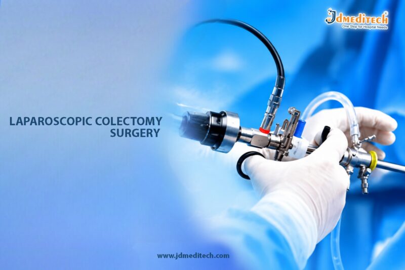
Laparoscopic colectomy surgery is an advanced minimally invasive procedure performed to remove part or all of the colon for the treatment of various colorectal diseases. The colon, also known as the large intestine, plays a major role in water absorption and waste elimination. However, conditions such as colorectal cancer, diverticular disease, inflammatory bowel disease, and bowel obstruction may require surgical intervention to restore digestive health and prevent serious complications.

Traditionally, colectomy procedures were performed through large abdominal incisions, resulting in significant postoperative pain and prolonged recovery. However, modern laparoscopic techniques now allow surgeons to perform colon surgery using small incisions, high-definition cameras, and specialized endoscopic instruments. Consequently, patients benefit from reduced pain, faster healing, shorter hospital stays, and improved cosmetic outcomes.

Today, laparoscopic colectomy has become a preferred surgical approach for many colorectal conditions because it combines surgical precision with the advantages of minimally invasive surgery.

What Is Laparoscopic Colectomy Surgery?

Laparoscopic colectomy surgery is a minimally invasive procedure used to remove diseased portions of the colon through small abdominal incisions with the assistance of a laparoscope.

A laparoscope is a thin camera-equipped instrument that provides magnified views of the abdominal cavity. During the procedure, surgeons use specialized instruments to separate and remove the affected section of the colon while preserving healthy surrounding tissue whenever possible.

After removal of the diseased segment, the remaining healthy ends of the intestine are usually reconnected to restore normal bowel function.

Ultimately, the primary goal of laparoscopic colectomy is to effectively treat colorectal disease while minimizing surgical trauma and improving recovery.

Why Is Laparoscopic Colectomy Performed?

Laparoscopic colectomy is recommended for a wide range of colorectal diseases that cannot be managed effectively with medications or non-surgical treatments.

Common Indications for Surgery Include:
  • Colorectal cancer
  • Diverticulitis
  • Crohn’s disease
  • Ulcerative colitis
  • Colon polyps
  • Bowel obstruction
  • Gastrointestinal bleeding
  • Colon perforation
  • Ischemic bowel disease
  • Familial polyposis syndromes

In addition, early surgical treatment may help prevent disease progression and serious abdominal complications.

Understanding Colorectal Diseases

Several disorders affecting the colon may require colectomy surgery.

Colorectal Cancer

Colorectal cancer is one of the most common reasons for colectomy. Surgical removal of the cancerous portion of the colon often provides the best chance for effective treatment.

Diverticulitis

Diverticulitis occurs when small pouches in the colon wall become inflamed or infected. Recurrent or severe cases may require surgery.

Inflammatory Bowel Disease

Conditions such as Crohn’s disease and ulcerative colitis can cause chronic inflammation and severe bowel damage.

Colon Polyps

Large or precancerous polyps may also require surgical removal to reduce cancer risk.

Therefore, timely diagnosis and appropriate treatment are extremely important for long-term digestive health.

Types of Laparoscopic Colectomy Procedures

Different types of colectomy procedures may be performed depending on the location and severity of disease.

Procedure Type Portion Removed Common Indications
Right Hemicolectomy Right side of colon Right-sided colon cancer
Left Hemicolectomy Left side of colon Diverticulitis or cancer
Sigmoid Colectomy Sigmoid colon Diverticular disease
Total Colectomy Entire colon Ulcerative colitis
Partial Colectomy Limited colon segment Localized disease

Ultimately, the surgeon selects the most appropriate procedure based on the patient’s condition.

Who Is a Suitable Candidate for Laparoscopic Colectomy?

Generally, laparoscopic colectomy may be recommended for:

  • Patients with colorectal cancer
  • Individuals with severe diverticular disease
  • Patients with inflammatory bowel disease
  • Individuals with recurrent bowel obstruction
  • Patients medically fit for minimally invasive surgery

Importantly, patient suitability depends on overall health, disease severity, and surgical complexity.

Preoperative Evaluation Before Surgery

Before laparoscopic colectomy, the surgical team performs detailed assessments to plan safe and effective treatment.

Medical History Review

Initially, the doctor reviews:

  • Digestive symptoms
  • Bowel habits
  • Weight loss
  • Previous surgeries
  • Family history of colorectal disease
  • Medication history
Physical Examination

Furthermore, abdominal examination helps identify tenderness, masses, or bowel abnormalities.

Diagnostic Investigations

Additional tests may include:

  • Colonoscopy
  • CT scan
  • MRI imaging
  • Blood investigations
  • Biopsy
  • Chest imaging

Consequently, these evaluations assist in surgical planning and disease staging.

How Laparoscopic Colectomy Surgery Is Performed

Typically, laparoscopic colectomy is performed under general anesthesia using advanced minimally invasive surgical equipment.

Step-by-Step Surgical Procedure
1. Creation of Small Incisions

First, the surgeon creates several small abdominal incisions for insertion of the laparoscope and surgical instruments.

2. Inflation of the Abdomen

Next, carbon dioxide gas is introduced into the abdomen to create working space.

3. Visualization of the Colon

Afterward, the laparoscope provides magnified views of the colon and surrounding organs.

4. Mobilization of the Diseased Colon Segment

Then, the surgeon carefully separates the affected portion of the colon from nearby tissues and blood vessels.

5. Removal of the Diseased Segment

Subsequently, the diseased colon segment is removed through a slightly enlarged incision.

6. Reconnection of the Intestine

The healthy ends of the intestine are then reconnected using stapling or suturing techniques.

7. Closure of Incisions

Finally, the surgical instruments are removed and the incisions are closed.

Overall, the procedure usually takes between 2 and 5 hours depending on complexity.

Benefits of Laparoscopic Colectomy Surgery

Laparoscopic colectomy offers several important advantages compared to traditional open colon surgery.

Minimally Invasive Technique

Because the procedure uses small incisions, postoperative pain and tissue trauma are significantly reduced.

Faster Recovery

Additionally, patients often return to normal activities sooner.

Reduced Blood Loss

Modern laparoscopic techniques help minimize surgical bleeding.

Lower Infection Risk

Smaller incisions may reduce wound-related complications.

Better Cosmetic Outcomes

Minimal scarring improves postoperative appearance.

Shorter Hospital Stay

Consequently, many patients experience earlier discharge from the hospital.

Recovery After Laparoscopic Colectomy

Recovery after laparoscopic colectomy varies depending on the extent of surgery and the patient’s overall health.

Common Postoperative Symptoms

Initially, patients may experience:

  • Mild abdominal discomfort
  • Fatigue
  • Temporary bloating
  • Changes in bowel habits
  • Reduced appetite

However, these symptoms usually improve gradually during recovery.

Recovery Guidelines

To support proper healing, patients are advised to:

  • Walk regularly after surgery
  • Stay hydrated
  • Follow dietary instructions
  • Avoid heavy lifting
  • Take medications as prescribed
  • Attend follow-up appointments

As a result, many individuals return to daily activities within several weeks.

Diet After Colectomy Surgery

Dietary management plays an important role after colon surgery.

Recommended Dietary Measures

Patients are encouraged to:

  • Start with soft foods
  • Eat smaller meals
  • Increase fiber gradually
  • Stay hydrated
  • Avoid highly processed foods initially

Consequently, these measures may help support healthy digestion and bowel recovery.

Risks and Complications

Although laparoscopic colectomy is considered safe, certain complications may occasionally occur.

Possible Complications Include:
  • Bleeding
  • Infection
  • Anastomotic leakage
  • Bowel obstruction
  • Blood clots
  • Injury to nearby organs
  • Hernia formation
  • Anesthesia-related complications

Nevertheless, complication rates are generally lower compared to traditional open surgery.

Success Rates of Laparoscopic Colectomy

Laparoscopic colectomy has excellent success rates for many colorectal diseases.

Factors Affecting Outcomes

Several factors influence surgical outcomes, including:

  • Underlying disease
  • Disease stage
  • Patient health condition
  • Surgical expertise
  • Postoperative care
General Outcomes

Modern laparoscopic colectomy typically provides:

  • Effective disease management
  • Faster recovery
  • Reduced pain
  • Lower complication rates
  • Improved quality of life

Therefore, early diagnosis and timely surgical intervention often improve long-term results.

Lifestyle Tips After Surgery

Healthy lifestyle habits may support long-term digestive health after colectomy.

Recommended Lifestyle Measures

Patients are encouraged to:

  • Maintain balanced nutrition
  • Exercise regularly
  • Avoid smoking
  • Limit alcohol consumption
  • Attend regular medical follow-ups
  • Maintain healthy body weight

Consequently, these habits may improve overall recovery and bowel health.

Choosing the Right Colorectal Surgeon

Successful laparoscopic colectomy requires expertise in minimally invasive colorectal surgery.

Patients Should Look For:
  • Experienced colorectal surgeons
  • Advanced laparoscopic surgical facilities
  • Expertise in gastrointestinal surgery
  • Comprehensive postoperative care
  • Strong patient safety standards

Ultimately, specialized surgical care improves both patient safety and long-term treatment outcomes.

Conclusion

Laparoscopic colectomy surgery is an advanced minimally invasive procedure for the treatment of various colorectal diseases, including cancer, diverticulitis, and inflammatory bowel disease. By combining modern laparoscopic technology with precise surgical techniques, the procedure offers effective disease management with reduced pain, faster recovery, and improved cosmetic outcomes.

Moreover, early diagnosis and timely surgical treatment can help prevent serious complications and improve long-term digestive health. Therefore, patients experiencing persistent colorectal symptoms should seek evaluation from an experienced colorectal surgeon.

With continuous advancements in minimally invasive surgery and specialized postoperative care, laparoscopic colectomy continues to remain a highly effective solution for colorectal disease management worldwide.


Explore More Product >>

Get Connected:
+91 79909 93062 | +91 63513 72032 | exports@jdmeditech.com

Laparoscopic Hernia Repair Surgery – Minimally Invasive Hernia Treatment

Laparoscopic hernia repair surgery is a modern minimally invasive procedure used to treat different types of hernias through small abdominal incisions. Hernias occur when an internal organ or tissue pushes through a weak area in the surrounding muscle or connective tissue. Although some hernias may initially cause mild discomfort, untreated hernias can gradually enlarge and lead to serious complications.

Traditionally, hernia repair required open surgery with larger incisions and longer recovery periods. However, advancements in laparoscopic surgery now allow surgeons to repair hernias using specialized endoscopic instruments and high-definition cameras. Consequently, patients benefit from reduced pain, faster recovery, shorter hospital stays, and minimal scarring.

Today, laparoscopic hernia repair is widely recognized as an effective and safe treatment option for many abdominal wall hernias. Therefore, it has become one of the most commonly performed minimally invasive surgical procedures worldwide.

What Is Laparoscopic Hernia Repair Surgery?

Laparoscopic hernia repair surgery is a minimally invasive procedure in which the surgeon repairs a hernia using small incisions, a laparoscope, and specialized surgical instruments.

A laparoscope is a thin tube equipped with a high-definition camera and light source. During the procedure, the surgeon inserts the laparoscope into the abdomen to obtain magnified views of the hernia and surrounding tissues.

Afterward, the protruding tissue is repositioned, and the weakened muscle wall is reinforced, usually with a surgical mesh. Ultimately, the procedure aims to restore abdominal wall strength, relieve symptoms, and prevent recurrence.

Understanding Hernias

A hernia develops when internal tissues push through a weak area in the abdominal wall or surrounding muscles.

Common Types of Hernias Include:
  • Inguinal hernia
  • Umbilical hernia
  • Femoral hernia
  • Incisional hernia
  • Hiatal hernia
  • Ventral hernia

Although hernias may vary in size and severity, surgical repair is often recommended to prevent complications.

Common Symptoms of Hernia

Symptoms may differ depending on the type and size of the hernia.

Frequently Reported Symptoms Include:
  • Visible abdominal or groin bulge
  • Pain or discomfort during lifting
  • Burning or aching sensation
  • Pressure in the abdomen
  • Swelling near the groin
  • Pain during coughing or straining

In severe cases, hernias may become trapped or strangulated, which requires emergency treatment.

Why Is Laparoscopic Hernia Repair Performed?

Laparoscopic hernia repair is performed to strengthen the abdominal wall and prevent worsening of the hernia.

Common Indications for Surgery Include:
  • Symptomatic hernia
  • Enlarging hernia
  • Recurrent hernia
  • Pain affecting daily activities
  • Strangulated hernia
  • Obstructed hernia
  • Weak abdominal wall after surgery

In addition, early surgical treatment may reduce the risk of emergency complications.

Who Is a Suitable Candidate for Laparoscopic Hernia Repair?

Generally, laparoscopic hernia repair may be recommended for:

  • Patients with inguinal hernias
  • Individuals with bilateral hernias
  • Patients with recurrent hernias
  • Individuals seeking faster recovery
  • Patients medically fit for minimally invasive surgery

However, the final surgical approach depends on hernia size, location, and patient health status.

Preoperative Evaluation Before Surgery

Before laparoscopic hernia repair, the surgeon performs a comprehensive assessment to evaluate the hernia and overall health condition.

Medical History Review

Initially, the doctor reviews:

  • Duration of symptoms
  • Previous surgeries
  • Hernia-related discomfort
  • Existing medical conditions
  • Medication use
  • Lifestyle factors
Physical Examination

Furthermore, physical examination helps determine:

  • Hernia location
  • Hernia size
  • Reducibility
  • Presence of complications
Diagnostic Investigations

Additional tests may include:

  • Ultrasound scan
  • CT scan
  • Blood investigations
  • Cardiac evaluation in selected patients

Consequently, these evaluations assist in surgical planning and patient safety.

How Laparoscopic Hernia Repair Surgery Is Performed

Typically, laparoscopic hernia repair is performed under general anesthesia using advanced minimally invasive surgical equipment.

Step-by-Step Surgical Procedure
1. Creation of Small Incisions

First, the surgeon creates several tiny abdominal incisions for insertion of the laparoscope and surgical instruments.

2. Inflation of the Abdomen

Next, carbon dioxide gas is introduced into the abdomen to create adequate working space.

3. Visualization of the Hernia

Afterward, the laparoscope provides magnified views of the hernia defect and surrounding tissues.

4. Repositioning of Herniated Tissue

Then, the protruding tissue or organ is gently moved back into its normal position.

5. Placement of Surgical Mesh

Subsequently, a synthetic mesh is placed over the weakened area to reinforce the abdominal wall.

6. Mesh Fixation

The mesh is secured using sutures, staples, or surgical adhesives.

7. Closure of Incisions

Finally, the instruments are removed and the small incisions are closed with sutures or surgical glue.

Overall, the procedure usually takes between 30 minutes and 2 hours depending on hernia complexity.

Types of Laparoscopic Hernia Repair Techniques

Several laparoscopic techniques are available depending on the type of hernia.

Procedure Type Common Use Key Advantage
TAPP Repair (Transabdominal Preperitoneal) Inguinal hernia Excellent visualization
TEP Repair (Totally Extraperitoneal) Inguinal hernia Avoids abdominal cavity entry
Laparoscopic Ventral Hernia Repair Ventral and incisional hernia Strong abdominal reinforcement
Hiatal Hernia Repair Hiatal hernia Improves reflux symptoms

Ultimately, the surgeon selects the most suitable approach based on individual patient needs.

Benefits of Laparoscopic Hernia Repair Surgery

Laparoscopic hernia repair offers several important advantages over traditional open surgery.

Minimally Invasive Technique

Because the procedure uses small incisions, tissue trauma and postoperative discomfort are significantly reduced.

Faster Recovery

Additionally, patients usually return to normal activities more quickly.

Reduced Pain

Smaller incisions often result in less postoperative pain.

Lower Risk of Infection

Furthermore, minimally invasive surgery may reduce wound-related complications.

Better Cosmetic Results

Minimal scarring improves cosmetic appearance after recovery.

Earlier Return to Work

Consequently, many patients resume work and daily activities sooner compared to open surgery.

Recovery After Laparoscopic Hernia Repair

Recovery after laparoscopic hernia repair is generally smooth and relatively quick.

Common Postoperative Symptoms

Initially, patients may experience:

  • Mild abdominal discomfort
  • Temporary bloating
  • Fatigue
  • Minor swelling near incision sites

However, these symptoms usually improve within several days.

Recovery Guidelines

To support proper healing, patients are advised to:

  • Walk regularly after surgery
  • Avoid heavy lifting temporarily
  • Maintain healthy bowel habits
  • Stay hydrated
  • Follow wound care instructions
  • Attend follow-up appointments

As a result, many individuals return to light activities within a few days and normal activities within several weeks.

Risks and Complications

Although laparoscopic hernia repair is considered very safe, certain complications may occasionally occur.

Possible Complications Include:
  • Bleeding
  • Infection
  • Hernia recurrence
  • Mesh-related complications
  • Injury to nearby organs
  • Chronic pain
  • Blood clots
  • Anesthesia-related complications

Nevertheless, complication rates are generally low when surgery is performed by experienced surgeons.

Preventing Hernia Recurrence

Following surgery, healthy lifestyle habits may help reduce the risk of hernia recurrence.

Preventive Measures Include:
  • Maintaining healthy body weight
  • Avoiding heavy lifting
  • Treating chronic cough
  • Preventing constipation
  • Exercising regularly
  • Following postoperative restrictions carefully

Consequently, these measures may strengthen abdominal muscles and support long-term surgical success.

Success Rates of Laparoscopic Hernia Repair

Laparoscopic hernia repair has excellent long-term success rates.

Factors Affecting Outcomes

Several factors influence recovery and surgical success, including:

  • Hernia type and size
  • Surgical technique
  • Patient health condition
  • Smoking status
  • Obesity
  • Surgeon expertise
General Outcomes

Modern laparoscopic hernia surgery typically provides:

  • Effective hernia repair
  • Faster healing
  • Reduced recurrence rates
  • Improved patient comfort
  • Excellent cosmetic outcomes

Therefore, early treatment often results in better long-term results.

Choosing the Right Surgeon

Successful laparoscopic hernia repair requires specialized expertise in minimally invasive surgery.

Patients Should Look For:
  • Experienced laparoscopic surgeons
  • Advanced surgical facilities
  • Expertise in hernia repair techniques
  • Comprehensive postoperative care
  • Strong patient safety protocols

Ultimately, experienced specialists improve surgical precision and patient recovery.

Conclusion

Laparoscopic hernia repair surgery is a modern minimally invasive solution for treating abdominal wall hernias safely and effectively. By combining advanced endoscopic technology with precise surgical techniques, the procedure offers reduced pain, faster recovery, minimal scarring, and excellent long-term outcomes.

Moreover, early diagnosis and timely treatment can prevent serious hernia-related complications such as obstruction and strangulation. Therefore, individuals experiencing hernia symptoms should consult an experienced surgeon for proper evaluation and personalized treatment planning.

With ongoing advancements in minimally invasive surgery and mesh repair technology, laparoscopic hernia repair continues to remain one of the most reliable and effective treatments for hernia management worldwide.


Explore More Product >>

Get Connected:
+91 79909 93062 | +91 63513 72032 | exports@jdmeditech.com 

Laparoscopic Diagnostic Surgery – Minimally Invasive Abdominal Evaluation Procedure

Laparoscopic diagnostic surgery, also known as diagnostic laparoscopy, is a modern minimally invasive procedure used to examine the organs inside the abdomen and pelvis. This advanced endoscopic technique helps surgeons accurately diagnose various abdominal and pelvic conditions when imaging studies or clinical evaluations are inconclusive.

Traditionally, exploratory abdominal surgery required large incisions and extended recovery periods. However, laparoscopic diagnostic surgery now allows doctors to directly visualize internal organs through small incisions using high-definition cameras and specialized surgical instruments. Consequently, patients benefit from less pain, faster recovery, shorter hospital stay, and reduced surgical trauma.

Today, laparoscopic diagnostic surgery plays a vital role in evaluating unexplained abdominal pain, infertility, pelvic disorders, abdominal masses, infections, and many other conditions. Therefore, it has become an important tool in modern minimally invasive surgery.

What Is Laparoscopic Diagnostic Surgery?

Laparoscopic diagnostic surgery is a minimally invasive procedure in which a laparoscope is inserted into the abdominal cavity through small incisions to examine internal organs and identify abnormalities.

A laparoscope is a thin tube equipped with a high-definition camera and light source. During the procedure, the camera transmits magnified images to a monitor, allowing surgeons to carefully inspect the abdominal and pelvic organs in real time.

In some cases, surgeons may also collect tissue samples or perform minor corrective procedures during the same surgery.

Ultimately, the primary goal of laparoscopic diagnostic surgery is to provide an accurate diagnosis while minimizing patient discomfort and surgical risk.

Why Is Laparoscopic Diagnostic Surgery Performed?

Laparoscopic diagnostic surgery is performed when non-invasive tests are unable to provide a clear diagnosis or when direct visualization of abdominal organs is necessary.

Common Reasons for Diagnostic Laparoscopy Include:
  • Unexplained abdominal pain
  • Chronic pelvic pain
  • Female infertility evaluation
  • Suspected endometriosis
  • Abdominal masses or tumors
  • Internal bleeding
  • Pelvic inflammatory disease
  • Suspected appendicitis
  • Liver disease assessment
  • Evaluation of abdominal trauma
  • Adhesion detection
  • Biopsy collection

Moreover, diagnostic laparoscopy often helps doctors avoid unnecessary open surgery.

Conditions Commonly Diagnosed Using Laparoscopy

Diagnostic laparoscopy is widely used in both general surgery and gynecology for evaluating multiple conditions.

Endometriosis

Laparoscopy is considered one of the most accurate methods for diagnosing endometriosis, a condition in which tissue similar to the uterine lining grows outside the uterus.

Pelvic Adhesions

Scar tissue formation inside the abdomen or pelvis may cause chronic pain and fertility problems. Therefore, laparoscopy helps identify these adhesions clearly.

Appendicitis

In uncertain cases, laparoscopy helps confirm appendicitis and may allow immediate appendix removal if necessary.

Ovarian Cysts and Masses

Laparoscopy allows direct visualization and evaluation of ovarian cysts, tumors, and pelvic masses.

Abdominal Infections

Infections or abscesses inside the abdomen may also be diagnosed and treated during the same procedure.

Who Is a Suitable Candidate for Laparoscopic Diagnostic Surgery?

Generally, laparoscopic diagnostic surgery may be recommended for:

  • Patients with unexplained abdominal symptoms
  • Women with infertility concerns
  • Individuals with chronic pelvic pain
  • Patients with abnormal imaging findings
  • Individuals requiring tissue biopsy
  • Patients medically fit for minimally invasive surgery

Importantly, detailed clinical evaluation helps determine whether laparoscopy is appropriate.

Preoperative Evaluation Before Surgery

Before laparoscopic diagnostic surgery, the surgeon performs comprehensive assessments to evaluate the patient’s health and surgical suitability.

Medical History Review

Initially, the doctor reviews:

  • Current symptoms
  • Previous surgeries
  • Medical conditions
  • Medication history
  • Menstrual and fertility history in women
  • Allergies
Physical Examination

Furthermore, physical examination helps assess abdominal tenderness, swelling, and other clinical findings.

Diagnostic Investigations

Additional tests may include:

  • Blood tests
  • Ultrasound scan
  • CT scan
  • MRI imaging
  • Urine analysis
  • Pregnancy testing in women

Consequently, these evaluations assist in safe surgical planning.

How Laparoscopic Diagnostic Surgery Is Performed

Typically, laparoscopic diagnostic surgery is performed under general anesthesia using advanced minimally invasive equipment.

Step-by-Step Surgical Procedure
1. Creation of Small Incisions

First, the surgeon creates one or more tiny abdominal incisions for insertion of the laparoscope and surgical instruments.

2. Inflation of the Abdomen

Next, carbon dioxide gas is introduced into the abdomen to create adequate working space.

3. Insertion of the Laparoscope

Afterward, the laparoscope is inserted through the incision to provide magnified visualization of internal organs.

4. Examination of Abdominal Organs

Then, the surgeon carefully examines organs such as:

  • Liver
  • Gallbladder
  • Appendix
  • Intestines
  • Reproductive organs
  • Kidneys
  • Pelvic structures
5. Tissue Biopsy or Minor Procedures

If abnormalities are identified, tissue samples may be collected or minor procedures may be performed simultaneously.

6. Closure of Incisions

Finally, the instruments are removed and the small incisions are closed with sutures or surgical glue.

Overall, the procedure usually takes between 30 minutes and 2 hours depending on the complexity of the evaluation.

Benefits of Laparoscopic Diagnostic Surgery

Laparoscopic diagnostic surgery offers several important advantages compared to traditional exploratory surgery.

Minimally Invasive Technique

Because the procedure uses small incisions, postoperative pain and tissue trauma are significantly reduced.

Faster Recovery

Additionally, patients usually recover more quickly and resume normal activities sooner.

Accurate Diagnosis

Direct visualization of internal organs provides highly accurate diagnostic information.

Shorter Hospital Stay

Many patients return home on the same day or within 24 hours.

Reduced Risk of Complications

Furthermore, minimally invasive surgery may reduce infection risk and postoperative complications.

Ability to Perform Immediate Treatment

In many cases, surgeons can diagnose and treat certain conditions during the same procedure.

Recovery After Laparoscopic Diagnostic Surgery

Recovery after diagnostic laparoscopy is generally smooth and uncomplicated.

Common Postoperative Symptoms

Initially, patients may experience:

  • Mild abdominal discomfort
  • Temporary bloating
  • Shoulder pain from surgical gas
  • Mild fatigue
  • Minor incision soreness

However, these symptoms usually improve within several days.

Recovery Guidelines

To support proper healing, patients are advised to:

  • Walk regularly after surgery
  • Stay hydrated
  • Avoid heavy lifting temporarily
  • Maintain incision hygiene
  • Take prescribed medications
  • Attend follow-up appointments

As a result, most individuals return to routine activities within a few days.

Risks and Complications

Although laparoscopic diagnostic surgery is considered very safe, certain complications may occasionally occur.

Possible Complications Include:
  • Bleeding
  • Infection
  • Injury to nearby organs
  • Blood clots
  • Adverse reaction to anesthesia
  • Hernia at incision sites

Nevertheless, complication rates are generally low when surgery is performed by experienced surgeons.

Diagnostic Laparoscopy in Female Infertility

Diagnostic laparoscopy plays an important role in fertility evaluation when other tests are inconclusive.

Fertility Conditions Commonly Evaluated Include:
  • Endometriosis
  • Tubal blockage
  • Pelvic adhesions
  • Ovarian cysts
  • Congenital abnormalities

Consequently, laparoscopy helps guide fertility treatment decisions more accurately.

Success Rates and Diagnostic Accuracy

Diagnostic laparoscopy has excellent accuracy for identifying abdominal and pelvic conditions.

Factors Influencing Diagnostic Success

Several factors affect outcomes, including:

  • Nature of the condition
  • Surgical expertise
  • Quality of imaging equipment
  • Patient anatomy
  • Extent of disease
General Outcomes

Modern diagnostic laparoscopy typically provides:

  • Precise diagnosis
  • Reduced need for open surgery
  • Faster treatment planning
  • Improved patient outcomes

Therefore, minimally invasive diagnostic surgery remains an essential tool in modern medicine.

Lifestyle Tips After Diagnostic Laparoscopy

Healthy postoperative habits may help support faster recovery and overall wellness.

Recommended Lifestyle Measures

Patients are encouraged to:

  • Rest adequately
  • Resume activity gradually
  • Follow balanced nutrition
  • Stay hydrated
  • Avoid smoking
  • Follow medical instructions carefully

Consequently, these measures may improve healing and recovery.

Choosing the Right Surgeon

Successful laparoscopic diagnostic surgery requires expertise in minimally invasive abdominal procedures.

Patients Should Look For:
  • Experienced laparoscopic surgeons
  • Advanced hospital facilities
  • Expertise in abdominal and pelvic disorders
  • Comprehensive postoperative care
  • Strong patient safety protocols

Ultimately, experienced specialists help ensure accurate diagnosis and safe surgical outcomes.

Conclusion

Laparoscopic diagnostic surgery is an advanced minimally invasive procedure that allows accurate evaluation of abdominal and pelvic conditions through small incisions and high-definition visualization. By combining modern endoscopic technology with precise surgical techniques, the procedure provides reliable diagnosis with reduced pain, faster recovery, and minimal scarring.

Moreover, diagnostic laparoscopy often helps detect conditions that may not be visible through conventional imaging studies. Therefore, patients with unexplained abdominal symptoms or pelvic disorders should consult experienced specialists to determine whether laparoscopic evaluation is appropriate.

With modern surgical advancements and expert clinical care, laparoscopic diagnostic surgery continues to remain a valuable tool for safe and effective abdominal assessment.


Explore More Product >>

Get Connected:
+91 79909 93062 | +91 63513 72032 | exports@jdmeditech.com 

Laparoscopic Salpingectomy Surgery – Minimally Invasive Treatment for Tubal Disorders

Laparoscopic salpingectomy surgery is a modern minimally invasive gynecological procedure performed to remove one or both fallopian tubes. This advanced endoscopic technique is commonly used for the treatment of various tubal pathologies, including ectopic pregnancy, hydrosalpinx, pelvic infections, damaged fallopian tubes, and certain gynecologic cancers.

Traditionally, salpingectomy required open abdominal surgery with larger incisions and prolonged recovery. However, advancements in laparoscopic surgery have transformed gynecologic care by allowing surgeons to perform fallopian tube removal through small incisions using specialized instruments and high-definition cameras.

As a result, laparoscopic salpingectomy offers reduced postoperative pain, faster healing, shorter hospital stay, minimal scarring, and improved surgical precision. Therefore, it has become a preferred surgical option for managing many fallopian tube-related conditions.

What Is Laparoscopic Salpingectomy Surgery?

Laparoscopic salpingectomy surgery is a minimally invasive procedure in which one or both fallopian tubes are surgically removed using laparoscopic instruments inserted through small abdominal incisions.

The fallopian tubes connect the ovaries to the uterus and play an important role in natural fertilization. However, certain diseases or structural abnormalities affecting the tubes may require surgical removal to protect reproductive health and prevent complications.

During the procedure, the surgeon uses a laparoscope, which is a thin camera-equipped instrument that provides magnified visualization of the pelvic organs. Subsequently, the affected fallopian tube is carefully separated and removed while preserving nearby reproductive structures whenever possible.

Ultimately, the procedure aims to safely treat tubal disease while minimizing surgical trauma and supporting overall gynecologic health.

Why Is Laparoscopic Salpingectomy Performed?

Laparoscopic salpingectomy is recommended for several gynecologic and reproductive conditions involving the fallopian tubes.

Common Indications for Surgery Include:
  • Ectopic pregnancy
  • Hydrosalpinx
  • Damaged or blocked fallopian tubes
  • Chronic pelvic inflammatory disease
  • Tubal infections
  • Tubal torsion
  • Endometriosis involving the tubes
  • Prevention of ovarian cancer in high-risk patients
  • Tubal tumors or suspicious growths
  • Permanent sterilization in selected cases

In addition, early surgical intervention may help prevent severe complications such as rupture, internal bleeding, chronic pelvic pain, and infertility.

Understanding Tubal Pathologies

Tubal pathologies refer to medical conditions affecting the fallopian tubes. These conditions may interfere with fertility, cause pain, or increase the risk of serious reproductive complications.

Ectopic Pregnancy

An ectopic pregnancy occurs when a fertilized egg implants outside the uterus, most commonly inside the fallopian tube. Consequently, this condition can become life-threatening if the tube ruptures.

Hydrosalpinx

Hydrosalpinx refers to fluid accumulation within a blocked fallopian tube. As a result, fertility may be significantly affected.

Pelvic Inflammatory Disease (PID)

Chronic infections may cause scarring and permanent damage to the fallopian tubes, leading to pain and infertility.

Therefore, timely diagnosis and treatment are essential for protecting reproductive health.

Who Is a Suitable Candidate for Laparoscopic Salpingectomy?

Generally, laparoscopic salpingectomy may be recommended for:

  • Women with tubal disease
  • Patients with ectopic pregnancy
  • Individuals with chronic pelvic pain related to tubal pathology
  • Women with severe tubal damage
  • Patients undergoing fertility treatment
  • Individuals medically fit for minimally invasive surgery

Importantly, treatment decisions depend on fertility goals, age, overall health, and the severity of the condition.

Preoperative Evaluation Before Surgery

Before laparoscopic salpingectomy, the gynecologist performs detailed clinical assessments to determine the most appropriate treatment plan.

Medical History Review

Initially, the doctor evaluates:

  • Menstrual history
  • Fertility history
  • Pelvic pain symptoms
  • Previous pelvic surgeries
  • History of infections
  • Pregnancy history
  • Current medications
Physical Examination

Furthermore, pelvic examination helps identify tenderness, masses, or signs of infection.

Diagnostic Investigations

Additional tests may include:

  • Pelvic ultrasound
  • Blood tests
  • Pregnancy testing
  • MRI or CT scan
  • Hysterosalpingography (HSG)
  • Diagnostic laparoscopy

Consequently, these investigations help confirm the diagnosis and guide surgical planning.

How Laparoscopic Salpingectomy Surgery Is Performed

Typically, laparoscopic salpingectomy is performed under general anesthesia using advanced minimally invasive surgical equipment.

Step-by-Step Surgical Procedure
1. Creation of Small Incisions

First, the surgeon creates several tiny abdominal incisions for insertion of the laparoscope and specialized surgical instruments.

2. Inflation of the Abdomen

Next, carbon dioxide gas is introduced into the abdomen to create adequate surgical working space.

3. Visualization of Pelvic Organs

Afterward, the laparoscope provides magnified views of the uterus, ovaries, fallopian tubes, and surrounding structures.

4. Identification of the Diseased Tube

Then, the affected fallopian tube is carefully identified and separated from surrounding tissues.

5. Removal of the Fallopian Tube

Subsequently, the blood supply to the tube is sealed, and the tube is safely removed through one of the small incisions.

6. Closure of Incisions

Finally, the instruments are removed and the incisions are closed with sutures or surgical glue.

Overall, the surgery usually takes between 30 minutes and 2 hours depending on surgical complexity.

Types of Salpingectomy Procedures

Several variations of salpingectomy may be performed depending on the patient’s condition.

Procedure Type Description Common Use
Unilateral Salpingectomy Removal of one fallopian tube Ectopic pregnancy or localized disease
Bilateral Salpingectomy Removal of both fallopian tubes Cancer prevention or severe bilateral disease
Partial Salpingectomy Removal of part of the tube Selected sterilization procedures
Salpingo-Oophorectomy Removal of tube and ovary Advanced gynecologic disease

Ultimately, the surgeon selects the most suitable approach based on the patient’s diagnosis and reproductive goals.

Benefits of Laparoscopic Salpingectomy Surgery

Laparoscopic salpingectomy offers several important advantages over traditional open surgery.

Minimally Invasive Technique

Because the procedure uses small incisions, tissue trauma and postoperative pain are significantly reduced.

Faster Recovery

Additionally, patients usually recover more quickly and return to normal activities sooner.

Reduced Blood Loss

Modern laparoscopic techniques help minimize intraoperative bleeding.

Better Cosmetic Results

Smaller incisions produce minimal visible scarring.

Improved Surgical Precision

Furthermore, magnified visualization allows accurate dissection and preservation of nearby structures.

Lower Risk of Infection

Consequently, minimally invasive surgery often reduces wound-related complications.

Recovery After Laparoscopic Salpingectomy

Recovery after laparoscopic salpingectomy is generally smooth and uncomplicated.

Common Postoperative Symptoms

Initially, patients may experience:

  • Mild abdominal discomfort
  • Temporary bloating
  • Shoulder pain from surgical gas
  • Mild fatigue
  • Minor vaginal spotting

However, these symptoms usually improve within several days.

Recovery Guidelines

To support proper healing, patients are advised to:

  • Walk regularly after surgery
  • Avoid heavy lifting temporarily
  • Stay hydrated
  • Follow postoperative dietary instructions
  • Maintain incision hygiene
  • Attend follow-up appointments

As a result, most patients return to normal activities within 1 to 2 weeks.

Fertility After Salpingectomy Surgery

Fertility outcomes after salpingectomy depend on whether one or both fallopian tubes are removed.

Important Fertility Considerations
  • Women with one healthy remaining tube may still conceive naturally.
  • Bilateral salpingectomy eliminates natural conception through the fallopian tubes.
  • IVF treatment may still be possible after bilateral salpingectomy.

Therefore, fertility counseling is important before surgery, especially for women planning future pregnancies.

Risks and Complications

Although laparoscopic salpingectomy is considered very safe, certain complications may occur in rare cases.

Possible Complications Include:
  • Bleeding
  • Infection
  • Injury to nearby organs
  • Adhesion formation
  • Blood clots
  • Anesthesia-related complications

Nevertheless, complication rates are generally low when surgery is performed by experienced gynecologic surgeons.

Success Rates of Laparoscopic Salpingectomy

Laparoscopic salpingectomy has excellent surgical success rates and favorable patient outcomes.

Factors Affecting Outcomes

Several factors influence recovery and success, including:

  • Severity of tubal disease
  • Presence of infection
  • Fertility goals
  • Patient health condition
  • Surgical expertise
General Outcomes

Modern laparoscopic salpingectomy typically provides:

  • Effective disease treatment
  • Reduced pelvic pain
  • Faster postoperative recovery
  • Low complication rates
  • Improved quality of life

Therefore, timely surgical intervention often leads to excellent long-term results.

Lifestyle Tips After Surgery

Healthy lifestyle habits may support faster healing and long-term reproductive wellness.

Recommended Lifestyle Measures

Patients are encouraged to:

  • Maintain balanced nutrition
  • Avoid smoking
  • Stay physically active
  • Follow medical advice carefully
  • Attend routine gynecologic checkups
  • Manage stress effectively

Consequently, these habits may improve recovery and overall reproductive health.

Choosing the Right Gynecologic Surgeon

Successful laparoscopic salpingectomy requires expertise in minimally invasive gynecologic surgery.

Patients Should Look For:
  • Experienced laparoscopic gynecologic surgeons
  • Advanced hospital facilities
  • Expertise in reproductive surgery
  • Comprehensive postoperative care
  • Strong patient safety standards

Ultimately, specialized surgical care improves both safety and treatment outcomes.

Conclusion

Laparoscopic salpingectomy surgery is an advanced minimally invasive procedure for treating various tubal pathologies, including ectopic pregnancy, hydrosalpinx, chronic infections, and damaged fallopian tubes. By combining modern laparoscopic technology with precise surgical techniques, the procedure offers effective disease management with reduced pain, faster recovery, and minimal scarring.

Moreover, early diagnosis and timely intervention can help prevent serious reproductive complications and improve long-term gynecologic health. Therefore, women experiencing symptoms related to fallopian tube disorders should seek evaluation from an experienced gynecologic surgeon.

With modern surgical advancements and comprehensive postoperative care, laparoscopic salpingectomy continues to remain a safe and highly effective treatment option for tubal disease management.


Explore More Product >>

Get Connected:
+91 79909 93062 | +91 63513 72032 | exports@jdmeditech.com 

Laparoscopic Appendectomy Surgery – Minimally Invasive Treatment for Acute Appendicitis

Laparoscopic appendectomy surgery is a modern minimally invasive procedure performed to remove the appendix in patients diagnosed with acute appendicitis. Acute appendicitis is one of the most common surgical emergencies and occurs when the appendix becomes inflamed or infected. If left untreated, the appendix may rupture and lead to serious abdominal complications. Therefore, timely surgical intervention is essential.

Traditionally, appendectomy was performed through a large abdominal incision. However, advancements in laparoscopic surgery now allow surgeons to remove the appendix using small incisions, specialized instruments, and high-definition cameras. Consequently, patients experience less pain, faster recovery, shorter hospital stays, and improved cosmetic outcomes.

Today, laparoscopic appendectomy is widely regarded as the preferred surgical treatment for acute appendicitis because of its safety, effectiveness, and minimally invasive nature.

What Is Laparoscopic Appendectomy Surgery?

Laparoscopic appendectomy surgery is a minimally invasive procedure used to remove the inflamed appendix through small abdominal incisions with the help of a laparoscope.

The appendix is a small finger-shaped pouch attached to the large intestine. Although its exact function remains unclear, inflammation of the appendix can rapidly become dangerous if infection spreads within the abdomen.

During laparoscopic appendectomy, the surgeon inserts a camera-equipped laparoscope and specialized instruments into the abdomen. Afterward, the appendix is carefully separated and removed while preserving nearby organs and tissues.

Ultimately, the primary goal of the surgery is to eliminate infection, prevent rupture, and restore abdominal health safely.

Understanding Acute Appendicitis

Acute appendicitis occurs when the appendix becomes blocked, inflamed, and infected. In many cases, the blockage may result from hardened stool, infection, swelling, or lymphatic tissue enlargement.

Common Symptoms of Acute Appendicitis Include:
  • Sudden abdominal pain
  • Pain starting near the navel and shifting to the lower right abdomen
  • Fever
  • Nausea and vomiting
  • Loss of appetite
  • Abdominal swelling
  • Difficulty passing gas
  • Pain during movement or coughing

Therefore, immediate medical evaluation is important whenever appendicitis is suspected.

Why Is Laparoscopic Appendectomy Performed?

Laparoscopic appendectomy is performed to remove the infected appendix before complications develop.

Common Indications for Surgery Include:
  • Acute appendicitis
  • Recurrent appendicitis
  • Ruptured appendix
  • Appendiceal abscess
  • Chronic appendiceal inflammation
  • Uncertain abdominal diagnosis requiring exploration

In addition, early surgery significantly reduces the risk of appendix rupture and severe abdominal infection.

Who Is a Suitable Candidate for Laparoscopic Appendectomy?

Generally, laparoscopic appendectomy may be recommended for:

  • Patients with confirmed appendicitis
  • Individuals with recurrent abdominal pain related to the appendix
  • Children and adults with acute appendicitis
  • Patients medically fit for minimally invasive surgery
  • Individuals requiring emergency abdominal surgery

Importantly, suitability depends on the patient’s overall condition and severity of infection.

Preoperative Evaluation Before Surgery

Before laparoscopic appendectomy, the surgeon performs detailed clinical assessments to confirm the diagnosis and prepare for surgery.

Medical History Review

Initially, the doctor evaluates:

  • Nature and duration of abdominal pain
  • Fever and digestive symptoms
  • Previous abdominal surgeries
  • Medication history
  • Existing medical conditions
Physical Examination

Furthermore, abdominal examination helps identify:

  • Right lower abdominal tenderness
  • Guarding or rigidity
  • Rebound tenderness
  • Signs of infection
Diagnostic Investigations

Additional tests may include:

  • Blood tests
  • Urine analysis
  • Abdominal ultrasound
  • CT scan
  • MRI in selected patients

Consequently, these investigations help confirm appendicitis and guide treatment planning.

How Laparoscopic Appendectomy Surgery Is Performed

Typically, laparoscopic appendectomy is performed under general anesthesia using advanced minimally invasive surgical equipment.

Step-by-Step Surgical Procedure
1. Creation of Small Incisions

First, the surgeon creates several tiny abdominal incisions for insertion of the laparoscope and surgical instruments.

2. Inflation of the Abdomen

Next, carbon dioxide gas is introduced to create working space inside the abdomen.

3. Visualization of the Appendix

Afterward, the laparoscope provides magnified visualization of the inflamed appendix and surrounding structures.

4. Separation of the Appendix

Then, the appendix is carefully separated from nearby tissues and blood vessels.

5. Removal of the Appendix

Subsequently, the appendix is sealed, divided, and removed through one of the small incisions.

6. Cleaning of the Abdominal Cavity

If infection or rupture is present, the surgeon may clean the abdominal cavity thoroughly.

7. Closure of Incisions

Finally, the surgical instruments are removed and the incisions are closed with sutures or surgical glue.

Overall, the procedure usually takes between 30 minutes and 1 hour depending on surgical complexity.

Benefits of Laparoscopic Appendectomy Surgery

Laparoscopic appendectomy offers several important advantages over traditional open surgery.

Minimally Invasive Technique

Because the procedure uses small incisions, postoperative pain and tissue trauma are significantly reduced.

Faster Recovery

Additionally, patients usually recover more quickly and return to daily activities sooner.

Shorter Hospital Stay

Many patients are discharged within 24 to 48 hours after surgery.

Reduced Risk of Wound Infection

Smaller incisions lower the risk of wound complications.

Better Cosmetic Results

Furthermore, minimal scarring improves cosmetic appearance after recovery.

Improved Visualization

The laparoscopic camera provides magnified surgical views, allowing greater precision during surgery.

Recovery After Laparoscopic Appendectomy

Recovery after laparoscopic appendectomy is generally smooth and uncomplicated.

Common Postoperative Symptoms

Initially, patients may experience:

  • Mild abdominal discomfort
  • Temporary bloating
  • Shoulder pain due to surgical gas
  • Mild fatigue
  • Nausea

However, these symptoms usually improve within several days.

Recovery Guidelines

To support proper healing, patients are advised to:

  • Walk regularly after surgery
  • Stay hydrated
  • Follow dietary recommendations
  • Avoid heavy lifting temporarily
  • Maintain incision hygiene
  • Take prescribed medications

As a result, most patients resume normal activities within 1 to 2 weeks.

Diet After Appendectomy Surgery

Following surgery, temporary dietary adjustments may support digestive recovery.

Recommended Dietary Measures

Patients are encouraged to:

  • Start with light meals
  • Eat easily digestible foods
  • Stay hydrated
  • Increase fiber gradually
  • Avoid excessively greasy foods initially

Consequently, these measures may help improve digestive comfort during recovery.

Risks and Complications

Although laparoscopic appendectomy is considered very safe, certain complications may occasionally occur.

Possible Complications Include:
  • Bleeding
  • Infection
  • Injury to nearby organs
  • Abscess formation
  • Hernia at incision sites
  • Blood clots
  • Anesthesia-related complications

Nevertheless, complication rates are generally low when surgery is performed promptly by experienced surgeons.

Complicated Appendicitis and Ruptured Appendix

In some cases, delayed treatment may cause the appendix to rupture, leading to serious abdominal infection.

Signs of Complicated Appendicitis May Include:
  • Severe abdominal pain
  • High fever
  • Generalized abdominal swelling
  • Pus formation
  • Sepsis symptoms

Therefore, emergency surgical management becomes critically important in such situations.

Success Rates of Laparoscopic Appendectomy

Laparoscopic appendectomy has very high success rates and excellent long-term outcomes.

Factors Affecting Outcomes

Several factors influence recovery and surgical success, including:

  • Timing of surgery
  • Severity of appendicitis
  • Presence of rupture
  • Patient health condition
  • Surgical expertise
General Outcomes

Modern laparoscopic appendectomy typically provides:

  • Complete symptom resolution
  • Rapid recovery
  • Minimal scarring
  • Low complication rates
  • Excellent patient satisfaction

Therefore, early diagnosis and treatment are highly beneficial.

Lifestyle Tips After Appendectomy Surgery

Healthy habits may support faster recovery and overall wellness after surgery.

Recommended Lifestyle Measures

Patients are encouraged to:

  • Resume physical activity gradually
  • Follow balanced nutrition
  • Avoid smoking
  • Maintain healthy body weight
  • Attend follow-up appointments
  • Follow postoperative instructions carefully

Consequently, these measures may improve healing and long-term recovery.

Choosing the Right Surgeon

Successful laparoscopic appendectomy requires expertise in minimally invasive abdominal surgery.

Patients Should Look For:
  • Experienced laparoscopic surgeons
  • Advanced hospital facilities
  • Emergency surgical expertise
  • Strong infection control protocols
  • Comprehensive postoperative care

Ultimately, experienced surgical teams improve patient safety and recovery outcomes.

Conclusion

Laparoscopic appendectomy surgery is an advanced minimally invasive procedure for the safe and effective treatment of acute appendicitis. By combining modern endoscopic technology with precise surgical techniques, the procedure provides rapid symptom relief, reduced postoperative pain, and faster recovery.

Moreover, early diagnosis and timely surgical intervention can prevent serious complications such as appendix rupture and abdominal infection. Therefore, individuals experiencing symptoms of appendicitis should seek immediate medical attention for proper evaluation and treatment.

With modern laparoscopic advancements and specialized surgical care, laparoscopic appendectomy continues to remain the preferred treatment for acute appendicitis worldwide.


Explore More Product >>

Get Connected:
+91 79909 93062 | +91 63513 72032 | exports@jdmeditech.com 

Laparoscopic Cholecystectomy Surgery – Minimally Invasive Gallbladder Removal

Laparoscopic cholecystectomy surgery is a modern minimally invasive procedure performed to remove the gallbladder, primarily in patients suffering from symptomatic cholelithiasis, commonly known as gallstones. Gallstones can cause severe abdominal pain, nausea, digestive disturbances, and serious complications if left untreated. Therefore, timely surgical management is often recommended for long-term relief and prevention of further health problems.

Traditionally, gallbladder removal required a large abdominal incision and prolonged recovery. However, advancements in laparoscopic surgery have transformed gallbladder treatment by allowing surgeons to perform the procedure through small incisions using specialized endoscopic instruments.

As a result, laparoscopic cholecystectomy has become the gold standard treatment for symptomatic gallstones because it offers less pain, faster recovery, shorter hospital stay, and excellent surgical outcomes.

What Is Laparoscopic Cholecystectomy Surgery?

Laparoscopic cholecystectomy surgery is a minimally invasive procedure used to remove the gallbladder through small abdominal incisions with the help of a laparoscope.

The gallbladder is a small organ located beneath the liver that stores bile, which helps digest fats. However, when gallstones develop, they may block bile flow and cause inflammation, pain, or infection.

During laparoscopic cholecystectomy, the surgeon inserts a camera-equipped laparoscope and specialized surgical instruments into the abdomen. Subsequently, the gallbladder is carefully separated from surrounding structures and removed safely.

Ultimately, the surgery aims to eliminate symptoms, prevent complications, and improve digestive comfort.

Understanding Symptomatic Cholelithiasis

Cholelithiasis refers to the formation of gallstones within the gallbladder. Although some gallstones remain asymptomatic, others may lead to significant discomfort and complications.

Common Symptoms of Gallstones Include:
  • Upper abdominal pain
  • Pain after fatty meals
  • Nausea and vomiting
  • Indigestion
  • Bloating
  • Shoulder or back pain
  • Fever in case of infection

Therefore, patients with persistent or severe symptoms often require surgical treatment.

Why Is Laparoscopic Cholecystectomy Performed?

Laparoscopic cholecystectomy is recommended when gallstones cause symptoms or complications that affect daily life and digestive health.

Common Indications for Surgery Include:
  • Symptomatic gallstones
  • Acute cholecystitis
  • Chronic gallbladder inflammation
  • Gallstone pancreatitis
  • Gallbladder polyps
  • Bile duct obstruction
  • Recurrent biliary colic

In addition, early surgery can help prevent serious complications such as infection, pancreatitis, or bile duct injury.

Who Is a Suitable Candidate for Laparoscopic Cholecystectomy?

Generally, laparoscopic cholecystectomy may be recommended for:

  • Patients with symptomatic gallstones
  • Individuals with recurrent abdominal pain
  • Patients with gallbladder inflammation
  • Individuals medically fit for surgery
  • Patients with gallstone-related complications

Importantly, a thorough medical evaluation helps determine the safest treatment approach.

Preoperative Evaluation Before Surgery

Before laparoscopic cholecystectomy, the surgeon performs detailed assessments to confirm the diagnosis and evaluate overall health.

Medical History Review

Initially, the doctor evaluates:

  • Nature and duration of symptoms
  • Digestive complaints
  • Previous abdominal surgeries
  • Existing medical conditions
  • Medication history
  • Dietary habits
Physical Examination

Furthermore, physical examination helps assess abdominal tenderness and signs of gallbladder inflammation.

Diagnostic Investigations

Additional tests may include:

  • Abdominal ultrasound
  • Liver function tests
  • Blood investigations
  • CT scan
  • MRI or MRCP imaging

Consequently, these investigations help guide surgical planning.

How Laparoscopic Cholecystectomy Surgery Is Performed

Typically, laparoscopic cholecystectomy is performed under general anesthesia using advanced minimally invasive surgical equipment.

Step-by-Step Surgical Procedure
1. Creation of Small Incisions

First, the surgeon creates several tiny incisions in the abdomen for insertion of the laparoscope and surgical instruments.

2. Inflation of the Abdomen

Next, carbon dioxide gas is introduced to create working space inside the abdominal cavity.

3. Visualization of the Gallbladder

Afterward, the laparoscope provides magnified visualization of the gallbladder and surrounding organs.

4. Dissection of Gallbladder Structures

Then, the cystic duct and cystic artery are carefully identified, clipped, and divided.

5. Gallbladder Removal

Subsequently, the gallbladder is separated from the liver and removed through one of the small incisions.

6. Closure of Incisions

Finally, the instruments are removed and the incisions are closed with sutures or surgical glue.

Overall, the surgery usually takes between 30 minutes and 2 hours depending on complexity.

Benefits of Laparoscopic Cholecystectomy Surgery

Laparoscopic cholecystectomy offers several important advantages compared to traditional open surgery.

Minimally Invasive Technique

Because the procedure uses small incisions, tissue trauma and postoperative pain are significantly reduced.

Faster Recovery

Additionally, most patients recover more quickly and resume normal activities sooner.

Shorter Hospital Stay

Many patients are discharged within 24 hours after surgery.

Reduced Scarring

Smaller incisions lead to improved cosmetic outcomes with minimal visible scars.

Lower Risk of Infection

Furthermore, minimally invasive surgery may reduce wound-related complications.

Improved Quality of Life

Most importantly, surgery provides long-term relief from gallstone-related pain and digestive symptoms.

Recovery After Laparoscopic Cholecystectomy

Recovery after laparoscopic cholecystectomy is generally smooth and uncomplicated.

Common Postoperative Symptoms

Initially, patients may experience:

  • Mild abdominal discomfort
  • Temporary bloating
  • Shoulder pain due to surgical gas
  • Mild fatigue
  • Nausea

However, these symptoms usually improve within a few days.

Recovery Guidelines

To support proper healing, patients are advised to:

  • Walk regularly after surgery
  • Stay hydrated
  • Follow a light diet initially
  • Avoid heavy lifting temporarily
  • Maintain incision hygiene
  • Take prescribed medications

As a result, most individuals return to work and routine activities within 1 to 2 weeks.

Diet After Gallbladder Removal

Although most patients digest food normally after surgery, temporary dietary adjustments may help during recovery.

Recommended Dietary Tips

Patients are encouraged to:

  • Eat smaller meals
  • Avoid very fatty foods initially
  • Increase fiber gradually
  • Stay hydrated
  • Limit fried and spicy foods during early recovery

Consequently, these measures may improve digestive comfort after surgery.

Risks and Complications

Although laparoscopic cholecystectomy is considered very safe, certain complications may occur in rare cases.

Possible Complications Include:
  • Bleeding
  • Infection
  • Bile leakage
  • Injury to bile ducts
  • Injury to surrounding organs
  • Blood clots
  • Anesthesia-related complications

Nevertheless, complication rates are generally low when surgery is performed by experienced surgeons.

Success Rates of Laparoscopic Cholecystectomy

Laparoscopic cholecystectomy has excellent success rates and is considered one of the safest abdominal surgeries.

Factors Influencing Outcomes

Several factors affect surgical outcomes, including:

  • Severity of gallbladder disease
  • Presence of infection
  • Patient health status
  • Obesity
  • Surgical expertise
General Outcomes

Modern laparoscopic gallbladder surgery typically achieves:

  • Excellent symptom relief
  • Rapid recovery
  • Low complication rates
  • Improved digestive quality of life

Therefore, early surgical intervention often provides the best long-term outcomes.

Lifestyle Tips After Gallbladder Surgery

Healthy lifestyle habits may help support digestion and overall wellness after surgery.

Recommended Lifestyle Measures

Patients are encouraged to:

  • Maintain healthy body weight
  • Follow balanced nutrition
  • Exercise regularly
  • Avoid smoking
  • Limit excessive fatty food intake
  • Attend follow-up appointments

Consequently, these habits can support long-term digestive health.

Choosing the Right Surgeon

Successful laparoscopic cholecystectomy requires expertise in minimally invasive abdominal surgery.

Patients Should Look For:
  • Experienced laparoscopic surgeons
  • Advanced surgical facilities
  • Expertise in gallbladder surgery
  • Comprehensive postoperative care
  • Strong patient safety protocols

Ultimately, experienced surgeons help ensure safe procedures and optimal recovery.

Conclusion

Laparoscopic cholecystectomy surgery is a modern minimally invasive solution for symptomatic cholelithiasis and gallbladder-related complications. By combining advanced laparoscopic techniques with precise surgical care, the procedure provides excellent symptom relief, faster recovery, and improved quality of life.

Moreover, early diagnosis and timely treatment can prevent serious complications associated with gallstones and gallbladder disease. Therefore, individuals experiencing persistent gallstone symptoms should consult an experienced surgeon for proper evaluation and treatment planning.

With modern surgical advancements and expert postoperative care, laparoscopic cholecystectomy continues to remain the gold standard treatment for gallbladder removal worldwide.


Explore More Product >>

Get Connected:
+91 79909 93062 | +91 63513 72032 | exports@jdmeditech.com 

Laparoscopic Ureteric Reimplantation Surgery – Minimally Invasive Ureter Reconstruction

Laparoscopic ureteric reimplantation surgery is an advanced minimally invasive urologic procedure performed to reconstruct and reposition the ureter into the urinary bladder. The ureters are thin muscular tubes responsible for carrying urine from the kidneys to the bladder. However, when the ureter becomes blocked, damaged, narrowed, or improperly connected to the bladder, surgical correction may become necessary.

Traditionally, ureteric reconstruction required large open incisions and prolonged recovery. Today, laparoscopic ureteric reimplantation offers a modern alternative with smaller incisions, improved surgical precision, less postoperative discomfort, and faster healing.

Moreover, this minimally invasive procedure is widely used to treat vesicoureteral reflux, ureteral strictures, ureteral injuries, and congenital urinary tract abnormalities. As a result, laparoscopic ureteric reimplantation has become an effective and reliable surgical solution for restoring normal urinary flow and protecting kidney function.

What Is Laparoscopic Ureteric Reimplantation Surgery?

Laparoscopic ureteric reimplantation surgery is a minimally invasive procedure in which the ureter is detached from its abnormal or damaged location and surgically reconnected to the bladder in a healthier position.

The surgery is performed using a laparoscope, which is a thin camera-equipped instrument inserted through small abdominal incisions. Additionally, specialized laparoscopic instruments allow the surgeon to precisely reconstruct the urinary tract while minimizing trauma to surrounding tissues.

Ultimately, the primary goal of the procedure is to restore proper urine drainage from the kidney into the bladder while preventing complications such as reflux, obstruction, or kidney damage.

Why Is Laparoscopic Ureteric Reimplantation Performed?

Laparoscopic ureteric reimplantation is recommended when abnormalities in the ureter interfere with normal urinary function.

Common Indications for Surgery Include:
  • Vesicoureteral reflux (VUR)
  • Ureteral strictures
  • Congenital ureteral abnormalities
  • Ureteral injury after surgery
  • Distal ureteral obstruction
  • Recurrent urinary tract infections
  • Kidney swelling due to urinary blockage
  • Megaureter conditions

In addition, early treatment may help preserve kidney function and prevent long-term urinary complications.

Understanding Vesicoureteral Reflux (VUR)

One of the most common reasons for ureteric reimplantation is vesicoureteral reflux. In this condition, urine flows backward from the bladder into the ureters and kidneys.

Symptoms of Vesicoureteral Reflux May Include:
  • Recurrent urinary tract infections
  • Fever in children
  • Painful urination
  • Kidney infections
  • Bedwetting
  • Kidney swelling

Therefore, surgical correction may be necessary when conservative treatments fail or when kidney damage risk increases.

Who Is a Suitable Candidate for Laparoscopic Ureteric Reimplantation?

Generally, laparoscopic ureteric reimplantation may be recommended for:

  • Children with severe vesicoureteral reflux
  • Adults with ureteral obstruction
  • Patients with ureteral injuries
  • Individuals with recurrent urinary infections
  • Patients with failed prior endoscopic treatment
  • Individuals with ureteral narrowing or scarring

Importantly, a detailed urologic evaluation is necessary before surgery.

Preoperative Evaluation Before Surgery

Before laparoscopic ureteric reimplantation, the urologist performs comprehensive diagnostic assessments to determine the exact cause and severity of the condition.

Medical History Review

Initially, the doctor evaluates:

  • Urinary symptoms
  • Frequency of infections
  • Previous surgeries
  • Congenital urinary conditions
  • Kidney-related symptoms
  • Medication history
Physical Examination

Furthermore, physical examination helps assess general health and abdominal findings.

Diagnostic Investigations

Additional tests may include:

  • Ultrasound scan
  • Voiding cystourethrogram (VCUG)
  • CT urography
  • MRI imaging
  • Urine analysis
  • Kidney function tests
  • Cystoscopy

Consequently, these investigations assist in accurate surgical planning.

How Laparoscopic Ureteric Reimplantation Surgery Is Performed

Typically, laparoscopic ureteric reimplantation is performed under general anesthesia using advanced minimally invasive surgical equipment.

Step-by-Step Surgical Procedure
1. Creation of Small Incisions

First, the surgeon creates several small abdominal incisions for insertion of the laparoscope and specialized instruments.

2. Visualization of the Ureter and Bladder

Next, the laparoscope provides magnified visualization of the ureter, bladder, and surrounding structures.

3. Mobilization of the Ureter

Afterward, the affected ureter is carefully separated from nearby tissues.

4. Removal of Diseased Segment

If narrowing or damage is present, the abnormal ureteral segment may be removed.

5. Reimplantation Into the Bladder

Then, the healthy ureter is surgically reconnected to the bladder in a new position designed to allow normal urine flow and prevent reflux.

6. Placement of Ureteral Stent

In many cases, a temporary ureteral stent is inserted to support healing and urine drainage.

7. Closure of Incisions

Finally, the small incisions are closed with sutures.

Overall, the procedure usually takes between 2 and 4 hours depending on surgical complexity.

Types of Ureteric Reimplantation Techniques

Several surgical techniques may be used depending on the patient’s condition.

Procedure Type Common Use Key Advantage
Simple Ureteric Reimplantation Distal ureteral problems Standard reconstruction
Psoas Hitch Procedure Short ureter defects Reduces tension
Boari Flap Reconstruction Longer ureteral defects Extends bladder reach
Bilateral Reimplantation Both ureters affected Simultaneous correction

Ultimately, the surgeon selects the most appropriate reconstructive technique based on anatomical findings.

Benefits of Laparoscopic Ureteric Reimplantation Surgery

Laparoscopic ureteric reimplantation offers several important advantages over traditional open surgery.

Minimally Invasive Technique

Because the surgery uses small incisions, tissue trauma and postoperative discomfort are significantly reduced.

Faster Recovery

Additionally, patients usually experience shorter hospital stays and quicker return to normal activities.

Improved Surgical Precision

The laparoscopic camera provides magnified views that enhance surgical accuracy.

Reduced Blood Loss

Furthermore, minimally invasive techniques often reduce intraoperative bleeding.

Better Cosmetic Results

Smaller incisions produce minimal visible scarring.

Preservation of Kidney Function

Most importantly, restoring normal urine flow helps protect kidney health and prevent further damage.

Recovery After Laparoscopic Ureteric Reimplantation

Recovery after surgery is generally smooth when postoperative instructions are followed properly.

Common Postoperative Symptoms

Initially, patients may experience:

  • Mild abdominal discomfort
  • Temporary urinary frequency
  • Minor blood in urine
  • Fatigue
  • Mild swelling

However, these symptoms usually improve within several days.

Recovery Guidelines

To support healing, patients are advised to:

  • Stay well hydrated
  • Avoid heavy lifting
  • Take prescribed medications
  • Maintain proper hygiene
  • Attend follow-up appointments
  • Follow stent care instructions if applicable

As a result, most patients gradually resume routine activities within a few weeks.

Success Rates of Laparoscopic Ureteric Reimplantation

Laparoscopic ureteric reimplantation generally provides excellent long-term outcomes.

Factors Affecting Success

Several factors influence treatment outcomes, including:

  • Severity of ureteral damage
  • Kidney function status
  • Presence of infection
  • Surgical expertise
  • Patient age
General Outcomes

Modern laparoscopic ureteric reconstruction may achieve:

  • High reflux correction rates
  • Improved urinary drainage
  • Reduced infection recurrence
  • Preservation of kidney function
  • Low recurrence rates

Therefore, early diagnosis and timely surgical treatment are highly beneficial.

Risks and Complications

Although laparoscopic ureteric reimplantation is considered safe, certain complications may occur in rare situations.

Possible Complications Include:
  • Bleeding
  • Infection
  • Urinary leakage
  • Ureteral narrowing recurrence
  • Stent-related discomfort
  • Injury to nearby organs
  • Anesthesia-related risks

Nevertheless, complications are uncommon when surgery is performed by experienced urologic surgeons.

Long-Term Follow-Up After Surgery

Long-term follow-up remains important to monitor kidney function and urinary tract health.

Follow-Up May Include:
  • Ultrasound imaging
  • Urine analysis
  • Kidney function tests
  • Stent removal if required
  • Monitoring for recurrent symptoms

Consequently, regular follow-up helps ensure long-term surgical success.

Choosing the Right Urologic Surgeon

Successful laparoscopic ureteric reimplantation requires advanced expertise in minimally invasive reconstructive urology.

Patients Should Look For:
  • Experienced urologic surgeons
  • Expertise in laparoscopic reconstruction
  • Advanced hospital facilities
  • Specialized urinary tract surgery experience
  • Comprehensive postoperative care

Ultimately, experienced specialists improve both patient safety and surgical outcomes.

Conclusion

Laparoscopic ureteric reimplantation surgery is an advanced minimally invasive procedure used to restore normal urinary flow and reconstruct damaged or abnormal ureters. By combining modern laparoscopic techniques with precise reconstructive surgery, the procedure provides excellent outcomes with reduced pain, faster recovery, and improved long-term kidney protection.

Moreover, early diagnosis and timely intervention can prevent serious complications such as recurrent infections, kidney damage, and chronic urinary obstruction. Therefore, patients experiencing ureteral abnormalities should consult an experienced urologist to determine the most appropriate treatment plan.

With specialized surgical care and proper follow-up, laparoscopic ureteric reimplantation continues to offer safe and highly effective long-term results.


Explore More Product >>

Get Connected:
+91 79909 93062 | +91 63513 72032 | exports@jdmeditech.com 

Laparoscopic Donor Nephrectomy Surgery – Minimally Invasive Living Kidney Donation Procedure

Laparoscopic donor nephrectomy surgery is an advanced minimally invasive procedure performed to remove a healthy kidney from a living donor for kidney transplantation. Over the years, living donor kidney transplantation has become one of the most effective treatment options for patients with end-stage kidney disease. Consequently, laparoscopic donor nephrectomy has emerged as the preferred surgical approach because it offers improved donor safety, faster recovery, and reduced postoperative discomfort.

Traditionally, kidney donation surgery required a large open incision, which often resulted in significant pain and prolonged recovery. However, modern laparoscopic techniques now allow surgeons to remove the donor kidney through small incisions using specialized endoscopic instruments and high-definition cameras.

As a result, laparoscopic donor nephrectomy has significantly improved the overall donation experience while maintaining excellent transplant outcomes for recipients.

What Is Laparoscopic Donor Nephrectomy Surgery?

Laparoscopic donor nephrectomy surgery is a minimally invasive surgical procedure used to remove a healthy kidney from a living donor for transplantation into a recipient with kidney failure.

During the procedure, surgeons use a laparoscope, which is a thin camera-equipped instrument inserted through small abdominal incisions. Additionally, specialized surgical tools are used to carefully separate the kidney from surrounding tissues while preserving its blood supply and function.

Afterward, the donor kidney is safely removed through a slightly enlarged incision and prepared immediately for transplantation.

Ultimately, the primary goal of laparoscopic donor nephrectomy is to ensure maximum safety for the donor while providing a healthy functioning kidney for the recipient.

Why Is Laparoscopic Donor Nephrectomy Performed?

Kidney transplantation is often the best long-term treatment for patients with severe kidney failure. Therefore, living kidney donation plays an essential role in improving transplant availability and outcomes.

Common Reasons for Living Kidney Donation Include:
  • End-stage kidney disease in a family member
  • Chronic kidney failure requiring transplantation
  • Reduced waiting time for kidney transplant
  • Better transplant success rates
  • Improved recipient survival and quality of life

Moreover, kidneys from living donors often function better and last longer compared to kidneys obtained from deceased donors.

Who Can Become a Living Kidney Donor?

Not everyone is eligible to donate a kidney. Therefore, potential donors must undergo extensive medical and psychological evaluation.

Suitable Donor Criteria May Include:
  • Good overall physical health
  • Normal kidney function
  • Absence of major chronic illnesses
  • Compatible blood and tissue matching
  • Emotional readiness for donation
  • Healthy body weight
  • No uncontrolled hypertension or diabetes

Importantly, donor safety remains the highest priority throughout the evaluation and surgical process.

Preoperative Evaluation Before Donor Nephrectomy

Before surgery, transplant specialists conduct comprehensive assessments to confirm donor suitability and ensure long-term safety.

Medical History Review

Initially, doctors evaluate:

  • Past medical conditions
  • Surgical history
  • Family medical history
  • Lifestyle habits
  • Medication use
  • Kidney health status
Physical Examination

Furthermore, a detailed physical examination assesses the donor’s overall health and fitness for surgery.

Diagnostic Investigations

Additional tests may include:

  • Blood tests
  • Urine analysis
  • Kidney function tests
  • CT angiography
  • Tissue compatibility testing
  • Cardiac evaluation

Consequently, these assessments help ensure safe donation and successful transplantation.

How Laparoscopic Donor Nephrectomy Surgery Is Performed

Typically, laparoscopic donor nephrectomy is performed under general anesthesia using advanced minimally invasive surgical equipment.

Step-by-Step Surgical Procedure
1. Creation of Small Incisions

First, the surgeon creates several small abdominal incisions for insertion of the laparoscope and specialized instruments.

2. Visualization of the Kidney

Next, the laparoscope provides magnified high-definition views of the kidney and surrounding blood vessels.

3. Kidney Dissection

Afterward, the kidney is carefully separated from nearby tissues while preserving vital blood vessels and the ureter.

4. Blood Vessel Control

Then, the renal artery and renal vein are safely secured and divided.

5. Kidney Removal

Finally, the kidney is removed through a small protected incision and immediately preserved for transplantation.

Overall, the procedure generally takes between 2 and 4 hours depending on anatomical complexity.

Types of Donor Nephrectomy Procedures

Several surgical techniques are available for kidney donation surgery.

Procedure Technique Common Advantages
Laparoscopic Donor Nephrectomy Minimally invasive laparoscopy Faster recovery and less pain
Hand-Assisted Laparoscopic Nephrectomy Combination technique Improved surgical control
Open Donor Nephrectomy Traditional open surgery Used in selected complex cases
Robotic Donor Nephrectomy Robotic-assisted surgery Enhanced precision and visualization

Ultimately, the surgical approach is selected based on donor anatomy and surgeon expertise.

Benefits of Laparoscopic Donor Nephrectomy Surgery

Laparoscopic donor nephrectomy offers several important advantages for living kidney donors.

Minimally Invasive Technique

Because the procedure uses small incisions, postoperative pain and tissue trauma are significantly reduced.

Faster Recovery

Additionally, donors usually recover more quickly and return to normal activities sooner than with open surgery.

Reduced Hospital Stay

Most donors can leave the hospital within a few days after surgery.

Better Cosmetic Results

Smaller incisions lead to minimal visible scarring.

Lower Risk of Complications

Furthermore, minimally invasive surgery may reduce the risk of wound complications and infections.

Improved Donor Experience

Consequently, laparoscopic surgery makes kidney donation more comfortable and less physically demanding.

Recovery After Laparoscopic Donor Nephrectomy

Recovery after surgery is generally smooth when postoperative guidelines are followed carefully.

Common Postoperative Symptoms

Initially, donors may experience:

  • Mild abdominal discomfort
  • Temporary fatigue
  • Minor swelling
  • Shoulder pain from surgical gas

However, these symptoms usually improve within several days.

Recovery Guidelines

To promote proper healing, donors are advised to:

  • Avoid heavy lifting temporarily
  • Stay well hydrated
  • Walk regularly to improve circulation
  • Follow a healthy diet
  • Attend follow-up appointments
  • Avoid strenuous physical activity for several weeks

As a result, many donors return to work and normal routines within 2 to 6 weeks.

Risks and Complications

Although laparoscopic donor nephrectomy is considered very safe, certain complications may occasionally occur.

Possible Complications Include:
  • Bleeding
  • Infection
  • Injury to surrounding organs
  • Blood clots
  • Hernia formation
  • Anesthesia-related complications

Nevertheless, serious complications are uncommon in experienced transplant centers.

Long-Term Health After Kidney Donation

Most kidney donors continue to live healthy and active lives after donation. In general, the remaining kidney adapts and maintains adequate kidney function.

Important Long-Term Recommendations

Donors are encouraged to:

  • Maintain a healthy lifestyle
  • Monitor blood pressure regularly
  • Stay physically active
  • Avoid smoking
  • Maintain healthy body weight
  • Attend routine medical checkups

Consequently, proper long-term care supports excellent donor health outcomes.

Success Rates of Living Donor Kidney Transplantation

Living donor kidney transplantation typically achieves excellent success rates.

Advantages for Recipients Include:
  • Better kidney function
  • Reduced waiting time
  • Lower rejection risk
  • Improved long-term survival
  • Faster transplant recovery

Therefore, living kidney donation remains one of the most successful treatments for kidney failure.

Emotional and Psychological Aspects of Kidney Donation

Living kidney donation is both a medical and emotional journey. Therefore, psychological evaluation and support are important parts of the donor process.

Emotional Support Measures Include:
  • Pre-donation counseling
  • Family support
  • Psychological assessment
  • Postoperative emotional care
  • Long-term follow-up support

As a result, donors often feel emotionally prepared and supported throughout the donation process.

Choosing the Right Transplant Center

Successful laparoscopic donor nephrectomy requires advanced surgical expertise and comprehensive transplant care.

Patients Should Look For:
  • Experienced transplant surgeons
  • Accredited transplant centers
  • Advanced laparoscopic expertise
  • Comprehensive donor evaluation programs
  • Multidisciplinary transplant teams

Ultimately, specialized transplant centers help maximize both donor safety and recipient outcomes.

Conclusion

Laparoscopic donor nephrectomy surgery is an advanced minimally invasive procedure that has transformed the process of living kidney donation. By combining modern laparoscopic techniques with specialized transplant expertise, the procedure allows safe kidney removal with reduced pain, faster recovery, and excellent long-term outcomes.

Moreover, living kidney donation continues to save lives and significantly improve the quality of life for patients with end-stage kidney disease. Therefore, individuals considering kidney donation should consult experienced transplant specialists to fully understand the evaluation process, surgical procedure, and long-term health considerations.

With expert surgical care, proper follow-up, and comprehensive donor support, laparoscopic donor nephrectomy remains one of the safest and most effective procedures in modern transplant surgery.


Explore More Product >>

Get Connected:
+91 79909 93062 | +91 63513 72032 | exports@jdmeditech.com 

Laparoscopic Cystectomy Surgery – Minimally Invasive Bladder Removal Procedure

Laparoscopic cystectomy surgery is an advanced minimally invasive urologic procedure performed for the removal of the urinary bladder in patients with serious bladder conditions, particularly bladder cancer. Traditionally, bladder removal required large open incisions and prolonged recovery. However, modern laparoscopic techniques now allow surgeons to perform cystectomy with greater precision, reduced surgical trauma, and faster recovery.

Today, laparoscopic cystectomy is widely recognized as an effective treatment option for selected patients requiring partial or complete bladder removal. Moreover, the procedure offers several advantages, including smaller incisions, less postoperative pain, shorter hospital stay, and improved cosmetic outcomes.

Because this minimally invasive technique uses advanced laparoscopic instruments and high-definition visualization, surgeons can safely remove diseased tissue while preserving surrounding structures whenever possible.

What Is Laparoscopic Cystectomy Surgery?

Laparoscopic cystectomy surgery is a minimally invasive procedure used to remove part or all of the urinary bladder using specialized laparoscopic instruments inserted through small abdominal incisions.

Depending on the underlying condition, the surgery may involve:

  • Partial cystectomy
  • Radical cystectomy
  • Removal of nearby lymph nodes
  • Urinary diversion reconstruction

During the procedure, the surgeon uses a laparoscope, which is a thin camera-equipped instrument that provides magnified visualization of the surgical field. Consequently, this allows precise dissection and improved surgical accuracy.

Ultimately, the primary goal of laparoscopic cystectomy is to safely remove diseased bladder tissue while minimizing complications and preserving patient quality of life.

Why Is Laparoscopic Cystectomy Performed?

Laparoscopic cystectomy is commonly recommended for serious bladder diseases that cannot be adequately treated with conservative therapies alone.

Common Indications for Surgery Include:
  • Muscle-invasive bladder cancer
  • Recurrent bladder tumors
  • High-risk non-invasive bladder cancer
  • Severe bladder dysfunction
  • Radiation-damaged bladder
  • Congenital bladder abnormalities
  • Chronic painful bladder conditions in selected cases

In addition, early surgical intervention may help prevent disease progression and improve long-term outcomes.

Types of Cystectomy Procedures

The type of cystectomy performed depends on the severity and extent of the disease.

Partial Cystectomy

Partial cystectomy involves removal of only the diseased portion of the bladder while preserving the remaining healthy bladder tissue.

Radical Cystectomy

Radical cystectomy involves complete removal of the bladder and may also include nearby organs and lymph nodes depending on the patient’s condition.

In men, radical cystectomy may involve removal of:

  • Prostate gland
  • Seminal vesicles

In women, surgery may include removal of:

  • Uterus
  • Ovaries
  • Part of the vaginal wall

Therefore, surgical planning is individualized for each patient.

Who Is a Suitable Candidate for Laparoscopic Cystectomy?

Generally, laparoscopic cystectomy may be recommended for:

  • Patients with localized bladder cancer
  • Individuals requiring bladder removal
  • Patients medically fit for minimally invasive surgery
  • Individuals seeking faster postoperative recovery
  • Patients without extensive abdominal adhesions

Importantly, a thorough preoperative evaluation is necessary to determine surgical suitability.

Preoperative Evaluation Before Surgery

Before laparoscopic cystectomy, the urologist performs comprehensive diagnostic assessments to evaluate disease severity and overall health.

Medical History Review

Initially, the doctor reviews:

  • Urinary symptoms
  • Previous surgeries
  • Cancer history
  • Smoking history
  • Existing medical conditions
  • Current medications
Physical Examination

Furthermore, detailed physical examination helps assess general fitness for surgery.

Diagnostic Investigations

Additional investigations may include:

  • Cystoscopy
  • CT scan
  • MRI scan
  • Urine cytology
  • Blood tests
  • Biopsy confirmation

Consequently, these evaluations assist in surgical planning and staging.

How Laparoscopic Cystectomy Surgery Is Performed

Typically, laparoscopic cystectomy is performed under general anesthesia using advanced minimally invasive surgical equipment.

Step-by-Step Surgical Procedure
1. Creation of Small Incisions

First, the surgeon creates several tiny abdominal incisions for insertion of the laparoscope and surgical instruments.

2. Visualization of the Surgical Area

Next, the laparoscope provides magnified high-definition views of the bladder and surrounding organs.

3. Bladder Dissection

Afterward, the bladder is carefully separated from surrounding tissues while preserving important structures whenever possible.

4. Removal of Diseased Tissue

Then, the bladder or diseased bladder portion is removed through a protected incision.

5. Lymph Node Dissection

In cancer cases, nearby lymph nodes may also be removed for staging and disease control.

6. Urinary Diversion Reconstruction

Finally, the surgeon creates a new pathway for urine drainage using one of several urinary diversion techniques.

Overall, the procedure may take several hours depending on surgical complexity and reconstruction requirements.

Urinary Diversion After Bladder Removal

After complete bladder removal, urine must be redirected through an alternative urinary diversion system.

Ileal Conduit

A small segment of intestine is used to create a passage for urine to drain into an external collection bag.

Neobladder Reconstruction

A new bladder-like reservoir is created from intestinal tissue, allowing more natural urination.

Continent Urinary Reservoir

An internal urine storage pouch is surgically created and periodically emptied using catheterization.

Therefore, the choice of diversion depends on patient health, anatomy, and lifestyle preferences.

Benefits of Laparoscopic Cystectomy Surgery

Laparoscopic cystectomy offers several important advantages over traditional open surgery.

Minimally Invasive Technique

Because the procedure uses small incisions, surgical trauma and postoperative discomfort are significantly reduced.

Faster Recovery

Additionally, patients usually experience shorter hospital stays and quicker return to daily activities.

Reduced Blood Loss

Modern laparoscopic techniques often minimize intraoperative bleeding.

Better Cosmetic Results

Smaller incisions lead to less visible scarring.

Enhanced Surgical Precision

Furthermore, magnified visualization improves surgical accuracy and tissue preservation.

Lower Postoperative Pain

Consequently, patients generally require fewer pain medications after surgery.

Recovery After Laparoscopic Cystectomy

Recovery after laparoscopic cystectomy varies depending on the extent of surgery and urinary diversion performed.

Common Postoperative Symptoms

Initially, patients may experience:

  • Mild abdominal discomfort
  • Temporary fatigue
  • Reduced appetite
  • Swelling around incision sites

However, these symptoms gradually improve during recovery.

Recovery Guidelines

To support proper healing, patients are advised to:

  • Follow dietary instructions
  • Stay hydrated
  • Avoid heavy lifting
  • Maintain wound hygiene
  • Attend regular follow-up visits
  • Follow urinary diversion care instructions

As a result, most patients gradually regain normal activity levels over several weeks.

Risks and Complications

Although laparoscopic cystectomy is considered safe, certain complications may occur in some patients.

Possible Complications Include:
  • Bleeding
  • Infection
  • Blood clots
  • Urinary leakage
  • Bowel injury
  • Urinary diversion complications
  • Erectile dysfunction in men
  • Changes in urinary function

Nevertheless, complication rates are generally lower when surgery is performed by experienced urologic surgeons.

Success Rates and Long-Term Outcomes

Laparoscopic cystectomy provides excellent outcomes in appropriately selected patients.

Factors Affecting Outcomes

Several factors influence long-term success, including:

  • Cancer stage
  • Overall patient health
  • Surgical expertise
  • Lymph node involvement
  • Type of urinary diversion
General Benefits

Modern laparoscopic cystectomy may provide:

  • Effective cancer control
  • Improved postoperative recovery
  • Reduced hospital stay
  • Better quality of life outcomes

Therefore, early diagnosis and specialized surgical care are extremely important.

Lifestyle and Rehabilitation After Surgery

Following recovery, healthy lifestyle modifications can support long-term wellness and postoperative adaptation.

Recommended Lifestyle Measures

Patients are encouraged to:

  • Maintain regular follow-up care
  • Stop smoking completely
  • Follow balanced nutrition
  • Stay physically active
  • Monitor urinary function
  • Practice urinary diversion care properly

Consequently, these measures may improve recovery and long-term health outcomes.

Choosing the Right Urologic Surgeon

Laparoscopic cystectomy is a highly specialized procedure requiring advanced surgical expertise.

Patients Should Look For:
  • Experienced urologic oncology surgeons
  • Expertise in laparoscopic and minimally invasive surgery
  • Advanced hospital infrastructure
  • Comprehensive cancer care services
  • Multidisciplinary treatment support

Ultimately, experienced surgical teams significantly improve patient safety and treatment success.

Conclusion

Laparoscopic cystectomy surgery is an advanced minimally invasive procedure for safe and effective bladder removal in patients with serious bladder conditions, particularly bladder cancer. By combining precise laparoscopic techniques with modern reconstructive methods, the procedure offers excellent surgical outcomes with reduced postoperative discomfort and faster recovery.

Moreover, advancements in minimally invasive urologic surgery continue to improve patient safety, quality of life, and long-term treatment success. Therefore, individuals diagnosed with bladder disease should seek timely evaluation from an experienced urologic surgeon to determine the most appropriate treatment plan.

With proper surgical care, postoperative management, and long-term follow-up, many patients achieve excellent recovery and improved health outcomes after laparoscopic cystectomy surgery.


Explore More Product >>

Get Connected:
+91 79909 93062 | +91 63513 72032 | exports@jdmeditech.com 

Laparoscopic Varicocelectomy Surgery – Advanced Treatment for Varicocele and Male Infertility

Laparoscopic varicocelectomy surgery is a modern minimally invasive procedure used to treat varicocele, a condition characterized by enlarged and dilated veins within the scrotum. Varicoceles commonly affect male fertility and may also cause pain, testicular discomfort, or testicular shrinkage. Therefore, timely diagnosis and proper treatment are important for preserving reproductive health and improving quality of life.

Today, laparoscopic varicocelectomy is widely preferred because it offers enhanced surgical precision, smaller incisions, reduced postoperative discomfort, and faster recovery. Moreover, this advanced urologic procedure allows surgeons to accurately identify and seal abnormal veins while preserving nearby structures such as arteries and lymphatic vessels.

As a result, laparoscopic varicocelectomy has become an effective and reliable treatment option for men with symptomatic varicoceles and infertility-related concerns.

What Is Laparoscopic Varicocelectomy Surgery?

Laparoscopic varicocelectomy surgery is a minimally invasive urologic procedure performed to remove or block enlarged veins in the scrotum known as varicoceles. The surgery is carried out using a laparoscope, which is a thin camera-equipped instrument inserted through small abdominal incisions.

During the procedure, the surgeon identifies the enlarged testicular veins and seals them using clips, cauterization, or surgical division. Consequently, blood flow is redirected into healthy veins, thereby reducing venous pressure within the scrotum.

Ultimately, the procedure aims to improve testicular function, relieve discomfort, and support male fertility.

Understanding Varicocele

A varicocele occurs when veins inside the scrotum become enlarged due to faulty blood flow. In most cases, the condition develops on the left side because of anatomical differences in venous drainage.

Common Symptoms of Varicocele

Although some men may remain asymptomatic, others may experience:

  • Scrotal pain or heaviness
  • Visible enlarged veins
  • Testicular discomfort after prolonged standing
  • Reduced fertility
  • Testicular shrinkage
  • Abnormal semen parameters

Therefore, medical evaluation is important when symptoms or fertility issues develop.

Why Is Laparoscopic Varicocelectomy Performed?

Laparoscopic varicocelectomy is recommended to improve symptoms and restore normal testicular function.

Common Indications for Surgery Include:
  • Male infertility associated with varicocele
  • Persistent scrotal pain
  • Abnormal semen analysis
  • Testicular atrophy
  • Bilateral varicocele
  • Failed previous varicocele treatment

In addition, surgery may improve sperm count, sperm motility, and testosterone production in selected patients.

Who Is a Suitable Candidate for Laparoscopic Varicocelectomy?

Generally, laparoscopic varicocelectomy may be recommended for:

  • Men with symptomatic varicocele
  • Patients with infertility and abnormal semen parameters
  • Adolescents with testicular growth impairment
  • Individuals with bilateral varicoceles
  • Patients experiencing chronic scrotal discomfort

Importantly, proper clinical evaluation helps determine whether surgery is the most appropriate treatment option.

Preoperative Evaluation Before Surgery

Before surgery, the urologist performs a detailed assessment to confirm the diagnosis and evaluate fertility status.

Medical History Review

Initially, the doctor reviews:

  • Fertility history
  • Duration of symptoms
  • Prior scrotal surgeries
  • Pain severity
  • Lifestyle and medical conditions
Physical Examination

Furthermore, physical examination helps assess:

  • Size of the varicocele
  • Testicular volume
  • Presence of tenderness
  • Bilateral involvement
Diagnostic Investigations

Additional tests may include:

  • Scrotal ultrasound
  • Semen analysis
  • Hormonal evaluation
  • Doppler ultrasound imaging

Consequently, these evaluations guide surgical planning and fertility counseling.

How Laparoscopic Varicocelectomy Surgery Is Performed

Typically, laparoscopic varicocelectomy is performed under general anesthesia using advanced minimally invasive surgical equipment.

Step-by-Step Surgical Procedure
1. Creation of Small Incisions

First, the surgeon creates tiny abdominal incisions to insert the laparoscope and surgical instruments.

2. Visualization of Testicular Veins

Next, the laparoscope provides magnified visualization of the enlarged veins and surrounding structures.

3. Identification of Abnormal Veins

Afterward, the dilated testicular veins are carefully identified while preserving arteries and lymphatic channels.

4. Vein Ligation

Then, the abnormal veins are clipped, sealed, or divided to stop abnormal blood flow.

5. Completion of Procedure

Finally, the instruments are removed and the incisions are closed with small sutures.

Overall, the surgery usually takes between 30 minutes and 1 hour depending on the complexity of the case.

Types of Varicocelectomy Procedures

Several surgical techniques are available for varicocele treatment.

Procedure Technique Common Advantages
Laparoscopic Varicocelectomy Minimally invasive laparoscopy Faster recovery and bilateral access
Microsurgical Varicocelectomy Operating microscope High precision and low recurrence
Open Varicocelectomy Traditional surgery Widely available
Percutaneous Embolization Radiologic technique No surgical incision

Ultimately, treatment selection depends on patient condition, surgeon expertise, and fertility goals.

Benefits of Laparoscopic Varicocelectomy Surgery

Laparoscopic varicocelectomy offers several important benefits compared to traditional surgical methods.

Minimally Invasive Procedure

Because the surgery uses small incisions, postoperative pain and scarring are significantly reduced.

Faster Recovery

Additionally, patients usually return to normal activities more quickly compared to open surgery.

Better Visualization

The laparoscopic camera provides magnified surgical views, allowing precise identification of abnormal veins.

Bilateral Treatment Capability

Furthermore, both sides can often be treated during the same procedure when necessary.

Improved Fertility Outcomes

In many patients, surgery improves:

  • Sperm count
  • Sperm motility
  • Sperm morphology
  • Testosterone levels

Consequently, laparoscopic varicocelectomy may improve natural fertility potential.

Recovery After Laparoscopic Varicocelectomy

Recovery after surgery is generally smooth and uncomplicated.

Common Postoperative Symptoms

Initially, patients may experience:

  • Mild abdominal discomfort
  • Minor scrotal swelling
  • Temporary bruising
  • Mild fatigue

However, these symptoms usually improve within several days.

Recovery Guidelines

To promote proper healing, patients are advised to:

  • Avoid heavy lifting temporarily
  • Wear supportive undergarments
  • Maintain incision hygiene
  • Limit strenuous physical activity
  • Attend follow-up appointments

As a result, most individuals return to work and daily activities within 1 to 2 weeks.

Success Rates of Laparoscopic Varicocelectomy

Laparoscopic varicocelectomy generally provides excellent outcomes when performed by experienced urologic surgeons.

Factors Affecting Success

Several factors influence surgical outcomes, including:

  • Severity of varicocele
  • Duration of infertility
  • Testicular function
  • Patient age
  • Surgical expertise
General Outcomes

Modern laparoscopic varicocelectomy may achieve:

  • Significant pain relief
  • Improved semen quality
  • Enhanced fertility potential
  • Low recurrence rates

Therefore, early treatment can provide long-term reproductive benefits.

Risks and Complications

Although laparoscopic varicocelectomy is considered safe, certain complications may occur in rare situations.

Possible Complications Include:
  • Bleeding
  • Infection
  • Hydrocele formation
  • Recurrence of varicocele
  • Injury to nearby structures
  • Anesthesia-related risks

Nevertheless, complications are uncommon when surgery is performed by skilled specialists.

Fertility Improvement After Varicocelectomy

Many patients undergo surgery specifically to improve fertility outcomes.

Potential Fertility Benefits Include:
  • Increased sperm concentration
  • Better sperm movement
  • Improved sperm quality
  • Enhanced testosterone production
  • Increased chances of natural conception

However, fertility improvement may take several months because sperm production cycles require time to recover.

Lifestyle Tips After Varicocelectomy Surgery

Healthy lifestyle habits may further improve fertility and recovery after surgery.

Recommended Lifestyle Measures

Patients are encouraged to:

  • Maintain a healthy body weight
  • Avoid smoking and excessive alcohol
  • Follow a nutritious diet
  • Reduce stress levels
  • Avoid excessive heat exposure

Consequently, these measures may support reproductive health and long-term surgical success.

Choosing the Right Urologic Surgeon

Successful laparoscopic varicocelectomy requires specialized expertise in minimally invasive urologic surgery.

Patients Should Look For:
  • Experienced urologic surgeons
  • Expertise in laparoscopic procedures
  • Advanced surgical facilities
  • Male infertility specialization
  • Comprehensive postoperative care

Ultimately, experienced surgeons help improve surgical precision, fertility outcomes, and patient safety.

Conclusion

Laparoscopic varicocelectomy surgery is an advanced minimally invasive solution for the treatment of varicocele and related fertility concerns. By sealing enlarged scrotal veins through precise laparoscopic techniques, the procedure helps improve blood circulation, relieve symptoms, and support reproductive health.

Moreover, modern surgical advancements have made laparoscopic varicocelectomy safer, more effective, and associated with faster recovery. Therefore, men experiencing varicocele-related pain or infertility should seek early evaluation from an experienced urologist to explore the most appropriate treatment options.

With timely intervention and expert surgical care, many patients can achieve significant improvement in both symptoms and fertility outcomes.


Explore More Product >>

Get Connected:
+91 79909 93062 | +91 63513 72032 | exports@jdmeditech.com