Laparoscopic Undescended Testis Surgery – Advanced Orchiopexy Treatment

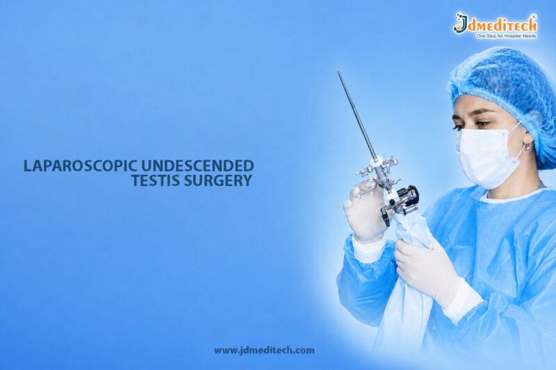
Undescended testis, also known as cryptorchidism, is a condition in which one or both testicles fail to move into the scrotum before birth. It is one of the most common congenital conditions affecting male infants. Although some testes descend naturally within the first few months of life, others require surgical correction to prevent complications related to fertility, hormonal function, and overall testicular health.

Fortunately, modern pediatric urology now offers laparoscopic undescended testis surgery as a highly effective minimally invasive treatment option. This advanced endoscopic technique allows surgeons to locate and reposition the undescended testis with precision while minimizing surgical trauma and shortening recovery time.

In this blog, we will discuss laparoscopic undescended testis surgery, its benefits, indications, surgical procedure, recovery process, risks, and long-term outcomes.

What Is an Undescended Testis?

An undescended testis occurs when a testicle does not descend normally from the abdomen into the scrotum during fetal development. The condition may affect:

  • One testicle (unilateral cryptorchidism)
  • Both testicles (bilateral cryptorchidism)

In many cases, the undescended testis may remain located:

  • Inside the abdomen
  • In the inguinal canal
  • Near the groin region

As a result, the testicle may not develop properly. Moreover, if left untreated, the condition can increase the risk of infertility, testicular torsion, inguinal hernia, and testicular cancer later in life.

What Is Laparoscopic Undescended Testis Surgery?

Laparoscopic undescended testis surgery, also known as laparoscopic orchiopexy, is a minimally invasive pediatric urologic procedure performed to locate and reposition the undescended testicle into the scrotum.

During the surgery, the surgeon uses a laparoscope and specialized instruments to carefully mobilize the testis and secure it inside the scrotum. In certain situations, however, removal may be necessary if the testicle is severely underdeveloped or nonfunctional.

Because the procedure uses tiny incisions, patients generally experience less pain, faster healing, and improved cosmetic results compared to traditional open surgery.

Causes of Undescended Testis

Several factors may contribute to cryptorchidism, including:

  • Premature birth
  • Low birth weight
  • Hormonal abnormalities
  • Genetic conditions
  • Family history
  • Abnormal fetal development

However, in many children, the exact cause remains unknown.

Symptoms of Undescended Testis

The most common sign of cryptorchidism is the absence of one or both testicles from the scrotum. Additionally, other findings may include:

  • Empty scrotal sac
  • Smaller scrotum on one side
  • Palpable lump in the groin
  • Associated inguinal hernia

Usually, the condition is identified during routine newborn or pediatric examinations.

Why Early Treatment Is Important

Timely treatment is extremely important because prolonged undescended testis may lead to several complications. Therefore, doctors usually recommend surgical correction between 6 and 18 months of age.

Early treatment helps:

  • Preserve fertility potential
  • Support normal testicular development
  • Reduce cancer risk
  • Prevent testicular torsion
  • Improve cosmetic appearance

Furthermore, early intervention allows easier long-term monitoring of testicular health and development.

Who Needs Laparoscopic Undescended Testis Surgery?

Doctors may recommend laparoscopic surgery when:

  • The testis cannot be felt during examination
  • The testicle is located inside the abdomen
  • Previous surgery was unsuccessful
  • Bilateral undescended testes are present
  • Imaging suggests intra-abdominal testis

In particular, laparoscopy is highly useful for diagnosing and treating non-palpable testes.

Benefits of Laparoscopic Undescended Testis Surgery
Minimally Invasive Technique

Since the surgery involves small incisions, tissue trauma and postoperative discomfort are significantly reduced.

Better Visualization

Additionally, the laparoscope provides magnified images that help surgeons identify the testis more accurately.

Faster Recovery

Compared to open surgery, children usually recover faster and can resume normal activities sooner.

Reduced Pain

Because of the minimally invasive approach, postoperative pain is generally mild.

Minimal Scarring

Furthermore, tiny incisions provide excellent cosmetic outcomes.

High Success Rate

Most importantly, laparoscopic orchiopexy offers excellent long-term success in repositioning the testis properly.

Diagnostic Evaluation Before Surgery

Before surgery, the pediatric urologist may perform several investigations, including:

  • Physical examination
  • Ultrasound scan
  • MRI in selected cases
  • Hormonal evaluation
  • Diagnostic laparoscopy

These investigations help confirm the location, condition, and viability of the undescended testis.

Step-by-Step Procedure of Laparoscopic Undescended Testis Surgery
Administration of General Anesthesia

First, the child is given general anesthesia to ensure comfort throughout the procedure.

Creation of Small Incisions

Next, tiny incisions are made in the abdomen for insertion of the laparoscope and surgical instruments.

Identification of the Testis

The surgeon then carefully locates the undescended testicle and evaluates its blood supply and mobility.

Mobilization of the Testis

Afterward, the surrounding tissues are gently released to allow adequate movement of the testis.

Placement Inside the Scrotum

Subsequently, the testicle is moved into the scrotum and secured in the correct position.

Closure of Incisions

Finally, the instruments are removed and the small incisions are closed carefully.

Recovery After Laparoscopic Orchiopexy

Recovery after laparoscopic surgery is usually smooth and relatively quick.

Immediate Recovery Period

Initially, children may experience:

  • Mild discomfort
  • Temporary swelling
  • Minor bruising
  • Mild fatigue

However, these symptoms usually improve within a few days.

Return to Normal Activities
  • Light activities: Within a few days
  • School attendance: Usually within 1 week
  • Full recovery: Approximately 2–3 weeks

Nevertheless, parents should carefully follow postoperative care instructions to support proper healing.

Risks and Complications

Although laparoscopic orchiopexy is generally considered safe, certain risks may still occur, including:

  • Bleeding
  • Infection
  • Testicular re-ascent
  • Injury to surrounding structures
  • Reduced blood supply to the testis
  • Testicular atrophy in rare cases

Fortunately, complication rates remain low when the surgery is performed by experienced pediatric urologists.

Long-Term Outcomes of Laparoscopic Undescended Testis Surgery

Most children experience excellent long-term outcomes after surgery. As a result, they may benefit from:

  • Improved fertility potential
  • Better hormonal function
  • Reduced cancer risk
  • Normal scrotal appearance
  • Easier future testicular examination

Additionally, regular follow-up visits help monitor healthy testicular growth and development over time.

Laparoscopic Surgery vs Open Surgery for Undescended Testis
Feature Laparoscopic Surgery Open Surgery
Incision Size Small Larger
Pain Level Less More
Recovery Time Faster Longer
Visualization Better Limited
Cosmetic Results Excellent Moderate
Hospital Stay Short Longer

Therefore, laparoscopic orchiopexy is widely preferred for intra-abdominal undescended testes because of its multiple advantages.

Preventing Complications of Undescended Testis

Early diagnosis and timely surgery are essential for preventing long-term complications. Therefore, parents should seek medical evaluation if:

  • One or both testes are absent from the scrotum
  • The scrotum appears underdeveloped
  • Groin swelling is noticed
  • The child experiences pain or discomfort

Moreover, regular pediatric checkups play an important role in early detection and timely treatment.

Conclusion

Laparoscopic undescended testis surgery is an advanced minimally invasive pediatric urologic procedure that effectively corrects cryptorchidism and helps preserve long-term testicular health. Because of its precision, faster recovery, reduced pain, and excellent success rates, laparoscopic orchiopexy has become the preferred treatment for many children with non-palpable or intra-abdominal testes.

Most importantly, early treatment supports fertility preservation, healthy development, and reduced future complications. Therefore, parents should consult an experienced pediatric urologist for timely diagnosis and personalized treatment planning.


Explore More Product >>

Get Connected:
+91 79909 93062 | +91 63513 72032 | exports@jdmeditech.com 


Laparoscopic PUJ Obstruction Surgery – Advanced Treatment for Hydronephrosis

Pelvi-Ureteric Junction (PUJ) obstruction is a urologic condition in which the flow of urine from the kidney to the ureter becomes blocked or narrowed. As a result, urine accumulates inside the kidney and causes hydronephrosis, which refers to swelling of the kidney. If left untreated, this condition may gradually damage kidney function and lead to severe pain, infections, or kidney stones.

Fortunately, advancements in minimally invasive urology have significantly improved treatment outcomes. Today, laparoscopic PUJ obstruction surgery is considered one of the most effective surgical options for restoring urine flow and preserving kidney health. Moreover, this modern procedure offers smaller incisions, less pain, faster recovery, and excellent long-term success rates compared to traditional open surgery.

In this blog, we will discuss laparoscopic PUJ obstruction surgery, its benefits, indications, procedure details, recovery process, and long-term outcomes.

What Is PUJ Obstruction?

Pelvi-Ureteric Junction obstruction occurs when the connection between the renal pelvis and ureter becomes narrowed or blocked. Consequently, urine cannot drain properly from the kidney into the bladder.

The condition may either be congenital or develop later in life due to secondary causes. In both situations, obstruction increases pressure inside the kidney and may eventually impair renal function.

What Is Laparoscopic PUJ Obstruction Surgery?

Laparoscopic PUJ obstruction surgery, also known as laparoscopic pyeloplasty, is a minimally invasive urologic procedure performed to correct the blockage at the pelvi-ureteric junction.

During the surgery, the surgeon uses a laparoscope and specialized instruments to remove the narrowed segment and reconstruct the urinary drainage pathway. As a result, urine can flow normally from the kidney to the bladder again.

Furthermore, laparoscopic pyeloplasty is widely recognized as the gold standard treatment for symptomatic PUJ obstruction because of its high success rate and minimal surgical trauma.

Causes of PUJ Obstruction

Several factors may contribute to PUJ obstruction, including:

  • Congenital narrowing of the PUJ
  • Crossing blood vessels compressing the ureter
  • Scar tissue formation
  • Kidney stones
  • Previous urinary tract surgery
  • Recurrent urinary infections

In children, congenital abnormalities are the most common cause. However, adults may develop the condition later because of acquired urologic problems.

Symptoms of PUJ Obstruction

Symptoms can vary depending on the severity of the blockage. Common symptoms include:

  • Flank or abdominal pain
  • Swelling of the kidney (hydronephrosis)
  • Recurrent urinary tract infections
  • Blood in urine
  • Nausea and vomiting
  • Kidney stones
  • Fever in case of infection

Sometimes, patients may remain asymptomatic. In such cases, the condition is often detected incidentally during imaging studies performed for unrelated reasons.

Who Needs Laparoscopic PUJ Obstruction Surgery?

Doctors may recommend surgery if:

  • Significant hydronephrosis is present
  • Kidney drainage is severely impaired
  • Persistent flank pain occurs
  • Recurrent infections develop
  • Kidney function begins to decline
  • Kidney stones are associated with obstruction

Importantly, early surgical intervention helps preserve long-term kidney function and prevents irreversible damage.

Benefits of Laparoscopic PUJ Obstruction Surgery
Minimally Invasive Technique

Since the procedure uses small incisions, patients experience less tissue trauma and reduced postoperative discomfort.

Faster Recovery

Compared to open surgery, laparoscopic pyeloplasty allows patients to recover more quickly and resume daily activities sooner.

Reduced Pain

Because of the minimally invasive approach, postoperative pain is usually much lower.

Smaller Scars

Additionally, the tiny incisions provide improved cosmetic outcomes.

Shorter Hospital Stay

Most patients can return home within a few days after surgery.

High Success Rate

Laparoscopic pyeloplasty provides excellent long-term success in restoring urine flow and relieving obstruction.

Kidney Function Preservation

Most importantly, timely surgery helps prevent further kidney damage and improves urinary drainage.

Diagnostic Evaluation Before Surgery

Before surgery, the urologist may perform several investigations, including:

  • Ultrasound examination
  • CT scan or MRI
  • Intravenous urography (IVU)
  • Renal nuclear scan
  • Blood tests
  • Urine analysis

These diagnostic tests help determine the severity of the obstruction and evaluate kidney function accurately.

Step-by-Step Procedure of Laparoscopic PUJ Obstruction Surgery
Administration of General Anesthesia

First, the patient is given general anesthesia to ensure comfort throughout the procedure.

Creation of Small Incisions

Next, several tiny incisions are made in the abdomen for insertion of laparoscopic instruments.

Identification of the Obstruction

The surgeon then carefully identifies the narrowed PUJ segment and evaluates nearby structures.

Removal of Narrowed Segment

Afterward, the obstructed portion is removed to eliminate the blockage.

Reconstruction of the Urinary Pathway

The healthy ureter is subsequently reconnected to the renal pelvis to restore smooth urine drainage.

Placement of Ureteric Stent

In many cases, a temporary internal stent is placed to support healing and maintain drainage.

Closure of Incisions

Finally, after ensuring proper urine flow and bleeding control, the instruments are removed and the incisions are closed.

Recovery After Laparoscopic PUJ Obstruction Surgery

Recovery is generally smoother and faster than traditional open surgery.

Immediate Recovery Period

Initially, patients may experience:

  • Mild abdominal discomfort
  • Temporary urinary symptoms
  • Fatigue
  • Minor bloating

However, these symptoms usually improve within a few days.

Hospital Stay

Typically, patients remain hospitalized for about 2–4 days depending on their recovery progress.

Return to Normal Activities
  • Light activities: Within 1–2 weeks
  • Office work: Usually within 2–3 weeks
  • Full recovery: Approximately 4–6 weeks

Nevertheless, recovery duration may vary depending on the patient’s health condition and surgical complexity.

Risks and Complications

Although laparoscopic pyeloplasty is generally considered safe, certain risks may occur, including:

  • Bleeding
  • Infection
  • Urine leakage
  • Injury to nearby organs
  • Recurrence of obstruction
  • Stent-related discomfort

Fortunately, choosing an experienced laparoscopic urologist can significantly reduce complication risks.

Long-Term Outcomes of Laparoscopic Pyeloplasty

Most patients experience excellent long-term outcomes after surgery. These benefits may include:

  • Improved kidney drainage
  • Relief from flank pain
  • Reduced infection risk
  • Preservation of kidney function
  • Better quality of life

Additionally, follow-up imaging studies are usually performed to confirm successful correction of the obstruction.

Laparoscopic Surgery vs Open Surgery for PUJ Obstruction
Feature Laparoscopic Surgery Open Surgery
Incision Size Small Large
Pain Level Less More
Recovery Time Faster Longer
Hospital Stay Short Extended
Cosmetic Results Better Visible Scar
Blood Loss Minimal Higher

Therefore, laparoscopic pyeloplasty has become the preferred surgical option in many advanced urology centers.

Preventing Kidney Damage from PUJ Obstruction

Early diagnosis and treatment are extremely important for preventing permanent kidney damage. Therefore, patients should seek medical evaluation if they experience:

  • Persistent flank pain
  • Recurrent urinary infections
  • Kidney swelling
  • Blood in urine
  • Repeated kidney stones

Moreover, regular follow-up after surgery helps monitor kidney function and ensures long-term treatment success.

Conclusion

Laparoscopic PUJ obstruction surgery is an advanced minimally invasive urologic procedure that effectively treats hydronephrosis and restores normal urinary drainage. Because of its precision, faster recovery, reduced pain, and excellent success rates, laparoscopic pyeloplasty has become the preferred treatment for PUJ obstruction.

Most importantly, timely intervention helps preserve kidney function and prevents serious complications. Therefore, individuals experiencing symptoms of urinary obstruction should consult an experienced urologist for proper diagnosis and personalized treatment planning.


Explore More Product >>

+91 79909 93062 | +91 63513 72032 | exports@jdmeditech.com 


Laparoscopic Ectopic Pregnancy Surgery – Advanced Emergency Tubal Treatment

An ectopic pregnancy is a serious medical condition in which a fertilized egg implants outside the uterus, most commonly in the fallopian tube. Since the pregnancy cannot develop normally, immediate medical attention is necessary to prevent life-threatening complications such as tubal rupture and internal bleeding. Modern gynecologic advancements now allow safer and faster treatment through laparoscopic ectopic pregnancy surgery.

Laparoscopic ectopic pregnancy surgery is a minimally invasive emergency procedure designed to remove the ectopic pregnancy while preserving reproductive health whenever possible. Compared to traditional open surgery, laparoscopy offers reduced pain, smaller incisions, quicker recovery, and shorter hospital stays.

This blog explains the procedure, indications, benefits, recovery process, risks, and fertility outcomes associated with laparoscopic surgery for ectopic pregnancy.

What Is Laparoscopic Ectopic Pregnancy Surgery?

Laparoscopic ectopic pregnancy surgery is an advanced endoscopic gynecologic procedure used to treat ectopic pregnancies through small abdominal incisions. A laparoscope, which is a thin camera-equipped instrument, allows the surgeon to visualize the pelvic organs and remove the abnormal pregnancy tissue safely.

Depending on the severity and condition of the fallopian tube, the surgeon may perform:

  • Salpingostomy (removal of pregnancy while preserving the tube)
  • Salpingectomy (removal of the affected fallopian tube)
  • Control of internal bleeding
  • Removal of damaged tissue

The procedure is commonly performed as an emergency surgery when ectopic pregnancy poses immediate health risks.

Understanding Ectopic Pregnancy

In a normal pregnancy, the fertilized egg implants inside the uterus. In ectopic pregnancy, implantation occurs outside the uterus, most often in:

  • Fallopian tube
  • Ovary
  • Cervix
  • Abdominal cavity

Since these areas cannot support fetal growth, the pregnancy becomes dangerous if untreated.

Symptoms of Ectopic Pregnancy

Common warning signs include:

  • Severe lower abdominal pain
  • Vaginal bleeding
  • Missed menstrual period
  • Shoulder pain
  • Dizziness or fainting
  • Pelvic discomfort

A ruptured ectopic pregnancy may lead to severe internal bleeding and requires emergency surgical intervention.

Causes and Risk Factors

Several factors may increase the risk of ectopic pregnancy, including:

  • Previous ectopic pregnancy
  • Pelvic inflammatory disease
  • Fallopian tube damage
  • Infertility treatments
  • Endometriosis
  • Prior pelvic surgery
  • Smoking

However, ectopic pregnancy can also occur without obvious risk factors.

Who Needs Laparoscopic Ectopic Pregnancy Surgery?

Doctors may recommend surgery when:

  • The ectopic pregnancy is large
  • Internal bleeding is suspected
  • The fallopian tube has ruptured
  • Medical treatment fails
  • Severe abdominal pain is present
  • The patient is medically unstable

In many emergency situations, laparoscopy becomes the preferred surgical approach due to its effectiveness and faster recovery.

Benefits of Laparoscopic Ectopic Pregnancy Surgery
Minimally Invasive Procedure

Small incisions reduce surgical trauma and improve cosmetic results.

Faster Recovery

Most patients recover more quickly than with open abdominal surgery.

Less Postoperative Pain

Minimally invasive techniques help reduce discomfort after surgery.

Shorter Hospital Stay

Many patients are discharged within 24 to 48 hours.

Reduced Blood Loss

Laparoscopic surgery provides better visualization and bleeding control.

Fertility Preservation

Whenever possible, surgeons attempt to preserve the healthy fallopian tube and reproductive organs.

Preoperative Evaluation

Before surgery, doctors may perform:

  • Pelvic examination
  • Pregnancy blood tests (hCG levels)
  • Ultrasound scan
  • Blood pressure monitoring
  • Blood grouping and hemoglobin testing

Rapid diagnosis is essential to prevent complications.

Step-by-Step Procedure of Laparoscopic Ectopic Pregnancy Surgery
Administration of Anesthesia

The procedure is performed under general anesthesia.

Small Abdominal Incisions

Tiny incisions are made for insertion of the laparoscope and surgical instruments.

Visualization of Pelvic Organs

The surgeon carefully examines the uterus, ovaries, and fallopian tubes.

Removal of Ectopic Pregnancy

The abnormal pregnancy tissue is removed using minimally invasive surgical techniques.

Tube Preservation or Removal

Depending on the extent of damage:

  • The tube may be preserved through salpingostomy
  • The damaged tube may be removed through salpingectomy
Bleeding Control and Closure

After controlling bleeding, the instruments are removed and the incisions are closed.

Recovery After Laparoscopic Ectopic Pregnancy Surgery

Recovery is usually smooth and relatively fast.

Immediate Recovery Period

Patients may experience:

  • Mild abdominal discomfort
  • Shoulder pain due to surgical gas
  • Fatigue
  • Light vaginal bleeding
Return to Routine Activities
  • Light activities: Within a few days
  • Office work: Usually within 1–2 weeks
  • Full recovery: Approximately 2–4 weeks

Recovery time depends on the patient’s overall health and severity of the ectopic pregnancy.

Fertility After Ectopic Pregnancy Surgery

Many women can conceive successfully after treatment. Fertility outcomes depend on:

  • Health of the remaining fallopian tube
  • Severity of tubal damage
  • Presence of underlying fertility issues
  • Early diagnosis and treatment

Doctors may advise waiting several months before attempting another pregnancy.

Risks and Complications

Although laparoscopic surgery is generally safe, possible complications include:

  • Bleeding
  • Infection
  • Injury to nearby organs
  • Scar tissue formation
  • Future ectopic pregnancy risk
  • Anesthesia-related complications

Choosing an experienced laparoscopic gynecologic surgeon helps minimize these risks.

Laparoscopic Surgery vs Open Surgery
Feature Laparoscopic Surgery Open Surgery
Incision Size Small Large
Pain Level Less More
Recovery Time Faster Longer
Hospital Stay Short Extended
Scarring Minimal More Visible
Blood Loss Lower Higher

Because of these advantages, laparoscopy is widely preferred whenever the patient is medically stable.

Preventing Future Ectopic Pregnancy

Although not all ectopic pregnancies can be prevented, certain measures may reduce risk:

  • Early treatment of pelvic infections
  • Smoking cessation
  • Regular gynecologic checkups
  • Timely management of reproductive health conditions

Women with a history of ectopic pregnancy should seek early ultrasound evaluation during future pregnancies.

When to Seek Emergency Medical Attention

Immediate medical care is necessary if you experience:

  • Sudden severe abdominal pain
  • Heavy vaginal bleeding
  • Dizziness or fainting
  • Shoulder pain
  • Weakness or collapse

Prompt treatment can prevent life-threatening complications.

Conclusion

Laparoscopic ectopic pregnancy surgery is a modern minimally invasive gynecologic procedure that provides safe and effective emergency treatment for ectopic pregnancy. With smaller incisions, faster recovery, reduced pain, and fertility-preserving benefits, laparoscopy has become the preferred surgical approach for many patients.

Early diagnosis and timely intervention are essential for protecting reproductive health and preventing serious complications. Women experiencing symptoms of ectopic pregnancy should seek immediate medical attention from a qualified gynecologic specialist.


Explore More Product >>

+91 79909 93062 | +91 63513 72032 | exports@jdmeditech.com 

Laparoscopic Endometriosis Surgery – Advanced Treatment for Pain Relief & Fertility Preservation

Endometriosis is a chronic gynecological condition that affects millions of women worldwide. It occurs when tissue similar to the lining of the uterus grows outside the uterus, leading to pelvic pain, painful periods, infertility, and discomfort during daily activities. Fortunately, advances in minimally invasive gynecology have transformed treatment outcomes. Among the most effective treatment options is laparoscopic endometriosis surgery, a modern surgical technique designed to relieve symptoms, remove endometriosis lesions, and preserve reproductive health.

This advanced procedure offers faster recovery, minimal scarring, and improved fertility outcomes compared to traditional open surgery. In this blog, we will explore the procedure, benefits, indications, recovery process, and why laparoscopic surgery has become the preferred treatment for endometriosis.

What Is Laparoscopic Endometriosis Surgery?

Laparoscopic endometriosis surgery is a minimally invasive gynecologic procedure performed to diagnose and treat endometriosis. During the surgery, a surgeon inserts a thin camera called a laparoscope through small abdominal incisions to visualize and remove abnormal endometrial tissue.

The procedure may involve:

  • Excision of endometriosis implants
  • Removal of ovarian cysts known as endometriomas
  • Separation of pelvic adhesions
  • Restoration of normal pelvic anatomy
  • Preservation of reproductive organs whenever possible

Because the surgery uses small incisions, patients generally experience less pain and quicker healing.

Understanding Endometriosis

Endometriosis commonly affects:

  • Ovaries
  • Fallopian tubes
  • Pelvic lining
  • Bladder
  • Bowel
  • Uterine ligaments

Common symptoms include:

  • Severe menstrual cramps
  • Chronic pelvic pain
  • Pain during intercourse
  • Heavy menstrual bleeding
  • Painful urination or bowel movements
  • Difficulty conceiving

In many cases, symptoms worsen over time if left untreated.

Who Needs Laparoscopic Endometriosis Surgery?

Doctors may recommend laparoscopic surgery when:

  • Pelvic pain becomes severe
  • Medical therapy fails to provide relief
  • Ovarian cysts are present
  • Fertility is affected
  • Endometriosis causes organ involvement
  • Suspicion of advanced-stage disease exists

Additionally, surgery may be advised for women planning pregnancy who want to improve fertility potential.

Benefits of Laparoscopic Endometriosis Surgery
Effective Pain Relief

One of the primary advantages of laparoscopic surgery is long-term relief from pelvic pain and menstrual discomfort.

Fertility Preservation

The procedure helps restore normal pelvic anatomy while preserving healthy ovarian tissue and reproductive organs.

Minimally Invasive Approach

Small incisions reduce trauma to surrounding tissues and improve cosmetic outcomes.

Faster Recovery

Patients typically recover more quickly compared to open surgery.

Reduced Hospital Stay

Most women can return home within 24 hours after surgery.

Better Visualization

The laparoscope provides magnified images that help surgeons precisely identify and remove endometriosis lesions.

Preoperative Evaluation

Before surgery, the gynecologist may perform:

  • Detailed pelvic examination
  • Ultrasound imaging
  • MRI scan in complex cases
  • Blood investigations
  • Fertility assessment if required

Patients are also counseled regarding surgical goals, fertility planning, and recovery expectations.

Step-by-Step Procedure of Laparoscopic Endometriosis Surgery
Anesthesia Administration

The surgery is performed under general anesthesia.

Creation of Small Incisions

Tiny incisions are made in the abdomen for insertion of the laparoscope and surgical instruments.

Pelvic Examination

The surgeon carefully examines the pelvic organs to identify endometriosis lesions, adhesions, and cysts.

Removal of Endometriosis Tissue

Abnormal tissue is removed using advanced surgical techniques such as excision, laser therapy, or electrosurgery.

Adhesiolysis

Scar tissue and adhesions are separated to restore normal pelvic anatomy.

Completion of Surgery

After ensuring bleeding control, the instruments are removed and the incisions are closed.

Types of Endometriosis Treated with Laparoscopy
Superficial Endometriosis

Small lesions located on pelvic surfaces.

Ovarian Endometriomas

Chocolate cysts affecting the ovaries.

Deep Infiltrating Endometriosis

Advanced disease involving deeper tissues, bowel, bladder, or ligaments.

Adhesive Disease

Scar tissue causing pelvic organ distortion.

Recovery After Laparoscopic Endometriosis Surgery

Recovery is generally smooth and quicker than traditional surgery.

Immediate Postoperative Period

Patients may experience:

  • Mild abdominal discomfort
  • Shoulder pain from surgical gas
  • Temporary fatigue
  • Minor bloating
Return to Daily Activities
  • Light activities: Within a few days
  • Office work: Usually within 1–2 weeks
  • Full recovery: Approximately 2–4 weeks

However, recovery time may vary depending on disease severity and extent of surgery.

Fertility Outcomes After Surgery

Laparoscopic surgery plays an important role in fertility preservation and improvement. By removing endometriosis lesions and restoring pelvic anatomy, the procedure may enhance natural conception chances.

Women with infertility related to endometriosis often benefit significantly from early surgical management.

Risks and Complications

Although laparoscopic endometriosis surgery is generally safe, potential risks include:

  • Bleeding
  • Infection
  • Injury to surrounding organs
  • Adhesion recurrence
  • Endometriosis recurrence
  • Temporary urinary or bowel symptoms

Choosing an experienced laparoscopic gynecologic surgeon significantly reduces these risks.

Preventing Endometriosis Recurrence

After surgery, doctors may recommend:

  • Hormonal therapy
  • Lifestyle modifications
  • Regular gynecologic follow-up
  • Fertility planning support

Early treatment and ongoing monitoring help reduce recurrence rates.

Why Choose Laparoscopic Surgery Over Open Surgery?

Compared to conventional open surgery, laparoscopy offers:

Feature Laparoscopic Surgery Open Surgery
Incision Size Small Large
Pain Level Less More
Recovery Time Faster Longer
Hospital Stay Short Extended
Blood Loss Minimal Higher
Cosmetic Results Better Visible Scar

Because of these advantages, laparoscopy is considered the gold standard for endometriosis treatment.

When to Consult a Gynecologic Specialist

Seek medical consultation if you experience:

  • Severe menstrual pain
  • Chronic pelvic pain
  • Difficulty conceiving
  • Pain during intercourse
  • Heavy menstrual bleeding
  • Persistent gastrointestinal symptoms during periods

Early diagnosis and timely treatment can prevent complications and improve quality of life.

Conclusion

Laparoscopic endometriosis surgery is an advanced and effective gynecologic procedure that offers significant pain relief, fertility preservation, and faster recovery. With modern minimally invasive techniques, surgeons can precisely remove endometriosis tissue while protecting healthy reproductive organs.

Women suffering from chronic pelvic pain or infertility due to endometriosis should consult an experienced gynecologic specialist to explore personalized treatment options. Early intervention not only improves symptoms but also enhances long-term reproductive and overall health outcomes.


Explore More Product >>

Get Connected:
+91 79909 93062 | +91 63513 72032 | exports@jdmeditech.com 

Laparoscopic Tubal Ligation Surgery – Minimally Invasive Female Sterilization Procedure

Laparoscopic tubal ligation surgery is a widely performed minimally invasive gynecologic procedure used for permanent female sterilization. The procedure involves blocking, sealing, or cutting the fallopian tubes to prevent pregnancy by stopping the egg and sperm from meeting. Because it provides long-term contraception without affecting hormonal balance, laparoscopic tubal ligation has become a highly effective and reliable birth control option for women who no longer wish to conceive.

Traditionally, sterilization procedures required larger abdominal incisions and longer recovery periods. However, advancements in laparoscopic surgery now allow gynecologic surgeons to perform tubal ligation through small abdominal incisions using high-definition cameras and specialized endoscopic instruments. Consequently, patients benefit from reduced postoperative discomfort, faster recovery, shorter hospital stays, and minimal scarring.

Today, laparoscopic tubal ligation remains one of the most commonly chosen permanent contraception procedures worldwide due to its safety, effectiveness, and minimally invasive advantages.

What Is Laparoscopic Tubal Ligation Surgery?

Laparoscopic tubal ligation surgery is a minimally invasive procedure performed to permanently block or seal the fallopian tubes.

The fallopian tubes are responsible for carrying eggs from the ovaries to the uterus. During tubal ligation, the surgeon interrupts the pathway of the tubes using clips, rings, cauterization, or surgical cutting techniques. As a result, fertilization cannot occur.

The procedure is performed using a laparoscope, which is a thin camera-equipped instrument inserted through small abdominal incisions. This provides magnified visualization of the reproductive organs and allows highly precise surgical treatment.

Ultimately, the primary goal of laparoscopic tubal ligation is to provide safe, permanent contraception with minimal surgical trauma.

Why Is Laparoscopic Tubal Ligation Performed?

Laparoscopic tubal ligation is performed for women seeking permanent birth control.

Common Reasons for Tubal Ligation Include:
  • Completion of family planning
  • Desire for permanent contraception
  • Medical conditions making pregnancy risky
  • Intolerance to hormonal contraception
  • Preference for long-term contraceptive solution
  • Genetic concerns regarding pregnancy

Additionally, the procedure eliminates the need for daily or temporary contraceptive methods.

Understanding Female Sterilization

Female sterilization is considered a permanent method of contraception.

How Tubal Ligation Prevents Pregnancy

After tubal ligation:

  • Eggs released from the ovaries cannot reach the uterus
  • Sperm cannot travel through blocked tubes
  • Fertilization is prevented

Importantly, tubal ligation does not affect menstrual cycles, hormonal production, or sexual function.

Types of Tubal Ligation Techniques

Several laparoscopic techniques may be used to occlude the fallopian tubes.

Technique Method Common Use
Tubal Clips Application of clips to tubes Minimally damaging approach
Tubal Rings Placement of silicone rings Common laparoscopic method
Electrocautery Sealing tubes with electrical energy Permanent occlusion
Partial Salpingectomy Removal of tube segments Long-term sterilization

Ultimately, the surgeon selects the most appropriate technique based on patient preference and clinical considerations.

Who Is a Suitable Candidate for Laparoscopic Tubal Ligation?

Generally, laparoscopic tubal ligation may be recommended for:

  • Women seeking permanent contraception
  • Individuals medically fit for minimally invasive surgery
  • Women certain about avoiding future pregnancy
  • Patients unable to use hormonal contraception
  • Women with completed family planning goals

Importantly, counseling is essential before surgery because tubal ligation is considered a permanent procedure.

Preoperative Evaluation Before Surgery

Before laparoscopic tubal ligation, the gynecologist performs detailed assessments to ensure patient safety and informed decision-making.

Medical History Review

Initially, the doctor evaluates:

  • Reproductive history
  • Current contraceptive methods
  • Previous pregnancies
  • Existing medical conditions
  • Medication use
  • Surgical history
Physical Examination

Furthermore, pelvic and general physical examinations help assess overall health and surgical fitness.

Diagnostic Investigations

Additional tests may include:

  • Pregnancy testing
  • Blood investigations
  • Pelvic ultrasound in selected cases
  • Pre-anesthesia evaluation

Consequently, these evaluations help ensure safe surgical planning.

How Laparoscopic Tubal Ligation Surgery Is Performed

Typically, laparoscopic tubal ligation is performed under general anesthesia using advanced minimally invasive surgical equipment.

Step-by-Step Surgical Procedure
1. Creation of Small Incisions

First, the surgeon creates one or more tiny abdominal incisions for insertion of the laparoscope and surgical instruments.

2. Inflation of the Abdomen

Next, carbon dioxide gas is introduced into the abdomen to create adequate working space.

3. Visualization of Reproductive Organs

Afterward, the laparoscope provides magnified views of the uterus, ovaries, and fallopian tubes.

4. Identification of the Fallopian Tubes

The surgeon carefully identifies both fallopian tubes and surrounding pelvic structures.

5. Tubal Occlusion Procedure

Then, the tubes are blocked using clips, rings, cauterization, or surgical division depending on the chosen technique.

6. Inspection for Bleeding or Complications

Furthermore, the pelvic cavity is inspected to ensure proper occlusion and bleeding control.

7. Closure of Incisions

Finally, the instruments are removed and the small incisions are closed with sutures or surgical glue.

Overall, the procedure usually takes between 20 minutes and 1 hour.

Benefits of Laparoscopic Tubal Ligation Surgery

Laparoscopic tubal ligation offers several important advantages compared to traditional open sterilization procedures.

Minimally Invasive Technique

Because the procedure uses small incisions, postoperative discomfort and tissue trauma are significantly reduced.

Permanent Birth Control

The procedure provides highly effective long-term contraception.

Faster Recovery

Additionally, patients usually recover quickly and resume daily activities sooner.

Minimal Scarring

Small incisions result in improved cosmetic appearance.

No Ongoing Maintenance

Unlike temporary contraceptive methods, tubal ligation requires no daily management.

Hormone-Free Contraception

Importantly, the procedure does not interfere with natural hormone production.

Recovery After Laparoscopic Tubal Ligation

Recovery after laparoscopic tubal ligation is generally smooth and relatively fast.

Common Postoperative Symptoms

Initially, patients may experience:

  • Mild abdominal discomfort
  • Temporary bloating
  • Fatigue
  • Mild pelvic cramping
  • Shoulder pain from surgical gas

However, these symptoms usually improve within several days.

Recovery Guidelines

To support proper healing, patients are advised to:

  • Walk regularly after surgery
  • Stay hydrated
  • Avoid heavy lifting temporarily
  • Follow incision care instructions
  • Attend follow-up appointments

As a result, many individuals return to normal activities within a few days.

Effectiveness of Tubal Ligation

Laparoscopic tubal ligation is considered one of the most effective forms of permanent contraception.

Success and Failure Rates

The procedure has:

  • Very high contraceptive success rates
  • Low pregnancy risk after surgery
  • Long-term effectiveness without maintenance

Nevertheless, although rare, pregnancy may occasionally occur after tubal ligation.

Risks and Complications

Although laparoscopic tubal ligation is considered very safe, certain complications may occasionally occur.

Possible Complications Include:
  • Bleeding
  • Infection
  • Injury to nearby organs
  • Reaction to anesthesia
  • Tubal ligation failure
  • Ectopic pregnancy
  • Adhesion formation

However, complication rates are generally low when surgery is performed by experienced gynecologic surgeons.

Emotional and Reproductive Considerations

Because tubal ligation is considered permanent, emotional and reproductive counseling is extremely important before surgery.

Important Considerations Include:
  • Future pregnancy plans
  • Emotional readiness
  • Relationship factors
  • Understanding reversibility limitations
  • Alternative contraceptive options

Therefore, patients should make informed decisions after detailed counseling.

Lifestyle Tips After Surgery

Healthy lifestyle habits may support healing and overall reproductive wellness after surgery.

Recommended Lifestyle Measures

Patients are encouraged to:

  • Maintain balanced nutrition
  • Stay physically active
  • Avoid smoking
  • Follow medical instructions carefully
  • Attend routine gynecologic follow-ups

Consequently, these habits may improve recovery and long-term health.

Choosing the Right Gynecologic Surgeon

Successful laparoscopic tubal ligation requires expertise in minimally invasive gynecologic surgery.

Patients Should Look For:
  • Experienced laparoscopic gynecologic surgeons
  • Advanced surgical facilities
  • Comprehensive contraceptive counseling
  • Strong patient safety standards
  • Quality postoperative care

Ultimately, specialized surgical expertise improves both safety and patient satisfaction.

Conclusion

Laparoscopic tubal ligation surgery is a safe, effective, and minimally invasive solution for permanent female contraception. By combining modern laparoscopic technology with precise surgical techniques, the procedure offers reliable pregnancy prevention with reduced pain, faster recovery, and minimal scarring.

Moreover, proper preoperative counseling and individualized treatment planning help ensure informed decision-making and long-term patient satisfaction. Therefore, women considering permanent contraception should consult an experienced gynecologic surgeon to discuss the most suitable treatment approach.

With continuous advancements in minimally invasive gynecologic surgery, laparoscopic tubal ligation continues to remain one of the most trusted and effective sterilization procedures worldwide.


Explore More Product >>

Get Connected:
+91 79909 93062 | +91 63513 72032 | exports@jdmeditech.com 

Laparoscopic Salpingectomy Surgery – Minimally Invasive Fallopian Tube Removal

Laparoscopic salpingectomy surgery is an advanced minimally invasive gynecologic procedure performed to remove one or both fallopian tubes for the treatment of various tubal diseases and reproductive health conditions. The fallopian tubes play a crucial role in fertility by transporting eggs from the ovaries to the uterus. However, infections, ectopic pregnancy, tubal blockage, hydrosalpinx, endometriosis, and certain cancers may damage the tubes and require surgical removal.

Traditionally, salpingectomy procedures were performed through open abdominal surgery with larger incisions and prolonged recovery periods. However, modern laparoscopic techniques now allow surgeons to remove diseased fallopian tubes through small abdominal incisions using high-definition cameras and specialized endoscopic instruments. Consequently, patients benefit from reduced postoperative pain, minimal scarring, shorter hospital stays, and faster recovery.

Today, laparoscopic salpingectomy has become one of the preferred minimally invasive procedures for treating fallopian tube disorders while improving patient safety and recovery outcomes.

What Is Laparoscopic Salpingectomy Surgery?

Laparoscopic salpingectomy surgery is a minimally invasive procedure used to remove one or both fallopian tubes through small abdominal incisions with the assistance of a laparoscope.

A laparoscope is a thin camera-equipped instrument that provides magnified visualization of the pelvic organs during surgery. Specialized laparoscopic instruments are used to carefully separate the affected fallopian tube from surrounding tissues and blood vessels before safely removing it.

Depending on the medical condition, the procedure may involve:

  • Unilateral salpingectomy (removal of one fallopian tube)
  • Bilateral salpingectomy (removal of both fallopian tubes)

Ultimately, the primary goal of laparoscopic salpingectomy is to treat tubal disease effectively while minimizing surgical trauma and preserving overall pelvic health.

Why Is Laparoscopic Salpingectomy Performed?

Laparoscopic salpingectomy is recommended for several gynecologic and reproductive health conditions involving the fallopian tubes.

Common Indications for Surgery Include:
  • Ectopic pregnancy
  • Hydrosalpinx
  • Tubal infection
  • Fallopian tube blockage
  • Endometriosis
  • Tubal damage
  • Ovarian or tubal cancer
  • Risk reduction for ovarian cancer
  • Pelvic inflammatory disease
  • Permanent contraception

Additionally, early surgical intervention may help prevent serious complications such as rupture, infection spread, and infertility.

Understanding Fallopian Tube Disorders

Several conditions affecting the fallopian tubes may require salpingectomy surgery.

Ectopic Pregnancy

An ectopic pregnancy occurs when a fertilized egg implants outside the uterus, most commonly inside the fallopian tube. This condition can become life-threatening if untreated.

Hydrosalpinx

Hydrosalpinx refers to fluid-filled blockage and swelling of the fallopian tube, which may interfere with fertility and IVF success.

Pelvic Inflammatory Disease

Severe pelvic infections may permanently damage the fallopian tubes and cause chronic pelvic pain or infertility.

Endometriosis

Endometriosis affecting the fallopian tubes may lead to scarring, pain, and reproductive complications.

Therefore, timely diagnosis and treatment are extremely important.

Who Is a Suitable Candidate for Laparoscopic Salpingectomy?

Generally, laparoscopic salpingectomy may be recommended for:

  • Women with tubal disease
  • Patients with ectopic pregnancy
  • Individuals with severe hydrosalpinx
  • Women seeking permanent sterilization
  • Patients medically fit for minimally invasive surgery

Importantly, the final treatment plan depends on fertility goals, disease severity, and overall reproductive health.

Preoperative Evaluation Before Surgery

Before laparoscopic salpingectomy, the gynecologic surgeon performs comprehensive assessments to ensure safe surgical planning.

Medical History Review

Initially, the doctor evaluates:

  • Menstrual history
  • Fertility concerns
  • Pelvic pain symptoms
  • Previous pregnancies
  • Prior surgeries
  • History of infections
  • Medication use
Physical Examination

Furthermore, pelvic examination helps assess tenderness, masses, and associated abnormalities.

Diagnostic Investigations

Additional tests may include:

  • Pelvic ultrasound
  • Blood investigations
  • Pregnancy testing
  • MRI imaging
  • CT scan in selected cases
  • Fertility evaluation

Consequently, these evaluations help determine the safest and most appropriate surgical approach.

How Laparoscopic Salpingectomy Surgery Is Performed

Typically, laparoscopic salpingectomy is performed under general anesthesia using advanced minimally invasive surgical equipment.

Step-by-Step Surgical Procedure
1. Creation of Small Incisions

First, the surgeon creates several tiny abdominal incisions for insertion of the laparoscope and surgical instruments.

2. Inflation of the Abdomen

Next, carbon dioxide gas is introduced into the abdomen to create adequate working space.

3. Visualization of Pelvic Structures

Afterward, the laparoscope provides magnified views of the uterus, ovaries, fallopian tubes, and surrounding tissues.

4. Identification of the Diseased Fallopian Tube

The surgeon carefully examines the affected tube and evaluates nearby structures.

5. Separation and Removal of the Tube

Then, the diseased fallopian tube is carefully separated from surrounding tissues and blood vessels before removal.

6. Inspection for Bleeding or Complications

Furthermore, the pelvic cavity is inspected to ensure complete treatment and bleeding control.

7. Closure of Incisions

Finally, the instruments are removed and the small incisions are closed with sutures or surgical glue.

Overall, the procedure usually takes between 30 minutes and 2 hours depending on surgical complexity.

Benefits of Laparoscopic Salpingectomy Surgery

Laparoscopic salpingectomy offers several important advantages compared to traditional open surgery.

Minimally Invasive Technique

Because the procedure uses small incisions, postoperative pain and tissue trauma are significantly reduced.

Faster Recovery

Additionally, patients often return to normal activities sooner.

Reduced Blood Loss

Modern laparoscopic techniques help minimize surgical bleeding.

Lower Infection Risk

Smaller incisions may reduce wound-related complications.

Better Cosmetic Outcomes

Minimal scarring improves postoperative appearance.

Shorter Hospital Stay

Consequently, many patients return home within 24 hours after surgery.

Recovery After Laparoscopic Salpingectomy

Recovery after laparoscopic salpingectomy is generally smooth and relatively fast.

Common Postoperative Symptoms

Initially, patients may experience:

  • Mild abdominal discomfort
  • Fatigue
  • Temporary bloating
  • Light vaginal spotting
  • Shoulder pain from surgical gas

However, these symptoms usually improve within several days.

Recovery Guidelines

To support proper healing, patients are advised to:

  • Walk regularly after surgery
  • Stay hydrated
  • Avoid heavy lifting temporarily
  • Follow dietary recommendations
  • Maintain incision hygiene
  • Attend follow-up appointments

As a result, many individuals return to routine activities within 1 to 3 weeks.

Fertility Considerations After Salpingectomy

Fertility outcomes after salpingectomy depend on whether one or both fallopian tubes are removed.

Important Fertility Considerations
  • Removal of one tube may still allow natural pregnancy
  • Bilateral salpingectomy eliminates natural conception
  • IVF may remain an option after bilateral salpingectomy
  • Fertility counseling may be recommended before surgery

Therefore, discussing reproductive goals with a gynecologic specialist is extremely important.

Risks and Complications

Although laparoscopic salpingectomy is considered safe, certain complications may occasionally occur.

Possible Complications Include:
  • Bleeding
  • Infection
  • Injury to nearby organs
  • Adhesion formation
  • Blood clots
  • Anesthesia-related complications
  • Fertility changes

Nevertheless, complication rates are generally low when surgery is performed by experienced laparoscopic surgeons.

Success Rates of Laparoscopic Salpingectomy

Laparoscopic salpingectomy has excellent success rates for managing tubal diseases and gynecologic complications.

Factors Affecting Outcomes

Several factors influence surgical success, including:

  • Underlying tubal condition
  • Fertility status
  • Severity of disease
  • Patient health condition
  • Surgical expertise
General Outcomes

Modern laparoscopic salpingectomy typically provides:

  • Effective disease management
  • Reduced pelvic pain
  • Faster recovery
  • Lower complication rates
  • Improved quality of life

Therefore, minimally invasive salpingectomy remains a highly effective treatment option.

Lifestyle Tips After Surgery

Healthy lifestyle habits may support healing and reproductive health after surgery.

Recommended Lifestyle Measures

Patients are encouraged to:

  • Maintain balanced nutrition
  • Stay physically active after recovery
  • Avoid smoking
  • Manage stress effectively
  • Attend routine gynecologic follow-ups

Consequently, these habits may support long-term wellness and recovery.

Choosing the Right Gynecologic Surgeon

Successful laparoscopic salpingectomy requires expertise in minimally invasive gynecologic surgery.

Patients Should Look For:
  • Experienced laparoscopic gynecologic surgeons
  • Advanced surgical facilities
  • Expertise in fertility-related surgery
  • Comprehensive postoperative care
  • Strong patient safety standards

Ultimately, specialized surgical expertise improves patient safety and treatment outcomes.

Conclusion

Laparoscopic salpingectomy surgery is a modern minimally invasive solution for managing various fallopian tube disorders safely and effectively. By combining advanced laparoscopic technology with precise surgical techniques, the procedure offers reduced pain, faster recovery, minimal scarring, and excellent long-term outcomes.

Moreover, early diagnosis and timely surgical treatment can help prevent serious reproductive and pelvic complications. Therefore, women experiencing persistent pelvic symptoms, tubal disease, or ectopic pregnancy should seek evaluation from an experienced gynecologic surgeon for personalized treatment planning.

With continuous advancements in minimally invasive gynecologic surgery, laparoscopic salpingectomy continues to remain one of the safest and most effective procedures for tubal disease management worldwide.


Explore More Product >>

Get Connected:
+91 79909 93062 | +91 63513 72032 | exports@jdmeditech.com 

Laparoscopic Ovarian Cystectomy Surgery – Minimally Invasive Ovarian Cyst Removal

Laparoscopic ovarian cystectomy surgery is an advanced minimally invasive gynecologic procedure performed to remove ovarian cysts while preserving healthy ovarian tissue. Ovarian cysts are fluid-filled sacs that develop on or within the ovaries and are commonly seen in women of reproductive age. Although many ovarian cysts are harmless and resolve naturally, some may become large, painful, persistent, or potentially harmful, requiring surgical treatment.

Traditionally, ovarian cyst removal required open abdominal surgery with larger incisions and longer recovery periods. However, modern laparoscopic techniques now allow surgeons to remove ovarian cysts through small abdominal incisions using high-definition cameras and specialized endoscopic instruments. Consequently, patients experience reduced postoperative pain, faster recovery, minimal scarring, and shorter hospital stays.

Today, laparoscopic ovarian cystectomy has become one of the preferred treatment options for ovarian cyst management because it combines surgical precision with fertility preservation and minimally invasive advantages.

What Is Laparoscopic Ovarian Cystectomy Surgery?

Laparoscopic ovarian cystectomy surgery is a minimally invasive procedure used to remove ovarian cysts while preserving the ovary whenever possible.

During the procedure, surgeons use a laparoscope, which is a thin camera-equipped instrument inserted through small abdominal incisions. The laparoscope provides magnified visualization of the ovaries, uterus, fallopian tubes, and surrounding pelvic structures.

Specialized laparoscopic instruments are then used to carefully separate and remove the cyst from healthy ovarian tissue. Afterward, the ovary is reconstructed to maintain normal reproductive and hormonal function.

Ultimately, the primary goal of laparoscopic ovarian cystectomy is to relieve symptoms, prevent complications, and preserve fertility.

Understanding Ovarian Cysts

Ovarian cysts are common gynecologic conditions that may vary in size, appearance, and cause.

Common Types of Ovarian Cysts Include:
  • Functional cysts
  • Dermoid cysts
  • Endometriomas
  • Cystadenomas
  • Hemorrhagic cysts
  • Polycystic ovarian cysts

While many cysts are benign, some may require surgical removal due to symptoms, size, or risk of complications.

Symptoms of Ovarian Cysts

Symptoms often depend on the size and type of the ovarian cyst.

Common Symptoms Include:
  • Pelvic pain
  • Abdominal bloating
  • Irregular menstrual cycles
  • Pain during intercourse
  • Lower abdominal pressure
  • Frequent urination
  • Nausea or vomiting
  • Fertility problems

In severe cases, cyst rupture or ovarian torsion may cause sudden intense pain and require emergency treatment.

Why Is Laparoscopic Ovarian Cystectomy Performed?

Laparoscopic ovarian cystectomy is recommended when ovarian cysts become symptomatic, persistent, or potentially dangerous.

Common Indications for Surgery Include:
  • Large ovarian cysts
  • Persistent cysts
  • Painful ovarian cysts
  • Suspicion of endometriosis
  • Fertility-related cysts
  • Ovarian torsion risk
  • Ruptured cysts
  • Suspicion of malignancy

Additionally, early treatment may help prevent ovarian damage and reproductive complications.

Who Is a Suitable Candidate for Laparoscopic Ovarian Cystectomy?

Generally, laparoscopic ovarian cystectomy may be recommended for:

  • Women with symptomatic ovarian cysts
  • Patients seeking fertility preservation
  • Individuals medically fit for minimally invasive surgery
  • Women with recurrent ovarian cysts
  • Patients with benign or suspicious cystic ovarian masses

Importantly, the final surgical approach depends on cyst size, appearance, patient age, and overall reproductive goals.

Preoperative Evaluation Before Surgery

Before laparoscopic ovarian cystectomy, the gynecologist performs detailed assessments to evaluate the ovarian cyst and reproductive health.

Medical History Review

Initially, the doctor evaluates:

  • Menstrual history
  • Pelvic pain symptoms
  • Fertility concerns
  • Previous gynecologic conditions
  • Medication use
  • Family history of ovarian disease
Physical Examination

Furthermore, pelvic examination helps assess tenderness, pelvic masses, and associated abnormalities.

Diagnostic Investigations

Additional tests may include:

  • Pelvic ultrasound
  • MRI imaging
  • Blood investigations
  • Tumor marker tests
  • Pregnancy test
  • CT scan in selected cases

Consequently, these evaluations help guide safe surgical planning.

How Laparoscopic Ovarian Cystectomy Surgery Is Performed

Typically, laparoscopic ovarian cystectomy is performed under general anesthesia using advanced minimally invasive surgical equipment.

Step-by-Step Surgical Procedure
1. Creation of Small Incisions

First, the surgeon creates several tiny abdominal incisions for insertion of the laparoscope and surgical instruments.

2. Inflation of the Abdomen

Next, carbon dioxide gas is introduced into the abdomen to create adequate working space.

3. Visualization of Pelvic Organs

Afterward, the laparoscope provides magnified views of the ovaries, uterus, and surrounding pelvic structures.

4. Identification of the Ovarian Cyst

The surgeon carefully examines the cyst and evaluates nearby tissues.

5. Removal of the Ovarian Cyst

Then, the cyst is carefully separated from healthy ovarian tissue and removed while preserving the ovary whenever possible.

6. Reconstruction of the Ovary

The remaining ovarian tissue is repaired to maintain ovarian function and fertility.

7. Closure of Incisions

Finally, the instruments are removed and the small incisions are closed with sutures or surgical glue.

Overall, the surgery usually takes between 1 and 3 hours depending on cyst complexity.

Benefits of Laparoscopic Ovarian Cystectomy Surgery

Laparoscopic ovarian cystectomy offers several important advantages compared to traditional open surgery.

Fertility Preservation

Because healthy ovarian tissue is preserved, reproductive function is often maintained.

Minimally Invasive Technique

Small incisions significantly reduce tissue trauma and postoperative discomfort.

Faster Recovery

Additionally, patients usually recover more quickly and resume normal activities sooner.

Reduced Blood Loss

Modern laparoscopic techniques help minimize surgical bleeding.

Lower Infection Risk

Smaller incisions may reduce wound-related complications.

Better Cosmetic Outcomes

Minimal scarring improves postoperative appearance.

Recovery After Laparoscopic Ovarian Cystectomy

Recovery after laparoscopic ovarian cystectomy is generally smooth and relatively fast.

Common Postoperative Symptoms

Initially, patients may experience:

  • Mild abdominal discomfort
  • Temporary bloating
  • Fatigue
  • Light vaginal spotting
  • Shoulder pain from surgical gas

However, these symptoms usually improve within several days.

Recovery Guidelines

To support proper healing, patients are advised to:

  • Walk regularly after surgery
  • Stay hydrated
  • Avoid heavy lifting temporarily
  • Follow dietary recommendations
  • Maintain incision hygiene
  • Attend follow-up appointments

As a result, many individuals return to routine activities within 1 to 3 weeks.

Fertility Considerations After Surgery

Preserving reproductive health is one of the major advantages of laparoscopic ovarian cystectomy.

Important Fertility Considerations

Patients are encouraged to:

  • Attend fertility follow-up evaluations
  • Monitor menstrual cycles
  • Follow hormonal treatment if prescribed
  • Discuss pregnancy planning with specialists

In many cases, ovarian cystectomy may improve fertility outcomes by restoring normal ovarian function.

Risks and Complications

Although laparoscopic ovarian cystectomy is considered safe, certain complications may occasionally occur.

Possible Complications Include:
  • Bleeding
  • Infection
  • Adhesion formation
  • Recurrence of ovarian cysts
  • Injury to nearby organs
  • Ovarian damage
  • Blood clots
  • Anesthesia-related complications

Nevertheless, complication rates are generally low when surgery is performed by experienced laparoscopic gynecologic surgeons.

Preventing Ovarian Cyst Recurrence

Although some ovarian cysts may recur, regular monitoring and healthy lifestyle habits may reduce recurrence risk.

Preventive Measures Include:
  • Routine gynecologic examinations
  • Hormonal management when indicated
  • Maintaining healthy body weight
  • Managing endometriosis properly
  • Early evaluation of pelvic symptoms

Consequently, ongoing follow-up care may support long-term reproductive health.

Success Rates of Laparoscopic Ovarian Cystectomy

Laparoscopic ovarian cystectomy has excellent success rates for symptom relief and fertility preservation.

Factors Affecting Outcomes

Several factors influence surgical success, including:

  • Type of ovarian cyst
  • Size of the cyst
  • Fertility status
  • Patient age
  • Surgical expertise
General Outcomes

Modern laparoscopic ovarian cystectomy typically provides:

  • Effective cyst removal
  • Reduced pelvic pain
  • Improved fertility outcomes
  • Faster recovery
  • Better quality of life

Therefore, early diagnosis and treatment often improve long-term gynecologic outcomes.

Lifestyle Tips After Surgery

Healthy lifestyle habits may support healing and reproductive wellness after surgery.

Recommended Lifestyle Measures

Patients are encouraged to:

  • Maintain balanced nutrition
  • Stay physically active
  • Manage stress effectively
  • Avoid smoking
  • Attend regular gynecologic follow-ups

Consequently, these habits may improve recovery and overall reproductive health.

Choosing the Right Gynecologic Surgeon

Successful laparoscopic ovarian cystectomy requires expertise in minimally invasive gynecologic surgery.

Patients Should Look For:
  • Experienced laparoscopic gynecologic surgeons
  • Expertise in fertility-preserving surgery
  • Advanced surgical facilities
  • Comprehensive reproductive care
  • Strong patient safety standards

Ultimately, specialized surgical expertise improves both patient safety and reproductive outcomes.

Conclusion

Laparoscopic ovarian cystectomy surgery is an advanced minimally invasive treatment for ovarian cysts that effectively relieves symptoms while preserving ovarian function and fertility. By combining modern laparoscopic technology with precise surgical techniques, the procedure offers reduced pain, faster recovery, minimal scarring, and excellent long-term outcomes.

Moreover, early diagnosis and timely treatment can help prevent serious complications such as ovarian torsion, cyst rupture, infertility, and chronic pelvic pain. Therefore, women experiencing persistent pelvic symptoms or ovarian cyst-related problems should seek evaluation from an experienced gynecologic surgeon for personalized treatment planning.

With continuous advancements in minimally invasive gynecologic surgery, laparoscopic ovarian cystectomy continues to remain one of the safest and most effective fertility-preserving treatments for ovarian cyst management worldwide.


Explore More Product >>

Get Connected:
+91 79909 93062 | +91 63513 72032 | exports@jdmeditech.com 

Laparoscopic Myomectomy Surgery – Minimally Invasive Fibroid Removal Treatment

Laparoscopic myomectomy surgery is an advanced minimally invasive gynecologic procedure performed to remove uterine fibroids while preserving the uterus and future fertility potential. Uterine fibroids are noncancerous growths that develop within or around the uterus and commonly affect women during their reproductive years. Although some fibroids remain asymptomatic, others may cause heavy menstrual bleeding, pelvic pain, infertility, abdominal pressure, and recurrent pregnancy complications.

Traditionally, fibroid removal required open abdominal surgery with larger incisions and longer recovery periods. However, modern laparoscopic techniques now allow surgeons to remove fibroids using small abdominal incisions, high-definition cameras, and specialized endoscopic instruments. Consequently, patients benefit from reduced pain, minimal scarring, faster recovery, and shorter hospital stay.

Today, laparoscopic myomectomy has become one of the preferred fertility-preserving surgical treatments for symptomatic uterine fibroids because it combines surgical precision with the advantages of minimally invasive surgery.

What Is Laparoscopic Myomectomy Surgery?

Laparoscopic myomectomy surgery is a minimally invasive procedure used to remove uterine fibroids while preserving the uterus.

During the procedure, surgeons use a laparoscope, which is a thin tube equipped with a high-definition camera and light source. The laparoscope provides magnified visualization of the uterus and surrounding pelvic structures through small abdominal incisions.

Specialized laparoscopic instruments are then used to carefully separate and remove fibroids from the uterine wall. After fibroid removal, the uterus is reconstructed using advanced suturing techniques to preserve uterine strength and reproductive function.

Ultimately, the primary goal of laparoscopic myomectomy is to relieve fibroid-related symptoms while maintaining fertility and uterine integrity.

Understanding Uterine Fibroids

Uterine fibroids, also known as leiomyomas, are benign muscular tumors that develop within the uterus.

Common Types of Fibroids Include:
  • Intramural fibroids
  • Subserosal fibroids
  • Submucosal fibroids
  • Pedunculated fibroids

Fibroids may vary greatly in size, number, and location. While some women remain symptom-free, others may develop significant reproductive and pelvic health problems.

Symptoms of Uterine Fibroids

Symptoms often depend on the size, number, and location of fibroids.

Common Symptoms Include:
  • Heavy menstrual bleeding
  • Pelvic pain or pressure
  • Abdominal bloating
  • Frequent urination
  • Pain during intercourse
  • Constipation
  • Infertility
  • Recurrent miscarriage
  • Lower back pain

Therefore, persistent gynecologic symptoms should always be evaluated by a specialist.

Why Is Laparoscopic Myomectomy Performed?

Laparoscopic myomectomy is recommended when fibroids cause significant symptoms or interfere with fertility.

Common Indications for Surgery Include:
  • Symptomatic uterine fibroids
  • Heavy menstrual bleeding
  • Pelvic pain
  • Fertility problems
  • Recurrent pregnancy loss
  • Rapidly enlarging fibroids
  • Pressure symptoms affecting nearby organs

Additionally, laparoscopic myomectomy may improve reproductive outcomes in selected patients.

Who Is a Suitable Candidate for Laparoscopic Myomectomy?

Generally, laparoscopic myomectomy may be recommended for:

  • Women wishing to preserve fertility
  • Patients with symptomatic fibroids
  • Women seeking uterus-preserving treatment
  • Individuals medically fit for minimally invasive surgery
  • Patients with appropriately sized and located fibroids

Importantly, the final surgical approach depends on fibroid size, number, location, and overall reproductive goals.

Preoperative Evaluation Before Surgery

Before laparoscopic myomectomy, the gynecologic surgeon performs detailed assessments to evaluate the fibroids and reproductive health.

Medical History Review

Initially, the doctor evaluates:

  • Menstrual history
  • Fertility concerns
  • Pelvic pain symptoms
  • Previous pregnancies
  • Prior surgeries
  • Medication use
Physical Examination

Furthermore, pelvic examination helps assess uterine enlargement and fibroid-related abnormalities.

Diagnostic Investigations

Additional tests may include:

  • Pelvic ultrasound
  • MRI imaging
  • Blood investigations
  • Hysteroscopy
  • Fertility evaluation in selected cases

Consequently, these investigations help guide safe surgical planning.

How Laparoscopic Myomectomy Surgery Is Performed

Typically, laparoscopic myomectomy is performed under general anesthesia using advanced minimally invasive surgical equipment.

Step-by-Step Surgical Procedure
1. Creation of Small Incisions

First, the surgeon creates several tiny abdominal incisions for insertion of the laparoscope and surgical instruments.

2. Inflation of the Abdomen

Next, carbon dioxide gas is introduced into the abdomen to create adequate working space.

3. Visualization of the Uterus and Fibroids

Afterward, the laparoscope provides magnified views of the uterus and surrounding pelvic organs.

4. Identification and Removal of Fibroids

Then, the surgeon carefully separates and removes fibroids from the uterine wall while preserving healthy tissue.

5. Reconstruction of the Uterus

The uterine muscle is repaired using advanced suturing techniques to restore structural integrity.

6. Removal of Fibroid Tissue

Subsequently, fibroids are removed through small incisions using specialized extraction techniques.

7. Closure of Incisions

Finally, the instruments are removed and the small incisions are closed with sutures or surgical glue.

Overall, the procedure usually takes between 1 and 4 hours depending on the number and size of fibroids.

Benefits of Laparoscopic Myomectomy Surgery

Laparoscopic myomectomy offers several important advantages compared to traditional open surgery.

Fertility Preservation

Because the uterus is preserved, many women maintain future pregnancy potential.

Minimally Invasive Technique

Small incisions significantly reduce tissue trauma and postoperative discomfort.

Faster Recovery

Additionally, patients often return to normal activities more quickly.

Reduced Blood Loss

Modern laparoscopic techniques help minimize surgical bleeding.

Better Cosmetic Outcomes

Minimal scarring improves postoperative appearance.

Shorter Hospital Stay

Consequently, many patients are discharged within 1 to 2 days after surgery.

Recovery After Laparoscopic Myomectomy

Recovery after laparoscopic myomectomy is generally smoother compared to open abdominal surgery.

Common Postoperative Symptoms

Initially, patients may experience:

  • Mild abdominal discomfort
  • Fatigue
  • Temporary bloating
  • Light vaginal spotting
  • Shoulder pain from surgical gas

However, these symptoms usually improve within several days.

Recovery Guidelines

To support proper healing, patients are advised to:

  • Walk regularly after surgery
  • Avoid heavy lifting temporarily
  • Stay hydrated
  • Follow dietary recommendations
  • Maintain incision hygiene
  • Attend follow-up appointments

As a result, many individuals return to routine activities within 2 to 4 weeks.

Fertility and Pregnancy After Myomectomy

Many women undergo laparoscopic myomectomy specifically to improve fertility outcomes.

Important Fertility Considerations

Patients are advised to:

  • Allow proper uterine healing before pregnancy
  • Attend fertility follow-up evaluations
  • Discuss pregnancy planning with specialists
  • Follow medical recommendations carefully

In selected cases, myomectomy may significantly improve fertility and pregnancy success rates.

Risks and Complications

Although laparoscopic myomectomy is considered safe, certain complications may occasionally occur.

Possible Complications Include:
  • Bleeding
  • Infection
  • Adhesion formation
  • Uterine scar complications
  • Recurrence of fibroids
  • Injury to nearby organs
  • Blood clots
  • Anesthesia-related complications

Nevertheless, complication rates are generally lower with experienced laparoscopic surgeons.

Preventing Fibroid Recurrence

Although fibroids may recur after surgery, regular follow-up care is important.

Preventive Measures Include:
  • Routine gynecologic checkups
  • Hormonal management when indicated
  • Healthy body weight maintenance
  • Balanced nutrition
  • Early treatment of symptoms

Consequently, ongoing monitoring may help detect recurrence at an early stage.

Success Rates of Laparoscopic Myomectomy

Laparoscopic myomectomy has excellent success rates for symptom relief and fertility preservation.

Factors Affecting Outcomes

Several factors influence surgical success, including:

  • Fibroid size and number
  • Surgical expertise
  • Patient age
  • Fertility status
  • Overall reproductive health
General Outcomes

Modern laparoscopic myomectomy typically provides:

  • Effective symptom relief
  • Improved fertility outcomes
  • Reduced pelvic pain
  • Faster recovery
  • Better quality of life

Therefore, early treatment often improves long-term reproductive and gynecologic outcomes.

Lifestyle Tips After Surgery

Healthy lifestyle habits may support recovery and reproductive health after surgery.

Recommended Lifestyle Measures

Patients are encouraged to:

  • Maintain healthy body weight
  • Follow balanced nutrition
  • Exercise regularly after recovery
  • Manage stress effectively
  • Attend routine gynecologic follow-ups

Consequently, these habits may support long-term pelvic health.

Choosing the Right Gynecologic Surgeon

Successful laparoscopic myomectomy requires expertise in advanced minimally invasive gynecologic surgery.

Patients Should Look For:
  • Experienced laparoscopic gynecologic surgeons
  • Expertise in fertility-preserving surgery
  • Advanced surgical facilities
  • Comprehensive reproductive care
  • Strong patient safety standards

Ultimately, specialized surgical expertise improves both fertility preservation and long-term outcomes.

Conclusion

Laparoscopic myomectomy surgery is an advanced minimally invasive treatment for uterine fibroids that effectively relieves symptoms while preserving fertility and uterine function. By combining modern laparoscopic technology with precise surgical techniques, the procedure offers reduced pain, faster recovery, minimal scarring, and improved reproductive outcomes.

Moreover, early diagnosis and timely fibroid treatment can help prevent complications such as infertility, severe bleeding, and chronic pelvic pain. Therefore, women experiencing fibroid-related symptoms should seek evaluation from an experienced gynecologic surgeon for personalized treatment planning.

With continuous advancements in minimally invasive gynecologic surgery, laparoscopic myomectomy continues to remain one of the most effective fertility-preserving solutions for uterine fibroid management worldwide.


Explore More Product >>

Get Connected:
+91 79909 93062 | +91 63513 72032 | exports@jdmeditech.com

Laparoscopic Adhesiolysis Surgery – Minimally Invasive Treatment for Abdominal Adhesions

Laparoscopic adhesiolysis surgery is an advanced minimally invasive procedure performed to remove or separate abnormal scar tissue bands known as adhesions inside the abdomen or pelvis. Adhesions commonly develop after abdominal surgery, infections, inflammation, trauma, or radiation therapy. Although some adhesions remain asymptomatic, others may cause chronic abdominal pain, bowel obstruction, infertility, digestive problems, and restricted organ movement.

Traditionally, adhesion treatment often required open surgery with larger incisions and longer recovery periods. However, modern laparoscopic techniques now allow surgeons to perform adhesiolysis through small incisions using high-definition cameras and specialized endoscopic instruments. Consequently, patients benefit from reduced postoperative pain, shorter hospital stay, faster healing, and lower risk of additional adhesion formation.

Today, laparoscopic adhesiolysis has become a preferred surgical approach for managing many adhesion-related complications because of its precision, safety, and minimally invasive advantages.

What Is Laparoscopic Adhesiolysis Surgery?

Laparoscopic adhesiolysis surgery is a minimally invasive procedure used to carefully separate or remove internal scar tissue adhesions that abnormally connect organs, tissues, or abdominal structures.

Normally, abdominal organs move freely within the abdominal cavity. However, adhesions may form after tissue injury or inflammation, causing organs to stick together. As a result, patients may experience pain, bowel obstruction, or reproductive complications.

During the procedure, surgeons use a laparoscope, which is a thin camera-equipped instrument inserted through small abdominal incisions. Specialized laparoscopic instruments are then used to cut and release adhesions while minimizing trauma to surrounding tissues.

Ultimately, the primary goal of laparoscopic adhesiolysis is to restore normal organ movement, relieve symptoms, and improve abdominal function.

Why Is Laparoscopic Adhesiolysis Performed?

Laparoscopic adhesiolysis is recommended when adhesions cause persistent symptoms or serious complications.

Common Indications for Surgery Include:
  • Chronic abdominal pain
  • Intestinal obstruction
  • Pelvic pain
  • Female infertility caused by adhesions
  • Adhesion-related digestive symptoms
  • Bowel kinking or narrowing
  • Recurrent abdominal bloating
  • Complications after previous surgery

Additionally, timely surgical treatment may help prevent severe bowel complications and improve quality of life.

Understanding Abdominal Adhesions

Abdominal adhesions are bands of fibrous scar tissue that form between internal organs and surrounding tissues.

Common Causes of Adhesions Include:
  • Previous abdominal surgery
  • Pelvic surgery
  • Appendicitis
  • Endometriosis
  • Abdominal infections
  • Inflammatory bowel disease
  • Radiation therapy
  • Trauma or injury

In many cases, adhesions form naturally during the healing process after tissue inflammation or surgery.

Symptoms of Adhesion-Related Complications

Symptoms vary depending on the location and severity of adhesions.

Common Symptoms Include:
  • Chronic abdominal pain
  • Cramping
  • Bloating
  • Nausea and vomiting
  • Constipation
  • Difficulty passing gas
  • Pelvic pain
  • Infertility in women
  • Recurrent bowel obstruction

Therefore, persistent abdominal symptoms should be evaluated by an experienced surgeon.

Who Is a Suitable Candidate for Laparoscopic Adhesiolysis?

Generally, laparoscopic adhesiolysis may be recommended for:

  • Patients with symptomatic adhesions
  • Individuals with recurrent bowel obstruction
  • Women with infertility linked to pelvic adhesions
  • Patients with chronic unexplained abdominal pain
  • Individuals medically fit for minimally invasive surgery

Importantly, patient selection depends on symptom severity, previous surgical history, and overall health status.

Preoperative Evaluation Before Surgery

Before laparoscopic adhesiolysis, the surgeon performs comprehensive assessments to determine the extent of adhesions and plan treatment safely.

Medical History Review

Initially, the doctor reviews:

  • Previous surgeries
  • Abdominal pain history
  • Digestive symptoms
  • Fertility concerns
  • Prior infections
  • Medication history
Physical Examination

Furthermore, abdominal examination helps identify tenderness, distension, or signs of bowel obstruction.

Diagnostic Investigations

Additional tests may include:

  • CT scan
  • Abdominal ultrasound
  • MRI imaging
  • Blood investigations
  • X-rays
  • Colonoscopy in selected cases

Consequently, these evaluations help identify complications and guide surgical planning.

How Laparoscopic Adhesiolysis Surgery Is Performed

Typically, laparoscopic adhesiolysis is performed under general anesthesia using advanced minimally invasive surgical equipment.

Step-by-Step Surgical Procedure
1. Creation of Small Incisions

First, the surgeon creates several tiny abdominal incisions for insertion of the laparoscope and surgical instruments.

2. Inflation of the Abdomen

Next, carbon dioxide gas is introduced into the abdomen to create adequate working space.

3. Visualization of Adhesions

Afterward, the laparoscope provides magnified visualization of abdominal organs and adhesion bands.

4. Careful Separation of Adhesions

Then, specialized instruments are used to cut and release adhesions while preserving nearby tissues and organs.

5. Restoration of Organ Mobility

The surgeon carefully frees trapped organs and restores normal anatomical movement.

6. Inspection for Additional Problems

Furthermore, the abdominal cavity is examined for bowel injury, inflammation, or other abnormalities.

7. Closure of Incisions

Finally, the instruments are removed and the small incisions are closed with sutures or surgical glue.

Overall, the procedure usually takes between 1 and 3 hours depending on the severity of adhesions.

Benefits of Laparoscopic Adhesiolysis Surgery

Laparoscopic adhesiolysis offers several important advantages compared to traditional open surgery.

Minimally Invasive Technique

Because the procedure uses small incisions, tissue trauma and postoperative discomfort are significantly reduced.

Faster Recovery

Additionally, patients often recover more quickly and resume normal activities sooner.

Reduced Risk of New Adhesion Formation

Minimally invasive surgery may lower the risk of future adhesions compared to open surgery.

Improved Surgical Precision

Magnified visualization allows accurate identification and separation of adhesions.

Reduced Blood Loss

Furthermore, laparoscopic techniques often minimize intraoperative bleeding.

Better Cosmetic Results

Smaller incisions produce minimal visible scarring.

Recovery After Laparoscopic Adhesiolysis

Recovery after laparoscopic adhesiolysis is generally smoother compared to open abdominal surgery.

Common Postoperative Symptoms

Initially, patients may experience:

  • Mild abdominal discomfort
  • Temporary bloating
  • Fatigue
  • Mild nausea
  • Shoulder pain from surgical gas

However, these symptoms usually improve within several days.

Recovery Guidelines

To support proper healing, patients are advised to:

  • Walk regularly after surgery
  • Stay hydrated
  • Follow dietary instructions
  • Avoid heavy lifting temporarily
  • Maintain incision hygiene
  • Attend follow-up appointments

As a result, many individuals return to routine activities within 1 to 3 weeks.

Risks and Complications

Although laparoscopic adhesiolysis is considered safe, certain complications may occasionally occur.

Possible Complications Include:
  • Bleeding
  • Infection
  • Bowel injury
  • Organ perforation
  • Recurrence of adhesions
  • Blood clots
  • Anesthesia-related complications

Nevertheless, complication rates are generally lower when surgery is performed by experienced laparoscopic surgeons.

Adhesion Recurrence Prevention

Although adhesions may recur after surgery, several measures may help reduce recurrence risk.

Preventive Strategies Include:
  • Minimally invasive surgical techniques
  • Gentle tissue handling
  • Use of adhesion barriers
  • Early postoperative movement
  • Proper infection control

Consequently, these measures may improve long-term outcomes and reduce repeat surgery risk.

Success Rates of Laparoscopic Adhesiolysis

Laparoscopic adhesiolysis has favorable outcomes for carefully selected patients.

Factors Affecting Outcomes

Several factors influence surgical success, including:

  • Severity of adhesions
  • Presence of bowel obstruction
  • Number of previous surgeries
  • Patient health condition
  • Surgical expertise
General Outcomes

Modern laparoscopic adhesiolysis typically provides:

  • Improved pain relief
  • Better bowel function
  • Reduced obstruction episodes
  • Faster recovery
  • Enhanced quality of life

Therefore, early intervention often helps improve long-term results.

Lifestyle Tips After Surgery

Healthy lifestyle habits may support recovery and digestive health after surgery.

Recommended Lifestyle Measures

Patients are encouraged to:

  • Stay physically active
  • Maintain healthy bowel habits
  • Follow balanced nutrition
  • Stay hydrated
  • Avoid smoking
  • Follow postoperative instructions carefully

Consequently, these habits may improve healing and reduce complications.

Choosing the Right Surgeon

Successful laparoscopic adhesiolysis requires expertise in advanced minimally invasive abdominal surgery.

Patients Should Look For:
  • Experienced laparoscopic surgeons
  • Expertise in complex abdominal surgery
  • Advanced hospital facilities
  • Comprehensive postoperative care
  • Strong patient safety standards

Ultimately, experienced surgical teams improve both safety and long-term outcomes.

Conclusion

Laparoscopic adhesiolysis surgery is a modern minimally invasive solution for treating adhesion-related abdominal and pelvic complications. By combining advanced laparoscopic technology with precise surgical techniques, the procedure provides effective symptom relief, faster recovery, reduced pain, and improved quality of life.

Moreover, early diagnosis and timely surgical management can help prevent severe complications such as bowel obstruction and chronic pain. Therefore, patients experiencing persistent abdominal symptoms after previous surgery or infection should seek evaluation from an experienced laparoscopic surgeon.

With continuous advancements in minimally invasive surgery and postoperative care, laparoscopic adhesiolysis continues to remain an effective treatment option for adhesion-related disorders worldwide.


Explore More Product >>

Get Connected:
+91 79909 93062 | +91 63513 72032 | exports@jdmeditech.com 

Laparoscopic Fundoplication Surgery – Minimally Invasive GERD and Acid Reflux Treatment

Laparoscopic fundoplication surgery is an advanced minimally invasive procedure used to treat gastroesophageal reflux disease (GERD) and chronic acid reflux. GERD is a digestive disorder in which stomach acid repeatedly flows back into the esophagus, causing symptoms such as heartburn, chest discomfort, regurgitation, and difficulty swallowing. If left untreated, chronic acid reflux may lead to serious complications, including esophageal inflammation, ulcers, and Barrett’s esophagus.

Traditionally, severe reflux disease was managed mainly with medications and lifestyle modifications. However, when symptoms persist despite medical treatment, laparoscopic fundoplication offers an effective long-term surgical solution. Moreover, this minimally invasive procedure provides excellent symptom control with reduced postoperative pain, smaller incisions, and faster recovery.

Today, laparoscopic fundoplication is widely recognized as one of the most successful surgical treatments for GERD and acid reflux disease.

What Is Laparoscopic Fundoplication Surgery?

Laparoscopic fundoplication surgery is a minimally invasive procedure in which the upper part of the stomach, called the fundus, is wrapped around the lower esophagus to strengthen the lower esophageal sphincter (LES).

The lower esophageal sphincter normally acts as a valve between the esophagus and stomach. However, when this valve becomes weak, stomach acid can flow backward into the esophagus and cause reflux symptoms.

During laparoscopic fundoplication, surgeons use a laparoscope and specialized instruments inserted through small abdominal incisions. Consequently, the procedure restores the barrier between the stomach and esophagus, helping prevent acid reflux.

Ultimately, the surgery aims to reduce reflux symptoms, improve quality of life, and prevent long-term esophageal damage.

Understanding GERD and Acid Reflux

GERD occurs when stomach acid repeatedly enters the esophagus due to weakness of the lower esophageal sphincter.

Common Symptoms of GERD Include:
  • Frequent heartburn
  • Acid regurgitation
  • Chest discomfort
  • Difficulty swallowing
  • Chronic cough
  • Sore throat
  • Hoarseness
  • Bitter taste in the mouth

In severe cases, GERD may interfere with sleep, eating habits, and daily activities.

Why Is Laparoscopic Fundoplication Performed?

Laparoscopic fundoplication is recommended when acid reflux symptoms become chronic or fail to improve with conservative treatment.

Common Indications for Surgery Include:
  • Severe GERD
  • Chronic acid reflux
  • Inadequate response to medications
  • Hiatal hernia associated with reflux
  • Esophagitis
  • Barrett’s esophagus
  • Regurgitation symptoms
  • Long-term dependence on acid suppression medications

Additionally, surgery may help improve long-term symptom control and reduce medication dependence.

Who Is a Suitable Candidate for Laparoscopic Fundoplication?

Generally, laparoscopic fundoplication may be recommended for:

  • Patients with confirmed GERD
  • Individuals with persistent reflux symptoms
  • Patients with reflux-related complications
  • Individuals medically fit for surgery
  • Patients seeking long-term reflux control

Importantly, proper evaluation is necessary before deciding on surgical treatment.

Preoperative Evaluation Before Surgery

Before laparoscopic fundoplication, the surgeon performs detailed assessments to confirm GERD and evaluate esophageal function.

Medical History Review

Initially, the doctor reviews:

  • Frequency of heartburn
  • Severity of reflux symptoms
  • Medication history
  • Dietary habits
  • Previous abdominal surgeries
  • Associated respiratory symptoms
Physical Examination

Furthermore, physical examination helps assess general health and abdominal findings.

Diagnostic Investigations

Additional tests may include:

  • Upper GI endoscopy
  • Esophageal pH monitoring
  • Esophageal manometry
  • Barium swallow study
  • CT scan in selected cases

Consequently, these investigations help confirm reflux disease and guide surgical planning.

How Laparoscopic Fundoplication Surgery Is Performed

Typically, laparoscopic fundoplication is performed under general anesthesia using advanced minimally invasive surgical equipment.

Step-by-Step Surgical Procedure
1. Creation of Small Incisions

First, the surgeon creates several tiny abdominal incisions for insertion of the laparoscope and surgical instruments.

2. Inflation of the Abdomen

Next, carbon dioxide gas is introduced to create adequate working space inside the abdomen.

3. Visualization of the Esophagus and Stomach

Afterward, the laparoscope provides magnified views of the stomach, diaphragm, and lower esophagus.

4. Hiatal Hernia Repair if Present

If a hiatal hernia is present, the surgeon repairs the opening in the diaphragm.

5. Wrapping of the Stomach Fundus

Then, the upper part of the stomach is wrapped around the lower esophagus to strengthen the reflux barrier.

6. Securing the Wrap

The wrap is carefully secured using sutures to maintain proper positioning.

7. Closure of Incisions

Finally, the surgical instruments are removed and the small incisions are closed.

Overall, the surgery usually takes between 1 and 3 hours depending on complexity.

Types of Fundoplication Procedures

Several types of fundoplication may be performed based on patient condition and esophageal function.

Procedure Type Description Common Use
Nissen Fundoplication Complete 360-degree wrap Standard GERD treatment
Toupet Fundoplication Partial 270-degree wrap Reduced swallowing difficulty risk
Dor Fundoplication Anterior partial wrap Selected esophageal conditions
Hiatal Hernia Repair with Fundoplication Combined repair GERD with hiatal hernia

Ultimately, the surgeon selects the most appropriate technique for each patient.

Benefits of Laparoscopic Fundoplication Surgery

Laparoscopic fundoplication offers several important advantages over traditional open surgery and long-term medication use.

Minimally Invasive Technique

Because the procedure uses small incisions, postoperative pain and tissue trauma are significantly reduced.

Faster Recovery

Additionally, patients usually return to normal activities more quickly.

Reduced Acid Reflux Symptoms

Most patients experience significant improvement in heartburn and regurgitation.

Smaller Scars

Minimal incisions provide better cosmetic outcomes.

Reduced Dependence on Medication

Furthermore, many patients no longer require long-term acid reflux medications after surgery.

Improved Quality of Life

Consequently, surgery often improves sleep, eating comfort, and daily functioning.

Recovery After Laparoscopic Fundoplication

Recovery after laparoscopic fundoplication is generally smooth when postoperative instructions are followed properly.

Common Postoperative Symptoms

Initially, patients may experience:

  • Mild abdominal discomfort
  • Temporary bloating
  • Difficulty swallowing
  • Fatigue
  • Shoulder pain from surgical gas

However, these symptoms usually improve gradually during recovery.

Recovery Guidelines

To support proper healing, patients are advised to:

  • Follow a soft or liquid diet initially
  • Eat smaller meals
  • Avoid carbonated beverages temporarily
  • Walk regularly
  • Avoid heavy lifting
  • Attend follow-up appointments

As a result, most individuals return to normal activities within 2 to 4 weeks.

Diet After Fundoplication Surgery

Dietary management plays an important role during recovery after surgery.

Recommended Dietary Tips

Patients are encouraged to:

  • Eat soft foods initially
  • Chew food thoroughly
  • Avoid overeating
  • Eat slowly
  • Stay hydrated
  • Gradually reintroduce solid foods

Consequently, these measures help reduce swallowing discomfort and support healing.

Risks and Complications

Although laparoscopic fundoplication is considered safe, certain complications may occasionally occur.

Possible Complications Include:
  • Difficulty swallowing
  • Gas bloating
  • Bleeding
  • Infection
  • Wrap loosening
  • Recurrence of reflux
  • Injury to nearby organs
  • Anesthesia-related complications

Nevertheless, complication rates are generally low when surgery is performed by experienced surgeons.

Success Rates of Laparoscopic Fundoplication

Laparoscopic fundoplication has excellent long-term success rates for GERD management.

Factors Affecting Outcomes

Several factors influence surgical success, including:

  • Severity of reflux disease
  • Presence of hiatal hernia
  • Esophageal function
  • Patient compliance
  • Surgical expertise
General Outcomes

Modern laparoscopic fundoplication typically provides:

  • Significant symptom relief
  • Improved quality of life
  • Reduced acid exposure
  • Lower medication dependence
  • Long-term reflux control

Therefore, proper patient selection and timely treatment are extremely important.

Lifestyle Tips After Surgery

Healthy lifestyle habits may further improve long-term reflux control after surgery.

Recommended Lifestyle Measures

Patients are encouraged to:

  • Maintain healthy body weight
  • Avoid smoking
  • Limit alcohol intake
  • Avoid late-night meals
  • Exercise regularly
  • Follow balanced nutrition

Consequently, these habits may support long-term digestive health.

Choosing the Right Surgeon

Successful laparoscopic fundoplication requires expertise in minimally invasive upper gastrointestinal surgery.

Patients Should Look For:
  • Experienced laparoscopic surgeons
  • Expertise in GERD treatment
  • Advanced hospital facilities
  • Comprehensive postoperative care
  • Strong patient safety standards

Ultimately, experienced specialists improve surgical precision and long-term outcomes.

Conclusion

Laparoscopic fundoplication surgery is an advanced minimally invasive treatment for GERD and chronic acid reflux. By strengthening the lower esophageal sphincter through precise laparoscopic techniques, the procedure provides long-term symptom relief, reduced medication dependence, and improved quality of life.

Moreover, modern surgical advancements have made laparoscopic fundoplication safer, more effective, and associated with faster recovery. Therefore, patients with severe or persistent acid reflux symptoms should seek evaluation from an experienced gastrointestinal surgeon to determine the most appropriate treatment approach.

With expert surgical care and proper lifestyle management, laparoscopic fundoplication continues to remain one of the most reliable solutions for chronic reflux disease.


Explore More Product >>

Get Connected:
+91 79909 93062 | +91 63513 72032 | exports@jdmeditech.com