

Cystoscope Flexible Biopsy Forceps for Accurate Tissue Sampling

Cystoscope Flexible Biopsy Forceps play a critical role in modern urology procedures. These instruments are designed to collect tissue samples from the bladder and urinary tract with high precision. As a result, they support accurate diagnosis and effective treatment planning.

Moreover, with the growing demand for minimally invasive procedures, the importance of flexible biopsy instruments has increased significantly. Therefore, healthcare professionals rely on high-quality forceps to ensure safety, efficiency, and reliable outcomes.

What Are Cystoscope Flexible Biopsy Forceps?

Cystoscope Flexible Biopsy Forceps are specialized endoscopic instruments used through a cystoscope to obtain tissue samples. They are specifically designed to pass through the working channel of a flexible cystoscope.

In addition, these forceps feature a flexible shaft and precision-engineered jaws. Consequently, they allow surgeons to access difficult anatomical areas with ease and accuracy.

Key Features of Cystoscope Flexible Biopsy Forceps
1. High Flexibility for Easy Navigation

These forceps are designed with a highly flexible shaft. Therefore, they can smoothly navigate through curved and narrow urinary pathways.

2. Precision Jaw Design

The jaws are engineered for clean and accurate tissue capture. As a result, they help obtain high-quality biopsy samples.

3. Excellent Control and Handling

Ergonomic handle design ensures better grip and control. Moreover, it reduces hand fatigue during long procedures.

4. Compatibility with Flexible Cystoscopes

These instruments are compatible with most standard flexible cystoscopes. Thus, they provide versatility in various clinical settings.

5. Durable and Reliable Construction

Manufactured using high-quality materials, they offer long-lasting performance. In addition, they maintain consistent functionality even after repeated use.

Clinical Applications

Cystoscope Flexible Biopsy Forceps are widely used in urological procedures. For instance:

  • Bladder biopsy for tumor diagnosis
  • Sampling of suspicious lesions
  • Evaluation of chronic inflammation
  • Monitoring of bladder cancer recurrence
  • Diagnostic procedures in urinary tract disorders

Therefore, they are essential tools in both diagnostic and follow-up procedures.

Benefits of Using Cystoscope Flexible Biopsy Forceps
Accurate Tissue Sampling

These forceps ensure precise tissue collection. As a result, they improve diagnostic accuracy.

Minimally Invasive Procedure

Since they are used with flexible cystoscopes, the procedure is less invasive. Consequently, patient discomfort is reduced.

Enhanced Safety

Smooth edges and controlled operation minimize tissue damage. Therefore, patient safety is improved.

Improved Procedural Efficiency

Quick and effective tissue capture reduces procedure time. In addition, it enhances workflow in clinical settings.

Why Choose JDMeditech Cystoscope Flexible Biopsy Forceps?

JDMeditech offers advanced Cystoscope Flexible Biopsy Forceps designed to meet international standards. Moreover, the products are crafted for precision, durability, and ease of use.

In addition, JDMeditech ensures:

  • High-quality manufacturing standards
  • Reliable performance in critical procedures
  • Competitive pricing for global markets
  • Trusted supply across India, UAE, and international regions

Therefore, choosing JDMeditech ensures both quality and value.

Maintenance and Handling Tips

Proper care is essential for optimal performance. Therefore, follow these tips:

  • Clean immediately after use
  • Use recommended sterilization methods
  • Inspect jaws and shaft regularly
  • Store in a dry and sterile environment

As a result, the lifespan of the instrument is extended.

Conclusion

Cystoscope Flexible Biopsy Forceps are indispensable tools in modern urology. They provide precise, safe, and efficient tissue sampling, which is essential for accurate diagnosis.


Explore More Product >>

Get Connected:
+91 79909 93062 | +91 63513 72032 | exports@jdmeditech.com 

TURP Cable – Safe Energy Transfer for TURP Surgery

The TURP Cable plays a critical role in modern urology procedures. It ensures safe and stable energy transmission during TURP surgery. Surgeons rely on this essential component to achieve precise cutting and effective coagulation. Therefore, choosing a high-quality TURP Cable directly impacts surgical safety and performance.

Transurethral Resection of the Prostate (TURP) requires reliable electrical connectivity between the electrosurgical unit and the resectoscope. Without a dependable TURP Cable, energy delivery can become inconsistent. As a result, surgical outcomes may be compromised.

What is a TURP Cable?

A TURP Cable is a specialized medical cable designed to transmit electrical energy from an electrosurgical unit (ESU) to a TURP resectoscope. It enables smooth energy flow required for tissue resection and coagulation.

Moreover, this cable is engineered to meet strict medical safety standards. It ensures consistent performance even during long surgical procedures.

Importance of TURP Cable in TURP Surgery

The TURP Cable is not just an accessory. Instead, it is a vital component that ensures the success of the procedure.

First, it provides stable energy transmission. This allows surgeons to perform precise tissue removal.
Second, it enhances patient safety by preventing energy fluctuations.
In addition, it minimizes the risk of electrical interruptions during surgery.
Finally, it supports efficient workflow in the operation theatre.

Because of these advantages, every modern urology setup depends on a reliable TURP Cable.

  • Consistent energy delivery
  • Enhanced surgical precision
  • Reduced risk of complications
  • Improved safety standards
  • Longer product lifespan

Therefore, investing in a premium TURP Cable is a smart decision for any healthcare facility.

Key Features of High-Quality TURP Cable
High Conductivity

A premium TURP Cable ensures efficient energy transfer. As a result, surgeons experience consistent cutting and coagulation performance.

Durable Construction

The cable is built with medical-grade materials. Therefore, it offers long-lasting durability and resistance to wear and tear.

Secure Connectivity

Strong connectors ensure a firm and stable connection. This reduces the risk of accidental disconnection during surgery.

Heat Resistance

Since TURP procedures involve continuous energy flow, heat resistance is essential. A good TURP Cable maintains performance under high temperatures.

Compatibility

High-quality cables are compatible with various resectoscopes and ESU systems. This makes them versatile for different setups.

Applications of TURP Cable

The TURP Cable is widely used in urology procedures, including:

  • Transurethral Resection of the Prostate (TURP)
  • Bladder tumor resection (TURBT)
  • Endoscopic urology procedures
  • Electrosurgical interventions

Because of its versatility, it is a standard component in operation theatres and urology clinics.

How to Choose the Right TURP Cable

Selecting the right TURP Cable is essential for safety and performance. Consider the following factors:

  • Compatibility with your resectoscope and ESU
  • High insulation quality for safety
  • Durable and flexible design
  • Certified medical-grade materials
  • Reliable manufacturer and supplier

By choosing the right cable, you can improve both surgical efficiency and patient outcomes.

Benefits of Using a Premium TURP Cable

Using a high-quality TURP Cable offers several benefits:

  • Consistent energy delivery
  • Enhanced surgical precision
  • Reduced risk of complications
  • Improved safety standards
  • Longer product lifespan

Therefore, investing in a premium TURP Cable is a smart decision for any healthcare facility.

 Maintenance and Care Tips

Proper maintenance extends the life of your TURP Cable.

  • Always inspect the cable before use
  • Avoid excessive bending or twisting
  • Clean according to medical guidelines
  • Store in a dry and safe environment
  • Replace damaged cables immediately

Regular maintenance ensures uninterrupted performance during surgery.

Why Choose JDMeditech TURP Cable

JDMeditech offers high-quality TURP Cables designed for reliability and safety.

  • Precision-engineered design
  • Medical-grade materials
  • Excellent compatibility
  • Trusted by healthcare professionals
  • Global supply and export capabilities

For general information on TURP procedures, visit: https://www.urologyhealth.org/urology-a-z/t/transurethral-resection-of-the-prostate-(turp)

Conclusion

The TURP Cable is an essential component in TURP surgery. It ensures safe and stable energy transmission, which is crucial for successful outcomes. Moreover, a high-quality cable enhances precision, safety, and efficiency in the operation theatre.

Therefore, healthcare providers must choose reliable and durable TURP Cables. With the right product, surgeons can perform procedures confidently while ensuring patient safety.


Explore More Product >>

Get Connected:
+91 79909 93062 | +91 63513 72032 | exports@jdmeditech.com 
Ellik Evacuator for TURP – Efficient Bladder Evacuation Device

The Ellik Evacuator for TURP is a vital surgical device used during transurethral resection of the prostate. It plays a crucial role in removing resected prostate chips and debris from the bladder. As a result, surgeons can maintain clear visibility throughout the procedure. Moreover, efficient evacuation reduces complications and improves surgical precision.

In modern urology, the Ellik evacuator has become an indispensable tool. Therefore, choosing a high-quality evacuator ensures both safety and efficiency during TURP procedures.

What is an Ellik Evacuator for TURP?

The Ellik Evacuator for TURP is a manually operated suction device designed to flush and remove tissue fragments from the bladder. Typically, it consists of a transparent bulb, nozzle, and connector that attaches to the resectoscope sheath.

Because of its simple yet effective design, the device allows repeated suction and irrigation cycles. Consequently, surgeons can quickly clear the bladder without interrupting the procedure.

Why Ellik Evacuator is Essential in TURP Surgery

During TURP, prostate tissue is resected into small chips. These fragments must be removed to avoid obstruction and maintain visibility. This is where the Ellik Evacuator becomes essential.

  • Ensures continuous removal of tissue debris
  • Maintains a clear surgical field
  • Reduces procedure time
  • Improves patient safety
  • Minimizes risk of complications

Furthermore, efficient evacuation directly contributes to better surgical outcomes.

Key Features of Ellik Evacuator for TURP

A high-quality Ellik Evacuator offers several important features that enhance performance and usability:

  • Transparent chamber for clear visualization of removed tissue
  • Ergonomic bulb design for easy handling
  • Durable, medical-grade materials
  • Leak-proof connections for safe operation
  • Compatible with standard resectoscope systems

In addition, the lightweight structure allows smooth and controlled operation during long procedures.

How Ellik Evacuator Works

The working principle of the Ellik Evacuator for TURP is simple yet effective. First, the surgeon compresses the bulb to expel air. Then, when released, the bulb creates suction that draws fluid and tissue fragments from the bladder.

After that, the collected material is expelled by compressing the bulb again. This cycle is repeated until the bladder is completely cleared. As a result, the surgeon maintains optimal visibility throughout the surgery.

Clinical Benefits of Using Ellik Evacuator

The Ellik Evacuator provides several clinical advantages during TURP procedures:

  • Enhances surgical visibility
  • Supports efficient tissue removal
  • Reduces operative time
  • Lowers risk of clot retention
  • Improves overall procedural efficiency

Therefore, it is considered a must-have device in every urology operating room.

Applications in Urology

Although primarily used in TURP procedures, the Ellik Evacuator is also useful in:

  • Bladder tumor resection (TURBT)
  • Removal of blood clots from the bladder
  • Endoscopic urological procedures
  • Post-surgical bladder cleaning

Thus, its versatility makes it valuable across multiple urology interventions.

Why Choose JDMeditech Ellik Evacuator?

At JDMeditech, we provide high-quality Ellik Evacuators designed for precision and durability. Our products are manufactured using medical-grade materials and follow strict quality standards.

  • Reliable performance during critical procedures
  • Designed for surgeon comfort and efficiency
  • Compatible with standard urology systems
  • Suitable for hospitals and surgical centers worldwide

Additionally, we focus on delivering cost-effective solutions without compromising quality.

Best Practices for Use

To ensure optimal performance, follow these best practices:

  • Always check connections before use
  • Maintain proper sterilization protocols
  • Use gentle pressure to avoid tissue damage
  • Inspect the device for wear and tear

By following these steps, you can extend the device lifespan and ensure patient safety.

Conclusion

The Ellik Evacuator for TURP is an essential device that ensures efficient bladder evacuation during prostate surgery. Not only does it improve surgical visibility, but it also enhances safety and reduces complications. Therefore, investing in a high-quality evacuator is crucial for modern urology practices.


Explore More Product >>

Get Connected:
+91 79909 93062 | +91 63513 72032 | exports@jdmeditech.com 
Laser TURP Resectoscope Set – Safe Prostate Surgery

The Laser TURP Resectoscope Set is transforming the way surgeons perform prostate procedures. It combines advanced laser technology with precision instrumentation. As a result, it ensures safer and more effective treatment for benign prostatic hyperplasia (BPH). Moreover, this modern solution helps reduce complications and improves patient recovery time. Therefore, hospitals and urologists are rapidly adopting this advanced surgical system.

What is a Laser TURP Resectoscope Set?

A Laser TURP Resectoscope Set is a specialized urology instrument used in transurethral resection of the prostate (TURP) with laser energy. Unlike traditional monopolar or bipolar systems, it uses laser fibers to vaporize or enucleate prostate tissue. Consequently, it provides superior precision and controlled tissue removal.

In addition, the system includes a resectoscope sheath, working element, optical components, and laser compatibility features. Because of this, surgeons can perform minimally invasive procedures with enhanced visualization.

Key Components of Laser TURP Resectoscope Set
Resectoscope Sheath

The sheath allows smooth insertion into the urethra. It also ensures continuous irrigation during the procedure.

Optical Telescope

This component provides high-definition visualization. Therefore, surgeons can clearly identify prostate tissue and surgical areas.

Working Element

The working element holds and guides the laser fiber. As a result, it allows controlled movement and precise tissue targeting.

Laser Fiber Compatibility

The system supports different laser technologies such as Holmium and Thulium lasers. Hence, it offers flexibility for various surgical techniques.

Irrigation System

The irrigation system maintains a clear surgical field. Additionally, it helps in removing debris and improving visibility.

Advantages of Laser TURP Resectoscope Set
Enhanced Precision

Laser technology allows accurate tissue removal. Therefore, it minimizes damage to surrounding tissues.

Reduced Bleeding

Compared to traditional TURP, laser procedures significantly reduce bleeding. Consequently, it improves patient safety.

Faster Recovery

Patients experience shorter hospital stays. In addition, they recover faster due to the minimally invasive approach.

Lower Risk of Complications

The Laser TURP Resectoscope Set reduces risks such as TUR syndrome. Thus, it ensures better clinical outcomes.

Versatility

It supports multiple laser techniques like vaporization and enucleation. Hence, surgeons can choose the best approach based on patient needs.

Applications in Urology

The Laser TURP Resectoscope Set is widely used in urological procedures. It is primarily used for treating benign prostatic hyperplasia (BPH). However, it is also suitable for:

  • Prostate tissue vaporization
  • Laser enucleation procedures (HoLEP, ThuLEP)
  • Management of urinary obstruction
  • Treatment of recurrent prostate enlargement
Why Choose JDMeditech Laser TURP Resectoscope Set?

JDMeditech offers high-quality Laser TURP Resectoscope Sets designed for modern surgical needs. First of all, the instruments are manufactured using medical-grade materials. Furthermore, they ensure durability and long-term performance.

In addition, JDMeditech focuses on precision engineering. Therefore, surgeons benefit from smooth handling and accurate control. Moreover, the products meet international quality standards, making them suitable for global markets.

Safety and Efficiency in Modern Surgery

Modern urology demands both safety and efficiency. The Laser TURP Resectoscope Set delivers on both aspects. For instance, it reduces intraoperative risks while improving surgical outcomes. At the same time, it enhances workflow efficiency in operation theatres.

Furthermore, advanced laser compatibility ensures adaptability with evolving technologies. As a result, hospitals can upgrade their systems without replacing the entire setup.

Conclusion

The Laser TURP Resectoscope Set is a vital advancement in prostate surgery. It combines precision, safety, and efficiency in one system. Therefore, it has become a preferred choice for urologists worldwide.

If you are looking for reliable and advanced urology instruments, JDMeditech provides trusted solutions. Explore more at https://www.jdmeditech.com and upgrade your surgical capabilities today.

Explore More Product >>

Get Connected:
+91 79909 93062 | +91 63513 72032 | exports@jdmeditech.com 
Optical Urethrotomy Set for Urethral Stricture Treatment

The Optical Urethrotomy Set is a vital surgical instrument used in the treatment of urethral strictures. It allows urologists to perform internal urethrotomy with precision, safety, and clear visualization. As minimally invasive procedures continue to evolve, this device has become an essential part of modern urology practice. Moreover, it helps surgeons achieve better clinical outcomes while reducing patient recovery time.

Urethral strictures can cause significant discomfort and urinary complications. Therefore, using a reliable Optical Urethrotomy Set ensures effective incision and restoration of normal urine flow. In addition, the integration of optical technology enhances visibility, making the procedure more controlled and accurate.

What is an Optical Urethrotomy Set?

An Optical Urethrotomy Set is a specialized endoscopic instrument designed to treat urethral strictures through direct vision internal urethrotomy (DVIU). It consists of a urethrotome sheath, optical system, working element, and cutting blade.

Because of its advanced design, the surgeon can visualize the stricture clearly while performing the incision. As a result, it minimizes the risk of damage to surrounding tissues. Furthermore, the device is compatible with standard endoscopic systems, which makes it easy to integrate into existing setups.

Key Features of Optical Urethrotomy Set
High-Quality Optical System

The Optical Urethrotomy Set provides clear and magnified visualization. Therefore, surgeons can identify strictures accurately and perform precise incisions.

Precision Cutting Mechanism

It includes a sharp and controlled cutting blade. As a result, it ensures clean incision of fibrotic tissue with minimal trauma.

Ergonomic Design

The instrument is designed for comfortable handling. In addition, it allows smooth maneuverability during delicate procedures.

Durable Medical-Grade Materials

Manufactured using high-quality stainless steel, the set ensures long-lasting performance and resistance to corrosion.

Easy Sterilization

The Optical Urethrotomy Set is designed for repeated use. Therefore, it supports standard sterilization protocols for safe surgical practice.

Components of Optical Urethrotomy Set
Urethrotome Sheath

Provides access to the urethra and supports the optical system.

Telescope (Optics)

Offers high-resolution visualization of the surgical field.

Working Element

Controls the movement of the cutting blade.

Cutting Knife

Used to incise the urethral stricture precisely.

Applications in Urology

The Optical Urethrotomy Set is widely used in various urological procedures. For example:

  • Treatment of urethral strictures
  • Direct Vision Internal Urethrotomy (DVIU)
  • Minimally invasive urethral surgery

Because of its efficiency, it is commonly preferred in hospitals, urology clinics, and surgical centers.

Advantages of Using Optical Urethrotomy Set

First, it provides superior visualization, which improves surgical accuracy.
Second, it enables minimally invasive treatment, leading to faster recovery.
Moreover, it reduces the risk of complications compared to open surgery.
In addition, it enhances procedural efficiency and saves operating time.
Finally, it ensures consistent and reliable performance in every procedure.

Why Choose JDMeditech Optical Urethrotomy Set?

JDMeditech offers high-quality Optical Urethrotomy Sets designed to meet international standards. Our products are trusted by surgeons worldwide for their precision and durability.

  • Precision-engineered instruments
  • Advanced optical clarity
  • Strict quality control standards
  • Competitive pricing for global markets
  • Reliable export and supply capabilities
Maintenance and Handling Tips

To ensure long-term performance, proper care is essential. After each procedure, clean and sterilize the components thoroughly. Also, handle the optical parts with care to avoid damage. Regular inspection helps maintain safety and functionality.

Conclusion

The Optical Urethrotomy Set plays a crucial role in modern urology by offering precise and minimally invasive treatment for urethral strictures. With advanced visualization and controlled cutting, it enhances surgical outcomes and patient comfort. Therefore, investing in a high-quality Optical Urethrotomy Set is essential for any healthcare facility aiming to deliver effective urological care.


Explore More Product >>

Get Connected:
+91 79909 93062 | +91 63513 72032 | exports@jdmeditech.com 
Pediatric Urethrotomy Set for Urethral Stricture

The Pediatric Urethrotomy Set is a highly specialized surgical instrument designed for the effective treatment of urethral strictures in pediatric patients. Because children require delicate handling and precision, this set ensures safe, controlled, and minimally invasive procedures. Moreover, it helps urologists achieve excellent outcomes while reducing complications and recovery time.

What is a Pediatric Urethrotomy Set?

A Pediatric Urethrotomy Set is a collection of finely engineered instruments used to perform internal urethrotomy in children. This procedure involves incising a narrowed urethral segment to restore normal urine flow. Unlike adult instruments, pediatric sets are smaller, more precise, and specifically designed for the anatomy of young patients.

Key Features of Pediatric Urethrotomy Set
Precision-Engineered Instruments

Each component is crafted to provide superior control and accuracy during delicate pediatric procedures.

Pediatric-Specific Design

The instruments are smaller in size, ensuring safe navigation through the narrow urethral passage.

High-Quality Medical Materials

Manufactured using surgical-grade stainless steel, the set offers durability, corrosion resistance, and long-term performance.

Minimally Invasive Approach

The design supports minimally invasive techniques, which reduce trauma and improve patient recovery.

Easy Sterilization

All components are compatible with standard sterilization methods, ensuring high hygiene standards in operation theatres.

Components of Pediatric Urethrotomy Set

Typically, a Pediatric Urethrotomy Set includes:

  • Pediatric urethrotome sheath
  • Optical urethrotome
  • Working element with blade mechanism
  • Guide rods and dilators
  • Fiber optic light carrier compatibility
  • Connecting accessories

These components work together to provide smooth and efficient surgical performance.

Clinical Applications
Pediatric Urethral Stricture Treatment

Primarily used to treat urethral narrowing caused by congenital conditions, trauma, or infection.

Minimally Invasive Urological Surgery

Ideal for endoscopic procedures requiring precision and reduced tissue damage.

Hospital & Specialty Urology Centers

Widely used in pediatric urology departments for advanced surgical care.

Benefits of Using Pediatric Urethrotomy Set
  • Ensures high precision in delicate pediatric procedures
  • Reduces surgical complications and trauma
  • Supports faster recovery and shorter hospital stay
  • Improves surgical efficiency and outcomes
  • Provides excellent visualization when used with endoscopy systems
Why Choose JDMeditech Pediatric Urethrotomy Set?

At JDMeditech, we focus on delivering world-class urology instruments tailored for modern surgical needs. Our Pediatric Urethrotomy Set stands out because:

  • Manufactured with international quality standards
  • Designed for maximum safety and precision
  • Trusted by surgeons and healthcare professionals globally
  • Suitable for hospitals, clinics, and surgical centers
  • Competitive pricing with reliable global supply
Conclusion

The Pediatric Urethrotomy Set is an essential tool for safe and effective management of urethral strictures in children. With its precision design and pediatric-focused features, it ensures optimal surgical outcomes. Therefore, it is a preferred choice for urologists aiming to deliver high-quality, minimally invasive care.

Explore More Product >>

Get Connected:
+91 79909 93062 | +91 63513 72032 | exports@jdmeditech.com 
Pediatric TURP Loop – Precision Surgical Instrument

The Pediatric TURP Loop is a specialized surgical electrode designed for safe and precise transurethral procedures in pediatric patients. It plays a critical role in urology by enabling controlled tissue resection and coagulation during prostate and bladder surgeries.

Because pediatric anatomy is delicate and smaller in size, surgeons require instruments that offer enhanced precision and safety. Therefore, the Pediatric TURP Loop is engineered with fine craftsmanship to deliver accurate cutting, minimal tissue trauma, and excellent visibility throughout the procedure.

What is a Pediatric TURP Loop?

A Pediatric TURP Loop is a miniaturized electrosurgical loop used with a resectoscope during transurethral resection procedures. It is specifically designed for pediatric use, allowing surgeons to perform delicate operations with greater control.

Moreover, it ensures smooth tissue resection while maintaining optimal hemostasis. As a result, surgeons can achieve better outcomes with reduced complications.

Key Features of Pediatric TURP Loop
High Precision Cutting

The loop provides controlled and accurate tissue resection. Consequently, it minimizes damage to surrounding tissues.

Pediatric-Specific Design

It is specially designed for smaller anatomical structures. Therefore, it ensures safer handling in pediatric procedures.

Superior Conductivity

Made from high-quality conductive materials, it delivers consistent energy flow. This helps maintain efficient cutting and coagulation.

Smooth Surface Finish

The loop features a polished surface, which reduces friction. As a result, surgeons experience better maneuverability.

Compatibility

It is compatible with standard pediatric resectoscope systems. Hence, it integrates easily into existing surgical setups.

Applications in Pediatric Urology

The Pediatric TURP Loop is widely used in various minimally invasive procedures. For instance:

  • Pediatric transurethral resection procedures
  • Bladder tumor resection in children
  • Posterior urethral valve (PUV) management
  • Pediatric bladder neck incision
  • Removal of abnormal tissue growth

In addition, it supports procedures that demand precision and minimal invasiveness.

Benefits of Using Pediatric TURP Loop
Enhanced Surgical Safety

The instrument allows precise control, thereby reducing the risk of accidental injury.

Minimal Tissue Trauma

Because of its fine design, it ensures gentle handling of tissues.

Improved Visibility

Efficient cutting and coagulation help maintain a clear surgical field.

Faster Recovery

Minimally invasive techniques contribute to quicker patient recovery.

Reliable Performance

Consistent energy delivery ensures dependable surgical outcomes.

Why Choose JDMeditech Pediatric TURP Loop?

JDMeditech offers high-quality Pediatric TURP Loops designed to meet international standards. In addition, the products are manufactured using advanced technology and strict quality control processes.

  • Precision-engineered design
  • Durable and high-grade materials
  • Trusted by urologists worldwide
  • Suitable for global export markets
  • Consistent performance in critical procedures

Furthermore, JDMeditech focuses on delivering instruments that enhance both surgeon confidence and patient safety.

Best Practices for Usage

To achieve optimal results, it is important to follow proper usage guidelines:

  • Always ensure compatibility with the resectoscope system
  • Use appropriate power settings during electrosurgery
  • Handle with care to maintain loop integrity
  • Sterilize properly before and after use
  • Regularly inspect for wear or damage

By following these steps, healthcare professionals can ensure long-term efficiency and safety.

Conclusion

The Pediatric TURP Loop is an essential tool in modern pediatric urology. It combines precision, safety, and efficiency, making it ideal for delicate surgical procedures in children.

As technology continues to evolve, such specialized instruments play a vital role in improving surgical outcomes. Therefore, choosing a reliable and high-quality Pediatric TURP Loop from JDMeditech ensures superior performance and patient care.

Explore More Product >>

Get Connected:
+91 79909 93062 | +91 63513 72032 | exports@jdmeditech.com 
TURP Visual Obturator for Precision TURP Surgery

The TURP Visual Obturator is an essential instrument used in transurethral resection of the prostate (TURP). It is specifically designed to provide enhanced visualization during the insertion of the resectoscope sheath. As a result, surgeons can perform procedures with greater confidence, precision, and safety.

In modern urology, visualization plays a critical role. Therefore, the TURP Visual Obturator ensures clear guidance while minimizing the risk of urethral trauma during instrument insertion.

What is a TURP Visual Obturator?

A TURP Visual Obturator is a specialized component used with a resectoscope system. Unlike traditional obturators, it allows direct visualization during insertion. Consequently, it significantly improves procedural accuracy.

Moreover, it helps surgeons navigate the urethra safely while placing the outer sheath. This feature is especially important in delicate prostate surgeries where precision is vital.

Key Features of TURP Visual Obturator
1. Enhanced Visualization

The TURP Visual Obturator allows continuous visual monitoring during insertion. As a result, surgeons can avoid blind entry and reduce complications.

2. Smooth and Atraumatic Design

It features a rounded and polished tip. Therefore, it minimizes tissue damage and ensures smooth passage through the urethra.

3. High-Quality Medical Materials

Manufactured using medical-grade stainless steel, the TURP Visual Obturator offers durability, corrosion resistance, and long service life.

4. Compatibility with Resectoscope Systems

It is designed to fit standard TURP resectoscope sheaths. Thus, it integrates seamlessly into existing surgical setups.

5. Easy Handling and Control

The ergonomic design ensures a firm grip. In addition, it allows surgeons to maintain precise control during insertion.

Benefits of Using TURP Visual Obturator

The TURP Visual Obturator provides several advantages in urological procedures:

  • Improves surgical safety by enabling direct visualization
  • Reduces the risk of urethral injury and complications
  • Enhances procedural efficiency and accuracy
  • Supports better patient outcomes
  • Simplifies sheath insertion process

Furthermore, it helps reduce operative stress for surgeons by offering better control and visibility.

Applications in Urology

The TURP Visual Obturator is widely used in:

  • Transurethral resection of the prostate (TURP)
  • Endoscopic urology procedures
  • Prostate enlargement treatments
  • Bladder and urethral interventions

In addition, it plays a crucial role in improving the overall workflow of minimally invasive surgeries.

Why Visualization Matters in TURP Surgery

In TURP procedures, accurate placement of instruments is essential. However, traditional obturators rely on blind insertion. This can increase the risk of complications.

On the other hand, the TURP Visual Obturator provides real-time visibility. Therefore, it allows surgeons to identify anatomical structures clearly. As a result, it significantly reduces errors and enhances surgical precision.

Maintenance and Sterilization

The TURP Visual Obturator is designed for easy cleaning and sterilization. It is fully compatible with standard hospital sterilization methods such as autoclaving.

Moreover, its robust construction ensures repeated use without compromising performance. Proper maintenance further extends its lifespan and reliability.

Why Choose JDMeditech TURP Visual Obturator?

JDMeditech offers high-quality TURP Visual Obturators designed to meet international standards. Our products are trusted by healthcare professionals worldwide.

  • Precision-engineered design
  • Premium medical-grade materials
  • Strict quality control processes
  • Reliable performance in surgical environments
  • Global supply and export capability
Conclusion

The TURP Visual Obturator is a vital innovation in modern prostate surgery. By providing enhanced visualization, it improves safety, accuracy, and efficiency. Therefore, it has become an indispensable tool for urologists performing TURP procedures.

If you are looking to upgrade your surgical instruments, the TURP Visual Obturator is a smart and reliable choice.


Explore More Product >>

Get Connected:
+91 79909 93062 | +91 63513 72032 | exports@jdmeditech.com 
Monopolar TURP Resectoscope Set for Precision Prostate Resection

The Monopolar TURP Resectoscope Set is a critical instrument used in transurethral resection of the prostate (TURP). It allows surgeons to remove enlarged prostate tissue with high precision and control. As benign prostatic hyperplasia (BPH) cases continue to rise, this device plays an essential role in effective surgical management. Moreover, its proven technology ensures consistent results, making it a preferred choice in many urology setups.

What is a Monopolar TURP Resectoscope Set?

A Monopolar TURP Resectoscope Set is an endoscopic surgical system designed to resect prostate tissue using monopolar electrosurgical energy. It is inserted through the urethra, which eliminates the need for external incisions. Therefore, it reduces recovery time and improves patient comfort.

In addition, the system uses a non-conductive irrigation fluid, typically glycine or mannitol solution. This setup ensures proper visibility while enabling safe energy transmission during resection.

Key Components of Monopolar TURP Resectoscope Set
Resectoscope Sheath

The sheath provides a stable channel for instrument insertion. It also allows continuous irrigation, which helps maintain clear visibility during surgery.

Working Element

The working element controls the movement of the cutting loop. Furthermore, it enables precise tissue resection and coagulation.

Cutting Loop Electrode

This loop is responsible for removing prostate tissue. It uses monopolar current to cut and coagulate simultaneously, ensuring controlled bleeding.

Telescope (Optical System)

The telescope delivers high-quality visualization of the surgical field. As a result, surgeons can perform accurate and safe procedures.

Light Cable & Camera Compatibility

The system connects to external light sources and camera systems. Hence, it supports enhanced imaging and real-time monitoring.

How the Monopolar TURP Resectoscope Set Works

The Monopolar TURP Resectoscope Set operates by transmitting electrical current from the active electrode to a return electrode placed on the patient’s body. Consequently, the cutting loop generates heat, which removes prostate tissue while coagulating blood vessels.

At the same time, irrigation fluid flows continuously through the sheath. This process keeps the surgical field clear and prevents overheating. Therefore, surgeons can work efficiently and safely throughout the procedure.

Advantages of Monopolar TURP Resectoscope Set
Proven Surgical Technique

Monopolar TURP has been used for decades. Thus, it is widely trusted by urologists worldwide.

High Precision Cutting

The system provides accurate tissue removal. As a result, it minimizes damage to surrounding structures.

Effective Hemostasis

Simultaneous cutting and coagulation reduce intraoperative bleeding. Therefore, it improves surgical visibility.

Cost-Effective Solution

Compared to advanced laser systems, monopolar TURP is more affordable. Hence, it remains accessible for many hospitals.

Compatibility with Standard Equipment

The device works with commonly available electrosurgical units. This compatibility makes integration easy.

Applications in Urology
Treatment of Benign Prostatic Hyperplasia (BPH)

The Monopolar TURP Resectoscope Set is mainly used for treating enlarged prostate. It helps relieve urinary symptoms effectively.

Removal of Obstructive Prostate Tissue

It removes tissue that blocks urine flow. Therefore, patients experience improved urinary function.

Bladder Neck and Urethral Procedures

In some cases, it is used for minor resections in related urological conditions.

Monopolar vs Bipolar TURP Resectoscope

Although both systems are effective, there are key differences:

  • Monopolar TURP uses non-conductive irrigation fluid, while bipolar uses saline.
  • Bipolar systems reduce the risk of TUR syndrome, but monopolar remains widely practiced.
  • Monopolar systems are more cost-efficient and commonly available.

Therefore, the choice depends on surgical preference and clinical requirements.

Safety Considerations

While the Monopolar TURP Resectoscope Set is reliable, proper precautions are necessary. For example, monitoring fluid absorption is important to prevent TUR syndrome. In addition, correct placement of the return electrode ensures safe energy flow.

Furthermore, using high-quality components improves overall safety and performance.

Why Choose JDMeditech Monopolar TURP Resectoscope Set

JDMeditech offers high-quality Monopolar TURP Resectoscope Sets designed for precision and durability. Each product is manufactured using medical-grade materials and follows international quality standards. Moreover, the system ensures excellent compatibility with modern endoscopy equipment.

In addition, JDMeditech provides reliable global supply and competitive pricing. Therefore, hospitals and urologists can depend on consistent performance and long-term value.

Conclusion

The Monopolar TURP Resectoscope Set remains a gold standard instrument for prostate resection. It combines precision, efficiency, and cost-effectiveness in one system. As a result, it continues to be widely used in urological surgeries across the world.

If you are looking for a dependable solution for TURP procedures, this instrument delivers proven performance and excellent surgical outcomes.


Explore More Product >>

Get Connected:
+91 79909 93062 | +91 63513 72032 | exports@jdmeditech.com 
Bipolar TURP Resectoscope Set for Prostate Surgery

The Bipolar TURP Resectoscope Set is a modern surgical instrument designed for the effective treatment of benign prostatic hyperplasia (BPH). As medical technology continues to evolve, urologists increasingly prefer bipolar systems because they offer improved safety, precision, and efficiency during transurethral resection of the prostate (TURP).

Moreover, compared to traditional monopolar systems, bipolar TURP significantly reduces complications. Therefore, it has become a preferred choice in hospitals and urology centers worldwide.

What is a Bipolar TURP Resectoscope Set?

A Bipolar TURP Resectoscope Set is a specialized endoscopic instrument used to remove enlarged prostate tissue through the urethra. Unlike monopolar systems, it uses saline solution as the irrigation medium, which enhances patient safety.

In addition, the bipolar energy system allows electrical current to flow only between the electrodes on the resection loop. As a result, it minimizes the risk of surrounding tissue damage.

Key Components of Bipolar TURP Resectoscope Set

A complete set typically includes the following components:

1. Resectoscope Sheath

The sheath provides a working channel for inserting surgical instruments. It also allows continuous irrigation, which ensures clear visibility during surgery.

2. Working Element

The working element controls the movement of the cutting loop. Furthermore, it enables precise resection of prostate tissue.

3. Bipolar Loop Electrode

This electrode is responsible for cutting and coagulating tissue simultaneously. Therefore, it reduces bleeding and improves surgical efficiency.

4. Telescope (Endoscope)

The telescope offers high-definition visualization of the surgical area. As a result, surgeons can perform procedures with better accuracy.

5. Light Cable Connector

It connects the telescope to a light source, ensuring optimal illumination during the procedure.

6. Irrigation System

The irrigation system maintains a clear surgical field by continuously flushing saline solution.

Advantages of Bipolar TURP Technology
Improved Patient Safety

First and foremost, bipolar TURP eliminates the risk of TUR syndrome because it uses saline instead of non-conductive fluids.

Reduced Bleeding

Since the system allows simultaneous cutting and coagulation, it significantly minimizes blood loss.

Better Surgical Precision

Additionally, the confined electrical circuit ensures precise tissue removal without affecting surrounding areas.

Faster Recovery Time

Patients typically experience shorter hospital stays and quicker recovery compared to conventional methods.

Lower Complication Rates

Furthermore, complications such as electrolyte imbalance and fluid absorption are greatly reduced.

Applications in Urology

The Bipolar TURP Resectoscope Set is widely used for:

  • Treatment of benign prostatic hyperplasia (BPH)
  • Management of urinary obstruction
  • Resection of prostate tissue
  • Minimally invasive urological surgeries

In many cases, it serves as the gold standard treatment for moderate to severe prostate enlargement.

Why Choose Bipolar TURP Over Monopolar TURP?

Although monopolar TURP has been used for decades, bipolar technology offers several advantages. For instance, it allows the use of normal saline, which is safer for patients. Additionally, it reduces the risk of complications and improves overall surgical outcomes.

Consequently, many healthcare providers are transitioning to bipolar systems for enhanced performance and patient safety.

Features to Look for in a High-Quality Bipolar TURP Resectoscope Set

When selecting a resectoscope set, consider the following features:

  • High-grade stainless steel construction
  • Ergonomic and easy-to-use working element
  • Compatibility with standard electrosurgical units
  • High-definition optical system
  • Durable and reusable components
  • Efficient irrigation flow system

These features not only improve performance but also ensure long-term reliability.

Maintenance and Sterilization

Proper maintenance is essential for ensuring optimal performance and longevity. After each use, the instrument should be thoroughly cleaned and sterilized using standard hospital protocols.

Moreover, regular inspection of components such as electrodes and optical systems helps maintain efficiency and safety during procedures.

Future of Bipolar TURP Technology

As technology continues to advance, bipolar TURP systems are becoming more sophisticated. For example, modern systems now offer enhanced energy control, improved optics, and better ergonomics.

Therefore, the future of prostate surgery is increasingly shifting toward minimally invasive and highly efficient solutions like bipolar TURP.

Conclusion

The Bipolar TURP Resectoscope Set represents a significant advancement in urological surgery. Not only does it improve surgical precision, but it also enhances patient safety and recovery outcomes.

In conclusion, investing in high-quality bipolar TURP instruments is essential for hospitals and urology centers aiming to deliver superior patient care.


Explore More Product >>

Get Connected:
+91 79909 93062 | +91 63513 72032 | exports@jdmeditech.com 
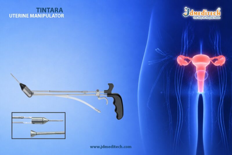
HoLEP Instrument Set for Prostate Surgery | JDMeditech

The HoLEP Instrument Set plays a vital role in modern urology. In particular, it is designed for Holmium Laser Enucleation of the Prostate (HoLEP), which is a highly effective treatment for prostate enlargement.

Today, the demand for minimally invasive procedures is growing rapidly. Therefore, surgeons need reliable and precise instruments. As a result, a high-quality HoLEP instrument set helps improve surgical accuracy and patient outcomes. In addition, it reduces complications and enhances overall efficiency.

At JDMeditech, we provide advanced HoLEP instruments that support safe and smooth surgical performance.

What is a HoLEP Instrument Set?

A HoLEP Instrument Set includes a range of specialized tools used during holmium laser prostate surgery. In simple terms, it helps surgeons remove enlarged prostate tissue with precision.

Moreover, this technique is widely considered the gold standard for treating Benign Prostatic Hyperplasia (BPH). During the procedure, laser energy is used to enucleate the prostate tissue. Consequently, patients experience better urine flow and symptom relief.

At the same time, the instrument set ensures that each surgical step is controlled and efficient.

Key Components of HoLEP Instrument Set

A complete HoLEP set consists of several essential instruments. Each component, therefore, plays a specific role.

Resectoscope Sheath

First, the resectoscope sheath allows continuous irrigation. As a result, the surgical area remains clear. In addition, it provides access for other instruments.

Telescope (Endoscope)

Next, the telescope offers clear and detailed visualization. Because of this, surgeons can perform precise movements. Furthermore, it improves overall surgical accuracy.

Laser Bridge / Working Element

The laser bridge guides the laser fiber smoothly. Thus, it allows controlled tissue removal. At the same time, it ensures stability during the procedure.

Morcellator and Morcellator Sheath

After enucleation, the tissue must be removed. Therefore, the morcellator breaks the tissue into small pieces. As a result, removal becomes faster and easier.

Irrigation System

Meanwhile, the irrigation system maintains a clean surgical field. In addition, it helps cool the area and improves visibility.

Accessories and Connectors

Finally, connectors and accessories support smooth operation. Without them, the system would not function efficiently.

Advantages of HoLEP Instrument Set

The HoLEP Instrument Set offers several advantages. Not only does it help surgeons, but it also benefits patients.

Minimally Invasive Approach

First of all, the procedure requires no large incisions. Therefore, patients experience less pain. In addition, recovery time is shorter.

High Precision

Because of advanced optics and laser compatibility, surgeons gain better control. As a result, surgical outcomes improve significantly.

Reduced Bleeding

The holmium laser seals blood vessels during surgery. Consequently, blood loss is minimized.

Faster Recovery

Since the procedure is less invasive, patients recover quickly. Therefore, they can return to daily activities sooner.

Suitable for Large Prostates

Unlike traditional methods, HoLEP works effectively for large prostates. Thus, it is a versatile solution.

Applications in Urology

The HoLEP Instrument Set is widely used in urology. For example, it is commonly used for:

  • Treatment of Benign Prostatic Hyperplasia (BPH)
  • Relief from urinary obstruction
  • Minimally invasive prostate surgery
  • Advanced urological procedures

In addition, it is suitable for both hospitals and specialized clinics.

Why Choose JDMeditech HoLEP Instrument Set?

Choosing the right instrument supplier is essential. Therefore, JDMeditech focuses on quality and performance.

Precision Manufacturing

First, all instruments are manufactured with strict quality standards. As a result, they deliver consistent performance.

High-Quality Materials

We use durable medical-grade materials. Therefore, the instruments last longer and resist corrosion.

Easy Handling

In addition, our instruments are ergonomically designed. This makes them easy to use and improves efficiency.

Global Supply

Finally, we supply products worldwide. Moreover, we ensure timely delivery and reliable support.

Maintenance and Sterilization

Proper maintenance is important. Otherwise, performance may decline over time.

  • First, clean instruments immediately after use
  • Then, use recommended sterilization methods
  • Also, inspect optical parts regularly
  • Finally, store instruments safely

By following these steps, you can extend the lifespan of the instruments.

Future of HoLEP Technology

HoLEP technology is evolving continuously. As new innovations emerge, procedures are becoming faster and more precise.

For instance, improved imaging systems enhance visibility. Similarly, advanced laser technologies improve accuracy. Therefore, patient outcomes continue to improve.

Conclusion

In conclusion, the HoLEP Instrument Set is essential for modern prostate surgery. Not only does it provide precision, but it also ensures safety and efficiency.

As a result, surgeons can achieve better outcomes, while patients benefit from faster recovery. Therefore, choosing a trusted provider like JDMeditech is important for long-term success.

Explore More Product >>

+91 79909 93062 | +91 63513 72032 | exports@jdmeditech.com 
 

One Cautery 400W Vessel Sealer – Advanced Surgical Power Unit

The One Cautery 400W Vessel Sealer is a cutting-edge surgical power unit designed to deliver precision, efficiency, and safety in modern surgical procedures. As hospitals and surgical centers increasingly demand reliable energy devices, this advanced electrosurgical system stands out for its high performance and consistent results.

Moreover, the integration of vessel sealing technology ensures effective hemostasis while minimizing tissue damage. Therefore, surgeons can perform complex procedures with greater confidence and control.

What is One Cautery 400W Vessel Sealer?

The One Cautery 400W Vessel Sealer is a high-powered electrosurgical unit that combines advanced bipolar vessel sealing with multifunctional surgical capabilities. It is specifically engineered to seal blood vessels securely while reducing bleeding during surgery.

In addition, the system supports multiple surgical modes, which makes it suitable for a wide range of specialties including general surgery, gynecology, urology, and laparoscopic procedures.

Key Features of One Cautery 400W Vessel Sealer
1. High Power Output (400W)

The device delivers up to 400 watts of power, ensuring efficient tissue cutting and coagulation. As a result, it enhances surgical speed and effectiveness.

2. Advanced Vessel Sealing Technology

It provides reliable sealing of vessels with minimal thermal spread. Consequently, this reduces the risk of complications and improves patient outcomes.

3. Intelligent Energy Control

The system automatically adjusts energy output based on tissue impedance. Therefore, it ensures consistent sealing and prevents overheating.

4. Multifunctional Surgical Modes

The unit supports monopolar, bipolar, and vessel sealing modes. Hence, surgeons can perform multiple procedures using a single device.

5. User-Friendly Interface

With an intuitive control panel and digital display, the system allows easy operation. Furthermore, it reduces the learning curve for medical professionals.

6. Safety Mechanisms

Built-in safety features prevent accidental burns and ensure controlled energy delivery. As a result, it enhances patient and operator safety.

Benefits of Using One Cautery 400W Vessel Sealer

The One Cautery 400W Vessel Sealer offers several advantages that make it an essential tool in modern operating rooms.

Firstly, it significantly reduces intraoperative bleeding. Secondly, it improves surgical precision and efficiency. Additionally, it shortens procedure time, which increases overall productivity in hospitals.

Moreover, the reduced thermal spread helps preserve surrounding tissues. Therefore, patients experience faster recovery and fewer complications.

Applications in Medical Procedures

This advanced surgical power unit is widely used across various medical specialties. For instance:

  • General surgery procedures
  • Laparoscopic surgeries
  • Gynecological operations
  • Urological procedures
  • Gastrointestinal surgeries

Because of its versatility, the device is suitable for both open and minimally invasive surgeries.

Why Choose JDMeditech for Surgical Power Units?

At JDMeditech, we provide high-quality medical equipment designed to meet international standards. Our products are trusted by healthcare professionals worldwide.

Key Reasons to Choose Us:
  • Precision-engineered medical devices
  • Reliable performance and durability
  • Competitive pricing
  • Global supply and export capabilities
  • Dedicated customer support

Furthermore, we ensure that all our products undergo strict quality checks before delivery.

Maintenance and Safety Tips

To ensure optimal performance of the One Cautery 400W Vessel Sealer, proper maintenance is essential.

  • Regularly inspect cables and electrodes
  • Follow manufacturer guidelines for cleaning
  • Avoid overloading the device
  • Ensure proper grounding during use

By following these steps, you can extend the lifespan of the equipment and maintain safety standards.

Future of Vessel Sealing Technology

With continuous advancements in medical technology, vessel sealing systems are becoming more efficient and intelligent. In the future, we can expect enhanced automation, improved precision, and integration with robotic surgical systems.

Therefore, investing in advanced devices like the One Cautery 400W Vessel Sealer ensures that healthcare facilities stay ahead in surgical innovation.

Conclusion

The One Cautery 400W Vessel Sealer is a powerful and reliable surgical unit that enhances precision, safety, and efficiency in the operating room. Its advanced features, combined with versatile applications, make it an indispensable tool for modern surgeons.

If you are looking for high-performance surgical equipment, JDMeditech offers trusted solutions tailored to your needs.


Explore More Product >>

Get Connected:
+91 79909 93062 | +91 63513 72032 | exports@jdmeditech.com 
 

Fiber Optic Cable for Endoscopy – High-Performance Light

In modern endoscopic procedures, clear visualization is essential for accurate diagnosis and successful treatment. One of the most critical components that ensures optimal illumination is the Fiber Optic Cable. This advanced medical accessory plays a key role in transmitting high-intensity light from the source to the endoscope, enabling surgeons to perform procedures with precision and confidence.

In this blog, we will explore the importance, features, applications, and benefits of using a fiber optic cable for endoscopy, along with why it is an indispensable part of modern surgical systems.

What is a Fiber Optic Cable in Endoscopy?

A Fiber Optic Cable is a specialized cable designed to transmit light efficiently using optical fibers. These fibers are made of high-quality glass or plastic and work on the principle of total internal reflection, ensuring minimal light loss.

In endoscopy, the fiber optic cable connects the light source (LED/Xenon) to the endoscope, delivering bright and consistent illumination inside the human body.

How Fiber Optic Cable Works

The working mechanism of a fiber optic cable for endoscopy is based on advanced optical technology. When light enters one end of the cable, it travels through multiple thin fibers by reflecting internally until it reaches the other end.

As a result:

  • Light transmission remains strong and stable
  • Minimal energy loss occurs
  • High brightness is maintained throughout procedures

Therefore, surgeons get a clear and detailed view of internal structures, which is crucial for accurate intervention.

Key Features of Fiber Optic Cable
1. High Light Transmission Efficiency

Fiber optic cables provide maximum light output with minimal loss, ensuring bright illumination during procedures.

2. Durable and Flexible Design

These cables are designed to be highly flexible, allowing easy handling in complex surgical environments.

3. Heat Resistance

Fiber optic cables can withstand high temperatures generated by powerful light sources such as Xenon and LED systems.

4. Universal Compatibility

Most fiber optic cables for endoscopy are compatible with various light sources and endoscope systems.

5. Sterilization-Friendly

They are built to endure repeated sterilization cycles without compromising performance.

Applications of Fiber Optic Cable in Medical Field

Fiber optic cables are widely used across different medical specialties, including:

  • Urology procedures (TURP, cystoscopy)
  • Gastroenterology (endoscopy, colonoscopy)
  • Laparoscopy surgeries
  • Arthroscopy procedures
  • ENT (Ear, Nose, Throat) examinations

Because of their versatility, fiber optic cables are considered essential in nearly all minimally invasive procedures.

Benefits of Using Fiber Optic Cable for Endoscopy
Enhanced Visualization

Bright and uniform light ensures better visibility, helping surgeons identify even small details.

Improved Surgical Accuracy

Clear imaging leads to more precise diagnosis and treatment.

Reduced Procedure Time

Efficient illumination minimizes the need for repeated adjustments, saving time during surgery.

Long Service Life

High-quality materials ensure durability and long-term performance.

Cost-Effective Solution

Reliable performance reduces maintenance and replacement costs over time.

Why Choose JDMeditech Fiber Optic Cable?

At JDMeditech, we provide high-quality fiber optic cables for endoscopy designed to meet international medical standards.

Our Advantages:
  • Premium-grade optical fibers
  • Excellent light transmission performance
  • Strong and durable construction
  • Compatibility with leading endoscopy systems
  • Trusted by healthcare professionals worldwide
Maintenance Tips for Fiber Optic Cable

To ensure long-lasting performance, proper care is essential:

  • Avoid sharp bending or twisting
  • Clean connectors regularly
  • Store in a dry and safe environment
  • Inspect for damage before use

Following these simple steps can significantly extend the life of your fiber optic cable.

Conclusion

The Fiber Optic Cable is a vital component in endoscopic systems, providing high-performance light transmission for clear and precise visualization. Its efficiency, durability, and compatibility make it indispensable in modern medical procedures.

Choosing a reliable supplier like JDMeditech ensures you receive top-quality products that enhance surgical performance and patient outcomes.


Explore More Product >>

+91 79909 93062 | +91 63513 72032 | exports@jdmeditech.com 
Xenon Light Source for Endoscopy – Bright Illumination

Modern endoscopic procedures rely heavily on high-quality illumination. Without proper lighting, even the most advanced endoscope cannot deliver accurate visualization. This is where the Xenon Light Source plays a crucial role. Known for its bright, white, and natural light, it has become a preferred choice in many hospitals and surgical centers.

In this detailed guide, you will understand how a xenon light source works, its benefits, applications, and why it remains an essential component in endoscopic imaging systems.

What is a Xenon Light Source?

A xenon light source is a medical illumination system that uses xenon gas to produce high-intensity white light. This light closely resembles natural daylight, which helps surgeons view tissues in their true colors during procedures.

Unlike traditional halogen lights, xenon systems offer superior brightness and clarity. As a result, they are widely used in endoscopy, laparoscopy, urology, and other minimally invasive surgeries.

How Does a Xenon Light Source Work?

A xenon light source operates by passing an electrical current through xenon gas inside a sealed bulb. This process creates a powerful arc of light that produces intense brightness.

The generated light is then transmitted through a fiber optic cable to the endoscope. Consequently, the internal organs and tissues are illuminated clearly, allowing precise diagnosis and treatment.

Key Features of Xenon Light Source
High-Intensity Illumination

Xenon light sources provide extremely bright light, which is essential for deep cavity visualization.

Natural Daylight Color

The light produced closely mimics daylight, ensuring accurate color reproduction of tissues.

Consistent Performance

These systems deliver stable illumination without flickering, which improves surgical efficiency.

Long Operational Life

Modern xenon bulbs are designed for durability, reducing the need for frequent replacements.

Compatibility with Endoscopic Systems

Xenon light sources are compatible with a wide range of rigid and flexible endoscopes.

Advantages of Using Xenon Light Source

One of the biggest advantages of a xenon light source is its superior brightness. This ensures that surgeons can clearly see even the smallest anatomical details.

Additionally, the natural color temperature helps in distinguishing between healthy and abnormal tissues. This is particularly important in procedures where accuracy is critical.

Moreover, xenon light sources improve workflow efficiency. Because of their stable output, surgeons can perform procedures without interruptions.

Another key benefit is enhanced patient safety. Better visualization reduces the chances of errors during surgery.

Applications of Xenon Light Source

Xenon light sources are widely used across multiple medical fields. Some common applications include:

Endoscopy

They provide clear illumination for gastrointestinal examinations and procedures.

Laparoscopy

Used in minimally invasive abdominal surgeries for better visualization.

Urology

Essential for procedures such as cystoscopy and ureteroscopy.

ENT Procedures

Helps in examining the ear, nose, and throat with high precision.

Gynecology

Used in hysteroscopy and other diagnostic procedures.

Xenon vs LED Light Source

Although LED light sources are becoming popular, xenon light sources still offer unmatched brightness in many cases.

Xenon systems provide a broader light spectrum and more natural color rendering. On the other hand, LED systems are more energy-efficient and require less maintenance.

However, for procedures that demand intense illumination and high accuracy, xenon light sources remain a strong choice.

Factors to Consider When Choosing a Xenon Light Source

When selecting a xenon light source, it is important to consider several factors.

First, check the brightness level and wattage. Higher wattage typically means better illumination.

Second, ensure compatibility with your existing endoscopic equipment.

Third, evaluate the lifespan of the xenon bulb and maintenance requirements.

Finally, consider safety features such as automatic shut-off and heat management systems.

Maintenance Tips for Xenon Light Source

Proper maintenance ensures optimal performance and longevity.

Regularly inspect the bulb and replace it as recommended.
Keep the ventilation system clean to avoid overheating.
Use high-quality fiber optic cables for efficient light transmission.
Always follow manufacturer guidelines for safe operation.

Why Choose Xenon Light Source for Endoscopy?

A xenon light source remains one of the most reliable solutions for endoscopic illumination. Its ability to deliver bright, natural light ensures accurate visualization during critical procedures.

For healthcare providers aiming to improve diagnostic accuracy and surgical outcomes, investing in a high-quality xenon light source is a smart decision.

Conclusion

The Xenon Light Source is a powerful and dependable illumination system designed for modern endoscopic procedures. With its high intensity, natural color output, and consistent performance, it continues to be a preferred choice among medical professionals.

As medical technology evolves, the demand for precise imaging will only increase. Therefore, choosing the right illumination system, such as a xenon light source, is essential for delivering safe and effective patient care.


Explore More Product >>

Get Connected:
+91 79909 93062 | +91 63513 72032 | exports@jdmeditech.com 
 

Portable Endoscope Camera – Compact Imaging for Surgery

In today’s fast-evolving medical landscape, precision, portability, and efficiency are essential for successful surgical outcomes. The Portable Endoscope Camera has emerged as a game-changing innovation, offering high-resolution imaging in a compact and user-friendly design.

Whether used in urology, laparoscopy, ENT, or gastroenterology, this advanced device ensures clear visualization, mobility, and seamless workflow integration. As a result, healthcare professionals can perform procedures with greater confidence and accuracy.

What is a Portable Endoscope Camera?

A Portable Endoscope Camera is a compact imaging system designed to capture and display internal body visuals during endoscopic procedures. Unlike traditional bulky systems, it is lightweight, easy to transport, and quick to set up, making it ideal for both hospitals and smaller clinics.

Moreover, it integrates camera control units, light sources, and display compatibility, allowing surgeons to achieve real-time imaging without complex installations.

Key Features of Portable Endoscope Camera
High-Resolution Imaging

These cameras deliver HD or Full HD image quality, ensuring sharp and detailed visuals during procedures.

Compact & Lightweight Design

Because of their portable structure, they are easy to carry and install, which significantly improves workflow efficiency.

Plug-and-Play Operation

Most systems offer quick setup with minimal technical expertise, saving valuable time in critical situations.

USB & HDMI Connectivity

Modern cameras support multiple output options, enabling connection with monitors, laptops, and recording systems.

Integrated LED Light Source

Some models include a built-in LED light source, which provides bright and consistent illumination.

Image & Video Recording

Surgeons can view procedures in real-time and record videos for documentation and training purposes.

Applications in Medical Fields
Urology

Portable endoscope cameras are widely used in cystoscopy, ureteroscopy, and TURP procedures, ensuring accurate diagnosis and treatment.

Laparoscopy

They play a crucial role in minimally invasive surgeries, offering clear internal visualization with minimal patient trauma.

ENT (Ear, Nose, Throat)

Doctors rely on these cameras for nasal, throat, and ear examinations, improving diagnostic precision.

Gastroenterology

In GI procedures, they assist in endoscopy, colonoscopy, and biopsy procedures, ensuring high-quality imaging.

Benefits of Using Portable Endoscope Camera
Enhanced Mobility

Because of their portability, these devices are perfect for multi-room usage, emergency setups, and field operations.

Cost-Effective Solution

Compared to traditional systems, portable units are more affordable, making them ideal for clinics and small hospitals.

Improved Efficiency

Quick setup and easy operation help reduce procedure time and increase patient throughput.

Better Patient Outcomes

Clear imaging allows surgeons to make accurate decisions, leading to improved treatment results.

Space-Saving Design

Since these systems are compact, they require minimal storage space, which is beneficial for modern healthcare facilities.

Why Choose JDMeditech Portable Endoscope Camera?

At JDMeditech, we focus on delivering advanced, reliable, and high-performance medical imaging solutions.

  • Precision-engineered imaging systems
  • High-quality medical-grade components
  • User-friendly interface for easy operation
  • Durable and long-lasting performance
  • Trusted by healthcare professionals globally
How to Choose the Right Portable Endoscope Camera

Before purchasing, consider the following factors:

  • Resolution: Opt for Full HD or higher for better clarity
  • Compatibility: Ensure it supports your existing endoscopy system
  • Connectivity: Check USB/HDMI options
  • Light Source: Integrated LED improves usability
  • Recording Features: Essential for documentation and training
Future of Portable Endoscopic Imaging

With ongoing advancements, portable endoscope cameras are becoming smarter and more efficient. Integration with AI-based diagnostics, wireless connectivity, and cloud storage will further enhance their capabilities.

Consequently, these devices will continue to play a vital role in modern, minimally invasive surgery.

Conclusion

The Portable Endoscope Camera is a compact yet powerful imaging solution that enhances surgical precision and efficiency. Thanks to its portability, high-resolution imaging, and ease of use, it has become an essential tool across multiple medical specialties.

For healthcare providers looking to upgrade their imaging systems, investing in a portable endoscope camera is a smart and future-ready decision.


Explore More Product >>

Get Connected:
+91 79909 93062 | +91 63513 72032 | exports@jdmeditech.com 
 

Medical Monitor – High-Resolution Display for Surgical Imaging

A Medical Monitor plays a critical role in modern surgical and diagnostic procedures. From urology and laparoscopy to endoscopy and general surgery, high-resolution medical displays ensure accurate visualization, better decision-making, and improved patient outcomes.

Unlike standard monitors, a medical-grade monitor is designed to deliver true color accuracy, sharp image quality, and real-time performance, which are essential in precision-driven environments like operating rooms.

What is a Medical Monitor?

A Medical Monitor is a specialized display system used in healthcare settings to visualize images from medical devices such as endoscopy cameras, laparoscopes, ultrasound machines, and surgical imaging systems.

These monitors are engineered to meet strict medical standards, ensuring consistent brightness, high contrast, and accurate image reproduction.

Why High-Resolution Matters in Medical Imaging

High-resolution displays are essential because they allow surgeons and clinicians to:

  • View fine anatomical details
  • Detect abnormalities with precision
  • Perform minimally invasive procedures safely
  • Improve diagnostic accuracy
  • Reduce the risk of surgical errors

Therefore, investing in a high-resolution medical monitor directly enhances both clinical efficiency and patient safety.

Key Features of a Medical Monitor
1. Full HD / 4K Resolution

Modern medical monitors offer Full HD (1920×1080) or 4K Ultra HD resolution, providing crystal-clear visuals for surgical precision.

2. High Brightness & Contrast Ratio

These monitors deliver consistent brightness levels and superior contrast, ensuring clear visibility even in complex procedures.

3. Wide Viewing Angle

A wide viewing angle ensures that multiple surgeons and assistants can clearly see the display from different positions.

4. True Color Reproduction

Accurate color representation is crucial for identifying tissues, blood vessels, and abnormalities.

5. Anti-Glare Screen

Medical monitors are equipped with anti-reflective coatings, reducing glare from operating room lights.

6. Multiple Connectivity Options

They support various inputs such as:

  • HDMI
  • DVI
  • SDI
  • VGA

This ensures compatibility with different medical devices.

7. Medical-Grade Compliance

Medical monitors are built to comply with healthcare safety standards, ensuring reliability and durability.

Applications of Medical Monitors
1. Urology

Medical monitors are widely used in procedures such as:

  • TURP (Transurethral Resection of Prostate)
  • Cystoscopy
  • Ureteroscopy

They provide clear visualization of the urinary tract, enabling accurate diagnosis and treatment.

2. Laparoscopy

In laparoscopic surgeries, surgeons rely heavily on high-quality displays to:

  • Navigate internal organs
  • Perform minimally invasive procedures
  • Improve surgical precision
3. Endoscopy

Medical monitors display real-time images during:

  • GI endoscopy
  • Bronchoscopy
  • Colonoscopy

Thus, they help doctors detect abnormalities quickly and efficiently.

4. General Surgery & Diagnostics

Medical monitors are also used in:

  • Operating rooms (OT)
  • ICU monitoring systems
  • Radiology and imaging departments
Benefits of Using a Medical Monitor
  • Enhanced Surgical Accuracy
  • Improved Workflow Efficiency
  • Better Team Collaboration
  • Reduced Eye Strain for Surgeons
  • Reliable Performance in Critical Environments

As a result, medical monitors significantly contribute to better patient outcomes and faster recovery times.

How to Choose the Right Medical Monitor

When selecting a medical monitor, consider the following factors:

1. Resolution Requirement

Choose Full HD or 4K depending on your application.

2. Screen Size

Common sizes include 21”, 24”, 27”, and 32”, depending on OT setup.

3. Compatibility

Ensure compatibility with your existing camera systems and surgical equipment.

4. Brightness & Contrast

Opt for monitors with high luminance and contrast ratio for better visibility.

5. Certifications

Look for medical-grade certifications to ensure safety and compliance.

Why Choose JDMeditech Medical Monitors?

At JDMeditech, we provide high-quality medical monitors designed for modern healthcare environments.

Our Advantages:
  • High-resolution display technology
  • Reliable and durable build
  • Compatible with multiple medical systems
  • Designed for precision and performance
  • Trusted by healthcare professionals
Conclusion

A Medical Monitor is an essential component in today’s healthcare ecosystem. With advancements in imaging technology, high-resolution monitors are transforming the way surgeries and diagnostics are performed.

Whether it’s urology, laparoscopy, or endoscopy, choosing the right medical monitor ensures clarity, precision, and better clinical outcomes.


Explore More Product >>

Get Connected:
+91 79909 93062 | +91 63513 72032 | exports@jdmeditech.com 
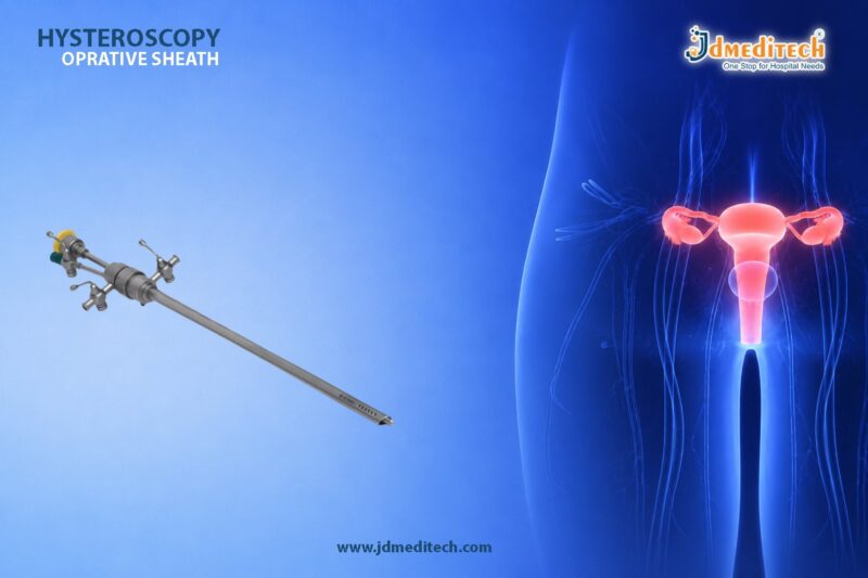
Wireless Endoscope Camera – Portable High-Resolution Imaging

In modern healthcare, precision and mobility are essential for accurate diagnosis and effective treatment. Therefore, the Wireless Endoscope Camera has emerged as a revolutionary solution in minimally invasive procedures. Unlike traditional wired systems, this advanced imaging device offers high-resolution visualization, seamless connectivity, and unmatched portability.

As a result, hospitals, clinics, and diagnostic centers are rapidly adopting wireless endoscopy systems to enhance workflow efficiency and patient outcomes.

What is a Wireless Endoscope Camera?

A Wireless Endoscope Camera is an advanced medical imaging device designed to capture and transmit real-time visuals during endoscopic procedures without the need for cumbersome cables. It connects wirelessly to monitors, tablets, or computers, providing clear and uninterrupted imaging.

Moreover, these systems are widely used in gastroenterology, urology, ENT, laparoscopy, and general surgery.

Key Features of Wireless Endoscope Camera
1. High-Resolution Imaging

First and foremost, wireless endoscope cameras deliver HD or Full HD image quality, ensuring accurate visualization of internal structures.

2. Wireless Connectivity

Additionally, the device uses Wi-Fi or Bluetooth technology for real-time image transmission, eliminating cable clutter.

3. Portable & Lightweight Design

Because of its compact size, it is easy to carry and ideal for point-of-care diagnostics and remote applications.

4. Long Battery Life

Furthermore, modern systems come with rechargeable batteries, allowing uninterrupted procedures.

5. Easy Integration

It seamlessly integrates with existing hospital systems, making it user-friendly for medical professionals.

Benefits of Wireless Endoscope Camera
Enhanced Mobility

Unlike traditional systems, doctors can move freely without cable restrictions. Consequently, this improves procedural efficiency.

Improved Workflow

Since there are fewer wires, setup time is reduced. Therefore, medical teams can focus more on patient care.

Better Infection Control

Wireless systems minimize physical contact points, thereby reducing contamination risks.

Remote Accessibility

In addition, images can be shared instantly for telemedicine and remote consultation, improving diagnostic accuracy.

Applications in Medical Field

Wireless endoscope cameras are versatile and used across multiple specialties:

  • Gastroenterology – GI tract examination
  • Urology – Bladder and urethral procedures
  • ENT– Ear, nose, and throat diagnosis
  • Laparoscopy – Minimally invasive surgeries
  • Veterinary Medicine – Animal diagnostics
Why Choose Wireless Endoscope Camera from JDMeditech?

At JDMeditech, we provide high-quality medical imaging solutions designed for precision and reliability.

Our Advantages:
  • Advanced high-resolution imaging technology
  • Durable and medical-grade components
  • Competitive pricing with global supply
  • Trusted by healthcare professionals worldwide
Technical Specifications (Typical)
  • Resolution: HD / Full HD
  • Connectivity: Wi-Fi / Bluetooth
  • Battery: Rechargeable Lithium-ion
  • Compatibility: Monitor / Laptop / Tablet
  • Design: Compact & Ergonomic
Future of Wireless Endoscopy

As technology evolves, wireless endoscopy is becoming more intelligent. For instance, integration with AI-based diagnostics and cloud storage is transforming patient care. Therefore, healthcare providers can expect faster, more accurate, and data-driven decisions.

Conclusion

In conclusion, the Wireless Endoscope Camera is a game-changing innovation in medical imaging. It offers portability, efficiency, and high-resolution visualization, making it an essential tool for modern healthcare facilities.

If you are looking for reliable and advanced endoscopy solutions, JDMeditech is your trusted partner.


Explore More Product >>

Get Connected:
+91 79909 93062 | +91 63513 72032 | exports@jdmeditech.com 
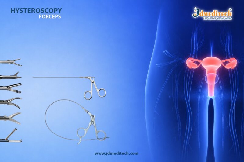
 

LED Light Source for Endoscopic Procedures | JDMeditech

In modern medical practice, visualization plays a critical role in accurate diagnosis and successful surgical outcomes. Therefore, a LED Light Source for Endoscopic Procedures has become an essential component in hospitals and diagnostic centers. It provides consistent, high-intensity illumination, which significantly enhances visibility during minimally invasive procedures.

Moreover, with advancements in medical technology, LED light sources are replacing traditional halogen and xenon systems due to their efficiency, durability, and superior performance.

What is an LED Light Source?

An LED Light Source is a medical illumination device designed to deliver bright, white light through fiber optic cables to endoscopic instruments. As a result, surgeons and physicians can clearly visualize internal organs and tissues.

Unlike conventional light systems, LED technology ensures:

  • Stable light intensity
  • Minimal heat generation
  • Long operational life

Thus, it is widely used in laparoscopy, urology, gastroenterology, and ENT procedures.

Why LED Light Source is Important in Endoscopy

During endoscopic procedures, proper illumination is crucial. Without sufficient lighting, visualization becomes difficult, which may lead to diagnostic errors.

Key reasons include:
  • Enhanced Visibility : Provides clear and detailed images
  • Accurate Diagnosis : Helps detect abnormalities effectively
  • Improved Surgical Precision : Supports minimally invasive techniques
  • Reduced Eye Strain : Ensures comfortable viewing for surgeons

Additionally, consistent lighting ensures uninterrupted procedures, which is critical in operation theaters.

Key Features of LED Light Source
1. High-Intensity Illumination

LED light sources deliver powerful brightness comparable to xenon lamps. Consequently, they provide excellent clarity even in deep cavities.

2. Long Lifespan

Unlike traditional bulbs, LED systems last up to 20,000–50,000 hours. Therefore, maintenance costs are significantly reduced.

3. Energy Efficient

LED technology consumes less power while delivering high output. As a result, hospitals can reduce energy expenses.

4. Low Heat Emission

Excess heat can damage tissues and equipment. However, LED light sources generate minimal heat, ensuring safety during procedures.

5. Compact & Portable Design

Modern LED units are lightweight and easy to integrate into existing endoscopy systems.

Applications of LED Light Source in Medical Field

The LED Light Source for Endoscopic Procedures is widely used across multiple specialties:

  • Urology : Cystoscopy, TURP procedures
  • Gastroenterology : Endoscopy, colonoscopy
  • Gynecology : Laparoscopic surgeries
  • ENT : Sinus and throat examinations
  • General Surgery : Minimally invasive procedures

Furthermore, its versatility makes it a standard component in advanced operation theaters.

LED vs Xenon Light Source – A Quick Comparison
Feature LED Light Source Xenon Light Source
Lifespan Very long Short
Heat Generation Low High
Maintenance Minimal Frequent
Energy Consumption Low High
Cost Efficiency High Moderate

Clearly, LED technology offers long-term benefits and reliability.

Benefits of Choosing LED Light Source

Choosing the right light source directly impacts performance. Therefore, LED systems provide several advantages:

  • Cost-effective in the long run
  • Eco-friendly and energy-saving
  • Consistent light output
  • Reduced downtime
  • Better patient safety

In addition, hospitals can improve workflow efficiency with reliable lighting systems.

Why Choose JDMeditech LED Light Source?

At JDMeditech, we focus on delivering high-quality medical equipment designed for precision and reliability.

Our advantages:
  • Premium-grade components
  • Advanced LED technology
  • Durable and robust design
  • Competitive pricing
  • Global supply capability

Therefore, our LED Light Source ensures optimal performance for modern healthcare facilities.

Maintenance Tips for LED Light Source

To maximize performance and lifespan, follow these simple steps:

  • Regularly clean fiber optic cables
  • Avoid bending cables sharply
  • Ensure proper ventilation
  • Perform routine system checks

As a result, your device will maintain consistent performance over time.

Conclusion

In conclusion, the LED Light Source – Bright & Reliable Illumination for Endoscopic Procedures is a vital tool in modern medicine. Not only does it enhance visualization, but it also improves surgical outcomes and operational efficiency.

Therefore, upgrading to LED technology is a smart investment for any healthcare facility aiming for precision and reliability.


Explore More Product >>

Get Connected:
+91 79909 93062 | +91 63513 72032 | exports@jdmeditech.com 
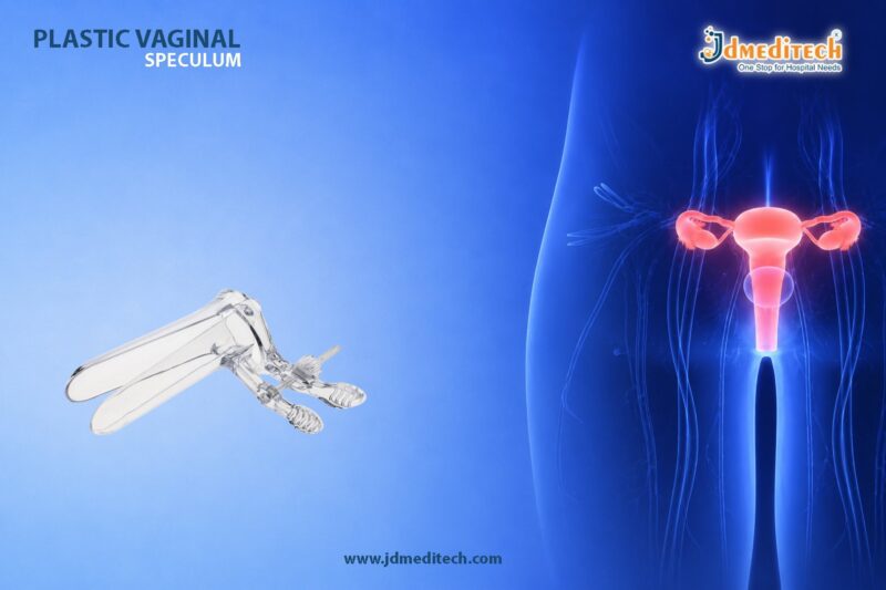
Camera Coupler – High-Precision Imaging for Endoscopic Procedures

A Camera Coupler plays a crucial role in modern endoscopic imaging systems. It acts as a bridge between the endoscope and the camera, ensuring that high-quality visuals are transmitted accurately. As surgical procedures demand greater precision, the importance of a reliable camera coupler for endoscopy continues to grow.

Moreover, advanced camera couplers improve image clarity, reduce distortion, and enhance overall surgical outcomes. Therefore, healthcare professionals rely on high-performance couplers to achieve consistent and precise imaging during procedures.

What is a Camera Coupler?

A camera coupler is an optical device that connects an endoscope to a medical camera. It ensures that the image captured through the endoscope is transferred efficiently to the camera system.

In addition, the coupler helps maintain image focus, brightness, and alignment. Without a proper coupler, even the best endoscopic camera system may fail to deliver accurate visuals.

Key Features of Camera Coupler
1. High-Precision Optics

Camera couplers are designed with advanced optical components. As a result, they deliver sharp and distortion-free images.

2. Adjustable Focus Mechanism

Most couplers come with a focus adjustment feature. Therefore, surgeons can easily fine-tune the image during procedures.

3. Universal Compatibility

Modern camera couplers for endoscopy are compatible with various endoscopes and camera systems. This flexibility makes them highly versatile in clinical settings.

4. Durable Construction

Typically made from medical-grade materials, these devices ensure long-lasting performance even with frequent use.

5. Lightweight & Ergonomic Design

A compact and lightweight design allows easy handling and quick setup in operation theaters.

Importance of Camera Coupler in Endoscopic Procedures
Enhanced Image Quality

A high-quality camera coupler ensures that images remain sharp and clear. Consequently, surgeons can perform procedures with greater confidence.

Improved Diagnostic Accuracy

Clear visualization helps in accurate diagnosis. In addition, it reduces the chances of errors during procedures.

Seamless Integration

Camera couplers allow smooth integration between different imaging components. Therefore, they support efficient workflow in hospitals.

Better Surgical Outcomes

When imaging is precise, procedures become safer and more effective. Thus, patient outcomes improve significantly.

Applications of Camera Coupler

Camera couplers are widely used across multiple medical specialties:

  • Urology – For cystoscopy and ureteroscopy procedures
  • Gastroenterology – For endoscopy and colonoscopy imaging
  • ENT (Otolaryngology) – For sinus and throat examinations
  • Gynecology – For hysteroscopy procedures
  • General Surgery – For minimally invasive surgeries
Types of Camera Couplers
Fixed Focus Camera Coupler

These couplers offer a preset focus, making them simple and easy to use.

Zoom Camera Coupler

Zoom couplers allow adjustable magnification. Therefore, they provide more flexibility during procedures.

C-Mount Camera Coupler

C-Mount couplers are widely used due to their compatibility with various camera systems.

How to Choose the Right Camera Coupler

Choosing the right camera coupler for endoscopy depends on several factors:

  • Compatibility with your endoscope and camera
  • Required image quality and resolution
  • Availability of focus or zoom features
  • Build quality and durability
  • Ease of sterilization and maintenance

In addition, selecting a trusted manufacturer ensures consistent performance and reliability.

Maintenance and Care Tips

Proper maintenance extends the lifespan of the camera coupler:

  • Clean lenses with approved medical-grade solutions
  • Avoid scratches on optical surfaces
  • Store in a protective case when not in use
  • Regularly inspect for alignment issues
Why Choose JDMeditech Camera Couplers?

At JDMeditech, we provide high-quality camera couplers for endoscopy designed for precision and reliability.

  • Advanced optical technology for clear imaging
  • Durable and medical-grade construction
  • Compatibility with leading endoscopy systems
  • Designed for consistent performance in surgical environments
Conclusion

A Camera Coupler is an essential component in endoscopic imaging systems. It ensures accurate image transmission, enhances clarity, and supports precise surgical procedures.

As medical technology continues to evolve, investing in a high-quality camera coupler for endoscopy is vital for achieving better diagnostics and improved patient outcomes.

Explore More Product >>

Get Connected:
+91 79909 93062 | +91 63513 72032 | exports@jdmeditech.com
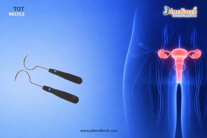


Endoscopy Video Recorder – HD Surgical Recording System

In modern healthcare, accurate surgical documentation is essential for diagnosis, training, and legal purposes. Therefore, the Endoscopy Video Recorder plays a vital role in capturing high-quality procedure videos. It allows surgeons and medical professionals to record, store, and review endoscopic procedures with clarity and precision.

Moreover, as hospitals increasingly adopt digital solutions, having a reliable recording system has become a necessity rather than a luxury. This is exactly where an advanced Endoscopy Video Recorder becomes indispensable.

What is an Endoscopy Video Recorder?

An Endoscopy Video Recorder is a medical device designed to capture and store video output from endoscopic camera systems. It records procedures such as gastroscopy, colonoscopy, cystoscopy, and laparoscopy in real time.

In addition, these systems support high-definition (HD) or even 4K recording, ensuring every detail is preserved. As a result, doctors can analyze procedures more effectively and maintain accurate patient records.

Key Features of Endoscopy Video Recorder
1. High-Definition Video Recording

First and foremost, modern Endoscopy Video Recorders provide HD or 4K video quality. Consequently, this ensures crystal-clear visualization of internal organs and tissues.

2. Real-Time Recording & Playback

Furthermore, the system enables real-time recording along with instant playback. This helps surgeons review procedures immediately when needed.

3. Multiple Storage Options

Most devices support USB drives, SSDs, or network storage. Therefore, managing and accessing recorded data becomes simple and efficient.

4. User-Friendly Interface

In addition, intuitive controls and easy navigation allow medical staff to operate the system without extensive training.

5. Compatibility with Endoscopy Systems

A good Endoscopy Video Recorder integrates seamlessly with various endoscopic camera systems, making it versatile for multiple specialties.

6. Image Capture Capability

Besides video recording, users can capture still images during procedures, which is useful for reports and documentation.

Applications of Endoscopy Video Recorder

The Endoscopy Video Recorder is widely used across multiple medical departments:

  • Gastroenterology – Recording colonoscopy and endoscopy procedures
  • Urology – Documenting cystoscopy and ureteroscopy
  • Gynecology – Capturing hysteroscopy procedures
  • ENT (Otolaryngology) – Recording sinus and throat examinations
  • General Surgery – Documenting laparoscopic procedures

Thus, it serves as a universal recording solution across various specialties.

Benefits of Using an Endoscopy Video Recorder
Enhanced Surgical Documentation

Accurate recording ensures that every procedure is documented properly for future reference.

Improved Training & Education

Medical students and professionals can review recorded procedures for learning and skill development.

Better Patient Communication

Doctors can explain procedures more effectively by showing recorded visuals to patients.

Legal & Compliance Support

Recorded data provides strong documentation for medico-legal cases and hospital compliance.

Efficient Data Management

With digital storage, hospitals can easily archive and retrieve patient records when required.

Why Choose JDMeditech Endoscopy Video Recorder?

At JDMeditech, we provide high-quality Endoscopy Video Recorders designed for reliability and performance.

Our Advantages:
  • Advanced HD recording technology
  • Durable and medical-grade components
  • Easy integration with existing systems
  • Competitive pricing with global supply
  • Trusted by hospitals and surgeons worldwide
Technical Specifications (Typical)
  • Video Resolution: HD / Full HD / 4K
  • Storage: USB / SSD / Network
  • Output Formats: HDMI / DVI
  • Recording Format: MP4 / AVI
  • Power Supply: Medical-grade compatible
Future of Surgical Recording Systems

As technology evolves, Endoscopy Video Recorders are becoming smarter and more efficient. For instance, integration with AI and cloud storage is improving data accessibility and analysis.

Additionally, hospitals are moving toward fully digital operation theaters, where recording systems play a central role. Therefore, investing in a modern Endoscopy Video Recorder ensures long-term efficiency and compliance.

Conclusion

In conclusion, the Endoscopy Video Recorder is an essential tool for modern surgical documentation. It not only improves accuracy and efficiency but also enhances training, patient care, and compliance.

Whether you run a hospital, clinic, or diagnostic center, choosing a reliable recording solution is crucial. With JDMeditech, you get a trusted partner delivering high-performance medical equipment tailored to your needs.


Explore More Product >>

Get Connected:
+91 79909 93062 | +91 63513 72032 | exports@jdmeditech.com
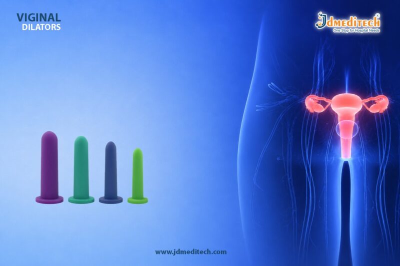
 

USB Camera System for Endoscopic Imaging – Compact & Clear Solution

In modern healthcare, clear visualization plays a critical role in accurate diagnosis and successful surgical procedures. Therefore, a USB Camera System for endoscopic imaging has become an essential tool for hospitals and clinics.

Unlike traditional bulky systems, this advanced camera solution offers compact design, easy connectivity, and high-quality imaging. As a result, it enhances workflow efficiency while maintaining precision in minimally invasive procedures.

What is a USB Camera System for Endoscopic Imaging?

A USB Camera System for endoscopic imaging is a digital imaging device designed to capture and display high-resolution images during endoscopic procedures. It connects directly to a computer or laptop via a USB interface, making it user-friendly and highly portable.

Moreover, this system eliminates the need for complex setups. Because of this, it is widely used in urology, gastroenterology, ENT, and laparoscopy.

Key Features of USB Camera Systems
1. Compact & Portable Design

First of all, the system is lightweight and compact. Therefore, it is easy to carry and install in any medical setting.

2. High-Resolution Imaging

In addition, it delivers crystal-clear images, which help doctors perform accurate diagnosis and procedures.

3. Plug-and-Play Connectivity

Unlike traditional systems, it connects directly via USB. As a result, it does not require complex installation or additional hardware.

4. Cost-Effective Solution

Furthermore, the USB Camera System for endoscopic imaging is more affordable compared to conventional camera systems, making it ideal for small clinics.

5. Real-Time Image Display

Because of advanced technology, it provides real-time visualization. Thus, surgeons can make immediate and precise decisions.

Applications in Medical Field

The USB Camera System for endoscopic imaging is widely used across multiple specialties. For example:

  • Urology : Cystoscopy and ureteroscopy procedures
  • Gastroenterology : Endoscopy and colonoscopy imaging
  • ENT : Ear, nose, and throat examinations
  • Gynecology : Minimally invasive diagnostic procedures

Therefore, its versatility makes it a valuable addition to any healthcare facility.

Advantages Over Traditional Camera Systems

Although traditional camera systems are effective, USB-based systems offer several advantages:

  • Easy setup and operation
  • Reduced equipment cost
  • Minimal maintenance requirements
  • Improved portability
  • Quick integration with digital systems

Consequently, healthcare providers prefer USB camera systems for their efficiency and convenience.

How to Choose the Right USB Camera System

When selecting a USB Camera System for endoscopic imaging, consider the following factors:

  • Resolution quality (HD or Full HD)
  • Compatibility with endoscopes
  • Ease of software integration
  • Durability and build quality
  • After-sales support and warranty

By evaluating these aspects, you can ensure optimal performance and long-term reliability.

Why Choose JDMeditech USB Camera Systems?

At JDMeditech, we provide advanced medical imaging solutions designed for precision and reliability.

  • High-quality medical-grade camera systems
  • Competitive pricing for global markets
  • Reliable performance and durability
  • Trusted by healthcare professionals
Future of Endoscopic Imaging

As technology continues to evolve, USB Camera Systems for endoscopic imaging are becoming more advanced. For instance, integration with AI, digital recording, and cloud storage is improving diagnostic capabilities.

Therefore, investing in modern imaging systems ensures better patient outcomes and efficient workflows.

Conclusion

In conclusion, the USB Camera System for endoscopic imaging is a compact, efficient, and cost-effective solution for modern medical practices. Not only does it provide high-quality visualization, but it also simplifies setup and operation.

Because of these advantages, it has become an essential tool for healthcare professionals seeking precision and convenience.


Explore More Product >>

Get Connected:
+91 79909 93062 | +91 63513 72032 | exports@jdmeditech.com
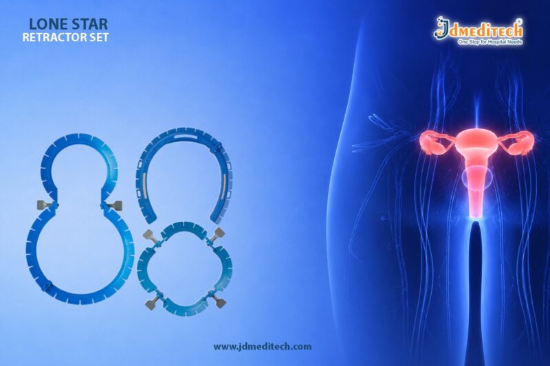
 

Full HD Endoscopy Camera System for Precision Surgery | HD Medical Imaging

In modern minimally invasive surgery, visualization plays a critical role in achieving successful outcomes. The Full HD Endoscopy Camera System has become an essential component in operating rooms, offering surgeons crystal-clear imaging and enhanced precision.

Because surgical procedures require accuracy at every step, high-definition imaging ensures better diagnosis, improved control, and safer patient outcomes. As a result, hospitals and surgical centers increasingly rely on advanced endoscopy camera systems.

What is a Full HD Endoscopy Camera System?

A Full HD Endoscopy Camera System is a medical imaging device used to capture and display high-resolution images during endoscopic procedures. It converts optical images from the endoscope into digital signals and displays them on a monitor in 1080p resolution.

Typically, the system includes:

  • HD camera head
  • Camera control unit (CCU)
  • High-resolution monitor
  • Light source
  • Recording system

Together, these components deliver real-time, detailed visualization of internal organs.

Key Features of Full HD Endoscopy Camera System
1. Crystal-Clear Full HD Resolution

The system provides 1080p high-definition imaging, allowing surgeons to see even the smallest anatomical structures clearly.

2. Advanced Color Reproduction

Accurate color balance helps differentiate tissues, blood vessels, and abnormalities, improving surgical precision.

3. High Frame Rate Imaging

Smooth and lag-free video ensures seamless operation during complex procedures.

4. Ergonomic and Lightweight Camera Head

Designed for ease of handling, reducing fatigue during long surgeries.

5. Digital Image Processing

Enhances sharpness, contrast, and brightness for superior visualization.

6. Compatibility with Multiple Endoscopes

The system supports rigid and flexible endoscopes across various specialties.

Applications in Medical Procedures

The Full HD Endoscopy Camera System is widely used across multiple specialties, including:

Urology
  • TURP procedures
  • Cystoscopy
  • Ureteroscopy
Gastroenterology
  • Endoscopy
  • Colonoscopy
  • ERCP procedures
Gynecology
  • Laparoscopy
  • Hysteroscopy
General Surgery
  • Minimally invasive surgeries
  • Diagnostic procedures

Because of its versatility, this system is a must-have in modern operating theaters.

Benefits of Using Full HD Endoscopy Camera System
1. Enhanced Surgical Precision

Clear visualization allows surgeons to perform procedures with higher accuracy.

2. Improved Patient Safety

Better imaging reduces the risk of complications and surgical errors.

3. Faster Procedures

Efficient visualization leads to quicker decision-making during surgery.

4. Better Training & Documentation

Recorded HD videos help in medical training and case review.

5. Reduced Eye Strain for Surgeons

High-quality imaging minimizes visual fatigue during long procedures.

Why Choose Full HD Over Standard Imaging?

While standard-definition systems provide basic imaging, Full HD Endoscopy Camera Systems offer:

  • Higher resolution
  • Better depth perception
  • Improved tissue differentiation
  • Superior surgical outcomes

Therefore, upgrading to Full HD is not just an improvement—it’s a necessity for modern healthcare facilities.

Latest Advancements in Endoscopy Camera Technology

Modern systems now include:

  • 3-chip CMOS sensors for superior image quality
  • LED light integration for consistent illumination
  • 4K upgrade compatibility
  • USB & HDMI recording options
  • Wireless data transfer features

These innovations continue to enhance surgical efficiency and patient care.

Maintenance & Care Tips

To ensure optimal performance:

  • Clean the camera head after every procedure
  • Use proper sterilization protocols
  • Avoid cable damage or bending
  • Regularly calibrate the system
  • Store in a dust-free environment

Proper maintenance increases lifespan and ensures consistent image quality.

Choosing the Right Endoscopy Camera System

When selecting a Full HD Endoscopy Camera System, consider:

  • Resolution quality (1080p minimum)
  • Compatibility with existing equipment
  • Ease of use
  • Service and support availability
  • Warranty and durability

Making the right choice ensures long-term value and performance.

Why Choose JDMeditech?

At JDMeditech, we provide advanced Full HD Endoscopy Camera Systems designed for precision, durability, and superior performance.

Our Advantages:
  • High-quality medical-grade technology
  • Reliable and cost-effective solutions
  • Global supply and export capabilities
  • Trusted by healthcare professionals
Conclusion

The Full HD Endoscopy Camera System has revolutionized minimally invasive surgery by delivering crystal-clear visualization and unmatched precision. As healthcare continues to evolve, adopting advanced imaging technology is essential for improving patient outcomes and surgical efficiency.

Therefore, investing in a high-quality endoscopy camera system is a strategic step toward modern, safe, and effective surgical care.


Explore More Product >>

Get Connected:
+91 79909 93062 | +91 63513 72032 | exports@jdmeditech.com
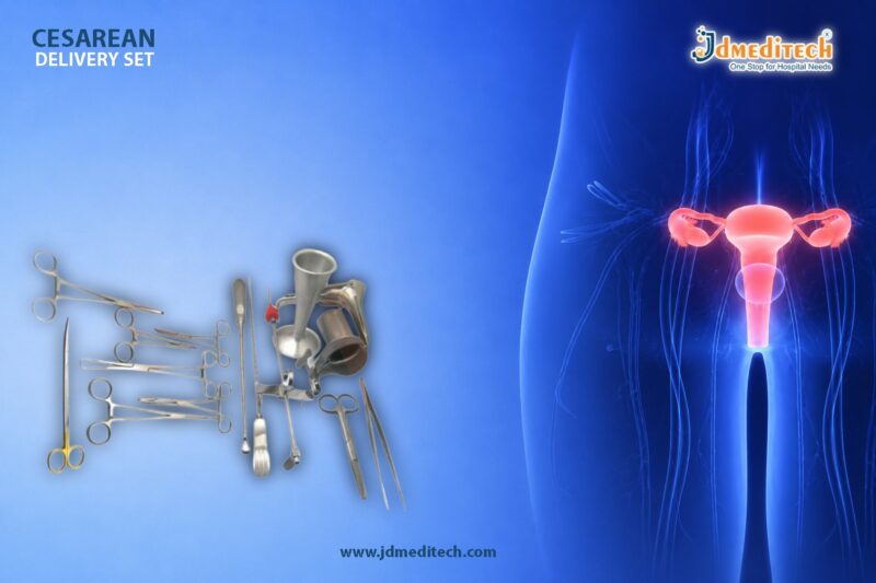


Bladder Scanner – Safe & Efficient Urological Diagnostic Tool

A Bladder Scanner is a non-invasive ultrasound device used to measure bladder volume and assess urinary retention. Today, healthcare professionals widely rely on this advanced tool because it provides accurate, quick, and safe diagnostics without the need for catheterization.

Moreover, with the growing demand for patient-friendly diagnostic solutions, the Bladder Scanner has become an essential instrument in hospitals, clinics, and urology centers. It not only improves workflow efficiency but also enhances patient comfort significantly.

What is a Bladder Scanner?

A Bladder Scanner is a portable ultrasound device that uses sound waves to estimate the amount of urine in the bladder. Unlike traditional methods, it does not require any invasive procedures.

Therefore, it reduces the risk of infections while delivering precise results within seconds.

How Does a Bladder Scanner Work?

The working principle of a Bladder Scanner is based on ultrasound imaging.

  • First, a probe is placed on the patient’s lower abdomen
  • Then, ultrasound waves are transmitted into the body
  • After that, the device calculates bladder volume using reflected signals
  • Finally, results are displayed instantly on the screen

As a result, healthcare providers can make quick and informed decisions.

Key Features of Bladder Scanner

A modern Bladder Scanner offers several advanced features:

  • Non-invasive and painless procedure
  • Portable and easy-to-use design
  • High accuracy in bladder volume measurement
  • Real-time imaging and results
  • Minimal training required for operation
  • Reduced risk of urinary tract infections (UTIs)

Additionally, many models come with touchscreen interfaces and automated measurement systems, which further simplify usage.

Applications of Bladder Scanner in Healthcare

The Bladder Scanner plays a crucial role across multiple medical settings:

1. Urology Departments

It helps diagnose urinary retention, bladder dysfunction, and post-void residual volume.

2. Emergency Care

Doctors use it to quickly evaluate bladder conditions in critical patients.

3. Post-Surgical Monitoring

After surgery, it ensures proper bladder emptying and prevents complications.

4. Geriatric Care

Elderly patients benefit greatly due to reduced need for catheterization.

5. Intensive Care Units (ICU)

Continuous monitoring of urine volume improves patient management.

Benefits of Using a Bladder Scanner

Using a Bladder Scanner offers multiple advantages:

  • Enhances patient comfort by eliminating invasive procedures
  • Reduces infection risks associated with catheter use
  • Saves time with instant results
  • Improves diagnostic accuracy
  • Cost-effective in long-term use

Furthermore, it supports better clinical decision-making, which leads to improved patient outcomes.

Bladder Scanner vs Traditional Catheterization
Feature Bladder Scanner Catheterization
Procedure Type Non-invasive Invasive
Infection Risk Very Low High
Patient Comfort High Low
Time Required Quick Time-consuming
Accuracy High Moderate

Clearly, the Bladder Scanner is a safer and more efficient alternative.

Why Choose JDMeditech for Bladder Scanner?

At JDMeditech, we provide high-quality medical devices designed for precision and reliability.

  • Premium quality and durable products
  • Advanced technology for accurate diagnostics
  • Competitive pricing
  • Trusted by healthcare professionals
  • Global supply capabilities

Additionally, our solutions are designed to meet international standards, ensuring safety and performance.

Future of Bladder Scanner Technology

The future of Bladder Scanner technology looks promising. With continuous innovation, devices are becoming more compact, accurate, and AI-enabled.

For instance, upcoming scanners may include:

  • AI-based volume analysis
  • Cloud data integration
  • Enhanced imaging capabilities

Therefore, healthcare providers can expect even better efficiency and diagnostic accuracy.

Conclusion

The Bladder Scanner is a revolutionary tool in modern urological diagnostics. Because it is non-invasive, accurate, and easy to use, it has replaced traditional methods in many clinical settings.

In conclusion, investing in a Bladder Scanner not only improves patient care but also enhances operational efficiency for healthcare providers.


Explore More Product >>

Get Connected:
+91 79909 93062 | +91 63513 72032 | exports@jdmeditech.com
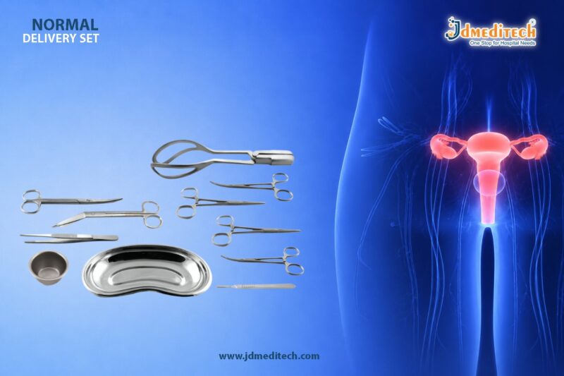
Uroflowmetry System for Urinary Flow Analysis

A Uroflowmetry System is an essential diagnostic device used to measure the flow rate of urine. It plays a critical role in evaluating lower urinary tract function. In modern urology, accurate urinary flow analysis helps detect conditions such as bladder outlet obstruction, prostate enlargement, and urinary retention.

Moreover, the Uroflowmetry System is non-invasive, quick, and highly effective. Therefore, it has become a standard tool in urology clinics and hospitals worldwide.

What is a Uroflowmetry System?

A Uroflowmetry System is a medical device designed to record the speed and volume of urine flow over time. Typically, the patient urinates into a specialized funnel connected to the system. Then, the device measures key parameters and generates a detailed flow curve.

In addition, the system provides precise data that helps urologists diagnose urinary disorders with confidence.

Key Components of Uroflowmetry System

A modern Uroflowmetry System includes the following components:

  • Collection Funnel – Designed for hygienic urine collection
  • Flow Sensor – Measures urine flow rate accurately
  • Electronic Control Unit – Processes and records data
  • Display & Printer – Shows real-time results and prints reports
  • Software Integration – Enables data storage and analysis

Furthermore, advanced systems come with digital connectivity for seamless hospital integration.

How Does a Uroflowmetry System Work?

The working principle of a Uroflowmetry System is simple yet highly effective:

  1. The patient urinates into the device.
  2. The system records urine flow continuously.
  3. It calculates parameters such as flow rate and voided volume.
  4. Finally, it generates a graphical flow curve for analysis.

As a result, doctors can quickly identify abnormalities in urinary flow patterns.

Important Parameters Measured

A Uroflowmetry System provides several critical measurements:

  • Maximum Flow Rate (Qmax) – Indicates peak urine flow
  • Average Flow Rate (Qavg) – Shows overall flow efficiency
  • Voided Volume – Total urine passed
  • Flow Time – Duration of urination
  • Flow Curve Pattern – Helps detect obstructions or weak bladder

Therefore, these parameters are essential for accurate diagnosis and treatment planning.

Clinical Applications of Uroflowmetry System

The Uroflowmetry System is widely used in various medical conditions:

  • Benign Prostatic Hyperplasia (BPH)
  • Urethral Stricture
  • Bladder Dysfunction
  • Neurogenic Bladder Disorders
  • Urinary Incontinence Evaluation

Additionally, it is used for pre-operative and post-operative assessments, ensuring better patient outcomes.

Advantages of Uroflowmetry System

There are several benefits of using a Uroflowmetry System:

  • Non-invasive and painless procedure
  • Quick and easy to perform
  • Provides accurate and reliable results
  • Enhances early diagnosis of urological disorders
  • Improves treatment monitoring

Thus, it is a highly preferred diagnostic tool in modern urology.

Why Choose JDMeditech Uroflowmetry System?

At JDMeditech, we offer advanced Uroflowmetry Systems designed for precision and reliability.

  • High-accuracy flow sensors
  • User-friendly interface
  • Durable and compact design
  • Advanced data analysis software
  • Suitable for hospitals and clinics

Furthermore, our systems meet international quality standards, ensuring consistent performance.

Future Trends in Uroflowmetry Technology

The future of Uroflowmetry Systems is evolving rapidly. For instance:

  • Integration with electronic medical records (EMR)
  • Wireless and portable devices
  • AI-based diagnostic support
  • Remote patient monitoring

Consequently, these innovations will enhance diagnostic accuracy and patient convenience.

Conclusion

The Uroflowmetry System is a vital diagnostic tool for evaluating urinary flow and detecting urological disorders. Because it is non-invasive, accurate, and efficient, it has become indispensable in modern healthcare.

If you are looking for reliable and advanced diagnostic solutions, JDMeditech Uroflowmetry Systems are the perfect choice for your medical facility.


Explore More Product >>

Get Connected:
+91 79909 93062 | +91 63513 72032 | exports@jdmeditech.com
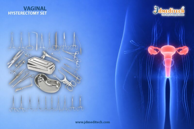
 

Urodynamic System – Advanced Diagnostics for Bladder Function

A Urodynamic System is a highly advanced diagnostic solution used to evaluate the function of the bladder and urethra. It plays a crucial role in identifying urinary disorders such as incontinence, overactive bladder, urinary retention, and neurogenic bladder conditions.

In modern urology, accurate diagnosis is essential. Therefore, healthcare professionals rely on a Urodynamic System to measure pressure, flow, and bladder activity in real time. As a result, clinicians can make precise treatment decisions and improve patient outcomes.

What is a Urodynamic System?

A Urodynamic System is a specialized medical device designed to assess how well the bladder, sphincters, and urethra store and release urine. It combines advanced sensors, catheters, and software to provide detailed functional analysis.

Moreover, this system helps doctors understand the root cause of urinary symptoms rather than relying only on patient history.

Key Components of a Urodynamic System

A modern Urodynamic System consists of several essential components. Each part contributes to accurate and reliable diagnosis:

  • Pressure Transducers – Measure bladder and abdominal pressure
  • Flow Meters – Record urine flow rate
  • Catheters & Sensors – Capture internal bladder data
  • Electromyography (EMG) – Evaluate muscle activity
  • Data Acquisition System – Processes and stores results
  • Analysis Software – Generates detailed reports

In addition, advanced systems offer digital interfaces and real-time monitoring, which significantly enhance diagnostic efficiency.

Types of Urodynamic Tests

A Urodynamic System supports multiple diagnostic tests. These tests help evaluate different aspects of urinary function:

1. Uroflowmetry

This test measures the speed and volume of urine flow. It is simple yet highly effective for detecting obstruction or weak bladder function.

2. Cystometry

Cystometry evaluates bladder capacity and pressure during filling. Consequently, it helps diagnose overactive bladder and reduced compliance.

3. Pressure Flow Study

This test analyzes bladder pressure during urination. Therefore, it is essential for diagnosing bladder outlet obstruction.

4. Electromyography (EMG)

EMG measures muscle activity around the bladder and urethra. As a result, it identifies nerve-related dysfunctions.

5. Video Urodynamics

This advanced test combines imaging with pressure measurements. Hence, it provides a comprehensive view of bladder behavior.

Applications of Urodynamic System

A Urodynamic System is widely used in various clinical settings. It is especially valuable in:

  • Diagnosing urinary incontinence
  • Evaluating overactive bladder (OAB)
  • Assessing neurogenic bladder disorders
  • Investigating urinary retention
  • Pre-surgical evaluation for urological procedures

Furthermore, it is frequently used in hospitals, urology clinics, and specialized diagnostic centers.

Advantages of Using a Urodynamic System

Using a Urodynamic System offers several important benefits:

  • Accurate Diagnosis – Provides objective data for precise evaluation
  • Improved Treatment Planning – Helps select the most effective therapy
  • Minimally Invasive Testing – Ensures patient comfort
  • Real-Time Monitoring – Enables immediate clinical decisions
  • Comprehensive Analysis – Combines multiple tests in one system

Therefore, clinicians can significantly enhance patient care and treatment outcomes.

How a Urodynamic Test is Performed

A Urodynamic System follows a structured testing procedure:

  1. First, the patient is prepared and positioned comfortably
  2. Then, catheters are inserted into the bladder and rectum
  3. Next, the bladder is filled with sterile fluid
  4. Meanwhile, pressure and flow data are recorded
  5. Finally, results are analyzed using specialized software

Although the procedure may sound complex, it is safe, quick, and well-tolerated by most patients.

Latest Innovations in Urodynamic Systems

Modern Urodynamic Systems are equipped with advanced features. For example:

  • Wireless and portable systems
  • High-resolution pressure sensors
  • AI-based diagnostic analysis
  • User-friendly touchscreen interfaces
  • Integrated reporting and data storage

As technology continues to evolve, these systems are becoming more efficient and easier to use.

Why Choose JDMeditech for Urodynamic Systems?

At JDMeditech, we provide high-quality Urodynamic Systems designed for precision and reliability.

Our Key Benefits:
  • Advanced diagnostic technology
  • High-accuracy sensors and components
  • User-friendly software interface
  • Durable and reliable systems
  • Global supply and support

Additionally, our systems are trusted by healthcare professionals for consistent performance and superior results.

Conclusion

A Urodynamic System is an essential tool for diagnosing bladder and urinary disorders. Not only does it provide detailed insights into urinary function, but it also helps clinicians deliver targeted and effective treatments.

Therefore, investing in a high-quality Urodynamic System is crucial for modern urology practices aiming to improve patient outcomes and diagnostic accuracy.


Explore More Product >>

Get Connected:
+91 79909 93062 | +91 63513 72032 | exports@jdmeditech.com