
Lymphadenectomy Surgery: Procedure, Types, Instruments, Benefits, Risks & Recovery

Lymphadenectomy Surgery is a surgical procedure that removes selected lymph nodes for examination or treatment. Surgeons commonly perform this operation during cancer care to assess whether malignant cells have spread through the lymphatic system.

Lymph nodes are small structures that filter lymph fluid and help the body respond to infections. However, cancer cells can sometimes travel from a primary tumor to nearby lymph nodes.

Therefore, examining these nodes can provide valuable information about cancer staging, prognosis, and treatment planning.

Depending on the diagnosis, specialists may remove a limited group of lymph nodes or perform a more extensive regional dissection.

This JDMeditech guide explains lymphadenectomy surgery, its indications, types, surgical instruments, potential benefits, risks, recovery, and equipment selection factors.

What Is Lymphadenectomy Surgery?

Lymphadenectomy Surgery, also called lymph node dissection, involves removing selected lymph nodes from a particular anatomical region.

After removal, a pathology laboratory examines the tissue for cancer cells or other relevant findings.

Furthermore, lymphadenectomy may help remove known cancer deposits in selected patients.

However, removing more lymph nodes does not automatically improve outcomes for every cancer.

Therefore, specialists follow cancer-specific guidelines when deciding whether lymph node removal is appropriate.

Understanding the Lymphatic System

The lymphatic system includes lymph nodes, lymphatic vessels, lymph fluid, and specialized immune tissues.

Its main functions include transporting excess tissue fluid and supporting immune responses.

Lymph nodes are located throughout the body, including the neck, armpits, chest, abdomen, pelvis, and groin.

Consequently, lymphadenectomy procedures vary according to the affected anatomical region.

Why Is Lymphadenectomy Surgery Performed?

Doctors may recommend lymphadenectomy to assess or treat cancer involving regional lymph nodes.

For example, selected gynecological cancers require pelvic or para-aortic lymph node assessment.

Similarly, some breast cancers, head and neck cancers, and other malignancies may require regional lymph node surgery.

However, modern cancer care increasingly uses less extensive procedures when evidence supports them.

Therefore, specialists consider tumor characteristics, imaging findings, sentinel node results, and treatment alternatives.

Common Indications for Lymphadenectomy Surgery
1. Cancer Staging

Lymphadenectomy can help determine whether cancer has spread to regional lymph nodes.

Consequently, pathological results may influence cancer stage and treatment planning.

2. Known Lymph Node Metastases

Some patients have confirmed cancer deposits within regional lymph nodes.

In selected cases, surgery may remove affected nodes as part of treatment.

3. Gynecological Cancers

Selected cervical, endometrial, ovarian, and vulvar cancers may require lymph node assessment.

However, the extent of surgery varies considerably by cancer type and stage.

4. Breast Cancer

Some breast cancer patients require axillary lymph node surgery.

Nevertheless, sentinel lymph node biopsy or omission of further surgery may be appropriate for many patients.

5. Head and Neck Cancers

Selected head and neck cancers may require cervical lymph node dissection.

Therefore, surgeons choose an operation based on the primary tumor and regional disease.

6. Urological Cancers

Selected prostate, bladder, penile, and testicular cancers may involve lymph node surgery.

However, the appropriate procedure depends on cancer-specific treatment guidelines.

Types of Lymphadenectomy Surgery
1. Pelvic Lymphadenectomy

Pelvic Lymphadenectomy removes selected lymph nodes within the pelvis.

For example, surgeons may evaluate nodes along the pelvic blood vessels during selected gynecological or urological cancer procedures.

2. Para-Aortic Lymphadenectomy

Para-Aortic Lymphadenectomy involves removing selected lymph nodes near the abdominal aorta.

It may form part of the management of certain gynecological or other cancers.

3. Axillary Lymph Node Dissection

Axillary lymph node dissection removes selected lymph nodes from the armpit.

However, it is no longer necessary for every patient with breast cancer.

4. Inguinal Lymphadenectomy

Inguinal lymphadenectomy removes selected lymph nodes from the groin.

For example, it may be considered in selected vulvar or penile cancers.

5. Cervical Lymph Node Dissection

Cervical lymph node dissection removes selected lymph nodes from the neck.

Therefore, it may support treatment of suitable head and neck malignancies.

6. Retroperitoneal Lymph Node Dissection

Retroperitoneal lymph node dissection removes selected nodes from the back of the abdominal cavity.

For example, specialists may use this procedure for certain testicular cancers.

7. Laparoscopic Lymphadenectomy

Laparoscopic lymphadenectomy uses small abdominal incisions and camera-assisted surgical instruments.

However, the suitability of this approach depends on the cancer type and operative plan.

8. Robotic-Assisted Lymphadenectomy

Robotic-assisted lymphadenectomy uses compatible robotic surgical equipment.

Nevertheless, patient selection, surgeon expertise, and clinical indications determine whether this approach is appropriate.

9. Regional or Complete Lymph Node Dissection

Regional dissection removes lymph nodes from a defined anatomical area.

Meanwhile, more extensive procedures may remove a larger group of nodes when indicated.

Therefore, the operation must follow the appropriate cancer-specific surgical plan.

Types of Lymphadenectomy and Their Applications
Type Anatomical Region Selected Applications
Pelvic Lymphadenectomy Pelvis Selected gynecological and urological cancers
Para-Aortic Lymphadenectomy Abdomen near aorta Selected gynecological cancers
Axillary Dissection Armpit Selected breast cancers
Inguinal Dissection Groin Selected vulvar and penile cancers
Cervical Neck Dissection Neck Selected head and neck cancers
Retroperitoneal Dissection Back of abdomen Selected testicular cancers
What Is Sentinel Lymph Node Biopsy?

Sentinel lymph node biopsy identifies and removes selected first-draining lymph nodes for pathological examination.

For example, specialists may use an approved radioactive tracer, blue dye, or fluorescence technique according to the cancer type and institutional protocol.

Furthermore, sentinel node assessment may help patients avoid a more extensive lymph node dissection.

However, a negative sentinel node result does not eliminate every possibility of cancer spread.

Therefore, the healthcare team interprets the results within the appropriate clinical context.

Sentinel Lymph Node Biopsy vs Lymphadenectomy
Feature Sentinel Node Biopsy Regional Lymphadenectomy
Nodes Removed Selected first-draining nodes Broader group of regional nodes
Main Purpose Targeted staging assessment Staging or treatment when indicated
Mapping Equipment May require approved tracer detection Not routinely required
Extent of Surgery Usually less extensive Generally more extensive
Lymphedema Risk Generally lower Generally higher
Suitability Selected cancers and patients Selected patients requiring broader removal

Importantly, the need for additional lymph node removal after sentinel node biopsy varies by cancer type and clinical findings.

Common Instruments Used in Lymphadenectomy Surgery

Lymphadenectomy requires instruments for tissue handling, surgical dissection, bleeding control, and specimen removal.

Furthermore, pelvic or para-aortic procedures may require laparoscopic equipment or specialized deep abdominal instruments.

The following examples describe common surgical instrument categories. However, this list is not a verified JDMeditech lymphadenectomy instrument-set packing list.

1. Surgical Scalpel

A surgical scalpel supports appropriate tissue incisions during open surgery.

Different compatible blade and handle designs serve different clinical requirements.

2. Metzenbaum Scissors

Metzenbaum scissors support delicate tissue dissection.

Therefore, surgeons may select suitable designs for soft tissue handling.

3. Mayo Scissors

Mayo scissors support cutting suitable tissues and surgical materials.

Furthermore, straight and curved configurations offer different handling characteristics.

4. Artery Forceps

Artery forceps help clamp selected blood vessels and tissues.

In addition, they support temporary bleeding control during suitable surgical tasks.

5. Mosquito Forceps

Mosquito forceps are small hemostatic instruments used for selected delicate clamping tasks.

However, instrument selection depends on tissue characteristics and intended use.

6. Allis Tissue Forceps

Allis forceps help hold suitable tissues securely.

Nevertheless, their toothed jaws require careful handling.

7. Babcock Forceps

Babcock forceps support gentler manipulation of selected tissues.

Therefore, surgeons choose an appropriate design for the planned procedure.

8. Dissecting Forceps

Dissecting forceps help manipulate and separate selected tissues.

Furthermore, toothed and non-toothed designs support different applications.

9. Right-Angle Forceps

Right-angle forceps may support selected dissection and vessel-handling tasks.

However, surgeons must select instruments appropriate for nearby anatomical structures.

10. Needle Holder

A needle holder helps surgeons control compatible surgical needles during suturing.

Different sizes and jaw configurations support different closure requirements.

11. Surgical Retractors

Surgical retractors help maintain exposure of the operative field.

For example, suitable handheld or self-retaining retractors may support open surgery.

12. Laparoscopic Grasping Forceps

Laparoscopic graspers support tissue manipulation during minimally invasive procedures.

However, jaw design and working length must match the intended application.

13. Laparoscopic Dissecting Forceps

Laparoscopic dissecting forceps support tissue separation under camera-assisted visualization.

Therefore, instrument compatibility remains important.

14. Laparoscopic Scissors

Laparoscopic scissors support selected cutting tasks during minimally invasive operations.

Furthermore, surgeons must select suitable lengths and tip designs.

15. Electrosurgical Instruments

Compatible electrosurgical instruments support tissue cutting and bleeding control.

However, the surgical team must verify generator compatibility and follow approved instructions for use.

16. Vessel-Sealing Instruments

Suitable vessel-sealing devices may support selected tissue and vessel management tasks.

Nevertheless, the device must be appropriate for its intended use.

17. Suction and Irrigation Instruments

Suction and irrigation instruments help maintain operative visibility.

Consequently, they may support selected open and minimally invasive procedures.

18. Specimen Retrieval Bag

Specimen retrieval bags may support contained removal of selected tissues during minimally invasive surgery.

However, specimen handling must follow the relevant oncology protocol.

19. Sentinel Node Detection Equipment

Selected procedures use gamma probes or compatible fluorescence imaging systems to locate sentinel nodes.

However, equipment selection depends on the approved tracer and clinical protocol.

20. Additional Surgical Accessories

Depending on the operation, hospitals may require sutures, clips, surgical drapes, suction tubing, and other accessories.

Therefore, purchasing teams should confirm the complete equipment requirements before ordering.

Lymphadenectomy Surgical Instruments and Functions
Instrument Primary Function
Surgical Scalpel Supports appropriate tissue incisions
Metzenbaum Scissors Support delicate tissue dissection
Mayo Scissors Cut suitable tissues and surgical materials
Artery Forceps Clamp selected vessels and tissues
Mosquito Forceps Support selected small-vessel clamping tasks
Allis Forceps Support secure tissue handling
Babcock Forceps Support gentle tissue manipulation
Dissecting Forceps Support tissue handling and dissection
Right-Angle Forceps Support selected deep dissection tasks
Needle Holder Supports suturing
Surgical Retractors Maintain operative exposure
Laparoscopic Graspers Support tissue manipulation
Laparoscopic Scissors Support compatible cutting tasks
Electrosurgical Instruments Support cutting and bleeding control
Vessel-Sealing Instruments Support selected vessel management
Suction and Irrigation Support operative visibility
Specimen Retrieval Bag Supports contained specimen removal
Sentinel Node Detection System Supports approved lymphatic mapping procedures

Importantly, the exact instruments depend on the cancer type, anatomical region, surgical approach, and manufacturer’s approved intended use.

How Is Lymphadenectomy Surgery Performed?

A qualified surgeon performs lymphadenectomy in an appropriately equipped healthcare facility.

First, the healthcare team reviews the patient’s diagnosis, imaging findings, and planned lymph node assessment.

Next, the anesthesia team prepares the patient for the selected procedure.

During surgery, the specialist accesses the appropriate anatomical region and removes the planned lymph nodes.

When indicated, the team may also perform surgery on the primary tumor.

Throughout the operation, clinicians monitor bleeding, nearby structures, and patient safety.

Afterward, the removed nodes are sent for pathological examination.

Finally, the oncology team reviews the results and determines whether additional treatment is necessary.

However, the exact surgical technique varies considerably. This overview does not replace professional surgical training.

Potential Benefits of Lymphadenectomy Surgery
1. Accurate Cancer Staging

Lymph node examination can reveal regional cancer spread.

Consequently, specialists may obtain more accurate staging information.

2. Treatment Planning

Pathological findings can help guide decisions about chemotherapy, radiation therapy, or other treatments.

Therefore, lymph node assessment may support individualized oncology care.

3. Removal of Selected Cancerous Nodes

In some patients, lymphadenectomy removes lymph nodes containing cancer.

However, the therapeutic benefit depends on the cancer type and clinical situation.

4. Pathological Examination

Laboratory examination provides information about the removed lymph nodes.

Furthermore, the findings may help estimate prognosis.

5. Regional Disease Assessment

Lymphadenectomy can provide information about cancer involvement in a particular anatomical region.

As a result, the oncology team may adjust the treatment plan.

6. Integration With Other Cancer Surgery

Surgeons may perform lymph node removal alongside appropriate cancer operations.

However, the extent of surgery must follow current clinical evidence and guidelines.

Open vs Laparoscopic Lymphadenectomy
Feature Open Surgery Laparoscopic Surgery
Access Open incision Small incisions
Visualization Direct exposure Camera-assisted view
Instruments Open surgical instruments Laparoscopic instruments
Early Recovery May be longer May be shorter
Suitability Selected patients and procedures Selected patients and procedures

However, cancer-specific outcomes and surgical safety are more important than incision size alone.

Risks and Complications of Lymphadenectomy Surgery

Lymphadenectomy is an invasive procedure with potential complications.

For example, patients may experience bleeding, infection, pain, or wound-healing problems.

In addition, surgery can injure nearby nerves, blood vessels, or other anatomical structures.

Some patients develop a seroma or lymphocele, which involves fluid accumulation after lymphatic disruption.

Furthermore, lymph node removal can increase the risk of lymphedema.

Therefore, the healthcare team must balance the expected benefits against the risks of removing additional nodes.

What Is Lymphedema After Lymphadenectomy?

Lymphedema is swelling caused by an accumulation of lymph fluid within body tissues.

For example, pelvic or groin lymph node removal may increase the risk of swelling in the legs.

Similarly, axillary lymph node surgery may affect lymph drainage from the arm.

However, not every patient develops lymphedema.

The risk depends on the location and extent of surgery, previous treatments, radiation therapy, and other factors.

Furthermore, lymphedema may develop months or years after cancer treatment.

Therefore, patients should report persistent swelling, heaviness, or tightness to their healthcare team.

Recovery After Lymphadenectomy Surgery

Recovery depends on the anatomical region, extent of lymph node removal, and surgical approach.

For example, a limited sentinel lymph node biopsy may require only a short recovery period.

However, extensive pelvic, abdominal, or regional lymph node dissection can require several weeks of recovery.

During healing, patients may experience discomfort, swelling, fatigue, or temporary activity limitations.

Furthermore, some patients require surgical drains or additional wound care.

Therefore, the healthcare team provides individualized instructions about activity, wound care, medication, and follow-up.

Patients should seek prompt medical assessment for fever, worsening redness, severe pain, sudden swelling, or breathing difficulty.

Long-Term Care After Lymphadenectomy

Long-term follow-up depends on the cancer diagnosis and the extent of surgery.

For example, some patients require additional oncology treatment after lymph node examination.

Furthermore, patients at risk of lymphedema may benefit from early symptom recognition and appropriate rehabilitation.

However, the recommended follow-up schedule varies by cancer type.

Therefore, patients should follow the surveillance plan provided by their oncology team.

How to Choose Lymphadenectomy Surgical Instruments

Hospitals and surgical departments should identify their clinical requirements before purchasing lymphadenectomy instruments.

First, determine the anatomical region and planned surgical approach.

Next, review the required instruments for tissue dissection, vessel management, and specimen handling.

Moreover, purchasing teams should confirm instrument dimensions, compatibility, and sterilization instructions.

Important Equipment Selection Factors
  • Approved intended use
  • Complete instrument-set packing list
  • Open or laparoscopic surgical requirements
  • Scalpel handle compatibility
  • Metzenbaum and Mayo scissors dimensions
  • Artery and mosquito forceps specifications
  • Allis and Babcock forceps configurations
  • Right-angle forceps design
  • Needle holder dimensions
  • Surgical retractor configuration
  • Laparoscopic grasper compatibility
  • Laparoscopic scissors working length
  • Electrosurgical system compatibility
  • Vessel-sealing device requirements
  • Suction and irrigation compatibility
  • Specimen retrieval accessories
  • Sentinel node detection requirements
  • Reusable or single-use classification
  • Cleaning and sterilization instructions
  • Manufacturer technical datasheet
  • Applicable regulatory documentation

Furthermore, hospitals should verify the exact supplied products before placing an order.

Surgical Instruments by JDMeditech

JDMeditech welcomes inquiries from hospitals, gynecology departments, urology departments, surgical centers, and international medical distributors regarding surgical instruments and related medical accessories.

For example, customers can inquire about available surgical forceps, scissors, clamps, needle holders, retractors, laparoscopic instruments, and compatible accessories.

In addition, the JDMeditech team can confirm available configurations, technical specifications, and applicable documentation for individual inquiries.

For product information and quotations, visit www.jdmeditech.com.

Frequently Asked Questions About Lymphadenectomy Surgery
1. What is lymphadenectomy surgery?

Lymphadenectomy is a procedure that removes selected lymph nodes for examination or treatment.

2. Why is lymphadenectomy performed?

Doctors may recommend it to assess regional cancer spread or remove selected lymph nodes containing cancer.

3. What is pelvic lymphadenectomy?

Pelvic lymphadenectomy removes selected lymph nodes from the pelvis during appropriate cancer procedures.

4. What is para-aortic lymphadenectomy?

Para-aortic lymphadenectomy removes selected lymph nodes located near the abdominal aorta.

5. What is the difference between sentinel node biopsy and lymphadenectomy?

Sentinel node biopsy removes selected first-draining nodes, while regional lymphadenectomy removes a broader group of nodes.

6. What instruments are used during lymphadenectomy?

Common instrument categories include surgical forceps, scissors, retractors, needle holders, electrosurgical instruments, and suitable specimen-handling accessories.

7. Is lymphadenectomy always necessary for cancer?

No. The need for lymph node surgery depends on the cancer type, stage, clinical findings, and treatment guidelines.

8. Can lymphadenectomy cause lymphedema?

Yes. Removing lymph nodes can disrupt lymph drainage and increase the risk of swelling in the affected area.

9. How long does recovery take?

Recovery ranges from a short period after limited node biopsy to several weeks or longer after extensive dissection.

10. Where can hospitals inquire about JDMeditech surgical instruments?

Hospitals and medical distributors can visit JDMeditech to request information about available instruments and accessories.

Conclusion

Lymphadenectomy Surgery plays an important role in the staging and selected treatment of cancer.

Moreover, appropriate surgical instruments support qualified healthcare professionals during lymph node dissection.

However, not every patient benefits from extensive lymph node removal.

Therefore, surgeons must select the appropriate procedure according to cancer-specific evidence, clinical requirements, and individual patient factors.

Contact JDMeditech for surgical instrument inquiries, compatible medical accessories, and international business requirements.

Medical Disclaimer: This article provides general educational information only. It does not replace professional medical advice, oncology treatment planning, or surgical training. Qualified healthcare professionals must make treatment and equipment decisions according to current clinical guidelines and approved manufacturer instructions.

Endometrial Cancer Staging Surgery: Procedure, Instruments, FIGO Stages & Recovery

Endometrial Cancer Staging Surgery is an important gynecologic oncology procedure that helps doctors determine the extent of cancer originating in the lining of the uterus. The operation can also remove the primary tumor and provide tissue for detailed pathological assessment.

Endometrial cancer develops in the endometrium, which forms the inner lining of the uterus. Although many patients receive a diagnosis at an early stage, some cancers spread into the uterine muscle, cervix, lymph nodes, or other organs.

Therefore, accurate staging helps specialists select appropriate treatment and estimate the risk of recurrence.

In many patients, surgery includes total hysterectomy and bilateral salpingo-oophorectomy. However, lymph node assessment and additional procedures depend on individual risk factors.

This JDMeditech guide explains endometrial cancer staging surgery, FIGO stages, surgical instruments, operative approaches, benefits, risks, recovery, and equipment selection.

What Is Endometrial Cancer Staging Surgery?

Endometrial Cancer Staging Surgery combines surgical treatment with assessment of the cancer’s extent.

During the operation, surgeons remove appropriate organs and collect selected tissues for pathological examination.

Furthermore, specialists may evaluate sentinel lymph nodes to determine whether cancer has spread beyond the uterus.

However, not every patient requires extensive lymph node dissection.

Therefore, the gynecologic oncologist selects the procedure according to the cancer’s characteristics and the patient’s medical condition.

Understanding Endometrial Cancer

Endometrial cancer begins in cells lining the uterus.

Endometrioid carcinoma is the most common histological type. However, other types include serous carcinoma, clear cell carcinoma, and carcinosarcoma.

Different cancer types can behave differently and require different treatment strategies.

Furthermore, molecular testing provides additional information about tumor biology.

Consequently, modern treatment planning considers anatomy, histology, grade, and molecular characteristics.

Common Symptoms of Endometrial Cancer

Abnormal uterine bleeding is a common warning sign of endometrial cancer.

For example, bleeding after menopause requires medical assessment.

Some patients experience unusual bleeding between menstrual periods or abnormal vaginal discharge.

However, these symptoms can also occur with noncancerous conditions.

Therefore, patients should consult a qualified healthcare professional for appropriate investigation.

Why Is Endometrial Cancer Staging Surgery Performed?

Doctors perform staging surgery to treat suitable endometrial cancers and obtain information about the extent of disease.

For example, examination of the uterus can reveal the depth of invasion into the uterine muscle.

Furthermore, tissue analysis can identify cervical involvement, lymphovascular space invasion, and other important findings.

Selected lymph node assessments may reveal regional cancer spread.

Therefore, surgical findings help determine whether additional treatment is necessary.

Common Indications for Endometrial Cancer Staging Surgery
1. Confirmed Endometrial Carcinoma

Many patients with confirmed endometrial carcinoma undergo surgical treatment when medically suitable.

However, the exact operation depends on disease characteristics and patient health.

2. Suspected Early-Stage Endometrial Cancer

Selected patients with apparently uterus-confined disease may undergo minimally invasive hysterectomy with appropriate staging.

Furthermore, the surgeon may perform sentinel lymph node assessment when indicated.

3. Higher-Risk Histological Types

Some tumor types have a greater risk of extrauterine spread.

Consequently, specialists may recommend additional staging procedures based on the cancer’s characteristics.

4. Suspected Lymph Node Involvement

Imaging or clinical findings may suggest cancer involvement in regional lymph nodes.

Therefore, the oncology team considers appropriate lymph node evaluation.

5. Selected Advanced Disease

Some patients with advanced endometrial cancer may benefit from surgery as part of multidisciplinary treatment.

However, systemic therapy or radiation may be more appropriate in other circumstances.

6. Treatment Planning After Cancer Diagnosis

Surgical pathology provides information that helps specialists estimate recurrence risk.

As a result, the oncology team can recommend suitable postoperative treatment and surveillance.

Types of Endometrial Cancer Staging Surgery
1. Total Hysterectomy

Total hysterectomy removes the uterus and cervix.

For many patients with endometrial cancer, it forms the central component of surgical treatment.

2. Bilateral Salpingo-Oophorectomy

Bilateral salpingo-oophorectomy removes both ovaries and fallopian tubes.

It commonly accompanies hysterectomy for endometrial cancer.

However, carefully selected younger patients with low-risk disease may be considered for ovarian preservation after specialist counseling.

3. Sentinel Lymph Node Mapping

Sentinel lymph node mapping identifies the first lymph nodes likely to receive drainage from the tumor region.

For example, surgeons may use an appropriate tracer and compatible imaging equipment to identify selected nodes.

However, mapping techniques and subsequent assessment must follow the institution’s approved oncology protocol.

4. Pelvic Lymph Node Dissection

Pelvic lymph node dissection removes selected lymph nodes for pathological examination.

However, systematic lymphadenectomy is not necessary for every patient.

Therefore, specialists consider tumor risk and the results of other staging assessments.

5. Para-Aortic Lymph Node Assessment

Selected patients require evaluation of lymph nodes near the abdominal aorta.

However, the need for para-aortic assessment depends on the cancer’s characteristics and surgical findings.

6. Laparoscopic Staging Surgery

Laparoscopic surgery uses small abdominal incisions and camera-assisted instruments.

For suitable patients with apparent early-stage disease, minimally invasive surgery is generally preferred.

7. Robotic-Assisted Staging Surgery

Robotic-assisted surgery uses compatible minimally invasive instruments controlled by a trained surgeon.

However, availability, patient characteristics, and surgical expertise influence selection.

8. Open Abdominal Staging Surgery

Open surgery uses an abdominal incision to access the pelvic and abdominal organs.

Specialists may choose this approach for selected extensive tumors, complex anatomy, or other clinical requirements.

9. Omental Assessment

Selected high-risk histological types may require assessment or removal of omental tissue.

However, routine omentectomy is not necessary for every endometrial cancer.

FIGO Staging of Endometrial Cancer

The International Federation of Gynecology and Obstetrics, known as FIGO, provides a staging system for endometrial cancer.

The 2023 FIGO system incorporates anatomical spread together with important pathological and molecular factors.

Therefore, the stage cannot always be determined from tumor location alone.

Overview of FIGO 2023 Stages
Stage General Description
Stage I Generally disease involving the uterine corpus, with certain narrowly defined uterus-and-ovary cases; classification also depends on histology and other factors
Stage II Cervical stromal invasion, substantial lymphovascular space invasion, or aggressive histology with myometrial invasion, depending on subtype
Stage III Local or regional spread, including selected adnexal, pelvic tissue, peritoneal, or regional lymph node involvement
Stage IV Bladder or bowel mucosal invasion, abdominal peritoneal metastasis beyond the pelvis, or distant metastasis

Importantly, these descriptions are simplified. The official FIGO 2023 classification includes multiple substages and molecular modifiers.

Furthermore, some clinical settings continue to report earlier staging systems alongside the updated FIGO system.

Therefore, patients should ask their oncology team which staging edition applies to their diagnosis.

Molecular Classification of Endometrial Cancer

Modern endometrial cancer assessment increasingly includes molecular testing.

For example, clinicians may evaluate POLE mutations, mismatch repair deficiency, and abnormal p53 expression.

Tumors without these defining molecular features may fall into the no-specific-molecular-profile category.

Furthermore, molecular classification can influence prognosis and treatment recommendations.

Therefore, oncology teams interpret molecular findings together with surgical pathology and clinical factors.

Common Instruments Used in Endometrial Cancer Staging Surgery

Endometrial cancer staging surgery requires instruments for pelvic access, tissue handling, dissection, bleeding control, and specimen retrieval.

Furthermore, selected procedures require specialized equipment for sentinel lymph node mapping or laparoscopic surgery.

The following instruments may support appropriate procedures. However, this list does not represent a confirmed JDMeditech instrument-set packing list.

1. Laparoscope Telescope

A laparoscope provides camera-assisted visualization during minimally invasive surgery.

Therefore, surgeons select compatible telescopes, camera systems, and light sources.

2. Laparoscopic Trocar and Cannula

Trocar and cannula systems provide access for compatible laparoscopic instruments.

Furthermore, the team must select suitable diameters and instrument lengths.

3. Laparoscopic Grasping Forceps

Grasping forceps support manipulation of suitable tissues during surgery.

However, the surgeon must select the appropriate jaw design.

4. Laparoscopic Dissecting Forceps

Dissecting forceps support controlled tissue separation.

Different designs provide different handling characteristics.

5. Laparoscopic Scissors

Laparoscopic scissors support tissue cutting during minimally invasive procedures.

Therefore, instrument compatibility and working length are important.

6. Electrosurgical Instruments

Compatible electrosurgical instruments support tissue cutting and bleeding control.

However, the surgical team must verify generator compatibility and follow approved instructions for use.

7. Vessel-Sealing Instruments

Suitable vessel-sealing devices may support selected vascular pedicle management tasks.

Nevertheless, the device must be appropriate for the intended clinical application.

8. Laparoscopic Suction and Irrigation Instruments

Suction and irrigation instruments support visibility by managing suitable fluids.

Consequently, they may help maintain the operative field.

9. Specimen Retrieval Bag

A specimen retrieval bag may support contained removal of selected surgical specimens.

Importantly, oncology teams should avoid unnecessary tumor disruption and spillage.

10. Surgical Scalpel

A surgical scalpel supports appropriate incisions during open surgery.

Furthermore, compatible blades and handles support different surgical tasks.

11. Mayo and Metzenbaum Scissors

Mayo scissors support cutting suitable tissues and surgical materials.

Meanwhile, Metzenbaum scissors support delicate tissue dissection.

12. Artery Forceps

Artery forceps help clamp selected vessels and tissues.

In addition, they support temporary bleeding control during appropriate tasks.

13. Allis and Babcock Forceps

Allis forceps provide firm handling of suitable tissues.

In contrast, Babcock forceps support gentler tissue manipulation.

14. Needle Holder

A needle holder helps surgeons control compatible surgical needles during suturing.

Different jaw configurations and lengths support different requirements.

15. Abdominal Retractors

Abdominal retractors help maintain exposure during open surgical procedures.

Therefore, the team selects suitable sizes and designs.

16. Sentinel Lymph Node Imaging Equipment

Selected procedures use near-infrared fluorescence imaging systems to visualize appropriate tracers during sentinel lymph node mapping.

However, the system and tracer must be approved and used according to the clinical protocol.

17. Lymph Node Dissection Instruments

Specialized forceps, scissors, and compatible energy instruments may support lymph node assessment.

However, exact requirements depend on the planned staging procedure.

18. Uterine Manipulation Instruments

Selected gynecological procedures may use compatible uterine manipulation instruments.

However, oncology teams must assess the potential for tumor disruption and select equipment according to their surgical protocol.

19. Surgical Sutures and Accessories

Suitable sutures and closure accessories support selected operative tasks.

Therefore, hospitals should verify material specifications and compatibility.

20. Additional Surgical Equipment

Complex staging procedures may require specialized equipment based on the surgical approach.

Consequently, purchasing teams should confirm the full instrument requirements before ordering.

Endometrial Cancer Staging Instruments and Functions
Instrument Primary Function
Laparoscope Telescope Provides camera-assisted visualization
Trocar and Cannula Support laparoscopic access
Laparoscopic Grasping Forceps Support tissue manipulation
Laparoscopic Dissecting Forceps Support tissue dissection
Laparoscopic Scissors Support tissue cutting
Electrosurgical Instruments Support compatible cutting and bleeding control
Vessel-Sealing Instruments Support selected vascular pedicle management
Suction and Irrigation Instruments Support operative visibility
Specimen Retrieval Bag Supports contained specimen retrieval
Surgical Scalpel Supports appropriate tissue incisions
Mayo and Metzenbaum Scissors Support cutting and dissection
Artery Forceps Clamp selected vessels and tissues
Allis and Babcock Forceps Support appropriate tissue handling
Needle Holder Supports suturing
Abdominal Retractors Maintain operative exposure
Fluorescence Imaging System Supports selected sentinel lymph node mapping
Lymph Node Dissection Instruments Support appropriate nodal assessment

Importantly, instrument selection depends on the surgical approach, intended use, and manufacturer instructions.

How Is Endometrial Cancer Staging Surgery Performed?

A qualified gynecologic oncology surgeon performs endometrial cancer staging surgery in an appropriately equipped hospital.

First, the healthcare team reviews the patient’s diagnosis, biopsy results, imaging findings, and medical history.

Next, the team determines the appropriate surgical approach and planned staging procedures.

During surgery, the specialist removes the uterus and other planned structures.

When indicated, the surgeon also performs sentinel lymph node mapping or appropriate lymph node assessment.

Throughout the operation, clinicians monitor bleeding, organ function, and patient safety.

Afterward, the surgeon completes the planned closure and arranges postoperative care.

Finally, the oncology team reviews the pathology findings, confirms the cancer stage, and determines whether further treatment is necessary.

However, the exact surgical technique varies. This overview does not replace professional surgical training.

Sentinel Lymph Node Mapping in Endometrial Cancer

Sentinel lymph node mapping helps identify selected lymph nodes that may be the first sites of regional cancer spread.

For example, compatible fluorescence imaging may support visualization after use of an appropriate tracer.

Furthermore, pathological examination can detect small tumor deposits that may not be visible during surgery.

However, successful mapping depends on the approved technique and institutional protocol.

Therefore, the oncology team decides whether sentinel mapping, additional lymph node dissection, or another assessment is appropriate.

Sentinel Lymph Node Mapping vs Lymphadenectomy
Feature Sentinel Lymph Node Mapping Lymphadenectomy
Purpose Identify and assess selected first-draining nodes Remove a broader group of lymph nodes
Extent Generally more limited Generally more extensive
Equipment May require compatible tracer imaging Requires suitable dissection instruments
Complication Risk Generally lower risk of some lymphatic complications Greater risk of lymphatic complications
Suitability Selected patients under an approved algorithm Selected patients requiring broader assessment

Therefore, lymph node assessment should reflect the patient’s cancer risk and the institution’s oncology protocol.

Laparoscopic vs Open Endometrial Cancer Staging Surgery
Feature Laparoscopic Surgery Open Surgery
Surgical Access Small abdominal incisions Larger abdominal incision
Visualization Camera-assisted view Direct surgical exposure
Instruments Laparoscopic surgical instruments Open surgical instruments
Hospital Stay Often shorter Generally longer
Recovery Often faster Generally longer
Typical Selection Preferred for many suitable uterus-confined cases Selected complex or extensive cases

However, the appropriate approach depends on cancer extent, patient characteristics, and surgical expertise.

Potential Benefits of Endometrial Cancer Staging Surgery
1. Removal of the Primary Cancer

Surgery removes the uterus containing the primary tumor in suitable patients.

Therefore, it may provide definitive local treatment for selected cancers.

2. Accurate Pathological Staging

Examination of surgical specimens provides important information about disease extent.

Consequently, specialists can develop a more precise treatment plan.

3. Assessment of Lymph Node Spread

Selected patients undergo appropriate regional lymph node assessment.

As a result, the oncology team can evaluate whether cancer has spread beyond the uterus.

4. Guidance for Additional Treatment

Staging findings help determine whether postoperative radiotherapy, chemotherapy, or other treatment is appropriate.

However, not every patient requires additional treatment.

5. Individualized Cancer Care

Modern oncology teams combine surgical findings with histological and molecular information.

Therefore, treatment recommendations can reflect the patient’s individual risk.

6. Minimally Invasive Options

Suitable patients may undergo laparoscopic or robotic-assisted staging procedures.

Consequently, some patients experience shorter hospitalization and faster early recovery.

Risks and Complications of Endometrial Cancer Staging Surgery

Endometrial cancer staging surgery is an invasive operation with potential complications.

For example, patients may experience bleeding, infection, wound problems, or blood clots.

In addition, surgery may injure nearby structures such as the bladder, ureters, bowel, or blood vessels.

Lymph node removal can also cause lymphocele or lower-limb lymphedema.

Furthermore, removal of both ovaries before natural menopause causes surgical menopause.

Therefore, careful patient selection, specialist surgical expertise, and appropriate postoperative care are essential.

Recovery After Endometrial Cancer Staging Surgery

Recovery depends on the surgical approach, patient’s health, and complexity of treatment.

For example, many patients return gradually to usual activities within approximately two to four weeks after uncomplicated minimally invasive hysterectomy.

However, complete recovery after open abdominal surgery commonly takes approximately six to eight weeks.

Furthermore, extensive staging procedures or postoperative complications may require longer recovery.

During recovery, patients may experience fatigue, abdominal discomfort, or temporary activity limitations.

Therefore, patients should follow their healthcare team’s instructions about wound care, exercise, lifting, medication, and follow-up.

Patients should seek prompt medical attention for fever, severe pain, heavy bleeding, leg swelling, or breathing difficulty.

Life After Endometrial Cancer Staging Surgery

Long-term outcomes depend on the final cancer stage, treatment received, and individual recovery.

For example, some patients require additional radiation therapy or systemic treatment after surgery.

Furthermore, removal of the uterus means the patient can no longer carry a pregnancy.

When both ovaries are removed before natural menopause, patients also experience surgical menopause.

Therefore, follow-up care should address cancer surveillance, physical recovery, hormonal health, and emotional well-being.

How to Choose Endometrial Cancer Staging Surgical Instruments

Hospitals and gynecologic oncology departments should identify their clinical requirements before purchasing staging instruments.

First, determine whether the planned operation involves laparoscopy, robotic-assisted surgery, or open abdominal surgery.

Next, review instrument dimensions, materials, working lengths, and system compatibility.

Moreover, purchasing teams should verify cleaning, sterilization, and maintenance requirements.

Important Equipment Selection Factors
  • Approved intended use
  • Complete instrument-set packing list
  • Laparoscopic or open surgical requirements
  • Laparoscope telescope diameter
  • Camera and light source compatibility
  • Trocar and cannula dimensions
  • Laparoscopic grasper configuration
  • Dissecting forceps design
  • Laparoscopic scissors specifications
  • Electrosurgical system compatibility
  • Vessel-sealing device requirements
  • Suction and irrigation compatibility
  • Specimen retrieval accessories
  • Sentinel lymph node imaging requirements
  • Lymph node dissection instruments
  • Open surgical forceps and retractors
  • Needle holder configuration
  • Reusable or single-use classification
  • Cleaning and sterilization instructions
  • Manufacturer technical datasheet
  • Applicable regulatory documentation

Furthermore, hospitals should confirm the exact supplied products before placing an order.

Gynecology Surgical Instruments by JDMeditech

JDMeditech welcomes inquiries from hospitals, gynecologic oncology departments, surgical centers, and international medical distributors regarding gynecological surgical instruments and related medical accessories.

For example, customers can inquire about available surgical forceps, scissors, clamps, needle holders, retractors, laparoscopic instruments, and compatible accessories.

In addition, the JDMeditech team can confirm available configurations, technical specifications, and applicable documentation for individual inquiries.

For product information and quotations, visit www.jdmeditech.com.

Frequently Asked Questions About Endometrial Cancer Staging Surgery
1. What is endometrial cancer staging surgery?

It is an operation that treats suitable endometrial cancers and provides tissue for determining the extent of disease.

2. Why is staging important in endometrial cancer?

Staging helps specialists estimate disease extent, assess recurrence risk, and plan additional treatment.

3. What organs are usually removed during staging surgery?

Standard surgery commonly removes the uterus, cervix, ovaries, and fallopian tubes, although selected patients may receive an individualized approach.

4. What is FIGO staging?

FIGO staging classifies endometrial cancer according to anatomical spread and, in the updated system, important pathological and molecular factors.

5. What is sentinel lymph node mapping?

It is a technique that identifies selected first-draining lymph nodes for pathological assessment.

6. Is lymph node dissection always required?

No. Specialists choose the appropriate lymph node assessment according to cancer risk and the clinical staging protocol.

7. What instruments are used during endometrial cancer staging?

Common instrument categories include laparoscopes, grasping forceps, scissors, retractors, needle holders, electrosurgical equipment, and selected lymph node assessment instruments.

8. Is minimally invasive surgery possible?

Yes. Laparoscopic or robotic-assisted surgery is generally preferred for many suitable patients with apparent uterus-confined disease.

9. How long does recovery take?

Recovery after uncomplicated minimally invasive surgery may take several weeks, while open abdominal surgery commonly requires around six to eight weeks.

10. Where can hospitals inquire about JDMeditech surgical instruments?

Hospitals and medical distributors can visit JDMeditech to request information about available gynecology instruments and accessories.

Conclusion

Endometrial Cancer Staging Surgery plays an important role in diagnosing the extent of disease and treating suitable patients with uterine cancer.

Moreover, modern staging combines surgical findings with histological and molecular information.

However, successful treatment requires careful patient assessment, specialist surgical expertise, appropriate equipment, and postoperative care.

Therefore, hospitals should select instruments according to clinical requirements, professional guidelines, and approved manufacturer instructions.
Contact JDMeditech for gynecology surgical instrument inquiries, compatible medical accessories, and international business requirements.

Medical Disclaimer: This article provides general educational information only. It does not replace professional medical advice, oncology treatment planning, or surgical training. Qualified healthcare professionals must make treatment and equipment decisions according to clinical judgment, current professional guidelines, and approved manufacturer instructions.
Ovarian Cancer Debulking Surgery: Procedure, Instruments, Benefits, Risks & Recovery

Ovarian Cancer Debulking Surgery is a major gynecologic oncology procedure that aims to remove as much visible cancer as safely possible from the abdomen and pelvis. Medical professionals also call this operation cytoreductive surgery.

Ovarian cancer can spread beyond the ovaries to the abdominal lining, omentum, and nearby organs. Therefore, treatment may require more extensive surgery than routine ovarian tumor removal.

In selected patients, surgeons perform debulking surgery before chemotherapy. However, others receive chemotherapy first and undergo surgery afterward.

The primary surgical goal is often complete gross resection, meaning that no visible cancer remains at the end of the operation.

This JDMeditech guide explains ovarian cancer debulking surgery, its types, indications, instruments, treatment approaches, benefits, risks, recovery, and equipment selection factors.

What Is Ovarian Cancer Debulking Surgery?

Ovarian Cancer Debulking Surgery is an operation designed to reduce the amount of visible cancer in the abdomen and pelvis.

Depending on the disease, surgeons may remove the ovaries, fallopian tubes, uterus, omentum, and selected affected tissues.

Furthermore, some patients require bowel resection or additional procedures when cancer involves nearby structures.

However, the surgical team balances extensive tumor removal against the risk of serious complications.

Therefore, gynecologic oncologists develop an individualized treatment plan based on the patient’s cancer and overall health.

Understanding Ovarian Cancer and Its Spread

Ovarian cancer includes several types of malignant tumors that affect ovarian tissue.

Epithelial ovarian cancer is the most common broad category. In addition, related cancers can develop in the fallopian tubes or primary peritoneal tissue.

These cancers may spread across the peritoneal surfaces within the abdomen.

For example, cancer deposits may develop on the omentum, pelvic lining, diaphragm, or bowel surfaces.

Consequently, advanced ovarian cancer often requires a multidisciplinary treatment approach.

Why Is Ovarian Cancer Debulking Surgery Performed?

Doctors recommend cytoreductive surgery to remove visible cancer when an operation is likely to provide meaningful treatment benefit.

For example, selected patients with advanced epithelial ovarian cancer may undergo surgery followed by chemotherapy.

However, some patients benefit from chemotherapy before surgery when immediate extensive resection is unlikely to be appropriate.

Therefore, the oncology team evaluates imaging findings, cancer characteristics, surgical feasibility, and the patient’s medical condition.

Common Indications for Ovarian Cancer Debulking Surgery
1. Advanced Epithelial Ovarian Cancer

Patients with selected stage II, III, or IV epithelial ovarian cancers may require cytoreductive surgery.

However, the timing and extent of surgery depend on the individual treatment plan.

2. Fallopian Tube Cancer

Fallopian tube cancer often follows treatment principles similar to epithelial ovarian cancer.

Consequently, selected patients may undergo cytoreductive surgery and systemic therapy.

3. Primary Peritoneal Cancer

Primary peritoneal cancer affects tissue lining the abdominal cavity.

In suitable patients, treatment may include extensive surgical removal of visible disease.

4. Cancer Suitable for Complete Gross Resection

The possibility of removing all visible disease is an important factor in surgical planning.

Therefore, specialists assess whether complete gross resection appears achievable with acceptable risk.

5. Selected Patients After Neoadjuvant Chemotherapy

Some patients receive chemotherapy before surgery to reduce disease burden.

After an appropriate response or stable disease, the team may recommend interval debulking surgery.

6. Selected Recurrent Ovarian Cancer

Some patients with recurrent ovarian cancer may benefit from secondary cytoreductive surgery.

However, this approach is suitable only for carefully selected patients and is not routine for every recurrence.

Types of Ovarian Cancer Debulking Surgery
1. Primary Debulking Surgery

Primary Debulking Surgery is performed before the main course of chemotherapy.

The surgeon aims to remove all visible cancer when feasible.

Afterward, the patient commonly receives platinum-based chemotherapy according to the oncology treatment plan.

2. Interval Debulking Surgery

Interval Debulking Surgery takes place after an initial course of neoadjuvant chemotherapy.

For example, selected patients may receive three or four cycles of chemotherapy before reassessment for surgery.

Following the operation, additional systemic treatment may be recommended.

3. Secondary Cytoreductive Surgery

Secondary cytoreductive surgery may be considered for selected patients with recurrent ovarian cancer.

However, specialists carefully evaluate the likelihood of complete resection and the benefits of alternative treatments.

4. Complete Gross Cytoreduction

Complete gross cytoreduction means that the surgeon leaves no visible tumor at the end of the operation.

Importantly, microscopic cancer cells may still remain, so additional systemic treatment may be necessary.

5. Optimal and Suboptimal Debulking

Historically, optimal debulking often meant leaving no residual tumor deposit larger than one centimeter.

However, complete gross resection is generally the preferred surgical objective when safely achievable.

In contrast, suboptimal debulking describes a greater amount of residual disease.

Primary vs Interval Debulking Surgery
Feature Primary Debulking Interval Debulking
Timing Before chemotherapy After initial chemotherapy
Initial Treatment Surgery Neoadjuvant chemotherapy
Surgical Goal Remove visible cancer Remove remaining visible cancer
Patient Selection Selected patients suitable for upfront surgery Selected patients benefiting from chemotherapy first
Further Treatment Usually chemotherapy Usually additional chemotherapy

Both approaches are established treatment options. Therefore, the oncology team determines the appropriate sequence for each patient.

Common Procedures Performed During Ovarian Cancer Debulking
1. Hysterectomy

Hysterectomy removes the uterus.

In many standard cytoreductive operations, surgeons remove the uterus as part of extensive cancer treatment.

2. Bilateral Salpingo-Oophorectomy

Bilateral salpingo-oophorectomy removes both ovaries and both fallopian tubes.

However, the exact organ removal depends on the cancer diagnosis and individualized treatment requirements.

3. Omentectomy

Omentectomy removes part or all of the omentum, a fatty tissue layer within the abdomen.

Because ovarian cancer commonly involves the omentum, surgeons may remove affected tissue during cytoreduction.

4. Peritoneal Tumor Resection

Surgeons may remove selected cancer deposits from the peritoneal surfaces.

However, the extent of resection depends on the location of disease and surgical safety.

5. Bowel Resection

Some ovarian cancers involve the bowel or nearby supporting tissues.

Consequently, selected patients may require bowel resection or reconstruction.

6. Diaphragmatic or Upper Abdominal Procedures

Advanced disease may involve the diaphragm or upper abdominal structures.

Therefore, the operation may require additional specialist surgical expertise.

7. Lymph Node Assessment

Surgeons may assess suspicious lymph nodes when clinically indicated.

However, systematic removal of clinically normal lymph nodes is not routinely recommended during complete cytoreduction for advanced ovarian cancer.

Common Instruments Used in Ovarian Cancer Debulking Surgery

Ovarian cancer debulking surgery requires instruments for surgical access, tissue dissection, bleeding control, specimen handling, and closure.

Furthermore, complex operations may involve gynecological, colorectal, urological, or upper abdominal surgical instruments.

The following instruments may support selected procedures. However, this list does not represent a confirmed JDMeditech surgical-set packing list.

1. Surgical Scalpel

A surgical scalpel supports appropriate tissue incisions during open abdominal surgery.

Different compatible blades and handles serve different clinical requirements.

2. Mayo Scissors

Mayo scissors support cutting suitable tissues and surgical materials.

Furthermore, straight and curved designs offer different handling characteristics.

3. Metzenbaum Scissors

Metzenbaum scissors support delicate tissue dissection.

Therefore, surgeons may select suitable designs for particular tissue-handling tasks.

4. Artery Forceps

Artery forceps help clamp selected vessels and tissues.

In addition, they support temporary bleeding control during appropriate surgical tasks.

5. Allis Tissue Forceps

Allis forceps provide a secure grip on suitable tissues.

However, their toothed jaws require careful handling.

6. Babcock Forceps

Babcock forceps support gentler handling of selected tissues.

For example, compatible designs may be used during suitable abdominal procedures.

7. Kocher Forceps

Kocher forceps support firm handling of selected tissues.

Nevertheless, surgeons must consider the instrument’s gripping characteristics.

8. Dissecting Forceps

Dissecting forceps support tissue manipulation and surgical dissection.

Furthermore, toothed and non-toothed designs serve different applications.

9. Needle Holder

A needle holder helps surgeons control compatible surgical needles during suturing.

Different lengths and jaw designs support different closure requirements.

10. Abdominal Retractors

Abdominal retractors help maintain exposure of the operative field.

For example, suitable self-retaining retractors may support extensive open abdominal surgery.

11. Deep Pelvic Retractors

Deep pelvic retractors support visualization and access to selected pelvic structures.

Consequently, they may assist the surgical team during complex operations.

12. Electrosurgical Instruments

Compatible electrosurgical instruments support tissue cutting and bleeding control.

However, the team must verify generator compatibility and follow approved instructions for use.

13. Vessel-Sealing Instruments

Suitable vessel-sealing instruments may support selected vascular and tissue management tasks.

Nevertheless, the device must be approved and appropriate for its intended application.

14. Suction Instruments

Suction instruments help remove blood and other fluids from the operative field.

As a result, the surgical team can maintain suitable visibility.

15. Vascular Clamps

Selected vascular clamps may support blood vessel management during complex surgery.

However, specialized instruments require appropriate clinical expertise.

16. Colorectal Surgical Instruments

Selected operations require bowel-related surgical instruments.

For example, compatible stapling or suturing systems may support bowel reconstruction when indicated.

17. Urological Instruments

Urological instruments may be necessary when cancer involves urinary structures.

For example, selected cases may require compatible catheters, stents, or other accessories.

18. Specimen Handling Accessories

Suitable accessories support collection and transfer of surgical specimens for pathological examination.

Furthermore, teams must follow relevant specimen-handling protocols.

19. Surgical Sutures and Accessories

Different procedures require suitable sutures, needle configurations, and closure accessories.

Therefore, hospitals should verify the exact materials needed for the planned operation.

20. Additional Surgical Equipment

Complex cytoreductive procedures may require specialized equipment from multiple surgical disciplines.

Consequently, purchasing teams should confirm the full equipment requirements with the operating team.

Ovarian Cancer Debulking Instruments and Functions
Instrument Primary Function
Surgical Scalpel Supports appropriate tissue incisions
Mayo Scissors Cut suitable tissues and surgical materials
Metzenbaum Scissors Support delicate tissue dissection
Artery Forceps Clamp selected vessels and tissues
Allis Forceps Support secure tissue handling
Babcock Forceps Support gentle tissue manipulation
Kocher Forceps Hold selected tissues firmly
Dissecting Forceps Support tissue handling and dissection
Needle Holder Supports surgical suturing
Abdominal Retractors Maintain operative exposure
Deep Pelvic Retractors Support access to deep pelvic structures
Electrosurgical Instruments Support compatible cutting and bleeding control
Vessel-Sealing Instruments Support selected vessel management
Suction Instruments Remove fluids from the operative field
Vascular Clamps Support selected vascular tasks
Colorectal Instruments Support selected bowel procedures
Urological Instruments Support selected urinary tract procedures

Importantly, the required instrument configuration depends on the extent of surgery and the manufacturer’s approved intended use.

How Is Ovarian Cancer Debulking Surgery Performed?

A qualified gynecologic oncology team performs cytoreductive surgery in an appropriately equipped hospital.

First, the team reviews the patient’s diagnosis, imaging results, pathology findings, and treatment history.

Next, the specialists determine the appropriate timing and extent of surgery.

During the operation, surgeons assess the abdominal and pelvic disease and remove selected cancerous tissues.

When necessary, the operation may involve additional organs or specialist procedures.

Throughout surgery, the team monitors bleeding, organ function, and patient safety.

Afterward, the surgeons complete the planned closure and arrange postoperative monitoring.

Finally, the oncology team reviews pathology findings and determines the next treatment steps.

However, the exact operation varies considerably. This overview does not replace professional surgical training.

Potential Benefits of Ovarian Cancer Debulking Surgery
1. Reduction of Visible Tumor Burden

Cytoreductive surgery aims to remove visible cancer deposits.

Therefore, it may reduce the amount of disease remaining after surgery.

2. Improved Outcomes in Selected Patients

Complete gross resection is associated with better outcomes in suitable patients.

However, prognosis also depends on cancer biology, stage, treatment response, and overall health.

3. Integration With Chemotherapy

Surgery commonly forms part of a broader treatment plan that includes chemotherapy.

Consequently, specialists coordinate the timing of these treatments.

4. Pathological Assessment

Examination of removed tissue provides important diagnostic information.

Furthermore, pathology findings may help guide additional treatment.

5. Individualized Cancer Treatment

Gynecologic oncologists tailor surgery according to disease extent and patient characteristics.

Therefore, not every patient requires the same procedures.

6. Management of Selected Cancer-Related Problems

In appropriate circumstances, surgery may help address selected complications caused by tumor involvement.

However, the expected benefit must be balanced against surgical risk.

Ovarian Cancer Debulking Surgery vs Routine Ovarian Tumor Removal
Feature Cancer Debulking Surgery Routine Ovarian Mass Removal
Main Goal Remove visible cancer across affected areas Remove a selected ovarian mass
Typical Disease Selected ovarian malignancies Often benign or localized masses
Extent May involve multiple organs Often limited to the ovary or adnexa
Surgical Team Gynecologic oncology specialists Appropriate gynecological specialists
Additional Treatment Often chemotherapy Depends on pathology
Recovery Often more extensive Depends on the procedure

Therefore, ovarian cancer debulking surgery should not be confused with routine ovarian cyst removal.

Chemotherapy Before and After Debulking Surgery

Chemotherapy is an important part of treatment for many patients with epithelial ovarian cancer.

For example, platinum-based combinations such as carboplatin and paclitaxel are commonly used.

Some patients receive chemotherapy after primary surgery.

However, others receive neoadjuvant chemotherapy before interval debulking.

Furthermore, selected patients may receive maintenance treatment based on cancer characteristics and molecular testing.

Therefore, a medical oncologist and gynecologic oncologist should coordinate the treatment plan.

What Is Complete Gross Resection?

Complete gross resection means that no visible tumor remains at the end of surgery.

This outcome is an important goal of ovarian cancer cytoreduction when safely achievable.

However, microscopic cancer cells may remain even when the surgeon sees no residual tumor.

Consequently, chemotherapy and other systemic treatments may still be necessary.

Risks and Complications of Ovarian Cancer Debulking Surgery

Ovarian cancer debulking is a major operation with potential complications.

For example, patients may experience significant bleeding, infection, blood clots, or wound-healing problems.

In addition, extensive surgery may cause injury to the bowel, bladder, ureters, blood vessels, or other organs.

Some patients require bowel reconstruction or a temporary or permanent stoma.

Furthermore, complications may include bowel leakage, abscesses, prolonged hospitalization, or additional surgery.

Serious complications and death are possible, particularly after extensive procedures.

Therefore, careful patient selection and experienced specialist care are essential.

Recovery After Ovarian Cancer Debulking Surgery

Recovery depends on the extent of surgery, the patient’s health, and postoperative complications.

For example, patients undergoing extensive open abdominal surgery may require several days to more than a week in the hospital.

However, bowel resection or other complex procedures can extend hospitalization.

Initial physical recovery often takes approximately six to eight weeks, while recovery from extensive operations may take longer.

Furthermore, fatigue can persist during subsequent chemotherapy.

Therefore, the healthcare team provides individualized guidance about wound care, nutrition, physical activity, and follow-up.

Life After Ovarian Cancer Debulking Surgery

Life after surgery depends on the cancer diagnosis, treatment response, and extent of organ removal.

For example, removal of both ovaries before natural menopause causes surgical menopause.

In addition, patients may experience fatigue, changes in bowel function, or emotional concerns.

Furthermore, many patients require chemotherapy, maintenance therapy, or ongoing cancer surveillance.

Therefore, follow-up care should address physical recovery, cancer treatment, and quality of life.

How to Choose Ovarian Cancer Debulking Surgical Instruments

Hospitals and gynecologic oncology departments should identify their clinical requirements before purchasing instruments for cytoreductive surgery.

First, determine the planned surgical approach and expected complexity.

Next, review instrument categories for abdominal, pelvic, colorectal, and urological procedures.

Moreover, purchasing teams should verify dimensions, materials, compatibility, and sterilization instructions.

Important Equipment Selection Factors
  • Approved intended use
  • Complete instrument-set packing list
  • Open abdominal surgical requirements
  • Surgical scalpel compatibility
  • Mayo and Metzenbaum scissors dimensions
  • Artery forceps specifications
  • Allis and Babcock forceps configurations
  • Kocher forceps design
  • Needle holder dimensions
  • Abdominal and deep pelvic retractors
  • Electrosurgical system compatibility
  • Vessel-sealing device requirements
  • Suction instrument configuration
  • Vascular instrument requirements
  • Colorectal instrument requirements
  • Urological instrument requirements
  • Surgical specimen-handling accessories
  • Reusable or single-use classification
  • Cleaning and sterilization instructions
  • Manufacturer technical datasheet
  • Applicable regulatory documentation

Furthermore, hospitals should verify the exact supplied products before placing an order.

Gynecology and Urology Surgical Instruments by JDMeditech

JDMeditech welcomes inquiries from hospitals, gynecologic oncology departments, urology departments, surgical centers, and international medical distributors regarding surgical instruments and related medical accessories.

For example, customers can inquire about available surgical forceps, scissors, clamps, needle holders, retractors, urological accessories, and other compatible products.

In addition, the JDMeditech team can confirm available product configurations, technical specifications, and applicable documentation for individual inquiries.

For product information and quotations, visit www.jdmeditech.com.

Frequently Asked Questions About Ovarian Cancer Debulking Surgery
1. What is ovarian cancer debulking surgery?

It is a major cancer operation that aims to remove as much visible ovarian cancer as safely possible.

2. What is cytoreductive surgery?

Cytoreductive surgery is another term for surgery that reduces the amount of cancer present in the body.

3. What is primary debulking surgery?

Primary debulking surgery takes place before the main course of chemotherapy.

4. What is interval debulking surgery?

Interval debulking surgery takes place after an initial course of chemotherapy in selected patients.

5. What is the goal of ovarian cancer debulking?

The preferred goal is often complete gross resection, meaning no visible cancer remains after surgery.

6. What organs may be removed during debulking?

Depending on disease extent, surgery may involve the ovaries, fallopian tubes, uterus, omentum, bowel, or other selected tissues.

7. What instruments are used during ovarian cancer debulking?

Common instrument categories include surgical forceps, scissors, retractors, needle holders, electrosurgical equipment, suction instruments, and selected colorectal or urological instruments.

8. Is chemotherapy required after debulking surgery?

Most patients with advanced epithelial ovarian cancer receive systemic chemotherapy, although the exact plan depends on the diagnosis and previous treatment.

9. How long does recovery take?

Initial recovery after extensive open abdominal surgery often takes six to eight weeks, but complex operations and additional cancer treatment may require longer recovery.

10. Where can hospitals inquire about JDMeditech surgical instruments?

Hospitals and medical distributors can visit JDMeditech to request information about available surgical instruments and accessories.

Conclusion

Ovarian Cancer Debulking Surgery is an important treatment option for selected patients with epithelial ovarian, fallopian tube, and primary peritoneal cancers.

Moreover, the operation often requires coordinated expertise in gynecologic oncology and other surgical specialties.

However, treatment success depends on cancer characteristics, appropriate surgical planning, patient selection, and subsequent oncology care.

Therefore, hospitals should select surgical instruments according to clinical requirements, professional guidelines, and approved manufacturer instructions.
Contact JDMeditech for inquiries about gynecology, urology, and compatible surgical instruments for hospitals and international medical distributors.

Medical Disclaimer: This article provides general educational information only. It does not replace professional medical advice, oncology treatment planning, or surgical training. Qualified healthcare professionals must make treatment and equipment decisions according to clinical judgment, current guidelines, and approved manufacturer instructions.

Pelvic Exenteration Surgery: Procedure, Instruments, Types, Risks & Recovery

Pelvic Exenteration Surgery is a highly specialized operation used to treat selected patients with advanced or recurrent pelvic cancer. During the procedure, surgeons remove cancerous tissue together with affected pelvic organs when necessary to achieve the planned treatment goals.

The pelvis contains important reproductive, urinary, digestive, vascular, and nervous structures. Consequently, cancers that involve several pelvic organs can require complex treatment.

For example, selected patients with recurrent cervical cancer may undergo pelvic exenteration when the disease remains localized and complete surgical removal appears possible.

However, this operation is not suitable for every patient. Therefore, a multidisciplinary cancer team must carefully evaluate the diagnosis, imaging findings, previous treatment, and overall health.

This JDMeditech guide explains pelvic exenteration surgery, its types, indications, surgical instruments, reconstruction options, benefits, risks, recovery, and equipment selection factors.

What Is Pelvic Exenteration Surgery?

Pelvic Exenteration Surgery is an extensive pelvic operation that removes multiple organs or tissues affected by cancer.

Depending on the disease location, the surgeon may remove reproductive organs, the bladder, the rectum, or other selected structures.

Furthermore, patients may require urinary diversion, bowel diversion, or reconstructive surgery after organ removal.

Unlike a routine hysterectomy, pelvic exenteration can involve several surgical specialties and major changes to urinary, bowel, and reproductive function.

Therefore, experienced multidisciplinary teams generally perform this operation at specialized cancer centers.

Understanding the Pelvic Organs

The pelvis contains several organs that perform essential functions.

In female reproductive anatomy, these structures include the uterus, cervix, ovaries, fallopian tubes, and vagina.

Meanwhile, the urinary system includes the bladder and ureters, which transport urine from the kidneys.

The rectum and lower bowel support the storage and passage of stool.

Furthermore, major blood vessels, nerves, muscles, and connective tissues lie close to these organs.

Consequently, pelvic exenteration requires careful planning to manage the relationship between cancerous tissue and surrounding anatomy.

Why Is Pelvic Exenteration Performed?

Doctors may consider pelvic exenteration when a cancer involves several pelvic structures and other treatment options are unsuitable or unlikely to provide adequate local control.

For example, selected patients with recurrent cervical cancer may benefit from this operation when the tumor remains confined to an area that surgeons can completely remove.

However, some patients have disease that extends beyond the area suitable for surgical removal.

Therefore, the cancer team evaluates whether the operation offers a realistic possibility of benefit.

Common Indications for Pelvic Exenteration
1. Recurrent Cervical Cancer

Pelvic exenteration is an established treatment option for carefully selected patients with locally recurrent cervical cancer.

For example, doctors may consider the procedure when cancer returns after previous radiotherapy and remains surgically resectable.

However, treatment decisions require specialist oncology assessment.

2. Selected Advanced Cervical Cancer

Rarely, pelvic exenteration may be considered for selected locally advanced cervical cancers.

Nevertheless, chemoradiation and other treatments remain more appropriate for many patients.

3. Recurrent Vaginal Cancer

Some recurrent vaginal cancers involve nearby pelvic structures.

Consequently, selected patients may require extensive surgical treatment after multidisciplinary evaluation.

4. Selected Vulvar Cancers

Locally advanced or recurrent vulvar cancer may occasionally require extensive pelvic surgery.

However, the appropriate operation depends on the tumor’s location and extent.

5. Recurrent Endometrial Cancer

Selected patients with locally recurrent endometrial cancer may undergo pelvic exenteration.

Therefore, clinicians must assess the possibility of complete tumor removal and alternative treatments.

6. Selected Colorectal or Anal Cancers

Some locally advanced or recurrent colorectal cancers involve multiple pelvic organs.

As a result, a specialist colorectal and pelvic oncology team may consider multivisceral surgery.

7. Selected Bladder Cancers

Extensive bladder cancer may require removal of neighboring pelvic structures.

However, the exact surgical approach depends on the cancer diagnosis and specialist treatment plan.

Types of Pelvic Exenteration Surgery
1. Anterior Pelvic Exenteration

Anterior Pelvic Exenteration focuses on organs in the front portion of the pelvis.

Depending on the patient’s anatomy and disease, surgery may involve removal of the bladder and selected reproductive organs.

Consequently, patients who undergo bladder removal require a suitable urinary diversion.

2. Posterior Pelvic Exenteration

Posterior Pelvic Exenteration focuses on structures in the back portion of the pelvis.

For example, surgery may involve removal of the rectum together with selected reproductive organs.

Furthermore, bowel reconstruction or a colostomy may be necessary.

3. Total Pelvic Exenteration

Total Pelvic Exenteration is the most extensive of the three commonly described categories.

It typically involves removal of both the bladder and rectum, along with relevant reproductive organs and affected tissues.

Therefore, patients usually require both urinary and bowel diversion.

4. Extended Pelvic Exenteration

Some cancers involve additional structures beyond the central pelvic organs.

Consequently, selected specialist teams may consider extended operations involving pelvic sidewall tissues or other structures.

However, these procedures require particularly careful assessment of surgical feasibility and risk.

5. Pelvic Exenteration With Reconstruction

Some patients require reconstructive procedures after removal of pelvic organs.

For example, surgeons may plan vaginal reconstruction, urinary diversion, or pelvic tissue reconstruction.

Therefore, reconstructive specialists often participate in treatment planning.

Pelvic Exenteration Types and Organs Involved
Procedure Type Common Structures Involved Possible Reconstruction
Anterior Exenteration Bladder and selected reproductive organs Urinary diversion
Posterior Exenteration Rectum and selected reproductive organs Bowel reconstruction or colostomy
Total Exenteration Bladder, rectum, and selected reproductive organs Urinary and bowel diversion
Extended Exenteration Additional selected pelvic structures Individualized reconstruction

Importantly, the exact organs removed depend on the patient’s anatomy, cancer location, and operative plan.

Preoperative Assessment for Pelvic Exenteration

Careful assessment is essential before considering this major operation.

First, the oncology team reviews the patient’s cancer diagnosis and previous treatments.

Next, imaging studies such as MRI, CT, or PET-CT may help assess disease extent.

Furthermore, clinicians evaluate the patient’s nutritional status, kidney function, general health, and ability to tolerate major surgery.

In addition, patients may meet colorectal, urological, reconstructive, and stoma-care specialists.

Therefore, the final treatment plan usually follows multidisciplinary discussion and detailed informed consent.

Common Instruments Used in Pelvic Exenteration Surgery

Pelvic exenteration requires specialized instruments for surgical exposure, tissue handling, dissection, bleeding control, and reconstruction.

Moreover, the procedure may involve equipment from gynecology, urology, colorectal surgery, and reconstructive surgery.

The following instruments may support selected operations. However, this list does not represent a confirmed JDMeditech pelvic exenteration instrument-set packing list.

1. Surgical Scalpel

A surgical scalpel supports appropriate tissue incisions during open abdominal surgery.

Furthermore, compatible blades and handles support different surgical applications.

2. Mayo Scissors

Mayo scissors support cutting suitable tissues and surgical materials.

Depending on the planned procedure, surgeons may use straight or curved designs.

3. Metzenbaum Scissors

Metzenbaum scissors support delicate tissue dissection.

Therefore, surgeons select appropriate instruments when working near sensitive structures.

4. Artery Forceps

Artery forceps help clamp selected vessels and tissues.

In addition, they support temporary bleeding control during suitable surgical tasks.

5. Kocher Forceps

Kocher forceps provide a firm grip on selected tissues.

However, their toothed jaws require careful handling.

6. Allis Tissue Forceps

Allis forceps support secure handling of suitable tissues.

For example, surgeons may use compatible designs during selected open procedures.

7. Babcock Forceps

Babcock forceps support gentler handling of selected tissues.

Therefore, the surgical team chooses suitable designs according to tissue characteristics.

8. Tissue Dissecting Forceps

Dissecting forceps help surgeons hold and manipulate tissues.

Furthermore, toothed and non-toothed designs serve different surgical requirements.

9. Needle Holder

A needle holder helps control compatible surgical needles during suturing.

Moreover, different jaw configurations support different closure requirements.

10. Abdominal Retractors

Abdominal retractors help maintain exposure during open pelvic surgery.

For example, selected self-retaining retractors may support complex abdominal procedures.

11. Deep Pelvic Retractors

Deep pelvic retractors support exposure of selected structures within the pelvis.

Consequently, they may assist the surgical team during complex pelvic operations.

12. Electrosurgical Equipment

Compatible electrosurgical instruments support tissue cutting and bleeding control.

However, the surgical team must verify generator compatibility and follow approved manufacturer instructions.

13. Vessel-Sealing Instruments

Suitable vessel-sealing devices may support selected vascular and tissue management tasks.

Nevertheless, the device must be appropriate for the intended use and vessel characteristics.

14. Suction Instruments

Suction instruments help remove blood and other fluids from the operative field.

As a result, surgeons can maintain appropriate visibility.

15. Vascular Clamps

Selected vascular clamps may support blood vessel management during complex operations.

However, specialist teams must choose appropriate instruments for the planned surgical tasks.

16. Urological Instruments

Urological instruments may support selected urinary tract procedures or reconstruction.

For example, suitable catheters, stents, and other accessories may be required according to the operative plan.

17. Colorectal Surgical Instruments

Selected colorectal instruments may support bowel resection and reconstruction.

Furthermore, suitable stapling or suturing systems may be necessary.

18. Specimen Handling Accessories

Appropriate surgical accessories help manage removed tissues and specimens.

However, oncology teams must follow relevant specimen-handling and pathology protocols.

19. Reconstructive Surgical Instruments

Reconstructive procedures may require additional instruments for tissue handling, suturing, and flap reconstruction.

Therefore, the surgical team should confirm the equipment required for the planned reconstruction.

20. Additional Surgical Accessories

Depending on the operation, hospitals may require sutures, surgical drapes, suction tubing, urinary catheters, and stoma-related accessories.

Consequently, purchasing teams should verify the complete equipment configuration before ordering.

Pelvic Exenteration Instruments and Functions
Instrument Primary Function
Surgical Scalpel Supports appropriate tissue incisions
Mayo Scissors Cut suitable tissues and surgical materials
Metzenbaum Scissors Support delicate tissue dissection
Artery Forceps Clamp selected vessels and tissues
Kocher Forceps Hold suitable tissues firmly
Allis Forceps Support tissue handling
Babcock Forceps Support gentle tissue manipulation
Dissecting Forceps Support tissue handling and dissection
Needle Holder Supports surgical suturing
Abdominal Retractors Maintain operative exposure
Deep Pelvic Retractors Support exposure of deep pelvic structures
Electrosurgical Equipment Supports compatible cutting and bleeding control
Vessel-Sealing Instruments Support selected tissue and vessel management
Suction Instruments Remove fluids from the operative field
Vascular Clamps Support selected vascular management tasks
Urological Instruments Support selected urinary procedures
Colorectal Instruments Support bowel-related surgical tasks
Reconstructive Instruments Support selected reconstructive procedures

Importantly, instrument selection depends on the exact operation, specialist requirements, and manufacturer instructions.

How Is Pelvic Exenteration Surgery Performed?

A multidisciplinary surgical team performs pelvic exenteration at an appropriately equipped specialist center.

First, the team reviews imaging, pathology, previous treatments, and the planned extent of surgery.

Next, the anesthesia team prepares the patient for a major operation.

During surgery, specialists access the pelvis and remove the planned organs and affected tissues.

When necessary, the team also performs urinary or bowel diversion and reconstructive procedures.

Throughout the operation, clinicians monitor bleeding, organ function, and patient safety.

Afterward, the team arranges intensive postoperative monitoring and appropriate rehabilitation.

Finally, the oncology specialists review the pathology results and determine the follow-up plan.

However, the exact surgical technique varies considerably. This overview does not replace specialist surgical training.

Urinary Diversion After Pelvic Exenteration

Patients who undergo bladder removal require a new pathway for urine drainage.

For example, surgeons may create an ileal conduit that directs urine through a stoma into an external collection pouch.

Alternatively, selected patients may qualify for another type of urinary diversion.

Therefore, a urology specialist and stoma-care team should discuss the available options before surgery.

Bowel Diversion After Pelvic Exenteration

Patients who undergo rectal removal may require bowel diversion.

For example, a colostomy allows stool to leave the body through an abdominal stoma.

However, the reconstruction plan depends on the extent of bowel removal and remaining anatomy.

Furthermore, patients require education about stoma care, skin protection, and suitable accessories.

Vaginal Reconstruction After Pelvic Exenteration

Some operations involve removal of part or all of the vagina.

In selected cases, reconstructive surgeons may create a neovagina using suitable tissue from another part of the body.

However, reconstruction is not possible or appropriate for every patient.

Therefore, specialists should discuss the expected function, risks, recovery, and alternatives before surgery.

Potential Benefits of Pelvic Exenteration Surgery
1. Potentially Curative Treatment in Selected Patients

Pelvic exenteration may offer a chance of long-term disease control or cure for selected patients with localized recurrent cancer.

However, treatment success depends on disease characteristics and the ability to achieve complete tumor removal.

2. Removal of Extensive Pelvic Disease

The operation allows specialists to remove selected organs and tissues affected by cancer.

Therefore, it may be appropriate when smaller operations cannot achieve the treatment goal.

3. Management of Selected Severe Symptoms

Advanced pelvic cancers may cause pain, bleeding, obstruction, or fistulas.

In carefully selected circumstances, extensive surgery may help manage some of these problems.

4. Multidisciplinary Reconstruction

Specialist teams can plan urinary, bowel, and selected pelvic reconstruction.

Consequently, treatment may include strategies for managing major changes in organ function.

5. Individualized Oncology Planning

Each operation reflects the cancer location, previous treatments, and patient’s goals.

Therefore, specialists can consider the balance between potential benefit and surgical risk.

Anterior vs Posterior vs Total Pelvic Exenteration
Feature Anterior Posterior Total
Main Area Front pelvis Back pelvis Front and back pelvis
Bladder Removal Usually Usually preserved Usually
Rectal Removal Usually preserved Usually Usually
Urinary Diversion Usually required Not routinely required Usually required
Bowel Diversion Not routinely required May be required Usually required
Complexity Major Major Highly extensive

However, the exact organ removal and reconstruction plan can vary from these typical patterns.

Pelvic Exenteration vs Radical Hysterectomy
Feature Pelvic Exenteration Radical Hysterectomy
Primary Goal Remove extensive selected pelvic cancer Remove selected reproductive organs and surrounding tissues
Bladder Removal May be required Not routine
Rectal Removal May be required Not routine
Urinary Diversion May be necessary Not routine
Bowel Diversion May be necessary Not routine
Surgical Extent Usually more extensive Generally less extensive
Typical Indications Selected advanced or recurrent pelvic cancers Selected early-stage cervical cancers

Therefore, pelvic exenteration and radical hysterectomy are different operations with distinct clinical indications.

Who May Not Be Suitable for Pelvic Exenteration?

Pelvic exenteration is not suitable for every patient with recurrent or advanced cancer.

For example, widespread distant metastases may make curative surgery inappropriate.

Furthermore, some patients have tumors that cannot be completely removed safely.

Severe medical conditions or poor functional status may also increase operative risk.

Therefore, the multidisciplinary team must assess the likelihood of meaningful benefit before recommending treatment.

Risks and Complications of Pelvic Exenteration

Pelvic exenteration is one of the most extensive operations used in pelvic cancer treatment.

For example, patients may experience major bleeding, infection, blood clots, or wound-healing problems.

In addition, complications may include bowel leakage, fistulas, abscesses, or urinary diversion problems.

Some patients require additional operations or prolonged hospitalization.

Furthermore, long-term effects may include changes in urinary function, bowel function, sexual health, fertility, and body image.

Serious complications and death are possible because of the magnitude of surgery.

Therefore, detailed informed consent and specialist postoperative support are essential.

Recovery After Pelvic Exenteration Surgery

Recovery after pelvic exenteration is generally longer than recovery after routine gynecological surgery.

For example, patients may remain in the hospital for approximately two to three weeks, although some require longer care.

Furthermore, full physical recovery can take several months and sometimes up to a year.

During recovery, patients may require physiotherapy, nutritional support, wound care, and stoma education.

In addition, psychological support can help patients adjust to changes in body function and appearance.

Therefore, rehabilitation should reflect the individual’s medical condition and progress.

Life After Pelvic Exenteration

Life after pelvic exenteration may involve major physical and emotional adjustments.

For example, patients with a urinary or bowel stoma must learn suitable daily care techniques.

Furthermore, some patients experience changes in sexual function, fertility, and body image.

However, specialized rehabilitation, stoma services, and psychological support can help patients adapt.

Therefore, long-term follow-up should address cancer surveillance, physical recovery, and quality of life.

How to Choose Pelvic Exenteration Surgical Instruments

Hospitals and specialist oncology centers should identify their clinical requirements before purchasing instruments for pelvic exenteration.

First, determine the planned surgical approach and participating specialties.

Next, review the required instruments for gynecological, urological, colorectal, and reconstructive procedures.

Moreover, purchasing teams should confirm technical specifications, compatibility, and sterilization requirements.

Important Equipment Selection Factors
  • Approved intended use
  • Complete instrument-set packing list
  • Open pelvic surgery instrument requirements
  • Surgical scalpel compatibility
  • Mayo and Metzenbaum scissors dimensions
  • Artery and vascular forceps specifications
  • Allis and Babcock forceps configurations
  • Needle holder dimensions
  • Abdominal retractor design
  • Deep pelvic retractor specifications
  • Electrosurgical equipment compatibility
  • Vessel-sealing system requirements
  • Suction instrument configuration
  • Urological instrument requirements
  • Colorectal surgical instrument requirements
  • Reconstructive surgery accessories
  • Urinary diversion accessories
  • Bowel diversion accessories
  • Reusable or single-use classification
  • Cleaning and sterilization instructions
  • Manufacturer technical datasheet
  • Applicable regulatory documentation

Furthermore, hospitals should verify the exact supplied instruments before placing an order.

Gynecology and Urology Surgical Instruments by JDMeditech

JDMeditech welcomes inquiries from hospitals, gynecologic oncology departments, urology departments, surgical centers, and international medical distributors regarding surgical instruments and related medical accessories.

For example, customers can inquire about available surgical forceps, scissors, clamps, needle holders, retractors, urological accessories, and other compatible products.

In addition, the JDMeditech team can confirm available configurations, technical specifications, and applicable documentation for individual inquiries.

For product details and quotations, visit www.jdmeditech.com.

Frequently Asked Questions About Pelvic Exenteration Surgery
1. What is pelvic exenteration surgery?

It is an extensive operation that removes multiple pelvic organs or tissues affected by selected advanced or recurrent cancers.

2. Why is pelvic exenteration performed?

Doctors may consider it for carefully selected patients with localized recurrent or advanced pelvic cancer when extensive surgery offers a realistic treatment benefit.

3. What is anterior pelvic exenteration?

Anterior pelvic exenteration removes selected organs in the front portion of the pelvis, typically including the bladder and affected reproductive structures.

4. What is posterior pelvic exenteration?

Posterior pelvic exenteration removes selected organs in the back portion of the pelvis, commonly including the rectum and affected reproductive structures.

5. What is total pelvic exenteration?

Total pelvic exenteration usually involves removal of the bladder, rectum, and selected reproductive organs, followed by appropriate diversion or reconstruction.

6. What instruments are used during pelvic exenteration?

Common instrument categories include surgical forceps, scissors, retractors, needle holders, electrosurgical equipment, vascular instruments, and selected urological or colorectal instruments.

7. Is a stoma always required?

Not always. However, removal of the bladder or rectum commonly requires urinary or bowel diversion, depending on the operation.

8. How long does recovery take?

Hospitalization may last approximately two to three weeks, while full recovery can take several months or longer.

9. What are the risks of pelvic exenteration?

Potential complications include major bleeding, infection, blood clots, fistulas, bowel or urinary problems, wound complications, and serious postoperative illness.

10. Where can hospitals inquire about JDMeditech surgical instruments?

Hospitals and medical distributors can visit JDMeditech to request information about available surgical instruments and accessories.

Conclusion

Pelvic Exenteration Surgery is a complex treatment option for carefully selected patients with advanced or recurrent pelvic cancer.

Moreover, the operation requires coordinated expertise across gynecologic oncology, urology, colorectal surgery, anesthesia, and reconstructive surgery.

However, treatment success depends on accurate assessment, suitable patient selection, specialized equipment, and careful postoperative care.

Therefore, hospitals should select instruments according to clinical requirements, professional guidelines, and approved manufacturer instructions.
Contact JDMeditech for inquiries about gynecology, urology, and compatible surgical instruments for hospitals and international medical distributors.

Medical Disclaimer: This article provides general educational information only. It does not replace professional medical advice, oncology treatment planning, or surgical training. Qualified specialists must make treatment and equipment decisions according to clinical judgment, current guidelines, and approved manufacturer instructions.
Radical Hysterectomy Surgery: Procedure, Instruments, Benefits, Risks & Recovery

Radical Hysterectomy Surgery is a major gynecologic oncology procedure that removes the uterus, cervix, surrounding supporting tissues, and a portion of the upper vagina. Surgeons may also assess or remove pelvic lymph nodes when clinically indicated.

Doctors primarily consider this operation for selected patients with cervical cancer. However, the appropriate treatment depends on the cancer stage, tumor characteristics, overall health, and individual treatment goals.

Unlike a simple hysterectomy, radical hysterectomy involves more extensive removal of tissues surrounding the cervix. Therefore, the procedure requires specialist surgical expertise and careful assessment of nearby organs.

Furthermore, the surgical team may use specialized instruments for pelvic exposure, dissection, tissue handling, bleeding control, and suturing.

This JDMeditech guide explains radical hysterectomy surgery, its indications, surgical approaches, commonly used instruments, potential benefits, risks, recovery, and equipment selection factors.

What Is Radical Hysterectomy Surgery?

Radical Hysterectomy Surgery is an operation that removes the uterus and cervix together with selected surrounding tissues, including the parametria and part of the upper vagina.

Depending on the diagnosis, the surgical team may also perform pelvic lymph node assessment or removal.

However, removal of the ovaries and fallopian tubes is not automatically necessary in every radical hysterectomy.

Therefore, the gynecologic oncologist develops an individualized treatment plan based on the patient’s diagnosis and clinical findings.

Understanding the Uterus, Cervix, and Parametrial Tissues

The uterus is a muscular reproductive organ located within the pelvis.

Meanwhile, the cervix forms the lower portion of the uterus and connects to the vagina.

The parametrial tissues lie alongside the cervix and contain connective tissues, blood vessels, and other important anatomical structures.

Furthermore, the bladder, ureters, rectum, and pelvic nerves lie close to the surgical area.

Consequently, radical hysterectomy requires careful planning and specialized anatomical knowledge.

Why Is Radical Hysterectomy Performed?

Doctors may recommend radical hysterectomy for selected patients with cervical cancer when surgery offers an appropriate treatment option.

For example, certain early-stage cervical cancers may be suitable for radical hysterectomy with appropriate lymph node assessment.

However, some small, low-risk tumors may qualify for less extensive surgery, while more advanced cancers often require chemoradiation or other treatment.

Therefore, a gynecologic oncology team must assess the cancer stage and other risk factors before recommending surgery.

Common Indications for Radical Hysterectomy
1. Selected Early-Stage Cervical Cancer

Radical hysterectomy may be appropriate for selected patients with early-stage invasive cervical cancer.

However, the treatment decision depends on tumor size, stage, and pathological features.

2. Cervical Cancer Requiring Wider Tissue Removal

Some cervical cancers require removal of tissues surrounding the cervix to achieve the planned surgical margins.

Therefore, a specialist may recommend radical rather than simple hysterectomy.

3. Selected Cases Requiring Pelvic Lymph Node Assessment

Cervical cancer can spread to nearby pelvic lymph nodes.

Consequently, surgeons may perform sentinel lymph node assessment or lymph node dissection according to the treatment plan.

4. Selected Gynecological Malignancies

Some other gynecological cancers may require an extensive pelvic operation in carefully selected circumstances.

However, radical hysterectomy is not routine treatment for every uterine, ovarian, or vaginal cancer.

5. Selected Patients Following Multidisciplinary Assessment

A multidisciplinary oncology team considers imaging, pathology, examination findings, and patient health.

As a result, the team can determine whether surgery, radiation, chemotherapy, or another approach offers appropriate treatment.

Types of Radical Hysterectomy Surgery
1. Open Abdominal Radical Hysterectomy

Open radical hysterectomy uses an abdominal incision to access the pelvic organs.

For most patients requiring radical hysterectomy for cervical cancer, the open approach is generally preferred because clinical studies found worse cancer outcomes with minimally invasive approaches.

2. Modified Radical Hysterectomy

Modified radical hysterectomy removes less surrounding tissue than a standard radical hysterectomy.

Therefore, it may suit selected patients whose cancer characteristics do not require more extensive tissue removal.

3. Type C Radical Hysterectomy

Type C radical hysterectomy describes a more extensive operation within the Querleu-Morrow classification system.

Furthermore, specialists distinguish nerve-sparing and non-nerve-sparing variations according to the planned extent of surgery.

4. Nerve-Sparing Radical Hysterectomy

Nerve-sparing techniques aim to preserve selected pelvic autonomic nerves when oncologically appropriate.

As a result, surgeons may reduce some postoperative bladder dysfunction.

However, cancer safety remains the primary consideration.

5. Minimally Invasive Radical Hysterectomy

Laparoscopic and robotic-assisted techniques use small abdominal incisions.

Although these approaches may offer short-term recovery advantages, randomized evidence has shown poorer cancer outcomes compared with open radical hysterectomy for early-stage cervical cancer.

Therefore, minimally invasive radical hysterectomy is not the routine preferred approach for cervical cancer and requires careful specialist selection and counseling.

Common Instruments Used in Radical Hysterectomy Surgery

Radical hysterectomy requires instruments for pelvic exposure, tissue handling, dissection, bleeding control, and surgical closure.

Moreover, the operation may involve specialized instruments for lymph node assessment and dissection.

The following instruments may support selected open gynecologic oncology operations. However, this overview does not represent a confirmed JDMeditech surgical instrument-set packing list.

1. Surgical Scalpel

A surgical scalpel supports appropriate tissue incisions during open abdominal surgery.

Furthermore, compatible blades and handles serve different surgical requirements.

2. Mayo Scissors

Mayo scissors support cutting suitable tissues and surgical materials.

Depending on the procedure, the surgeon may select straight or curved designs.

3. Metzenbaum Scissors

Metzenbaum scissors support delicate tissue dissection.

Therefore, surgeons may use compatible designs when working around selected pelvic structures.

4. Artery Forceps

Artery forceps help surgeons clamp suitable blood vessels and tissues.

In addition, they support temporary bleeding control during selected surgical tasks.

5. Heaney Hysterectomy Clamps

Heaney clamps are specialized instruments associated with hysterectomy procedures.

They may support clamping of suitable tissue pedicles during selected operations.

However, the surgical team must choose an appropriate instrument design and size.

6. Kocher Forceps

Kocher forceps provide a firm grip on selected tissues.

Nevertheless, their toothed jaws require careful handling to avoid unnecessary tissue damage.

7. Allis Tissue Forceps

Allis forceps help hold and manipulate suitable tissues.

For example, they may support tissue positioning during selected open procedures.

8. Babcock Forceps

Babcock forceps support gentler handling of selected tissues.

However, the surgeon must select an appropriate instrument for the tissue involved.

9. Tissue Dissecting Forceps

Dissecting forceps help surgeons hold and manipulate suitable tissues.

Furthermore, toothed and non-toothed designs support different surgical requirements.

10. Needle Holder

A needle holder helps the surgeon control compatible surgical needles during suturing.

Moreover, different instrument configurations support different surgical tasks.

11. Surgical Retractors

Surgical retractors help maintain exposure of the operative field.

For example, selected handheld or self-retaining abdominal retractors may support open pelvic surgery.

12. Electrosurgical Equipment

Compatible electrosurgical instruments support tissue cutting and bleeding control.

However, the surgical team must verify generator compatibility and follow approved manufacturer instructions.
13. Suction Instruments

Suction instruments help remove blood and other fluids from the operative field.

Consequently, surgeons can maintain suitable visibility during selected procedures.

14. Vascular Instruments

Selected vascular instruments may support tissue and blood vessel management during complex pelvic surgery.

Nevertheless, surgeons must choose devices according to the intended procedure.

15. Lymph Node Dissection Instruments

Specialized forceps, scissors, and compatible energy devices may support pelvic lymph node assessment and dissection.

However, the exact requirements depend on the oncological plan.

16. Vaginal Speculum and Retractors

Vaginal speculums and retractors may support selected examinations and vaginal aspects of gynecological surgery.

However, a vaginal speculum alone does not constitute a radical hysterectomy surgical instrument set.

17. Additional Surgical Accessories

Depending on the operation, hospitals may require suitable sutures, suction tubing, surgical drapes, and other accessories.

Therefore, purchasing teams should verify the complete equipment configuration before ordering.

Radical Hysterectomy Instruments and Functions
Instrument Primary Function
Surgical Scalpel Supports appropriate tissue incisions
Mayo Scissors Cut suitable tissues and surgical materials
Metzenbaum Scissors Support delicate tissue dissection
Artery Forceps Clamp selected vessels and tissues
Heaney Hysterectomy Clamps Support clamping of selected tissue pedicles
Kocher Forceps Hold suitable tissues firmly
Allis Forceps Support tissue handling
Babcock Forceps Support gentle tissue manipulation
Dissecting Forceps Support tissue dissection and handling
Needle Holder Supports surgical suturing
Surgical Retractors Maintain exposure of the operative field
Electrosurgical Equipment Supports compatible cutting and bleeding control
Suction Instruments Remove fluids from the operative field
Vascular Instruments Support selected vessel management tasks
Lymph Node Dissection Instruments Support suitable lymph node assessment and removal
Vaginal Speculum Supports selected vaginal examinations and access

Importantly, the exact instrument requirements depend on the planned operation, surgeon preference, and manufacturer instructions.

How Is Radical Hysterectomy Surgery Performed?

A qualified gynecologic oncology surgeon performs radical hysterectomy in an appropriately equipped medical facility.

First, the healthcare team reviews the patient’s cancer diagnosis, staging results, imaging findings, and treatment plan.

Next, the anesthesia team prepares the patient and provides suitable anesthesia.

During the operation, the surgeon accesses the pelvic organs through the selected surgical approach.

The surgical team then removes the uterus, cervix, and planned surrounding tissues according to the oncological requirements.

When indicated, the team also performs appropriate pelvic lymph node assessment.

Throughout the operation, clinicians monitor bleeding, patient safety, and nearby structures.

Afterward, the surgeon completes the necessary surgical closure and arranges postoperative care.

Finally, the oncology team reviews the pathology results and determines whether additional treatment is necessary.

However, the exact surgical technique varies. This overview does not replace professional surgical training.

Potential Benefits of Radical Hysterectomy
1. Treatment of Selected Cervical Cancers

Radical hysterectomy can provide definitive local surgical treatment for selected cervical cancers.

However, outcomes depend on the cancer stage and other pathological factors.

2. Removal of Surrounding At-Risk Tissues

The operation removes selected tissues surrounding the cervix.

Therefore, it may help achieve appropriate surgical margins in suitable patients.

3. Pathological Assessment

Examination of the removed tissues provides important information about the cancer.

Consequently, the oncology team can evaluate whether further treatment is necessary.

4. Lymph Node Assessment

Selected patients undergo sentinel lymph node assessment or pelvic lymph node dissection.

As a result, specialists may obtain important information about disease spread.

5. Individualized Oncology Treatment

Gynecologic oncologists can adapt the treatment plan to the patient’s cancer characteristics.

However, not every patient requires the same extent of surgery.

6. Potential Curative Treatment

For appropriately selected early-stage cancers, radical hysterectomy may form part of treatment with curative intent.

Nevertheless, no operation can guarantee cure or prevent recurrence in every patient.

Radical Hysterectomy vs Total Hysterectomy
Feature Radical Hysterectomy Total Hysterectomy
Uterus Removal Yes Yes
Cervix Removal Yes Yes
Parametrial Tissue Removal More extensive Not routinely extensive
Upper Vaginal Tissue Removal Usually includes a portion Not routinely extensive
Typical Indications Selected gynecological cancers Various benign or malignant conditions
Surgical Complexity Generally greater Depends on indication and approach

Therefore, radical hysterectomy is not interchangeable with a routine total hysterectomy.

Open vs Minimally Invasive Radical Hysterectomy
Feature Open Radical Hysterectomy Minimally Invasive Radical Hysterectomy
Access Abdominal incision Small abdominal incisions
Visualization Direct surgical exposure Camera-assisted visualization
Typical Equipment Open gynecologic oncology instruments Laparoscopic or robotic instruments
Recovery Generally longer Often shorter initially
Cervical Cancer Outcomes Preferred approach for most patients requiring radical hysterectomy Associated with worse oncological outcomes in major clinical studies
Selection Standard option in most eligible cases Restricted specialist consideration

Importantly, short-term recovery advantages do not outweigh cancer-control considerations. Therefore, patients should discuss surgical approach and oncological outcomes with their gynecologic oncologist.

Radical Hysterectomy and Pelvic Lymph Nodes

Pelvic lymph nodes play an important role in cervical cancer staging and treatment planning.

For example, surgeons may assess sentinel lymph nodes in suitable patients.

In other cases, the treatment plan may require a more extensive lymph node dissection.

However, removing lymph nodes can increase the risk of complications such as lymphocele or lower-limb lymphedema.

Therefore, the oncology team selects the appropriate assessment method according to the clinical findings.

Radical Hysterectomy and Ovarian Preservation

Radical hysterectomy does not automatically require removal of both ovaries.

For selected patients, surgeons may preserve healthy ovaries when oncologically appropriate.

However, the decision depends on cancer type, age, and other clinical factors.

Furthermore, removal of both ovaries before natural menopause causes surgical menopause.

Therefore, patients should discuss hormonal effects and ovarian preservation before surgery.

Fertility After Radical Hysterectomy

Radical hysterectomy removes the uterus.

Consequently, the patient can no longer carry a pregnancy after the operation.

However, some carefully selected patients with early-stage cervical cancer may qualify for fertility-sparing treatment such as radical trachelectomy.

Therefore, patients who wish to preserve fertility should discuss available options with a gynecologic oncologist before treatment.

Risks and Complications of Radical Hysterectomy

Radical hysterectomy is a major surgical procedure with potential complications.

For example, patients may experience bleeding, infection, wound problems, or blood clots.

In addition, surgery may injure nearby structures such as the bladder, ureters, bowel, or blood vessels.

Bladder dysfunction and difficulty emptying the bladder may occur because of the extent of pelvic dissection.

Furthermore, patients may experience lymphocele, lymphedema, sexual-function changes, or vaginal healing problems.

Therefore, careful patient selection, specialist surgical expertise, and appropriate postoperative care are essential.

Recovery After Radical Hysterectomy Surgery

Recovery depends on the surgical approach, patient’s health, and complexity of treatment.

For example, recovery after open abdominal hysterectomy commonly takes approximately six to eight weeks.

However, radical hysterectomy may require additional recovery time because of extensive pelvic dissection and possible bladder-function changes.

During recovery, patients may experience fatigue, abdominal discomfort, or temporary activity limitations.

Furthermore, some patients require a urinary catheter for a longer period while bladder function recovers.

The healthcare team provides instructions about wound care, activity, lifting, medication, and follow-up.

Patients should seek prompt medical attention for fever, severe pain, heavy bleeding, inability to pass urine, leg swelling, or breathing difficulty.

Life After Radical Hysterectomy

Long-term outcomes depend on the cancer diagnosis, treatment received, and individual recovery.

For example, some patients require additional radiotherapy or chemotherapy after surgery.

In addition, patients may experience changes in bladder function, sexual health, or hormonal symptoms.

Furthermore, the loss of fertility can create important emotional concerns.

Therefore, follow-up care should address cancer surveillance, rehabilitation, physical health, and emotional well-being.

How to Choose Radical Hysterectomy Surgical Instruments

Hospitals and gynecologic oncology departments should identify their clinical requirements before purchasing radical hysterectomy instruments.

First, determine the planned surgical approach and required instrument categories.

Next, review instrument dimensions, materials, jaw designs, and compatibility requirements.

Moreover, purchasing teams should verify cleaning, sterilization, and maintenance instructions.

Important Equipment Selection Factors
  • Approved intended use
  • Complete instrument-set packing list
  • Open abdominal surgical requirements
  • Surgical instrument material
  • Scalpel handle compatibility
  • Mayo and Metzenbaum scissors configuration
  • Heaney hysterectomy clamp design
  • Artery and vascular forceps specifications
  • Allis and Babcock forceps dimensions
  • Needle holder configuration
  • Abdominal retractor type and dimensions
  • Electrosurgical equipment compatibility
  • Suction instrument requirements
  • Lymph node dissection instrument requirements
  • Vaginal speculum and retractor configuration
  • Reusable or single-use classification
  • Cleaning and sterilization instructions
  • Manufacturer technical datasheet
  • Applicable regulatory documentation

Furthermore, hospitals should confirm the exact supplied instruments before placing an order.

Gynecology Surgical Instruments by JDMeditech

JDMeditech welcomes inquiries from hospitals, gynecology departments, surgical centers, and international medical distributors regarding gynecological surgical instruments and related medical accessories.

For example, customers can inquire about available surgical forceps, scissors, clamps, needle holders, retractors, and other compatible instruments.

In addition, the JDMeditech team can confirm available product configurations, technical specifications, and applicable documentation for individual inquiries.

For product details and quotations, visit www.jdmeditech.com.

Frequently Asked Questions About Radical Hysterectomy Surgery
1. What is radical hysterectomy surgery?

It is a major gynecologic oncology operation that removes the uterus, cervix, selected surrounding tissues, and part of the upper vagina.

2. Why is radical hysterectomy performed?

Doctors primarily recommend it for selected patients with cervical cancer when extensive surgical removal offers appropriate treatment.

3. What is the difference between radical and total hysterectomy?

Radical hysterectomy removes more surrounding tissue than a routine total hysterectomy.

4. Are pelvic lymph nodes always removed?

Not always. The oncology team determines whether sentinel lymph node assessment or lymph node dissection is necessary.

5. What instruments are used during radical hysterectomy?

Common instruments include surgical scissors, Heaney clamps, artery forceps, tissue forceps, needle holders, retractors, and compatible electrosurgical equipment.

6. Is open surgery preferred for cervical cancer?

Yes. For most patients requiring radical hysterectomy for cervical cancer, open surgery is preferred because major studies found better cancer outcomes than with minimally invasive radical hysterectomy.

7. Are the ovaries always removed?

No. Surgeons may preserve healthy ovaries in selected patients when oncologically appropriate.

8. Can pregnancy occur after radical hysterectomy?

No. Because the uterus is removed, the patient cannot carry a pregnancy after radical hysterectomy.

9. How long does recovery take?

Recovery after open abdominal surgery commonly takes around six to eight weeks. However, radical hysterectomy recovery varies according to surgical complexity and individual factors.

10. Where can hospitals inquire about JDMeditech gynecology surgical instruments?

Hospitals and medical distributors can visit JDMeditech to request available product information and quotations.

Conclusion

Radical Hysterectomy Surgery remains an important treatment option for selected patients with cervical cancer.

Moreover, compatible surgical instruments support qualified gynecologic oncology specialists during complex pelvic procedures.

However, successful treatment requires accurate staging, appropriate surgical planning, specialist expertise, and careful postoperative follow-up.

Therefore, hospitals should select instruments according to clinical requirements, professional guidelines, and approved manufacturer instructions.
Contact JDMeditech for gynecology surgical instrument inquiries, medical accessories, and international business requirements.

Medical Disclaimer: This article provides general educational information only. It does not replace professional medical advice, oncology treatment planning, or surgical training. Qualified healthcare professionals must make treatment and equipment decisions according to clinical judgment, current professional guidelines, and approved manufacturer instructions.
Pelvic Floor Repair Surgery: Procedure, Instruments, Benefits, Risks & Recovery

Pelvic Floor Repair Surgery is a group of gynecological and urogynecological procedures designed to restore support to pelvic organs affected by prolapse. Depending on the condition, surgery may repair the vaginal walls or support the uterus and vaginal vault.

The pelvic floor consists of muscles, ligaments, and connective tissues that help support the bladder, uterus, vagina, and rectum. However, pregnancy, childbirth, aging, and other factors can weaken these supporting structures.

As a result, some patients experience pelvic pressure, vaginal bulging, urinary symptoms, or bowel-related difficulties.

Fortunately, several treatment options are available. For example, pelvic floor muscle training and vaginal pessaries may help selected patients without surgery.

When symptoms remain troublesome, specialists may recommend an appropriate surgical repair.

This JDMeditech guide explains pelvic floor repair surgery, its indications, surgical techniques, instruments, potential benefits, risks, recovery, and equipment selection factors.

What Is Pelvic Floor Repair Surgery?

Pelvic Floor Repair Surgery refers to surgical procedures that correct selected pelvic support defects and treat symptomatic pelvic organ prolapse.

During treatment, a qualified specialist may repair weakened vaginal tissues or provide additional support to prolapsed pelvic organs.

However, the operation depends on which structures require treatment. Therefore, anterior vaginal repair, posterior vaginal repair, and apical prolapse surgery involve different surgical goals.

In addition, some patients require treatment for more than one area of prolapse.

Understanding the Pelvic Floor and Pelvic Organ Prolapse

The pelvic floor forms a supportive structure at the base of the pelvis.

Its muscles and connective tissues help maintain the position of the bladder, uterus, vagina, and rectum.

Furthermore, these structures contribute to urinary control, bowel function, and pelvic support.

When support becomes insufficient, one or more pelvic organs may descend toward or into the vagina.

This condition is called pelvic organ prolapse.

Common Types of Pelvic Organ Prolapse
1. Anterior Vaginal Wall Prolapse

Anterior vaginal wall prolapse occurs when the front vaginal wall loses support.

As a result, the bladder may bulge toward the vagina. Clinicians commonly use the term cystocele for this condition.

2. Posterior Vaginal Wall Prolapse

Posterior vaginal wall prolapse occurs when the back vaginal wall loses support.

Consequently, the rectum may bulge toward the vagina, creating a rectocele.

3. Uterine Prolapse

Uterine prolapse occurs when weakened pelvic support allows the uterus to descend.

Depending on severity, patients may experience a vaginal bulge or pelvic pressure.

4. Vaginal Vault Prolapse

Vaginal vault prolapse occurs when the upper portion of the vagina descends after hysterectomy.

Therefore, treatment may require a procedure that restores support to the vaginal apex.

5. Enterocele

An enterocele involves herniation of the peritoneal sac, sometimes containing small bowel, toward the vaginal wall.

However, the exact anatomy requires specialist assessment.

Why Is Pelvic Floor Repair Surgery Performed?

Doctors may recommend pelvic floor repair when prolapse causes significant symptoms or affects daily activities.

For example, some patients experience a sensation of heaviness or a visible vaginal bulge.

Others may develop urinary difficulties, bowel symptoms, or discomfort during sexual activity.

However, surgery is not necessary for every patient with prolapse.

Therefore, specialists consider symptom severity, examination findings, medical history, patient preferences, and non-surgical alternatives.

Common Indications for Pelvic Floor Repair Surgery
1. Symptomatic Cystocele

A cystocele may cause vaginal bulging or problems with bladder emptying.

In selected cases, anterior vaginal repair may improve support.

2. Symptomatic Rectocele

A rectocele may cause a vaginal bulge or difficulty with bowel movements.

However, surgery is not always necessary because some symptoms respond to conservative treatment.

3. Uterine Prolapse

Uterine prolapse may create pelvic pressure or discomfort.

Therefore, specialists may discuss uterine-preserving procedures or surgery involving hysterectomy when appropriate.

4. Vaginal Vault Prolapse

After hysterectomy, the upper vagina may lose support.

Consequently, some patients require an apical suspension procedure.

5. Persistent Vaginal Bulging

Patients with bothersome prolapse may experience a bulging sensation during standing or physical activity.

When conservative treatment does not provide suitable relief, surgery may become an option.

6. Selected Urinary Symptoms

Pelvic organ prolapse can sometimes affect bladder emptying or urinary function.

However, prolapse repair does not guarantee improvement in every urinary symptom.

Therefore, the specialist must assess bladder function before treatment.

7. Symptoms Affecting Quality of Life

Some patients experience persistent discomfort or limitations in everyday activities.

Consequently, a specialist may recommend surgery after discussing expected benefits and potential risks.

Types of Pelvic Floor Repair Surgery
1. Anterior Colporrhaphy

Anterior colporrhaphy is a vaginal procedure that repairs selected defects in the anterior vaginal wall.

For example, surgeons may use this approach to treat symptomatic cystocele.

The repair generally uses sutures to reinforce native supporting tissues.

2. Posterior Colporrhaphy

Posterior colporrhaphy repairs selected defects in the posterior vaginal wall.

Therefore, it may be appropriate for symptomatic rectocele.

However, the surgeon must consider bowel symptoms and other pelvic conditions before recommending treatment.

3. Perineorrhaphy

Perineorrhaphy repairs selected weakened tissues of the perineal body.

In some cases, surgeons perform this procedure alongside posterior vaginal repair.

4. Sacrospinous Ligament Fixation

Sacrospinous ligament fixation provides support to the upper vagina or uterus in selected cases.

During the operation, the surgeon uses suitable sutures to attach the prolapsed structure to a supporting ligament.

5. Uterosacral Ligament Suspension

Uterosacral ligament suspension supports the vaginal apex using selected pelvic ligaments.

However, the procedure requires careful assessment because the ureters lie near the operative area.

6. Sacrocolpopexy

Sacrocolpopexy is a procedure that supports the vaginal vault using an abdominal or laparoscopic approach.

It typically uses surgical mesh to connect the upper vagina to the sacrum.

However, mesh use requires careful counseling, regulatory compliance, and consideration of potential complications.

7. Sacrohysteropexy

Sacrohysteropexy is a uterus-preserving procedure for selected patients with uterine prolapse.

Therefore, it may be an option when the patient wishes to retain the uterus.

8. Combined Pelvic Floor Repair

Some patients have prolapse affecting multiple vaginal compartments.

Consequently, the surgeon may combine suitable procedures during one operation.

Nevertheless, the surgical plan must reflect the patient’s symptoms and clinical findings.

Non-Surgical Treatment Options

Not every patient with pelvic organ prolapse requires surgery.

For example, supervised pelvic floor muscle training may help selected patients improve symptoms.

Furthermore, a vaginal pessary can provide mechanical support without an operation.

In some patients, clinicians may recommend lifestyle changes or suitable vaginal estrogen treatment.

Therefore, the healthcare team should discuss conservative and surgical options before treatment.

Common Instruments Used in Pelvic Floor Repair Surgery

Pelvic floor repair requires instruments for vaginal exposure, tissue handling, dissection, bleeding control, and suturing.

Moreover, some procedures require specialized instruments for apical suspension or laparoscopic access.

The following instruments may support selected gynecological operations. However, this overview does not represent a confirmed JDMeditech instrument-set packing list.

1. Vaginal Speculum

A vaginal speculum helps provide access and visibility during selected vaginal procedures.

Furthermore, different designs support different clinical requirements.

2. Auvard Weighted Speculum

An Auvard weighted speculum may help maintain vaginal exposure during selected operations.

Therefore, the surgeon must choose the appropriate size and configuration.

3. Sims Vaginal Speculum

A Sims speculum supports vaginal wall retraction during suitable examinations and surgical procedures.

However, the exact instrument choice depends on the operative approach.

4. Vaginal Retractors

Vaginal retractors help maintain exposure of the operative field.

As a result, the surgical team can access selected vaginal tissues more effectively.

5. Allis Tissue Forceps

Allis forceps help surgeons hold suitable tissues during vaginal surgery.

However, their toothed jaws require careful handling.

6. Tissue Dissecting Forceps

Dissecting forceps support the handling and manipulation of suitable tissues.

Furthermore, toothed and non-toothed designs serve different purposes.

7. Mayo Scissors

Mayo scissors support cutting suitable tissues and surgical materials.

Depending on their design, they may have straight or curved blades.

8. Metzenbaum Scissors

Metzenbaum scissors support delicate tissue dissection.

Therefore, surgeons select an appropriate configuration for the planned procedure.

9. Artery Forceps

Artery forceps help clamp selected blood vessels and tissues.

In addition, they support temporary bleeding control during suitable surgical tasks.

10. Needle Holder

A needle holder helps the surgeon control compatible surgical needles during suturing.

Moreover, different designs support different surgical applications.

11. Tenaculum Forceps

Tenaculum forceps may support secure grasping of suitable cervical tissue during selected procedures.

However, clinicians must use them carefully to reduce tissue injury.

12. Sponge-Holding Forceps

Sponge-holding forceps help hold suitable surgical swabs during operative preparation and selected tasks.

Furthermore, surgical teams must follow established swab-counting protocols.

13. Electrosurgical Instruments

Compatible electrosurgical instruments support tissue cutting and bleeding control.

However, surgeons must verify generator compatibility and follow approved instructions for use.

14. Suction Instruments

Suction instruments help remove blood and other fluids from the operative field.

Consequently, they can support visualization during appropriate surgical tasks.

15. Laparoscopic Instruments

Selected pelvic reconstruction procedures use laparoscopic instruments.

For example, compatible laparoscopes, graspers, scissors, and suturing instruments may support abdominal or laparoscopic prolapse repair.

16. Additional Surgical Accessories

Depending on the procedure, hospitals may require suitable sutures, drapes, catheters, and other accessories.

Therefore, purchasing teams should verify the complete equipment configuration before ordering.

Pelvic Floor Repair Instruments and Functions
Instrument Primary Function
Vaginal Speculum Supports vaginal exposure and access
Auvard Weighted Speculum Helps maintain operative exposure
Sims Speculum Supports vaginal wall retraction
Vaginal Retractors Maintain exposure of selected tissues
Allis Forceps Hold suitable tissues
Dissecting Forceps Support tissue handling
Mayo Scissors Cut suitable tissues and surgical materials
Metzenbaum Scissors Support delicate tissue dissection
Artery Forceps Clamp selected vessels and tissues
Needle Holder Supports surgical suturing
Tenaculum Forceps Support cervical tissue handling when appropriate
Sponge-Holding Forceps Hold suitable surgical swabs
Electrosurgical Instruments Support compatible cutting and bleeding control
Suction Instruments Remove fluids from the operative field
Laparoscopic Instruments Support selected minimally invasive procedures

Importantly, instrument selection depends on the exact procedure, surgeon preference, and manufacturer instructions.
How Is Pelvic Floor Repair Surgery Performed?

A qualified gynecologist or urogynecologist performs pelvic floor repair in an appropriately equipped medical facility.

First, the healthcare team reviews the patient’s symptoms, medical history, pelvic examination findings, and treatment preferences.

Next, the anesthesia team prepares the patient and provides suitable anesthesia.

During surgery, the specialist accesses the affected pelvic structures through the selected operative approach.

The surgeon then repairs or supports the appropriate tissues according to the planned procedure.

Throughout the operation, the healthcare team monitors bleeding, patient safety, and other important clinical factors.

Afterward, the surgeon completes the necessary closure and arranges postoperative care.

Finally, the healthcare team provides instructions about activity, pelvic floor rehabilitation, and follow-up.

However, the exact surgical technique varies. This overview does not replace professional surgical training.

Potential Benefits of Pelvic Floor Repair Surgery
1. Improved Pelvic Organ Support

Pelvic floor repair aims to restore support to selected prolapsed organs.

As a result, patients may experience reduced vaginal bulging or pressure.

2. Relief From Prolapse-Related Symptoms

Some patients experience improvement in discomfort caused by prolapse.

However, symptom relief depends on the affected structures and the chosen procedure.

3. Improved Daily Comfort

Successful treatment may reduce symptoms that interfere with everyday activities.

Consequently, some patients experience improved quality of life.

4. Different Surgical Approaches

Specialists can select vaginal, abdominal, or laparoscopic procedures according to clinical requirements.

Therefore, treatment can reflect the patient’s anatomy and preferences.

5. Uterus-Preserving Options

Some prolapse operations preserve the uterus.

For example, selected hysteropexy procedures may provide support without hysterectomy.

6. Treatment of Multiple Prolapse Compartments

Some patients require repair of more than one pelvic support defect.

Consequently, specialists may combine suitable procedures when clinically appropriate.

Anterior vs Posterior Pelvic Floor Repair
Feature Anterior Repair Posterior Repair
Common Procedure Anterior colporrhaphy Posterior colporrhaphy
Target Area Front vaginal wall Back vaginal wall
Common Condition Cystocele Rectocele
Main Goal Improve anterior vaginal support Improve posterior vaginal support
Typical Access Vaginal Vaginal
Common Instruments Speculums, forceps, scissors, needle holders Speculums, forceps, scissors, needle holders

However, some patients have defects in more than one compartment. Therefore, specialists may recommend combined treatment.

Pelvic Floor Repair Surgery vs Non-Surgical Treatment
Feature Surgical Repair Non-Surgical Treatment
Main Approach Repair or support pelvic structures Manage symptoms without surgery
Examples Colporrhaphy, apical suspension Pelvic floor exercises, pessary
Anesthesia Usually required Usually not required
Recovery Requires postoperative healing No surgical recovery
Potential Risks Surgical complications Treatment-specific discomfort or limitations
Suitability Selected symptomatic patients Many patients, including those avoiding surgery

Importantly, surgery is not always necessary for pelvic organ prolapse.

Native Tissue Repair vs Surgical Mesh

Native tissue repair uses the patient’s own supporting tissues and sutures.

For example, anterior and posterior colporrhaphy commonly use native tissue repair techniques.

In contrast, certain abdominal prolapse procedures may use surgical mesh.

However, mesh is not appropriate for every operation or patient.

Furthermore, transvaginal mesh for prolapse has important safety concerns and regulatory restrictions in several countries.

Therefore, specialists must consider applicable local regulations, clinical guidance, informed consent, and available alternatives before any mesh-based procedure.

Who May Need Additional Assessment Before Surgery?

Patients with urinary symptoms, bowel dysfunction, previous prolapse surgery, or complex pelvic conditions may need additional assessment.

For example, the specialist may evaluate bladder emptying and pelvic support before recommending treatment.

Furthermore, patients planning future pregnancy should discuss their reproductive goals.

Consequently, individualized assessment helps determine the most suitable treatment plan.

Risks and Complications of Pelvic Floor Repair Surgery

Pelvic floor repair is an invasive procedure with potential complications.

For example, patients may experience bleeding, infection, or postoperative discomfort.

In addition, surgery may injure nearby structures such as the bladder, ureters, bowel, or blood vessels.

Urinary retention, new urinary symptoms, constipation, or pain during sexual activity may also occur.

Furthermore, prolapse may return after surgery and require further treatment.

When surgical mesh is involved, additional complications may include mesh exposure, pain, or infection.

Therefore, patients should discuss procedure-specific risks with their specialist before treatment.

Recovery After Pelvic Floor Repair Surgery

Recovery depends on the procedure, patient’s health, and postoperative progress.

For example, many patients gradually resume usual activities over approximately four to six weeks after uncomplicated vaginal repair.

However, some procedures and individual circumstances require longer recovery.

During the first few weeks, patients may experience mild vaginal bleeding, discomfort, or fatigue.

Furthermore, clinicians may recommend early gentle movement and suitable pelvic floor rehabilitation.

Patients should follow their surgeon’s advice about lifting, exercise, sexual activity, and follow-up appointments.

However, fever, heavy bleeding, severe pain, inability to pass urine, or breathing difficulty requires prompt medical assessment.

Pelvic Floor Exercises After Surgery

Pelvic floor muscle exercises may support rehabilitation after suitable prolapse operations.

However, patients should follow instructions from their surgeon or pelvic health physiotherapist.

Furthermore, gradual activity and appropriate bowel care can help reduce unnecessary straining during recovery.

Therefore, rehabilitation should reflect the individual’s healing progress.

Can Pelvic Organ Prolapse Return After Surgery?

Pelvic floor repair aims to improve support, but it cannot guarantee that prolapse will never return.

For example, aging, connective tissue characteristics, and ongoing pressure on the pelvic floor may affect long-term results.

In addition, some patients develop prolapse in another vaginal compartment.

Consequently, follow-up and symptom assessment remain important after surgery.

How to Choose Pelvic Floor Repair Surgical Instruments

Hospitals and gynecology departments should identify their clinical requirements before purchasing pelvic floor repair instruments.

First, determine whether the planned operation involves anterior repair, posterior repair, apical suspension, or laparoscopic reconstruction.

Next, review instrument dimensions, materials, jaw designs, and compatibility requirements.

Moreover, purchasing teams should verify cleaning, sterilization, and maintenance instructions.

Important Equipment Selection Factors
  • Approved intended use
  • Complete instrument-set packing list
  • Vaginal speculum type and size
  • Auvard weighted speculum configuration
  • Sims speculum dimensions
  • Vaginal retractor design
  • Allis forceps configuration
  • Dissecting forceps type
  • Mayo and Metzenbaum scissors dimensions
  • Artery forceps design
  • Needle holder compatibility
  • Tenaculum forceps configuration
  • Electrosurgical equipment compatibility
  • Suction instrument requirements
  • Laparoscopic equipment compatibility
  • Reusable or single-use classification
  • Cleaning and sterilization instructions
  • Manufacturer technical datasheet
  • Applicable regulatory documentation

Furthermore, hospitals should confirm the exact supplied instruments before placing an order.

Gynecology Surgical Instruments by JDMeditech

JDMeditech welcomes inquiries from hospitals, gynecology departments, urogynecology specialists, surgical centers, and international medical distributors regarding gynecological surgical instruments and related medical accessories.

For example, customers can inquire about available vaginal speculums, surgical forceps, scissors, needle holders, retractors, and other compatible instruments.

In addition, the JDMeditech team can confirm available product configurations, technical specifications, and applicable documentation for individual inquiries.

For product details and quotations, visit www.jdmeditech.com.

Frequently Asked Questions About Pelvic Floor Repair Surgery
1. What is pelvic floor repair surgery?

It is a group of surgical procedures designed to improve support for pelvic organs affected by prolapse.

2. Why is pelvic floor repair performed?

Doctors may recommend surgery when pelvic organ prolapse causes significant symptoms and suitable non-surgical treatments do not provide adequate relief.

3. What is anterior vaginal repair?

Anterior vaginal repair, also called anterior colporrhaphy, treats selected defects in the front vaginal wall.

4. What is posterior vaginal repair?

Posterior vaginal repair, also called posterior colporrhaphy, treats selected defects in the back vaginal wall.

5. What instruments are used during pelvic floor repair?

Common instruments include vaginal speculums, retractors, surgical forceps, scissors, needle holders, and suitable electrosurgical equipment.

6. Is hysterectomy always required?

No. Many pelvic floor repair procedures preserve the uterus. The treatment plan depends on the patient’s condition and preferences.

7. Is surgical mesh always used?

No. Many repairs use native tissues and sutures. Mesh-based procedures require specific clinical assessment and consideration of local regulations.

8. How long does recovery take?

Many patients gradually resume usual activities within approximately four to six weeks after uncomplicated vaginal repair. However, recovery varies.

9. Can prolapse return after surgery?

Yes. Although surgery aims to restore support, pelvic organ prolapse may recur or develop in another area.

10. Where can hospitals inquire about JDMeditech pelvic floor repair instruments?

Hospitals and medical distributors can visit JDMeditech to request available product information and quotations.

Conclusion

Pelvic Floor Repair Surgery provides treatment options for selected patients with symptomatic pelvic organ prolapse.

Moreover, suitable gynecological instruments support qualified surgeons during vaginal and abdominal reconstruction procedures.

However, successful treatment requires careful assessment, appropriate equipment, surgical expertise, and postoperative care.

Therefore, hospitals should select instruments according to clinical requirements, professional guidelines, and approved manufacturer instructions.
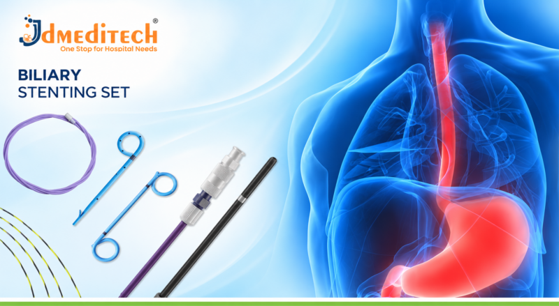
Contact JDMeditech for gynecology surgical instrument inquiries, medical accessories, and international business requirements.

Medical Disclaimer: This article provides general educational information only. It does not replace professional medical advice or surgical training. Qualified healthcare professionals must make treatment and equipment decisions according to clinical judgment, applicable guidelines, and approved manufacturer instructions.
Salpingo-Oophorectomy Surgery: Procedure, Instruments, Benefits, Risks & Recovery

Salpingo-Oophorectomy Surgery is a gynecological operation that removes an ovary and its associated fallopian tube. Depending on the patient’s medical condition, a surgeon may remove the structures on one side or both sides.

The ovaries produce reproductive hormones and contain eggs. Meanwhile, the fallopian tubes connect the region near the ovaries to the uterus and provide a pathway involved in reproduction.

Doctors may recommend salpingo-oophorectomy for selected ovarian tumors, complex adnexal masses, severe disease, or certain cancer-related conditions.

Furthermore, some patients with inherited high cancer risk may consider preventive surgery after specialist counseling.

This JDMeditech guide explains salpingo-oophorectomy surgery, its types, indications, surgical instruments, benefits, risks, recovery, and equipment selection factors.

What Is Salpingo-Oophorectomy Surgery?

Salpingo-Oophorectomy Surgery removes an ovary together with the fallopian tube on the same side.

The procedure may involve one side, called unilateral salpingo-oophorectomy, or both sides, called bilateral salpingo-oophorectomy.

However, this operation does not automatically include removal of the uterus. Hysterectomy is a separate procedure that surgeons may perform when clinically necessary.

Therefore, patients should understand exactly which organs their surgeon plans to remove.

Understanding the Ovaries and Fallopian Tubes

The ovaries are reproductive organs located on either side of the uterus. They produce hormones such as estrogen and progesterone and contain the eggs involved in reproduction.

Meanwhile, the fallopian tubes provide a pathway for an egg to travel toward the uterus.

During the reproductive years, these structures play important roles in fertility and hormonal health.

Consequently, removing one ovary has different effects from removing both ovaries.

Why Is Salpingo-Oophorectomy Performed?

Doctors may recommend salpingo-oophorectomy when an ovarian or tubal condition requires surgical treatment.

For example, certain ovarian masses may require removal of the affected ovary and tube for diagnosis or treatment.

In addition, severe damage or selected cancer-related conditions may require this operation.

However, surgery is not necessary for every ovarian cyst or pelvic condition. Therefore, specialists consider the patient’s diagnosis, age, symptoms, fertility goals, and treatment alternatives.

Common Indications for Salpingo-Oophorectomy
1. Suspicious Ovarian Masses

Some ovarian masses show concerning features during imaging or clinical assessment.

Consequently, a specialist may recommend removal of the affected ovary and fallopian tube.

However, suspected malignancy requires appropriate gynecologic oncology evaluation.

2. Selected Ovarian Tumors

Certain ovarian tumors require surgery because of their size, appearance, or associated symptoms.

For example, removal may be appropriate when preserving the affected ovary is not medically suitable.

3. Severe Endometriosis

Endometriosis may affect the ovaries and surrounding pelvic tissues.

In selected severe cases, removal of an affected ovary and tube may form part of treatment.

Nevertheless, clinicians should discuss less extensive options and the consequences of organ removal.

4. Ovarian Torsion With Nonviable Tissue

Ovarian torsion occurs when the ovary twists and may compromise its blood supply.

This condition requires urgent specialist assessment.

However, surgeons generally aim to preserve the ovary when feasible. Removal may become necessary in selected cases involving severely damaged or suspicious tissue.

5. Selected Tubal and Ovarian Conditions

Some conditions affect both the fallopian tube and ovary.

For example, severe infection or a complex tubo-ovarian abscess may occasionally require surgical removal when other treatments are unsuccessful.

6. Risk Reduction for Hereditary Cancer

Patients with certain inherited cancer-risk conditions may consider risk-reducing bilateral salpingo-oophorectomy.

For example, some individuals with pathogenic BRCA1 or BRCA2 variants may benefit from this approach after specialist counseling.

However, the timing and suitability depend on genetic risk, age, and reproductive plans.

7. Selected Gynecological Cancers

Certain ovarian, fallopian tube, or other gynecological cancers may require removal of one or both ovaries and fallopian tubes.

Furthermore, cancer treatment may involve additional procedures or therapies.

Therefore, a gynecologic oncology team should determine the appropriate treatment plan.

Types of Salpingo-Oophorectomy Surgery
1. Unilateral Salpingo-Oophorectomy

Unilateral Salpingo-Oophorectomy (USO) removes one ovary and its associated fallopian tube.

When the opposite ovary remains healthy, it may continue producing hormones and releasing eggs.

As a result, natural conception may remain possible if the remaining reproductive structures function adequately.

2. Bilateral Salpingo-Oophorectomy

Bilateral Salpingo-Oophorectomy (BSO) removes both ovaries and both fallopian tubes.

Consequently, the patient can no longer produce eggs naturally.

Furthermore, removal of both ovaries before natural menopause causes immediate surgical menopause.

3. Laparoscopic Salpingo-Oophorectomy

Laparoscopic surgery uses small abdominal incisions and a camera-assisted surgical system.

For suitable patients, this approach may allow faster recovery than open abdominal surgery.

4. Open Salpingo-Oophorectomy

Open surgery uses an abdominal incision to access the pelvic organs.

Specialists may choose this approach for selected large or suspicious masses, extensive disease, or complex operations.

5. Robotic-Assisted Salpingo-Oophorectomy

Robotic-assisted surgery uses laparoscopic instruments controlled by a trained surgeon through a robotic system.

However, its suitability depends on the clinical indication, available expertise, and surgical requirements.

6. Risk-Reducing Bilateral Salpingo-Oophorectomy

Risk-reducing BSO removes both ovaries and fallopian tubes to lower cancer risk in selected high-risk patients.

Nevertheless, surgery does not eliminate every risk of primary peritoneal cancer.

Therefore, specialist genetic counseling and informed consent are essential.

Common Instruments Used in Salpingo-Oophorectomy Surgery

Salpingo-oophorectomy requires surgical instruments for tissue handling, dissection, bleeding control, visualization, and specimen retrieval.

Moreover, the required equipment depends on whether the surgeon performs open or minimally invasive surgery.

The following instruments may support selected procedures. However, this list does not represent a confirmed JDMeditech surgical-set packing list.

1. Laparoscope Telescope

A laparoscope provides a camera-assisted view of the abdominal and pelvic organs.

Furthermore, it allows surgeons to assess the operative field during suitable minimally invasive procedures.

2. Laparoscopic Trocar and Cannula

Trocar and cannula systems provide access for compatible laparoscopic instruments.

Therefore, the surgical team must select suitable diameters and instrument configurations.

3. Laparoscopic Grasping Forceps

Grasping forceps help surgeons manipulate suitable tissues during laparoscopic surgery.

However, the surgeon must choose an appropriate jaw design and working length.

4. Laparoscopic Dissecting Forceps

Dissecting forceps support controlled tissue separation during selected procedures.

Moreover, compatible designs help surgeons work within the available operative space.

5. Laparoscopic Scissors

Laparoscopic scissors support tissue cutting during minimally invasive operations.

Depending on the procedure, the surgeon may require straight or curved designs.

6. Electrosurgical Instruments

Compatible electrosurgical instruments support tissue cutting and bleeding control.

However, surgeons must follow the approved manufacturer instructions and verify generator compatibility.

7. Vessel-Sealing Instruments

Suitable vessel-sealing devices may support tissue and vascular pedicle management during selected procedures.

Nevertheless, device selection depends on the intended use and approved technical specifications.

8. Laparoscopic Suction and Irrigation Instruments

Suction and irrigation instruments help maintain visibility in the operative field.

As a result, the surgical team can manage appropriate fluids during the operation.

9. Specimen Retrieval Bag

A specimen retrieval bag may support contained removal of suitable surgical specimens.

Importantly, suspected malignancy requires appropriate precautions to minimize unnecessary tumor rupture or spillage.

10. Surgical Scalpel

A surgical scalpel supports appropriate tissue incisions during open surgery.

Furthermore, compatible blades and handles support different surgical requirements.

11. Mayo and Metzenbaum Scissors

Mayo scissors support cutting suitable tissues and surgical materials.

Meanwhile, Metzenbaum scissors support more delicate tissue dissection.

12. Artery Forceps

Artery forceps help surgeons clamp selected blood vessels and tissues.

In addition, they support temporary bleeding control during suitable surgical tasks.

13. Allis and Babcock Forceps

Allis forceps provide a firm grip on suitable tissues.

In contrast, Babcock forceps support gentler handling of selected structures.

14. Surgical Retractors

Surgical retractors help maintain exposure during open abdominal operations.

Therefore, the surgical team selects appropriate shapes and dimensions.

15. Needle Holder

A needle holder helps surgeons control compatible surgical needles during suturing.

Moreover, different instrument lengths and jaw configurations support different procedures.

16. Additional Surgical Accessories

Depending on the operation, hospitals may require suitable sutures, suction tubing, surgical drapes, and other accessories.

Consequently, purchasing teams should verify the complete equipment configuration before ordering.

Salpingo-Oophorectomy Instruments and Functions
Instrument Primary Function
Laparoscope Telescope Provides camera-assisted visualization
Trocar and Cannula Support laparoscopic instrument access
Laparoscopic Grasping Forceps Support suitable tissue manipulation
Laparoscopic Dissecting Forceps Support tissue dissection
Laparoscopic Scissors Support tissue cutting
Electrosurgical Instruments Support compatible cutting and bleeding control
Vessel-Sealing Instruments Support selected vascular pedicle management
Suction and Irrigation Instruments Support operative visibility and fluid management
Specimen Retrieval Bag Supports contained retrieval of suitable specimens
Surgical Scalpel Supports open surgical incisions
Mayo and Metzenbaum Scissors Support cutting and dissection
Artery Forceps Clamp selected vessels and tissues
Allis and Babcock Forceps Support appropriate tissue handling
Surgical Retractors Maintain exposure during open surgery
Needle Holder Supports surgical suturing

Importantly, the exact instrument requirements depend on the operative approach and manufacturer instructions.
How Is Salpingo-Oophorectomy Surgery Performed?

A qualified gynecological surgeon performs salpingo-oophorectomy in an appropriately equipped operating room.

First, the healthcare team reviews the patient’s diagnosis, medical history, imaging results, and surgical plan.

Next, the anesthesia team prepares the patient and provides suitable anesthesia.

During the operation, the surgeon accesses the pelvic organs through the selected surgical approach.

The surgical team then removes the planned ovary and associated fallopian tube while protecting nearby structures.

When malignancy is suspected, the team follows appropriate oncological precautions and specimen-handling requirements.

Afterward, the surgeon completes the necessary closure and arranges postoperative care.

Finally, the healthcare team reviews pathology findings and determines whether further treatment or follow-up is necessary.

However, the exact surgical technique depends on the patient’s condition. This overview does not replace professional surgical training.

Potential Benefits of Salpingo-Oophorectomy
1. Treatment of Selected Ovarian and Tubal Conditions

Removing an affected ovary and tube may treat certain tumors, complex masses, or severely damaged tissues.

However, outcomes depend on the underlying diagnosis.

2. Pathological Diagnosis

Examination of removed tissue can help establish the final diagnosis.

Consequently, specialists can plan additional treatment when necessary.

3. Cancer Risk Reduction in Selected Patients

Risk-reducing BSO can substantially lower ovarian and fallopian tube cancer risk in selected high-risk patients.

Nevertheless, it does not remove every possibility of primary peritoneal cancer.

4. Treatment of Selected Painful Conditions

Some ovarian or tubal conditions cause persistent pain.

In appropriate cases, surgical treatment may help relieve related symptoms.

5. Preservation of the Opposite Ovary in Unilateral Surgery

Unilateral salpingo-oophorectomy preserves the opposite ovary when it remains in place.

As a result, hormone production and fertility potential may continue.

6. Different Surgical Approaches

Surgeons can select laparoscopic, robotic-assisted, or open surgery according to clinical requirements.

Therefore, the treatment plan can reflect the patient’s anatomy and diagnosis.

Unilateral vs Bilateral Salpingo-Oophorectomy
Feature Unilateral Surgery Bilateral Surgery
Ovaries Removed One Both
Fallopian Tubes Removed One Both
Hormone Production May continue through remaining ovary Ovarian hormone production ends
Natural Egg Production May continue Ends
Surgical Menopause Not usually immediate if opposite ovary functions Occurs if performed before natural menopause
Pregnancy Potential May remain possible Natural conception is no longer possible

Importantly, patients should discuss fertility and hormonal effects before surgery.

Laparoscopic vs Open Salpingo-Oophorectomy
Feature Laparoscopic Surgery Open Surgery
Surgical Access Small abdominal incisions Larger abdominal incision
Visualization Camera-assisted view Direct surgical view
Typical Instruments Laparoscopic instruments Open surgical instruments
Hospital Stay Often shorter Generally longer
Recovery Often faster Generally longer
Clinical Suitability Suitable uncomplicated or selected complex cases Selected large masses or extensive disease

Both approaches have important clinical roles. Therefore, specialists choose the method according to patient safety and treatment requirements.

Salpingo-Oophorectomy vs Salpingectomy

Salpingo-oophorectomy removes an ovary and its associated fallopian tube.

In contrast, salpingectomy removes one or both fallopian tubes while preserving the ovaries.

Consequently, bilateral salpingectomy does not directly cause surgical menopause when the ovaries remain healthy.

However, bilateral salpingectomy prevents natural conception through the fallopian tubes.

Therefore, these operations have different effects on fertility and hormone production.

Salpingo-Oophorectomy vs Oophorectomy

Oophorectomy removes an ovary without necessarily removing its associated fallopian tube.

Meanwhile, salpingo-oophorectomy removes both structures on the affected side.

However, the clinical indication determines which operation is appropriate.

Hormonal Effects After Bilateral Salpingo-Oophorectomy

Removing both ovaries before natural menopause causes immediate surgical menopause.

As a result, patients may experience hot flashes, night sweats, vaginal dryness, and sleep disturbances.

Furthermore, early loss of ovarian hormones can affect bone and cardiovascular health.

Therefore, clinicians may discuss menopausal hormone therapy or other suitable treatments.

However, hormone therapy is not appropriate for every patient, particularly in certain cancer-related situations.

Fertility After Salpingo-Oophorectomy

Fertility outcomes depend on whether the surgeon removes one or both ovaries and tubes.

After unilateral surgery, pregnancy may remain possible if the remaining reproductive structures function adequately.

However, bilateral surgery eliminates natural egg production and the tubal pathway for conception.

In selected circumstances, assisted reproduction may remain possible if the uterus is present and suitable reproductive options are available.

Therefore, patients who wish to preserve fertility should discuss their options before surgery whenever feasible.

Risks and Complications of Salpingo-Oophorectomy

Salpingo-oophorectomy is an invasive operation with potential complications.

For example, patients may experience bleeding, infection, or postoperative discomfort.

In addition, surgery may injure nearby structures such as the bladder, ureters, bowel, or blood vessels.

Blood clots and anesthesia-related complications are also possible.

Furthermore, removal of both ovaries can cause major hormonal and reproductive changes.

Consequently, careful patient assessment, qualified surgical expertise, and appropriate postoperative care are essential.

Recovery After Salpingo-Oophorectomy Surgery

Recovery depends on the surgical approach, the patient’s health, and the complexity of treatment.

For example, many patients resume usual daily activities within approximately two to four weeks after uncomplicated laparoscopic surgery.

However, open surgery and extensive procedures may require a longer recovery period.

During recovery, patients may experience fatigue, abdominal discomfort, or temporary activity limitations.

Furthermore, the healthcare team provides instructions about wound care, medication, lifting, driving, and follow-up.

Patients should seek prompt medical attention for severe pain, fever, heavy bleeding, wound problems, or breathing difficulty.

Life After Salpingo-Oophorectomy

Long-term effects depend on the type of surgery and the patient’s age.

For example, patients who retain one healthy ovary may continue producing reproductive hormones.

In contrast, premenopausal patients who undergo bilateral surgery experience surgical menopause.

Furthermore, patients undergoing cancer-related surgery may require additional treatment or surveillance.

Therefore, follow-up care should address pathology results, recovery, hormonal health, and fertility concerns.

How to Choose Salpingo-Oophorectomy Surgical Instruments

Hospitals and gynecology departments should identify their clinical requirements before purchasing salpingo-oophorectomy instruments.

First, determine whether the planned operation involves laparoscopy, open surgery, or a specialist oncological procedure.

Next, review instrument dimensions, materials, working lengths, and system compatibility.

Moreover, purchasing teams should verify cleaning, sterilization, and maintenance requirements.

Important Equipment Selection Factors
  • Approved intended use
  • Complete instrument-set packing list
  • Open or laparoscopic surgical approach
  • Laparoscope telescope diameter
  • Camera and light source compatibility
  • Trocar and cannula dimensions
  • Laparoscopic grasper design
  • Dissecting forceps configuration
  • Laparoscopic scissors specifications
  • Electrosurgical system compatibility
  • Vessel-sealing device requirements
  • Suction and irrigation compatibility
  • Specimen retrieval requirements
  • Open surgical forceps and retractors
  • Needle holder configuration
  • Reusable or single-use classification
  • Cleaning and sterilization instructions
  • Manufacturer technical datasheet
  • Applicable regulatory documentation

Furthermore, hospitals should confirm the exact supplied instruments before placing an order.

Gynecology Surgical Instruments by JDMeditech

JDMeditech welcomes inquiries from hospitals, gynecology departments, surgical centers, and international medical distributors regarding gynecological surgical instruments and related medical accessories.

For example, customers can inquire about available laparoscopic instruments, grasping forceps, scissors, clamps, needle holders, retractors, and compatible surgical accessories.

In addition, the JDMeditech team can confirm available product configurations, technical specifications, and applicable documentation for individual inquiries.

For product details and quotations, visit www.jdmeditech.com.

Frequently Asked Questions About Salpingo-Oophorectomy Surgery
1. What is salpingo-oophorectomy surgery?

It is a gynecological operation that removes an ovary and its associated fallopian tube on one or both sides.

2. What is unilateral salpingo-oophorectomy?

Unilateral salpingo-oophorectomy removes one ovary and one fallopian tube.

3. What is bilateral salpingo-oophorectomy?

Bilateral salpingo-oophorectomy removes both ovaries and both fallopian tubes.

4. Why is salpingo-oophorectomy performed?

Doctors may recommend it for selected ovarian tumors, severe ovarian or tubal disease, cancer-related conditions, or cancer risk reduction in suitable high-risk patients.

5. What instruments are used during salpingo-oophorectomy?

Common instruments include laparoscopes, grasping forceps, dissecting forceps, scissors, suction instruments, compatible energy devices, and open surgical instruments when required.

6. Does bilateral salpingo-oophorectomy cause menopause?

Yes. Removing both ovaries before natural menopause causes immediate surgical menopause.

7. Can pregnancy occur after unilateral salpingo-oophorectomy?

Pregnancy may remain possible if the remaining ovary, tube, uterus, and other reproductive factors allow conception.

8. How long does recovery take?

Many patients return to usual activities within two to four weeks after uncomplicated laparoscopic surgery. However, recovery after open or extensive surgery may take longer.

9. What are the risks of salpingo-oophorectomy?

Potential complications include bleeding, infection, blood clots, injury to nearby organs, and hormonal or fertility effects.

10. Where can hospitals inquire about JDMeditech gynecology instruments?

Hospitals and medical distributors can visit JDMeditech to request product information and quotations.

Conclusion

Salpingo-Oophorectomy Surgery is an important treatment option for selected ovarian and fallopian tube conditions.

Moreover, qualified surgeons use compatible instruments during laparoscopic and open procedures.

However, successful treatment requires careful diagnosis, suitable equipment, specialist expertise, and appropriate follow-up.

Therefore, hospitals should select instruments according to clinical requirements, professional guidelines, and approved manufacturer instructions.
Contact JDMeditech for gynecology surgical instrument inquiries, compatible medical accessories, and international business requirements.

Medical Disclaimer: This article provides general educational information only. It does not replace professional medical advice or surgical training. Qualified healthcare professionals must make treatment and equipment decisions according to clinical judgment, applicable guidelines, and approved manufacturer instructions.
Ovarian Tumor Removal Surgery: Procedure, Instruments, Benefits, Risks & Recovery

Ovarian Tumor Removal Surgery is a gynecological procedure that removes selected abnormal growths from an ovary or removes the affected ovary when clinically necessary. Doctors may recommend surgery for certain large, persistent, symptomatic, or suspicious ovarian masses.

Ovarian tumors vary in their appearance and biological behavior. Although many ovarian growths are benign, some are borderline or malignant.

Therefore, accurate diagnosis plays an essential role in treatment planning. A specialist considers ultrasound findings, symptoms, age, medical history, and other clinical factors before recommending surgery.

Furthermore, different surgical techniques allow doctors to manage ovarian masses according to their characteristics. Some procedures preserve healthy ovarian tissue, while others require removal of an ovary or additional organs.

This JDMeditech guide explains ovarian tumor removal surgery, its indications, surgical approaches, instruments, benefits, risks, recovery, and equipment selection factors.

What Is Ovarian Tumor Removal Surgery?

Ovarian Tumor Removal Surgery refers to surgical treatment that removes an ovarian mass or the affected ovary.

The operation depends on the tumor’s size, location, appearance, and suspected diagnosis.

For example, a surgeon may remove a suitable benign cyst while preserving the remaining ovary. In contrast, a suspicious ovarian mass may require removal of the entire affected ovary and fallopian tube.

Furthermore, confirmed ovarian cancer may require specialist staging procedures and additional treatment.

Therefore, ovarian tumor removal does not describe one fixed operation. Instead, the surgical plan depends on the individual patient’s condition.

Understanding the Ovaries and Ovarian Tumors

The ovaries are reproductive organs located on either side of the uterus. They contain eggs and produce hormones, including estrogen and progesterone.

During the reproductive years, ovarian follicles develop as part of the menstrual cycle.

However, different types of cysts and tumors may develop within or around the ovaries.

Some ovarian cysts disappear without treatment. Nevertheless, persistent or suspicious masses may require further investigation.

Consequently, clinicians use appropriate imaging and other tests to assess the ovarian mass before choosing treatment.

Common Types of Ovarian Masses
1. Functional Ovarian Cysts

Functional cysts commonly develop during the menstrual cycle.

Most are benign and may disappear without surgery. Therefore, clinicians often recommend observation when appropriate.

2. Ovarian Dermoid Cysts

Dermoid cysts, also known as mature cystic teratomas, may contain different tissue types.

Although most are benign, larger cysts may cause symptoms or increase the risk of ovarian torsion.

3. Ovarian Cystadenomas

Cystadenomas are ovarian tumors that may contain fluid or mucus-like material.

Some grow considerably larger over time. As a result, surgical removal may become necessary.

4. Ovarian Endometriomas

Endometriomas are ovarian cysts associated with endometriosis.

They may cause pelvic pain or affect fertility. However, surgery is not always necessary because removal can also reduce ovarian reserve.

5. Borderline Ovarian Tumors

Borderline ovarian tumors have biological features that differ from both benign tumors and invasive ovarian cancer.

Therefore, treatment requires specialist assessment and appropriate pathological evaluation.

6. Malignant Ovarian Tumors

Malignant ovarian tumors contain cancerous cells and may spread beyond the ovary.

Consequently, suspected ovarian cancer requires evaluation by an appropriate gynecologic oncology team.

Depending on the diagnosis, treatment may include surgery, chemotherapy, or other specialist care.

Why Is Ovarian Tumor Removal Performed?

Doctors may recommend ovarian tumor removal when a mass causes symptoms, continues growing, or raises concern for malignancy.

For example, some patients experience persistent pelvic pain or abdominal pressure.

In addition, large ovarian masses may affect nearby structures.

However, surgery is not necessary for every ovarian cyst. Therefore, clinicians consider observation and other appropriate management options before recommending an operation.

Common Indications for Ovarian Tumor Removal Surgery
1. Large Ovarian Masses

Large ovarian masses may cause abdominal swelling, pressure, or discomfort.

In selected cases, surgical removal may provide suitable treatment.

2. Persistent Ovarian Cysts

Some ovarian cysts remain present during follow-up examinations.

However, persistence alone does not always require surgery. The clinician also considers the cyst’s appearance, size, symptoms, and patient characteristics.

3. Suspicious Imaging Findings

Ultrasound may reveal solid areas, complex internal structures, or other concerning features.

Consequently, the patient may need additional assessment and specialist referral.

4. Pelvic Pain or Pressure

Certain ovarian masses cause persistent pelvic discomfort.

For example, a larger mass may create pressure on nearby organs.

Nevertheless, clinicians must investigate other possible causes of pain before recommending surgery.

5. Ovarian Torsion

Ovarian torsion occurs when an ovary twists around its supporting tissues and may lose its blood supply.

This condition can cause sudden, severe pelvic pain and requires urgent medical assessment.

However, surgeons often try to preserve the ovary when clinically appropriate.

6. Suspected Ovarian Malignancy

A suspicious ovarian mass may require surgical evaluation by a gynecologic oncology specialist.

Furthermore, the surgeon may need to remove tissue for pathological examination and cancer staging.

7. Selected Fertility-Related Concerns

Some ovarian conditions affect reproductive health.

Therefore, specialists may consider fertility-preserving treatment when medically appropriate.

However, ovarian surgery can affect ovarian reserve, so patients should discuss fertility goals before treatment.

Types of Ovarian Tumor Removal Surgery
1. Ovarian Cystectomy

Ovarian cystectomy removes a suitable ovarian cyst while preserving as much healthy ovarian tissue as possible.

This approach may suit selected benign ovarian masses.

Nevertheless, the surgeon must consider the cyst’s location and potential effects on ovarian function.

2. Unilateral Oophorectomy

Unilateral oophorectomy removes one ovary.

In selected cases, surgeons recommend this approach when preserving the affected ovary is not appropriate.

However, the remaining ovary may continue producing hormones and releasing eggs.

3. Bilateral Oophorectomy

Bilateral oophorectomy removes both ovaries.

Removing both ovaries before natural menopause causes surgical menopause.

Therefore, specialists carefully assess the medical indication and long-term effects before recommending this procedure.

4. Salpingo-Oophorectomy

Salpingo-oophorectomy removes an ovary and its associated fallopian tube.

Depending on the diagnosis, the surgeon may remove structures on one or both sides.

5. Laparoscopic Ovarian Tumor Removal

Laparoscopic surgery uses small abdominal incisions and a camera-assisted surgical system.

For suitable masses that appear benign, this approach may provide faster recovery than open surgery.

6. Open Ovarian Tumor Removal

Open surgery, also called laparotomy, uses an abdominal incision to access the pelvic organs.

This approach may be appropriate for selected very large or suspicious masses.

Furthermore, open surgery may help specialists remove a concerning mass intact when necessary.

7. Ovarian Cancer Staging or Cytoreductive Surgery

Confirmed or strongly suspected ovarian cancer may require more extensive surgery.

For example, specialist procedures may include removal of additional tissues, surgical staging, or cytoreduction.

However, these operations differ substantially from routine benign ovarian cyst removal.

Common Instruments Used in Ovarian Tumor Removal Surgery

Ovarian tumor surgery requires instruments for surgical access, tissue handling, dissection, bleeding control, and suturing.

Moreover, the equipment varies between open and laparoscopic approaches.

The following instruments may support selected gynecological operations. However, this overview does not represent a confirmed JDMeditech instrument-set packing list.

1. Surgical Scalpel

A surgical scalpel supports appropriate tissue incisions during open surgery.

Furthermore, compatible blades and handles allow different surgical applications.

2. Mayo Scissors

Mayo scissors support the cutting of suitable tissues and surgical materials.

Depending on their design, they may have straight or curved blades.

3. Metzenbaum Scissors

Metzenbaum scissors support delicate tissue dissection.

Therefore, surgeons select an appropriate instrument according to the intended use.

4. Artery Forceps

Artery forceps help surgeons clamp suitable vessels and tissues.

In addition, they support temporary bleeding control during selected surgical tasks.

5. Allis Tissue Forceps

Allis forceps provide a secure grip on suitable tissues.

However, their toothed jaws require careful handling.

6. Babcock Forceps

Babcock forceps support gentler handling of selected tissues.

For example, surgeons may use compatible designs for suitable tubular structures.

7. Tissue Dissecting Forceps

Dissecting forceps allow surgeons to hold and manipulate suitable tissues.

Furthermore, toothed and non-toothed designs serve different surgical requirements.

8. Needle Holder

A needle holder helps surgeons control compatible surgical needles during suturing.

Moreover, different jaw designs and lengths support different applications.

9. Surgical Retractors

Surgical retractors help maintain exposure of the operative field.

For example, suitable handheld or self-retaining retractors may support open abdominal surgery.

10. Electrosurgical Equipment

Compatible electrosurgical instruments support tissue cutting and bleeding control.

However, the surgical team must confirm generator compatibility and follow approved instructions for use.

11. Suction Instruments

Suction instruments help remove blood and other fluids from the operative field.

Consequently, surgeons can maintain a clearer view during suitable procedures.

12. Laparoscope and Camera System

A laparoscope provides a camera-assisted view of the pelvic organs during minimally invasive surgery.

Therefore, the camera, telescope, and light source must form a compatible system.

13. Laparoscopic Grasping Forceps

Compatible laparoscopic graspers help surgeons manipulate suitable tissues during minimally invasive operations.

However, the selected instrument must match the procedure and access system.

14. Laparoscopic Dissection Instruments

Suitable laparoscopic instruments support tissue dissection during selected operations.

Moreover, the surgeon may require compatible energy devices for bleeding control.

15. Specimen Retrieval Bag

A suitable specimen retrieval bag may help contain tissue during selected laparoscopic procedures.

Importantly, suspected malignancy requires careful planning to avoid unnecessary tumor rupture or spillage.

16. Additional Surgical Accessories

Depending on the procedure, hospitals may require compatible sutures, surgical drapes, suction tubing, and other accessories.

Therefore, purchasing teams should verify the exact equipment configuration before ordering.

Ovarian Tumor Removal Instruments and Functions
Instrument Primary Function
Surgical Scalpel Supports appropriate tissue incisions
Mayo Scissors Cut suitable tissues and surgical materials
Metzenbaum Scissors Support delicate tissue dissection
Artery Forceps Clamp selected vessels and tissues
Allis Forceps Hold suitable tissues
Babcock Forceps Support gentle tissue handling
Dissecting Forceps Support tissue manipulation
Needle Holder Supports surgical suturing
Surgical Retractors Maintain operative exposure
Electrosurgical Equipment Supports compatible cutting and bleeding control
Suction Instruments Remove fluids from the operative field
Laparoscope Provides camera-assisted visualization
Laparoscopic Graspers Support tissue manipulation
Dissection Instruments Support selected laparoscopic dissection tasks
Specimen Retrieval Bag Supports contained retrieval of suitable specimens

Importantly, surgeons must select instruments according to the intended operation and manufacturer instructions.
How Is Ovarian Tumor Removal Surgery Performed?

A qualified gynecological surgeon performs ovarian tumor removal in an appropriately equipped medical facility.

First, the healthcare team reviews the patient’s symptoms, medical history, imaging findings, and surgical plan.

Next, the specialist assesses the likelihood of malignancy and determines whether gynecologic oncology involvement is necessary.

During surgery, the surgeon accesses the affected ovary using the selected operative approach.

The surgical team then removes the planned cyst, mass, or affected organ according to the patient’s diagnosis.

When malignancy is suspected, the team takes appropriate precautions to avoid unnecessary tumor spillage and to support accurate pathological assessment.

Afterward, the surgeon completes the necessary surgical closure and arranges postoperative care.

Finally, the healthcare team reviews the pathology results and determines whether further treatment or follow-up is necessary.

However, the exact surgical technique varies considerably. This overview does not replace professional surgical training.

Potential Benefits of Ovarian Tumor Removal Surgery
1. Treatment of Symptomatic Ovarian Masses

Removing an appropriate ovarian mass may reduce pain, pressure, or other related symptoms.

However, the outcome depends on the underlying condition.

2. Pathological Diagnosis

Surgical tissue examination can help establish the final diagnosis.

As a result, clinicians can determine whether the mass is benign, borderline, or malignant.

3. Fertility Preservation in Selected Cases

Some procedures preserve healthy ovarian tissue.

Therefore, fertility may remain possible after treatment.

Nevertheless, surgery can reduce ovarian reserve, and fertility outcomes vary.

4. Treatment of Selected Large Tumors

Large ovarian masses may cause pressure or discomfort.

In suitable cases, surgical removal may help relieve these symptoms.

5. Appropriate Cancer Management

When ovarian cancer is present, specialist surgery may form an important part of treatment.

However, the overall care plan may also require chemotherapy or other treatments.

6. Individualized Surgical Options

Different approaches allow specialists to consider the patient’s diagnosis and reproductive goals.

Consequently, treatment can range from ovarian cystectomy to more extensive oncological surgery.

Open vs Laparoscopic Ovarian Tumor Removal
Feature Open Surgery Laparoscopic Surgery
Surgical Access Open abdominal incision Small abdominal incisions
Visualization Direct surgical view Camera-assisted view
Common Instruments Open surgical instruments Laparoscopic instruments
Recovery Generally longer Often faster
Hospital Stay Generally longer Often shorter
Clinical Suitability Selected large or suspicious masses Suitable masses, particularly those thought benign

Both approaches have important clinical roles. Therefore, specialists select the surgical method according to the mass characteristics and patient needs.

Ovarian Cystectomy vs Oophorectomy
Feature Ovarian Cystectomy Oophorectomy
Primary Goal Remove a suitable ovarian cyst Remove an ovary
Ovarian Preservation Preserves some ovarian tissue Removes the affected ovary
Potential Fertility Impact May reduce ovarian reserve Depends on whether the other ovary remains
Common Indications Selected benign cysts Selected tumors or severely affected ovaries
Hormonal Impact Usually preserves some function Depends on unilateral or bilateral removal

Importantly, removing both ovaries before natural menopause causes surgical menopause.

Ovarian Tumor Surgery and Fertility Preservation

Fertility is an important consideration for patients who may wish to become pregnant.

For example, a suitable ovarian cystectomy may preserve ovarian tissue.

However, surgery can affect ovarian reserve, particularly when healthy tissue must also be removed.

In selected early-stage ovarian cancers, fertility-sparing treatment may be possible under specialist guidance.

Therefore, patients should discuss reproductive goals and fertility preservation options before surgery whenever feasible.

Ovarian Tumor Removal and Cancer Risk

Not every ovarian mass is cancerous.

Nevertheless, certain imaging findings, clinical features, and blood test results may raise concern for malignancy.

For example, specialists may use ultrasound findings and selected tumor markers during evaluation.

However, no single blood test can reliably determine whether every ovarian mass is malignant.

Consequently, suspicious masses require appropriate specialist assessment and treatment planning.

Risks and Complications of Ovarian Tumor Removal

Ovarian tumor removal is an invasive procedure with potential complications.

For example, patients may experience bleeding, infection, or postoperative pain.

In addition, surgery may injure nearby structures such as the bladder, ureters, bowel, or blood vessels.

Blood clots and anesthesia-related complications are also possible.

Furthermore, ovarian surgery may affect fertility or hormone production.

When malignancy is suspected, tumor rupture or spillage may create additional concerns.

Consequently, careful patient selection, suitable surgical expertise, and appropriate postoperative care are essential.

Recovery After Ovarian Tumor Removal Surgery

Recovery depends on the surgical approach, patient’s health, and complexity of treatment.

For example, recovery after uncomplicated laparoscopic surgery is often faster than after open abdominal surgery.

However, extensive operations may require a longer hospital stay and recovery period.

During recovery, patients may experience fatigue, discomfort, or temporary activity limitations.

Furthermore, the healthcare team provides instructions about wound care, medications, lifting, and follow-up.

Patients should seek prompt medical attention for severe pain, fever, heavy bleeding, wound problems, or breathing difficulty.

Life After Ovarian Tumor Removal

Long-term outcomes depend on the final diagnosis and the type of surgery.

For example, patients who undergo removal of a benign cyst may need only routine or individualized follow-up.

In contrast, patients with borderline or malignant tumors may require specialist surveillance or additional treatment.

Furthermore, hormonal effects depend on whether one or both ovaries remain.

Therefore, follow-up care should address pathology findings, recovery, fertility, and any ongoing symptoms.

How to Choose Ovarian Tumor Removal Surgical Instruments

Hospitals and gynecology departments should identify their clinical requirements before purchasing ovarian surgery instruments.

First, determine whether the intended operation involves open surgery, laparoscopy, or specialist oncological treatment.

Next, review the instrument categories, dimensions, materials, and compatibility requirements.

Moreover, purchasing teams should verify cleaning, sterilization, and maintenance instructions.

Important Equipment Selection Factors
  • Approved intended use
  • Complete instrument-set packing list
  • Open or laparoscopic surgical approach
  • Surgical instrument material
  • Scalpel handle compatibility
  • Mayo and Metzenbaum scissors configuration
  • Artery forceps type and size
  • Allis and Babcock forceps dimensions
  • Needle holder configuration
  • Surgical retractor type and size
  • Laparoscope and camera compatibility
  • Laparoscopic grasper dimensions
  • Electrosurgical equipment compatibility
  • Suction instrument configuration
  • Specimen retrieval requirements
  • Reusable or single-use classification
  • Cleaning and sterilization instructions
  • Manufacturer technical datasheet
  • Applicable regulatory documentation

Furthermore, hospitals should confirm the exact supplied instruments before placing an order.

Gynecology Surgical Instruments by JDMeditech

JDMeditech welcomes inquiries from hospitals, gynecology departments, surgical centers, and international medical distributors regarding gynecological surgical instruments and related medical accessories.

For example, customers can inquire about available surgical forceps, scissors, clamps, needle holders, retractors, and compatible laparoscopic accessories.

In addition, the JDMeditech team can confirm available product configurations, technical specifications, and applicable documentation for individual inquiries.

For product details and quotations, visit www.jdmeditech.com.

Frequently Asked Questions About Ovarian Tumor Removal Surgery
1. What is ovarian tumor removal surgery?

It is a surgical procedure that removes a suitable ovarian mass or the affected ovary, depending on the diagnosis.

2. Are all ovarian tumors cancerous?

No. Many ovarian masses are benign, while others may be borderline or malignant.

3. Why is ovarian tumor removal performed?

Doctors may recommend surgery for selected large, symptomatic, persistent, or suspicious ovarian masses.

4. What is ovarian cystectomy?

Ovarian cystectomy removes a suitable cyst while preserving as much healthy ovarian tissue as possible.

5. What is oophorectomy?

Oophorectomy is the surgical removal of one or both ovaries.

6. What instruments are used during ovarian tumor removal?

Common instruments include surgical scissors, artery forceps, tissue forceps, needle holders, retractors, suction instruments, and compatible laparoscopic equipment.

7. Can pregnancy occur after ovarian tumor surgery?

Pregnancy may remain possible when reproductive organs and sufficient ovarian function remain. However, fertility outcomes depend on the diagnosis and treatment.

8. How long does recovery take?

Recovery varies according to the surgical approach and complexity. Generally, uncomplicated laparoscopic procedures allow faster recovery than open surgery.

9. What are the risks of ovarian tumor removal?

Potential complications include bleeding, infection, blood clots, injury to nearby organs, and possible effects on fertility or hormone production.

10. Where can hospitals inquire about JDMeditech gynecology instruments?

Hospitals and medical distributors can visit JDMeditech to request product information and quotations.

Conclusion

Ovarian Tumor Removal Surgery provides treatment options for selected benign, borderline, and malignant ovarian masses.

Moreover, appropriate surgical instruments support qualified specialists during open and laparoscopic gynecological procedures.

However, successful treatment requires accurate diagnosis, suitable equipment, specialist expertise, and appropriate follow-up.

Therefore, hospitals should select instruments according to clinical requirements, professional guidelines, and approved manufacturer instructions.
Contact JDMeditech for gynecology surgical instrument inquiries, compatible medical accessories, and international business requirements.

Medical Disclaimer: This article provides general educational information only. It does not replace professional medical advice or surgical training. Qualified healthcare professionals must make treatment and equipment decisions according to clinical judgment, applicable guidelines, and approved manufacturer instructions.
Open Myomectomy Surgery: Procedure, Instruments, Benefits, Risks & Recovery

Open Myomectomy Surgery is a gynecological procedure in which a surgeon removes uterine fibroids through an abdominal incision while preserving the uterus. Doctors may recommend this approach for selected patients with large, multiple, or difficult-to-access fibroids.

Uterine fibroids are usually noncancerous growths that develop in the muscular wall of the uterus. Although many fibroids cause no symptoms, others may lead to heavy menstrual bleeding, pelvic pressure, or discomfort.

In some cases, medication and less invasive treatments may not provide suitable relief. Therefore, a gynecologist may recommend surgical removal of the fibroids.

Unlike hysterectomy, myomectomy preserves the uterus. As a result, it may be an important option for patients who wish to retain their uterus or consider future pregnancy.

This JDMeditech guide explains open myomectomy surgery, its indications, surgical instruments, benefits, risks, recovery, and equipment selection factors.

What Is Open Myomectomy Surgery?

Open Myomectomy Surgery, also called abdominal myomectomy, is an operation that removes uterine fibroids through an incision in the abdomen.

During the procedure, a qualified gynecological surgeon accesses the uterus and removes suitable fibroids while preserving the remaining uterine tissue.

Furthermore, the surgeon repairs the uterus according to the location and extent of the surgical incisions.

However, the procedure does not guarantee that new fibroids will never develop. Therefore, patients may require further assessment or treatment in the future.

Understanding Uterine Fibroids

Uterine fibroids, also known as leiomyomas or myomas, are growths that develop from the muscular tissue of the uterus.

Most fibroids are benign. Nevertheless, their size, number, and location can vary considerably.

For example, some fibroids remain small and cause no symptoms. Others may enlarge the uterus or affect nearby pelvic organs.

Consequently, clinicians assess each patient’s symptoms and imaging results before recommending treatment.

Common Types of Uterine Fibroids
1. Intramural Fibroids

Intramural fibroids develop within the muscular wall of the uterus.

Depending on their size and location, they may cause heavy bleeding, pelvic pressure, or changes in uterine shape.

2. Subserosal Fibroids

Subserosal fibroids grow toward the outer surface of the uterus.

As a result, larger growths may create pressure on nearby pelvic structures.

3. Submucosal Fibroids

Submucosal fibroids extend toward or into the uterine cavity.

For example, these fibroids may contribute to abnormal menstrual bleeding.

However, selected submucosal fibroids may be suitable for hysteroscopic removal rather than open surgery.

4. Pedunculated Fibroids

Pedunculated fibroids attach to the uterus through a stalk-like structure.

They may develop toward the outer uterine surface or inside the uterine cavity.

Therefore, the surgeon selects treatment according to the fibroid’s position and clinical findings.

Why Is Open Myomectomy Performed?

Doctors may recommend open myomectomy when uterine fibroids cause significant symptoms or require surgical treatment.

For example, patients may experience persistent heavy menstrual bleeding, pelvic discomfort, or pressure symptoms.

Furthermore, some fibroids may affect the uterine cavity and contribute to fertility problems.

However, not every fibroid requires surgery. Therefore, specialists consider the patient’s symptoms, fertility goals, fibroid characteristics, and available alternatives before recommending treatment.

Common Indications for Open Myomectomy
1. Large Uterine Fibroids

Large fibroids may enlarge the uterus and cause pressure or discomfort.

In selected cases, an open abdominal approach provides suitable surgical access.

2. Multiple Uterine Fibroids

Some patients develop several fibroids in different areas of the uterus.

Consequently, open myomectomy may be appropriate when the number or location of fibroids makes less invasive surgery difficult.

3. Heavy Menstrual Bleeding

Fibroids may contribute to heavy or prolonged menstrual bleeding.

As a result, some patients develop iron deficiency or anemia.

When other treatments do not provide suitable relief, a specialist may consider myomectomy.

4. Pelvic Pain or Pressure

Larger fibroids can create pressure within the pelvis.

For example, patients may experience discomfort, urinary frequency, or pressure-related symptoms.

However, clinicians must investigate other possible causes before recommending surgery.

5. Selected Fertility Concerns

Certain fibroids may affect fertility, especially when they distort the uterine cavity.

Therefore, specialists may discuss myomectomy with selected patients who wish to become pregnant.

Nevertheless, surgery does not guarantee improved fertility or a successful pregnancy.

6. Fibroids Affecting Nearby Organs

Some large fibroids may press against the bladder or bowel.

Consequently, patients may experience urinary or bowel-related symptoms.

In suitable cases, fibroid removal may help reduce these symptoms.

7. Symptoms That Continue Despite Treatment

Medication and other treatments may help control fibroid-related symptoms.

However, some patients continue to experience significant problems.

Therefore, a gynecologist may recommend surgery after reviewing the available options.

Types of Myomectomy Surgery
1. Open Abdominal Myomectomy

Open abdominal myomectomy removes fibroids through an abdominal incision.

This approach may be suitable for selected large, numerous, or complex fibroids.

2. Laparoscopic Myomectomy

Laparoscopic myomectomy uses small abdominal incisions and a camera-assisted surgical system.

In suitable patients, this approach may offer less postoperative pain and a shorter hospital stay.

3. Robotic-Assisted Myomectomy

Robotic-assisted myomectomy uses a robotic surgical system controlled by a qualified surgeon.

However, its suitability depends on patient factors, surgical complexity, and available expertise.

4. Hysteroscopic Myomectomy

Hysteroscopic myomectomy removes selected fibroids that project into the uterine cavity.

Unlike abdominal myomectomy, this approach uses access through the cervix.

Therefore, it does not suit every fibroid type or location.

Common Instruments Used in Open Myomectomy Surgery

Open myomectomy requires instruments for surgical access, tissue handling, dissection, bleeding control, and suturing.

Moreover, the surgical team may use specialized instruments to support fibroid manipulation and uterine repair.

The following instruments commonly support selected open gynecological operations. However, this overview does not represent a confirmed JDMeditech instrument-set packing list.

1. Surgical Scalpel

A surgical scalpel allows the surgeon to make appropriate tissue incisions.

Furthermore, compatible blades and handles support different surgical requirements.

2. Myoma Screw

A myoma screw is a specialized instrument that may help a surgeon hold or manipulate suitable uterine fibroids.

For example, certain designs support controlled traction during selected open myomectomy procedures.

However, surgeons must choose the appropriate instrument according to the clinical situation.

3. Mayo Scissors

Mayo scissors support the cutting of suitable tissues and surgical materials.

Depending on their design, they may have straight or curved blades.

4. Metzenbaum Scissors

Metzenbaum scissors support delicate tissue dissection.

Therefore, surgeons select an appropriate design according to the intended use.

5. Artery Forceps

Artery forceps help surgeons clamp selected vessels and tissues.

In addition, they support temporary bleeding control during suitable surgical tasks.

6. Allis Tissue Forceps

Allis forceps help hold and manipulate suitable tissues.

However, their toothed jaws require careful handling to reduce tissue damage.

7. Babcock Forceps

Babcock forceps support gentler handling of selected tissues.

For example, surgeons may use compatible designs when manipulating suitable tubular structures.

8. Tissue Dissecting Forceps

Dissecting forceps allow surgeons to hold and manipulate suitable tissues.

Furthermore, toothed and non-toothed designs support different surgical requirements.

9. Needle Holder

A needle holder helps the surgeon control compatible surgical needles during suturing.

Moreover, appropriate needle holders support uterine repair and other closure requirements.

10. Surgical Retractors

Surgical retractors help maintain exposure of the operative field.

For example, Richardson, Deaver, and selected self-retaining retractors may support open abdominal surgery.

11. Electrosurgical Equipment

Compatible electrosurgical instruments support tissue cutting and bleeding control.

However, the surgical team must verify generator compatibility and follow the approved instructions for use.

12. Suction Instruments

Suction instruments help remove blood and other fluids from the operative field.

As a result, the surgeon can maintain a clearer view during appropriate surgical tasks.

13. Sponge-Holding Forceps

Sponge-holding forceps help hold suitable surgical swabs.

Furthermore, the surgical team must follow established swab-counting and safety protocols.

14. Hemostatic Instruments

Suitable hemostatic instruments help surgeons manage bleeding during open surgery.

Nevertheless, instrument selection depends on the planned procedure and surgical requirements.

15. Additional Surgical Accessories

Depending on the operation, hospitals may require sutures, suction tubing, surgical drapes, clamps, and other accessories.

Therefore, purchasing teams should confirm the complete instrument list before ordering.

Open Myomectomy Instruments and Functions
Instrument Primary Function
Surgical Scalpel Supports appropriate tissue incisions
Myoma Screw Supports manipulation of suitable fibroids
Mayo Scissors Cut suitable tissue and surgical materials
Metzenbaum Scissors Support delicate tissue dissection
Artery Forceps Clamp selected vessels and tissues
Allis Forceps Hold and manipulate suitable tissues
Babcock Forceps Support gentle tissue handling
Dissecting Forceps Support tissue manipulation
Needle Holder Supports surgical suturing
Surgical Retractors Maintain exposure of the operative field
Electrosurgical Equipment Supports compatible cutting and bleeding control
Suction Instruments Remove fluids from the operative field
Sponge-Holding Forceps Hold suitable surgical swabs
Hemostatic Instruments Support bleeding control

Importantly, instrument selection depends on the procedure, surgeon preference, and manufacturer instructions.
How Is Open Myomectomy Surgery Performed?

A qualified gynecological surgeon performs open myomectomy in an appropriately equipped operating room.

First, the healthcare team reviews the patient’s symptoms, medical history, imaging results, and surgical plan.

Next, the anesthesia team prepares the patient and provides suitable anesthesia.

During the operation, the surgeon makes an appropriate abdominal incision to access the uterus.

The surgical team then identifies the planned fibroids and removes them while preserving the uterus.

After removal, the surgeon repairs the uterine tissue and completes the necessary surgical closure.

Throughout the procedure, clinicians monitor bleeding, patient safety, and other important clinical factors.

Finally, the healthcare team monitors recovery and provides instructions about activity, wound care, medication, and follow-up.

However, the exact surgical technique depends on the patient’s condition. This overview does not replace professional surgical training.

Potential Benefits of Open Myomectomy
1. Uterus-Preserving Surgery

Open myomectomy removes selected fibroids while preserving the uterus.

Therefore, it may suit patients who wish to retain their uterus.

2. Treatment of Symptomatic Fibroids

Removing fibroids may reduce symptoms such as heavy bleeding and pelvic pressure.

However, results depend on the underlying condition and other clinical factors.

3. Access to Large or Multiple Fibroids

An open abdominal approach provides direct access to the uterus.

Consequently, it may be appropriate for selected large or complex fibroids.

4. Potential Fertility Preservation

Because myomectomy preserves the uterus, pregnancy may remain possible after recovery.

Nevertheless, fertility depends on many factors, and surgery does not guarantee pregnancy.

5. Direct Surgical Visualization

Open surgery allows the surgeon to examine and access the uterus directly.

As a result, the approach may support selected complex surgical requirements.

6. Relief From Pressure Symptoms

Large fibroids may place pressure on nearby pelvic organs.

In suitable cases, removing these fibroids may help reduce related discomfort.

Open Myomectomy vs Laparoscopic Myomectomy
Feature Open Myomectomy Laparoscopic Myomectomy
Surgical Access Open abdominal incision Small abdominal incisions
Visualization Direct surgical view Camera-assisted view
Common Instruments Open gynecological instruments Laparoscopic instruments
Recovery Generally longer Often faster
Hospital Stay Generally longer Often shorter
Clinical Suitability Selected large or complex fibroids Suitable fibroids based on size and location

Both approaches have important clinical roles. Therefore, the surgeon selects the most suitable method according to the patient’s anatomy, fibroid characteristics, and treatment goals.

Open Myomectomy vs Hysterectomy
Feature Open Myomectomy Hysterectomy
Primary Goal Remove selected uterine fibroids Remove the uterus
Uterus Preservation Yes No
Future Pregnancy May remain possible Cannot carry a pregnancy
Menstruation Usually continues Uterine menstruation ends
New Fibroid Development Possible Not possible in the removed uterus
Clinical Selection Depends on symptoms and uterus-preservation goals Depends on diagnosis and treatment goals

Importantly, myomectomy and hysterectomy have different long-term effects. Therefore, patients should discuss their fertility plans and treatment preferences before surgery.

Pregnancy After Open Myomectomy

Pregnancy may remain possible after myomectomy because the surgeon preserves the uterus.

However, the procedure can create scar tissue within the uterus or pelvis.

Furthermore, the depth and location of uterine incisions may affect future pregnancy care.

For this reason, some patients may require a planned cesarean delivery during a future pregnancy.

Therefore, patients should discuss pregnancy timing and delivery planning with their gynecologist.

Can Uterine Fibroids Return After Myomectomy?

Myomectomy removes selected existing fibroids.

However, new fibroids may develop after surgery.

Consequently, some patients may experience recurring symptoms or require further treatment.

The risk depends on several factors, including age and the number of fibroids.

Therefore, patients should seek medical assessment if symptoms return.

Risks and Complications of Open Myomectomy

Open myomectomy is a major surgical procedure with potential complications.

For example, patients may experience significant bleeding, infection, or wound-healing problems.

In addition, surgery may injure nearby structures such as the bladder, ureters, or bowel.

Blood clots and anesthesia-related complications are also possible.

Furthermore, surgery may cause scar tissue that affects pelvic organs or fertility.

In rare cases, severe bleeding or other complications may require an unplanned hysterectomy.

Consequently, careful patient selection, qualified surgical expertise, and appropriate postoperative care are essential.

Recovery After Open Myomectomy

Recovery depends on the patient’s health, fibroid characteristics, surgical complexity, and postoperative progress.

Generally, full recovery after open abdominal myomectomy may take approximately six to eight weeks.

During this period, patients may experience fatigue, abdominal discomfort, and reduced activity tolerance.

However, the healthcare team encourages suitable early movement to help reduce the risk of blood clots.

Patients should follow their surgeon’s instructions about wound care, lifting, exercise, medications, and follow-up appointments.

Furthermore, fever, worsening abdominal pain, heavy bleeding, wound problems, or breathing difficulty requires prompt medical attention.

Life After Open Myomectomy

After myomectomy, the uterus remains in place.

As a result, menstrual periods generally continue, although bleeding symptoms may improve.

Furthermore, some patients experience relief from pelvic pressure or discomfort.

However, recovery and long-term outcomes vary between individuals.

Therefore, follow-up care should address symptom improvement, fertility plans, and any new concerns.

How to Choose Open Myomectomy Surgical Instruments

Hospitals and gynecology departments should identify their clinical requirements before purchasing an open myomectomy instrument set.

First, review the planned procedure and required instrument categories.

Next, check instrument dimensions, jaw designs, materials, and compatibility with the surgical workflow.

Moreover, purchasing teams should verify cleaning, sterilization, and maintenance requirements.

Important Equipment Selection Factors
  • Approved intended use
  • Complete instrument-set packing list
  • Surgical instrument material
  • Myoma screw design and dimensions
  • Scalpel handle compatibility
  • Mayo and Metzenbaum scissors configuration
  • Artery forceps type and size
  • Allis and Babcock forceps dimensions
  • Needle holder configuration
  • Retractor type and dimensions
  • Electrosurgical equipment compatibility
  • Suction instrument configuration
  • Hemostatic instrument requirements
  • Reusable or single-use classification
  • Cleaning and sterilization instructions
  • Manufacturer technical datasheet
  • Applicable regulatory documentation

Furthermore, hospitals should confirm the exact supplied instruments before placing an order.

Gynecology Surgical Instruments by JDMeditech

JDMeditech welcomes inquiries from hospitals, gynecology departments, surgical centers, and international medical distributors regarding gynecological surgical instruments and related medical accessories.

For example, customers can inquire about available surgical forceps, scissors, clamps, needle holders, retractors, and other compatible instruments.

In addition, the JDMeditech team can confirm product configurations, technical specifications, and applicable documentation for individual inquiries.

For product details and quotations, visit www.jdmeditech.com.

Frequently Asked Questions About Open Myomectomy Surgery
1. What is open myomectomy surgery?

It is a gynecological operation in which a surgeon removes uterine fibroids through an abdominal incision while preserving the uterus.

2. Why is open myomectomy performed?

Doctors may recommend it for selected patients with large, multiple, or symptomatic fibroids that require surgical treatment.

3. Is open myomectomy the same as hysterectomy?

No. Myomectomy removes fibroids while preserving the uterus. In contrast, hysterectomy removes the uterus.

4. What is a myoma screw?

A myoma screw is a specialized surgical instrument that may help surgeons hold or manipulate suitable fibroids during selected procedures.

5. What instruments are used during open myomectomy?

Common instruments include a myoma screw, surgical scissors, artery forceps, tissue forceps, needle holders, retractors, and compatible electrosurgical equipment.

6. Can pregnancy occur after open myomectomy?

Yes. Pregnancy may remain possible because the uterus stays in place. However, fertility outcomes vary, and some patients may need special pregnancy care.

7. How long does open myomectomy recovery take?

Full recovery often takes approximately six to eight weeks. However, individual recovery times vary.

8. Can fibroids return after myomectomy?

Yes. Although the surgeon removes selected existing fibroids, new fibroids may develop later.

9. What are the risks of open myomectomy?

Potential complications include bleeding, infection, blood clots, injury to nearby organs, scar tissue, and rarely an unplanned hysterectomy.

10. Where can hospitals inquire about JDMeditech gynecology surgical instruments?

Hospitals and medical distributors can visit JDMeditech to request product information and quotations.

Conclusion

Open Myomectomy Surgery remains an important uterus-preserving treatment option for selected patients with symptomatic uterine fibroids.

Moreover, compatible surgical instruments help qualified surgeons perform appropriate open abdominal procedures.

However, successful treatment requires careful patient selection, suitable equipment, surgical expertise, and postoperative care.

Therefore, hospitals should select instruments according to clinical requirements, professional guidelines, and approved manufacturer instructions.
Contact JDMeditech for gynecology surgical instrument inquiries, medical accessories, and international business requirements.

Medical Disclaimer: This article provides general educational information only. It does not replace professional medical advice or surgical training. Qualified healthcare professionals must make treatment and equipment decisions according to clinical judgment, applicable guidelines, and approved manufacturer instructions.
Vaginal Hysterectomy Surgery: Procedure, Instruments, Benefits & Recovery

Vaginal Hysterectomy Surgery is a minimally invasive gynecological procedure in which a surgeon removes the uterus through the vagina. Unlike abdominal hysterectomy, this approach does not require an incision in the abdominal wall.

The uterus plays an important role in menstruation and pregnancy. However, conditions such as uterine prolapse, fibroids, and persistent abnormal bleeding may require surgical treatment.

For suitable patients, vaginal hysterectomy offers several potential advantages. For example, recovery may be faster than after open abdominal surgery.

Furthermore, modern gynecological instruments help surgeons perform vaginal procedures with suitable tissue handling and surgical access.

This JDMeditech guide explains vaginal hysterectomy surgery, its indications, types, surgical instruments, benefits, risks, recovery, and equipment selection factors.

What Is Vaginal Hysterectomy Surgery?

Vaginal Hysterectomy Surgery is a surgical procedure that removes the uterus through the vaginal canal.

During treatment, a qualified gynecological surgeon accesses the uterus through the vagina. Therefore, the procedure usually does not require external abdominal incisions.

Depending on the surgical plan, the specialist may also remove the fallopian tubes or ovaries.

However, ovarian removal is not a routine requirement for every hysterectomy. The decision depends on the patient’s diagnosis, age, medical history, and preferences.

In addition, the surgeon may perform other pelvic procedures when clinically necessary.

Understanding the Uterus and Female Reproductive System

The uterus is a muscular organ located in the female pelvis. It supports pregnancy and plays a central role in menstruation.

Meanwhile, the cervix connects the lower uterus to the vagina. The ovaries produce hormones and release eggs during the reproductive years.

Furthermore, the fallopian tubes provide a pathway between the ovaries and uterus.

Because these organs perform different functions, the effects of hysterectomy depend partly on which structures the surgeon removes.

Why Is Vaginal Hysterectomy Performed?

Doctors may recommend vaginal hysterectomy when a uterine condition requires surgical treatment and the vaginal approach is suitable.

For example, uterine prolapse can cause pelvic pressure and discomfort. In selected cases, hysterectomy may form part of the treatment plan.

Similarly, some patients experience severe abnormal bleeding or symptoms from uterine fibroids.

However, hysterectomy is not always the first treatment option. Therefore, specialists consider medical treatment, less invasive procedures, and the patient’s preferences before recommending surgery.

Common Indications for Vaginal Hysterectomy
1. Uterine Prolapse

Uterine prolapse occurs when weakened pelvic support allows the uterus to descend toward or into the vagina.

As a result, patients may experience pelvic pressure, a vaginal bulge, or discomfort.

Depending on the condition, a specialist may recommend pelvic support treatment, uterine-preserving surgery, or hysterectomy.

2. Abnormal Uterine Bleeding

Persistent abnormal uterine bleeding can affect daily activities and overall health.

Although medication and other procedures may help, some patients continue to experience severe symptoms.

Consequently, hysterectomy may provide a definitive treatment option when clinically appropriate.

3. Uterine Fibroids

Uterine fibroids are noncancerous growths that develop in the muscular wall of the uterus.

Some fibroids cause heavy bleeding, pelvic discomfort, or pressure symptoms.

However, the size and location of fibroids affect whether vaginal hysterectomy is suitable.

4. Adenomyosis

Adenomyosis occurs when tissue similar to the uterine lining grows within the muscular wall of the uterus.

As a result, patients may experience heavy periods and painful menstruation.

For selected patients who have completed childbearing, hysterectomy may provide definitive treatment for uterine adenomyosis.

5. Selected Benign Uterine Conditions

Some noncancerous uterine conditions require hysterectomy when other treatments cannot provide suitable relief.

However, the surgeon must assess uterine size, mobility, anatomy, and associated pelvic disease before choosing the vaginal approach.

6. Selected Pelvic Support Problems

Pelvic support problems may affect the uterus and nearby organs.

In some cases, the surgeon combines vaginal hysterectomy with an appropriate pelvic support procedure.

Nevertheless, treatment depends on the patient’s symptoms and pelvic examination findings.

7. Other Carefully Selected Conditions

Some complex gynecological conditions may require hysterectomy.

However, suspected cancer, extensive adhesions, or significant disease outside the uterus may require another surgical approach.

Therefore, specialists must evaluate each patient before selecting treatment.

Types of Vaginal Hysterectomy Surgery
1. Total Vaginal Hysterectomy

Total Vaginal Hysterectomy (TVH) removes the uterus and cervix through the vagina.

However, the surgeon may preserve the ovaries and fallopian tubes when appropriate.

2. Non-Descent Vaginal Hysterectomy

Non-Descent Vaginal Hysterectomy (NDVH) refers to vaginal hysterectomy in a patient without significant uterine prolapse.

In selected cases, experienced surgeons can perform this approach when uterine mobility and vaginal access are suitable.

Nevertheless, the patient’s anatomy and underlying condition determine eligibility.

3. Vaginal Hysterectomy With Pelvic Floor Repair

Some patients require treatment for pelvic organ prolapse along with hysterectomy.

Therefore, the surgeon may perform an appropriate pelvic support procedure during the same operation.

4. Vaginal Hysterectomy With Salpingectomy

Salpingectomy involves removing one or both fallopian tubes.

In selected cases, the surgeon can remove the fallopian tubes during vaginal hysterectomy while preserving the ovaries.

Furthermore, salpingectomy may reduce future ovarian cancer risk in some patients.

5. Laparoscopically Assisted Vaginal Hysterectomy

Laparoscopically Assisted Vaginal Hysterectomy (LAVH) combines laparoscopic surgical assistance with removal of the uterus through the vagina.

Unlike a purely vaginal hysterectomy, this approach also requires small abdominal incisions for laparoscopic instruments.

Therefore, the surgeon selects the approach according to the patient’s anatomy and clinical needs.

Common Instruments Used in Vaginal Hysterectomy Surgery

Vaginal hysterectomy requires surgical instruments for tissue handling, exposure, cutting, clamping, and suturing.

Moreover, the surgical team may require specialized gynecological instruments according to the planned procedure.

The following instruments commonly support selected vaginal surgical procedures. However, this overview does not represent a confirmed JDMeditech instrument-set packing list.

1. Vaginal Speculum

A vaginal speculum helps the surgeon view and access the operative area.

For example, different speculum designs may support selected gynecological procedures.

Therefore, the surgical team must choose a suitable instrument configuration.

2. Auvard Weighted Speculum

An Auvard weighted speculum supports vaginal exposure during selected gynecological operations.

Its design helps maintain access to the operative field.

However, the surgeon must select the appropriate size and configuration.

3. Heaney Hysterectomy Clamps

Heaney clamps are specialized surgical instruments associated with hysterectomy procedures.

They help surgeons clamp selected tissue pedicles during appropriate operations.

Furthermore, different designs and lengths support different surgical requirements.

4. Heaney Needle Holder

A Heaney needle holder supports suturing during selected gynecological procedures.

Its design helps the surgeon control compatible surgical needles.

Nevertheless, hospitals must confirm the required size and needle compatibility.

5. Artery Forceps

Artery forceps help clamp suitable vessels and tissues.

In addition, they support temporary bleeding control during selected surgical tasks.

6. Allis Tissue Forceps

Allis forceps help surgeons grasp and manipulate suitable tissues.

However, their toothed jaws require careful handling.

7. Babcock Forceps

Babcock forceps support gentler handling of selected tissues.

For example, surgeons may use compatible designs when manipulating suitable tubular structures.

8. Surgical Scissors

Surgical scissors support cutting and tissue dissection.

Depending on the procedure, the surgeon may use compatible Mayo or Metzenbaum scissors.

9. Tissue Dissecting Forceps

Dissecting forceps allow surgeons to hold and manipulate suitable tissues.

Moreover, toothed and non-toothed designs serve different purposes.

10. Vaginal Retractors

Vaginal retractors help maintain suitable exposure of the operative field.

As a result, the surgical team can access the required tissues more effectively.

11. Tenaculum Forceps

Tenaculum forceps provide a secure grip on suitable cervical tissue during selected procedures.

However, clinicians must use them carefully to reduce tissue injury.

12. Sponge-Holding Forceps

Sponge-holding forceps help hold suitable surgical swabs during operative preparation and selected procedures.

Furthermore, the surgical team must follow established swab-counting protocols.

13. Electrosurgical Equipment

Compatible electrosurgical instruments support tissue cutting and bleeding control.

However, the surgical team must verify generator compatibility and follow approved instructions for use.

14. Suction Instruments

Suction instruments help remove blood and other fluids from the operative field.

Consequently, the surgeon can maintain a clearer view during appropriate surgical tasks.

15. Additional Surgical Accessories

Depending on the operation, hospitals may require sutures, compatible clamps, suction tubing, surgical drapes, and other accessories.

Therefore, purchasing teams should verify the complete instrument list before ordering.

Vaginal Hysterectomy Instruments and Functions
Instrument Primary Function
Vaginal Speculum Supports vaginal examination and operative access
Auvard Weighted Speculum Helps maintain vaginal exposure
Heaney Hysterectomy Clamp Clamps selected surgical tissue pedicles
Heaney Needle Holder Supports suturing during suitable procedures
Artery Forceps Clamp selected vessels and tissues
Allis Forceps Hold and manipulate suitable tissues
Babcock Forceps Support gentle handling of selected tissues
Surgical Scissors Support tissue cutting and dissection
Dissecting Forceps Support tissue handling
Vaginal Retractors Maintain exposure of the operative field
Tenaculum Forceps Support secure grasping of suitable cervical tissue
Sponge-Holding Forceps Hold suitable surgical swabs
Electrosurgical Equipment Supports compatible cutting and bleeding control
Suction Instruments Remove fluids from the operative field

Importantly, instrument selection depends on the procedure, surgeon preference, and manufacturer instructions.

How Is Vaginal Hysterectomy Surgery Performed?

A qualified gynecological surgeon performs vaginal hysterectomy in an appropriately equipped operating room.

First, the healthcare team reviews the patient’s diagnosis, medical history, imaging results, and surgical plan.

Next, the anesthesia team prepares the patient and provides suitable anesthesia.

During the operation, the surgeon accesses the uterus through the vagina.

Using compatible instruments, the surgical team manages the required tissues and removes the uterus according to the planned procedure.

Throughout surgery, clinicians monitor bleeding, patient safety, and other important clinical factors.

After removal, the surgeon completes the necessary closure and arranges postoperative care.

Finally, the healthcare team monitors recovery and provides instructions about activity, medication, and follow-up.

However, the exact technique depends on the patient’s condition. This overview does not replace professional surgical training.

Potential Benefits of Vaginal Hysterectomy
1. No External Abdominal Incision

Vaginal hysterectomy removes the uterus through the vagina.

Therefore, a purely vaginal approach does not require an external abdominal incision.

2. Faster Recovery Than Open Abdominal Surgery

Many patients recover faster after vaginal hysterectomy than after open abdominal hysterectomy.

However, recovery depends on the patient’s health and surgical complexity.

3. Shorter Hospital Stay

Vaginal hysterectomy often requires a shorter hospital stay than abdominal hysterectomy.

Consequently, suitable patients may return home sooner.

4. Treatment of Selected Uterine Conditions

Hysterectomy provides definitive treatment for certain conditions affecting the uterus.

For example, removing the uterus eliminates uterine fibroids and ends normal uterine menstruation.

5. Suitable Option for Selected Prolapse Cases

Vaginal hysterectomy may form part of treatment for certain cases of uterine prolapse.

Furthermore, the surgeon may combine it with an appropriate pelvic support procedure.

6. Minimally Invasive Surgical Approach

Vaginal hysterectomy avoids a large abdominal incision.

As a result, it may offer advantages in postoperative comfort and recovery for suitable patients.

Vaginal Hysterectomy vs Abdominal Hysterectomy
Feature Vaginal Hysterectomy Abdominal Hysterectomy
Surgical Access Through the vagina Through an abdominal incision
External Abdominal Incision Not required Required
Common Instruments Vaginal surgical instruments Open abdominal instruments
Hospital Stay Often shorter Generally longer
Recovery Often faster Generally longer
Clinical Suitability Suitable uterine anatomy and access Selected complex or open surgical cases

Both approaches have important clinical roles. However, specialists generally prefer a minimally invasive route when it is safe and feasible.

Vaginal Hysterectomy vs Laparoscopic Hysterectomy
Feature Vaginal Hysterectomy Laparoscopic Hysterectomy
Surgical Access Vaginal canal Small abdominal incisions
Visualization Direct surgical exposure Camera-assisted visualization
Typical Equipment Vaginal surgical instruments Laparoscopic instruments
Abdominal Skin Incisions None Usually several small incisions
Recovery Often relatively fast Often relatively fast
Selection Depends on anatomy and clinical needs Depends on anatomy and clinical needs

Importantly, the surgeon must choose the approach that offers the safest and most appropriate treatment for the individual patient.

Vaginal Hysterectomy and Ovarian Preservation

Vaginal hysterectomy does not automatically require removal of the ovaries.

When surgeons preserve healthy ovaries, these organs may continue producing hormones.

However, removing both ovaries before natural menopause causes surgical menopause.

As a result, patients may experience hot flashes, vaginal dryness, and other hormonal changes.

Therefore, clinicians consider age, diagnosis, personal preferences, and cancer risk before recommending ovarian removal.

Who May Not Be Suitable for Vaginal Hysterectomy?

Vaginal hysterectomy is not appropriate for every patient.

For example, a very large uterus or extensive pelvic adhesions may make the vaginal approach difficult.

In addition, certain ovarian conditions or suspected malignancy may require another surgical route.

However, previous cesarean delivery or limited uterine descent does not automatically rule out vaginal hysterectomy.

Therefore, the surgeon must evaluate uterine mobility, vaginal access, associated disease, and clinical requirements.

Risks and Complications of Vaginal Hysterectomy

Vaginal hysterectomy is a major surgical procedure with potential complications.

For example, patients may experience bleeding, infection, or postoperative pain.

In addition, surgery may injure nearby structures such as the bladder, ureters, or bowel.

Blood clots and anesthesia-related complications are also possible.

Furthermore, some patients may develop problems with vaginal healing or pelvic support after surgery.

Consequently, careful patient selection, qualified surgical expertise, and appropriate postoperative care are essential.

Recovery After Vaginal Hysterectomy

Recovery depends on the patient’s health, surgical complexity, and postoperative progress.

Many patients recover faster after vaginal hysterectomy than after open abdominal surgery. However, full recovery may still take several weeks.

During this period, patients may experience fatigue, mild discomfort, or light vaginal bleeding.

Furthermore, the healthcare team may encourage suitable early movement to help lower the risk of blood clots.

Patients should follow their surgeon’s instructions about lifting, exercise, sexual activity, and follow-up appointments.

However, fever, worsening pelvic pain, heavy bleeding, or breathing difficulty requires prompt medical attention.

Life After Vaginal Hysterectomy

After hysterectomy, patients can no longer carry a pregnancy because the uterus is absent.

In addition, normal uterine menstrual periods stop after total vaginal hysterectomy.

However, hormonal changes depend on whether the ovaries remain in place.

Some patients experience relief from severe symptoms. Meanwhile, others need time to adjust physically or emotionally.

Therefore, follow-up care should address recovery, hormonal health, and individual concerns.

How to Choose Vaginal Hysterectomy Surgical Instruments

Hospitals and gynecology departments should identify their clinical requirements before purchasing vaginal hysterectomy instruments.

First, review the planned procedure and the required instrument categories.

Next, check instrument dimensions, jaw designs, materials, and compatibility with the surgical workflow.

Moreover, purchasing teams should verify cleaning, sterilization, and maintenance requirements.

Important Equipment Selection Factors
  • Approved intended use
  • Complete instrument-set packing list
  • Surgical instrument material
  • Vaginal speculum type and size
  • Auvard weighted speculum configuration
  • Heaney hysterectomy clamp design
  • Heaney needle holder dimensions
  • Artery forceps configuration
  • Allis and Babcock forceps sizes
  • Surgical scissors design
  • Vaginal retractor dimensions
  • Tenaculum forceps configuration
  • Electrosurgical equipment compatibility
  • Suction instrument configuration
  • Reusable or single-use classification
  • Cleaning and sterilization instructions
  • Manufacturer technical datasheet
  • Applicable regulatory documentation

Furthermore, hospitals should confirm the exact supplied instruments before placing an order.

Gynecology Surgical Instruments by JDMeditech

JDMeditech welcomes inquiries from hospitals, gynecology departments, surgical centers, and international medical distributors regarding gynecological surgical instruments and related medical accessories.

For example, customers can inquire about available surgical forceps, scissors, clamps, needle holders, retractors, and other compatible instruments.

In addition, the JDMeditech team can confirm product configurations, technical specifications, and applicable documentation for individual inquiries.

For product details and quotations, visit www.jdmeditech.com.

Frequently Asked Questions About Vaginal Hysterectomy Surgery
1. What is vaginal hysterectomy surgery?

It is a gynecological operation in which a surgeon removes the uterus through the vagina without making an abdominal incision.

2. Why is vaginal hysterectomy performed?

Doctors may recommend it for selected cases involving uterine prolapse, abnormal bleeding, fibroids, adenomyosis, or other suitable uterine conditions.

3. What is total vaginal hysterectomy?

Total vaginal hysterectomy involves removing the uterus and cervix through the vagina.

4. What is non-descent vaginal hysterectomy?

Non-descent vaginal hysterectomy is a vaginal hysterectomy performed in a patient without significant uterine prolapse.

5. What instruments are used during vaginal hysterectomy?

Common instruments include vaginal speculums, Heaney clamps, needle holders, surgical scissors, tissue forceps, retractors, and suitable electrosurgical equipment.

6. Is an abdominal incision required?

No. A purely vaginal hysterectomy does not require an abdominal incision. However, laparoscopically assisted procedures use small abdominal incisions.

7. How long does recovery take?

Recovery generally takes several weeks. However, individual recovery times vary according to the patient’s condition and surgical complexity.

8. Are the ovaries removed during vaginal hysterectomy?

Not necessarily. Surgeons may preserve the ovaries when clinically appropriate.

9. What are the risks of vaginal hysterectomy?

Potential complications include bleeding, infection, blood clots, injury to nearby organs, and problems with vaginal healing.

10. Where can hospitals inquire about JDMeditech vaginal hysterectomy instruments?

Hospitals and medical distributors can visit JDMeditech to request product information and quotations.

Conclusion

Vaginal Hysterectomy Surgery offers a minimally invasive treatment option for selected gynecological conditions.

Moreover, compatible vaginal surgical instruments help qualified surgeons perform appropriate procedures without an external abdominal incision.

However, successful treatment requires careful patient selection, suitable equipment, surgical expertise, and postoperative care.

Therefore, hospitals should select instruments according to clinical requirements, professional guidelines, and approved manufacturer instructions.

Contact JDMeditech for gynecology surgical instrument inquiries, medical accessories, and international business requirements.

Medical Disclaimer: This article provides general educational information only. It does not replace professional medical advice or surgical training. Qualified healthcare professionals must make treatment and equipment decisions according to clinical judgment, applicable guidelines, and approved manufacturer instructions.

Abdominal Hysterectomy Surgery: Procedure, Instruments, Benefits & Recovery

Abdominal Hysterectomy Surgery is a major gynecological operation in which a surgeon removes the uterus through an incision in the abdomen. Doctors may recommend this procedure for certain uterine conditions when other treatments cannot provide suitable results.

The uterus plays an important role in menstruation and pregnancy. However, conditions such as uterine fibroids, abnormal bleeding, and certain cancers may require surgical treatment.

In selected cases, abdominal hysterectomy offers direct access to the pelvic organs. As a result, the surgeon can manage conditions that may make other surgical approaches difficult.

This JDMeditech guide explains abdominal hysterectomy surgery, its types, indications, surgical instruments, benefits, risks, recovery, and equipment selection factors.

What Is Abdominal Hysterectomy Surgery?

Abdominal Hysterectomy Surgery is an open surgical procedure that removes the uterus through an incision in the lower abdomen.

During the operation, a qualified gynecological surgeon accesses the pelvic organs and removes the uterus according to the patient’s medical condition.

Depending on the surgical plan, the surgeon may also remove the cervix, fallopian tubes, or ovaries.

However, hysterectomy does not automatically involve removing the ovaries. Therefore, patients should discuss the exact procedure with their surgeon before treatment.

Understanding the Uterus and Female Reproductive System

The uterus is a muscular organ located in the female pelvis. It supports fetal development during pregnancy and plays a central role in menstruation.

In addition, the reproductive system includes the cervix, fallopian tubes, ovaries, and vagina.

Each structure has a different function. For example, the ovaries produce hormones and release eggs during the reproductive years.

Consequently, the effects of hysterectomy depend partly on which organs the surgeon removes.

Why Is Abdominal Hysterectomy Performed?

Doctors may recommend abdominal hysterectomy when a medical condition requires removal of the uterus.

For example, some patients experience severe symptoms from large uterine fibroids or persistent abnormal uterine bleeding.

Furthermore, certain cancers may require an open surgical approach as part of treatment.

Nevertheless, hysterectomy is not always the first treatment option. Therefore, specialists consider medications, less invasive procedures, and other suitable treatments before recommending surgery when appropriate.

Common Indications for Abdominal Hysterectomy
1. Uterine Fibroids

Uterine fibroids are noncancerous growths that develop in the muscular wall of the uterus.

Large fibroids may cause heavy bleeding, pelvic pressure, or discomfort. Consequently, some patients may require hysterectomy when other treatments are unsuitable or unsuccessful.

2. Abnormal Uterine Bleeding

Persistent abnormal uterine bleeding can affect daily life and overall health.

Although medication and less invasive treatments may help, some patients continue to experience severe symptoms. In these cases, a specialist may consider hysterectomy.

3. Adenomyosis

Adenomyosis occurs when tissue similar to the uterine lining grows within the muscular wall of the uterus.

As a result, patients may experience heavy periods and painful menstruation.

For selected patients who have completed childbearing, hysterectomy may provide definitive treatment for uterine adenomyosis.

4. Endometriosis

Endometriosis occurs when tissue similar to the endometrium grows outside the uterus.

Some patients experience chronic pelvic pain or other symptoms. However, hysterectomy does not guarantee complete relief because endometriosis can remain outside the uterus.

Therefore, specialists carefully evaluate treatment options before recommending surgery.

5. Gynecological Cancer

Certain uterine or cervical cancers may require hysterectomy as part of treatment.

Moreover, the surgeon may need to remove additional tissue depending on the cancer type and stage.

However, treatment decisions require specialist evaluation and an appropriate cancer care plan.

6. Selected Cases of Uterine Prolapse

Uterine prolapse occurs when weakened pelvic support allows the uterus to descend.

Although vaginal and other surgical approaches are often suitable, selected complex cases may require abdominal surgery.

7. Complex Pelvic Conditions

Some patients have extensive scar tissue, large pelvic masses, or difficult anatomy.

Consequently, an open abdominal approach may provide suitable surgical access when less invasive methods are not appropriate.

Types of Abdominal Hysterectomy Surgery
1. Total Abdominal Hysterectomy

Total Abdominal Hysterectomy (TAH) involves removing both the uterus and cervix through an abdominal incision.

However, the ovaries and fallopian tubes may remain in place unless the clinical plan includes their removal.

2. Subtotal Abdominal Hysterectomy

Subtotal hysterectomy removes the upper portion of the uterus while preserving the cervix.

As a result, patients may still require cervical cancer screening according to relevant guidelines.

3. Radical Abdominal Hysterectomy

Radical hysterectomy removes the uterus, cervix, and selected surrounding tissues.

Specialists may recommend this operation for certain gynecological cancers.

Nevertheless, the exact extent of surgery depends on the patient’s diagnosis and treatment plan.

4. Abdominal Hysterectomy With Salpingectomy

Salpingectomy means removing one or both fallopian tubes.

In selected cases, surgeons may remove the fallopian tubes while preserving the ovaries.

Furthermore, this approach may reduce future ovarian cancer risk for some patients.

5. Abdominal Hysterectomy With Salpingo-Oophorectomy

Salpingo-oophorectomy involves removing a fallopian tube and ovary on one or both sides.

However, surgeons do not routinely remove healthy ovaries in every hysterectomy.

Instead, the decision depends on factors such as age, medical history, cancer risk, and the underlying condition.

Common Instruments Used in Abdominal Hysterectomy Surgery

Abdominal hysterectomy requires instruments for tissue handling, surgical exposure, cutting, clamping, and suturing.

Moreover, the surgical team may require additional equipment according to the patient’s anatomy and the planned operation.

The following instruments commonly appear in open gynecological surgery. However, this overview does not represent a confirmed JDMeditech instrument-set packing list.

1. Surgical Scalpel

A surgical scalpel allows the surgeon to make precise tissue incisions.

Furthermore, compatible blades and handles support different surgical requirements.

2. Mayo Scissors

Mayo scissors support the cutting of suitable tissue or surgical materials.

Depending on their design, they may have straight or curved blades.

3. Metzenbaum Scissors

Metzenbaum scissors support delicate tissue dissection during surgical procedures.

Therefore, surgeons select an appropriate design according to the intended use.

4. Artery Forceps

Artery forceps help surgeons clamp suitable blood vessels and tissues.

In addition, they support temporary control of bleeding during selected surgical tasks.

5. Kocher Forceps

Kocher forceps provide a firm grip on suitable tissues.

However, their toothed jaws require careful use because they can damage delicate structures.

6. Allis Tissue Forceps

Allis forceps help hold and manipulate suitable tissue during surgery.

Consequently, surgeons may use them when a secure tissue grip is necessary.

7. Babcock Forceps

Babcock forceps feature a design that supports gentler handling of selected tissues.

For example, surgeons may use compatible Babcock forceps for appropriate tubular structures.

8. Heaney Hysterectomy Clamps

Heaney clamps are specialized instruments associated with hysterectomy procedures.

They help surgeons clamp selected tissue pedicles during appropriate open gynecological operations.

However, the exact clamp configuration varies according to surgical requirements.

9. Needle Holder

A needle holder provides control of compatible surgical needles during suturing.

Moreover, different jaw designs and instrument lengths support different clinical applications.

10. Surgical Retractors

Surgical retractors help maintain exposure of the operative field.

For example, Richardson, Deaver, and selected self-retaining retractors may support open abdominal procedures.

11. Tissue Dissecting Forceps

Dissecting forceps allow the surgeon to hold and manipulate suitable tissues.

Furthermore, toothed and non-toothed designs serve different surgical needs.

12. Electrosurgical Equipment

Compatible electrosurgical instruments support tissue cutting and bleeding control.

However, the surgical team must verify generator compatibility and follow the approved instructions for use.

13. Suction Instruments

Suction instruments help remove blood and other fluids from the operative field.

As a result, the surgeon can maintain a clearer view during appropriate surgical tasks.

14. Sponge-Holding Forceps

Sponge-holding forceps help hold suitable surgical swabs during operative preparation and selected procedures.

Nevertheless, the surgical team must follow established swab-counting and safety protocols.

15. Additional Surgical Accessories

Depending on the procedure, hospitals may require compatible sutures, suction tubing, drapes, clamps, and other accessories.

Therefore, purchasing teams should verify the complete instrument list before ordering.

Abdominal Hysterectomy Instruments and Functions
Instrument Primary Function
Surgical Scalpel Supports precise tissue incisions
Mayo Scissors Cut suitable tissue or surgical materials
Metzenbaum Scissors Support delicate tissue dissection
Artery Forceps Clamp selected vessels and tissues
Kocher Forceps Provide a firm grip on suitable tissues
Allis Forceps Hold and manipulate suitable tissues
Babcock Forceps Support gentle handling of selected tissues
Heaney Clamps Clamp selected hysterectomy tissue pedicles
Needle Holder Supports surgical suturing
Surgical Retractors Maintain exposure of the operative field
Dissecting Forceps Support tissue handling
Electrosurgical Equipment Supports compatible cutting and bleeding control
Suction Instruments Remove fluids from the surgical field
Sponge-Holding Forceps Hold suitable surgical swabs

Importantly, instrument selection depends on the procedure, surgeon preference, and manufacturer instructions.

How Is Abdominal Hysterectomy Surgery Performed?

A qualified gynecological surgeon performs abdominal hysterectomy in an appropriately equipped operating room.

First, the healthcare team reviews the patient’s diagnosis, medical history, imaging results, and surgical plan.

Next, the anesthesia team prepares the patient and provides suitable anesthesia.

During the operation, the surgeon makes an appropriate abdominal incision to access the pelvic organs.

The surgical team then uses compatible instruments to manage tissues and remove the uterus according to the planned procedure.

Throughout surgery, clinicians monitor patient safety, bleeding, and other important clinical factors.

Afterward, the surgeon completes the necessary closure and arranges postoperative care.

Finally, the healthcare team monitors recovery and provides instructions for wound care, activity, and follow-up.

However, the exact technique varies according to the patient’s condition. This overview does not replace professional surgical training.

Potential Benefits of Abdominal Hysterectomy
1. Treatment of Selected Uterine Conditions

Hysterectomy can provide definitive treatment for certain conditions affecting the uterus.

For example, removing the uterus eliminates uterine fibroids and prevents future uterine menstrual bleeding.

2. Direct Access to Pelvic Organs

The abdominal approach gives the surgeon direct access to the pelvic cavity.

Consequently, it may support treatment when a very large uterus or complex anatomy makes other approaches unsuitable.

3. Treatment of Selected Gynecological Cancers

Some gynecological cancers require hysterectomy as part of treatment.

However, the appropriate surgical approach depends on the diagnosis and specialist treatment plan.

4. Relief From Certain Severe Symptoms

Patients may experience improvement in symptoms caused by the condition that required hysterectomy.

Nevertheless, the outcome depends on the underlying diagnosis and any other pelvic conditions.

5. Permanent End of Uterine Menstruation

Removing the uterus ends normal uterine menstrual periods.

However, patients who retain their cervix may occasionally experience light cyclical bleeding.

6. Suitable Option for Selected Complex Cases

Open abdominal surgery may provide an appropriate option for patients with certain large masses, scar tissue, or complex surgical requirements.

Nevertheless, vaginal or laparoscopic hysterectomy often offers faster recovery when clinically suitable.

Abdominal Hysterectomy vs Laparoscopic Hysterectomy
Feature Abdominal Hysterectomy Laparoscopic Hysterectomy
Surgical Access Open abdominal incision Several smaller abdominal incisions
Visualization Direct view of pelvic organs Camera-assisted visualization
Common Instruments Open surgical instruments Laparoscopic instruments
Hospital Stay Generally longer Often shorter
Recovery Generally longer Often faster
Clinical Suitability Selected complex or open surgical cases Suitable cases based on patient and surgeon factors

Both approaches have important clinical roles. Therefore, the surgeon selects the method according to the patient’s diagnosis, anatomy, and surgical requirements.

Total Abdominal Hysterectomy vs Subtotal Abdominal Hysterectomy
Feature Total Hysterectomy Subtotal Hysterectomy
Uterus Removed Upper portion removed
Cervix Removed Preserved
Ovaries May remain May remain
Future Pregnancy Not possible Not possible
Menstrual Bleeding Normal uterine periods end Occasional light cyclical bleeding may occur
Cervical Screening Depends on history and indication Generally continues according to guidelines

Importantly, patients should discuss the planned procedure and organ preservation with their surgeon before surgery.

Abdominal Hysterectomy and Ovarian Preservation

Hysterectomy does not always require removal of the ovaries.

When surgeons preserve healthy ovaries, these organs may continue producing hormones.

However, removing both ovaries before natural menopause causes surgical menopause.

As a result, patients may experience symptoms such as hot flashes, vaginal dryness, and changes in bone health.

Therefore, the surgical team considers age, diagnosis, personal preferences, and cancer risk before deciding whether to remove the ovaries.

Risks and Complications of Abdominal Hysterectomy

Abdominal hysterectomy is a major operation with potential complications.

For example, patients may experience bleeding, infection, or wound-healing problems.

In addition, surgery may injure nearby structures such as the bladder, ureters, or bowel.

Blood clots, anesthesia-related complications, and postoperative pain are also possible.

Furthermore, some patients experience longer-term pelvic or emotional effects after surgery.

Consequently, careful assessment, qualified surgical expertise, and appropriate postoperative care are essential.

Recovery After Abdominal Hysterectomy

Recovery varies according to the patient’s health, surgical complexity, and postoperative progress.

Generally, full recovery after abdominal hysterectomy may take approximately six to eight weeks.

During this period, patients may experience fatigue, discomfort, and reduced activity tolerance.

However, the healthcare team encourages suitable early movement to help lower the risk of blood clots.

Patients should follow their surgeon’s instructions about wound care, medications, lifting, exercise, and sexual activity.

Furthermore, fever, worsening abdominal pain, heavy bleeding, wound problems, or breathing difficulty require prompt medical attention.

Life After Abdominal Hysterectomy

After hysterectomy, patients can no longer carry a pregnancy because the uterus is absent.

In addition, normal uterine menstruation ends after total hysterectomy.

However, hormonal changes depend on whether the ovaries remain in place.

Some patients experience relief from severe symptoms. Meanwhile, others need time to adjust physically or emotionally.

Therefore, follow-up care should address recovery, hormonal health, and individual concerns.

How to Choose Abdominal Hysterectomy Surgical Instruments

Hospitals and surgical departments should identify their clinical requirements before purchasing an abdominal hysterectomy instrument set.

First, review the planned procedure and required instrument categories.

Next, check instrument dimensions, jaw designs, materials, and compatibility with the surgical workflow.

Moreover, purchasing teams should verify cleaning, sterilization, and maintenance requirements.

Important Equipment Selection Factors
  • Approved intended use
  • Complete instrument-set packing list
  • Surgical instrument material
  • Scalpel handle compatibility
  • Scissors design and working length
  • Artery forceps configuration
  • Heaney clamp design
  • Allis and Babcock forceps sizes
  • Needle holder compatibility
  • Retractor type and dimensions
  • Electrosurgical system compatibility
  • Suction instrument configuration
  • Reusable or single-use classification
  • Cleaning and sterilization instructions
  • Manufacturer technical datasheet
  • Applicable regulatory documentation

Furthermore, hospitals should confirm the exact supplied instruments before placing an order.

Gynecology Surgical Instruments by JDMeditech

JDMeditech welcomes inquiries from hospitals, gynecology departments, surgical centers, and international medical distributors regarding gynecological surgical instruments and related medical accessories.

For example, customers can inquire about available surgical forceps, scissors, clamps, needle holders, retractors, and other compatible instruments.

In addition, the JDMeditech team can confirm product configurations, technical specifications, and applicable documentation for individual inquiries.

For product details and quotations, visit www.jdmeditech.com.

Frequently Asked Questions About Abdominal Hysterectomy Surgery
1. What is abdominal hysterectomy surgery?

It is an open gynecological operation in which a surgeon removes the uterus through an abdominal incision.

2. Why is abdominal hysterectomy performed?

Doctors may recommend it for selected cases involving uterine fibroids, abnormal bleeding, adenomyosis, certain cancers, or complex pelvic conditions.

3. What is total abdominal hysterectomy?

Total abdominal hysterectomy involves removing the uterus and cervix through an abdominal incision.

4. Are the ovaries always removed during hysterectomy?

No. Surgeons may preserve the ovaries depending on the patient’s diagnosis, age, and medical needs.

5. What instruments are used in abdominal hysterectomy?

Common instruments include surgical scissors, artery forceps, Heaney clamps, tissue forceps, needle holders, retractors, and suitable electrosurgical equipment.

6. How long does abdominal hysterectomy recovery take?

Full recovery often takes approximately six to eight weeks. However, individual recovery times vary.

7. Can pregnancy occur after hysterectomy?

A person cannot carry a pregnancy after hysterectomy because the uterus has been removed.

8. Is abdominal hysterectomy better than laparoscopic hysterectomy?

Neither approach is best for every patient. Although laparoscopic surgery often allows faster recovery, open abdominal surgery may suit certain complex conditions.

9. What are the risks of abdominal hysterectomy?

Potential complications include bleeding, infection, blood clots, wound problems, and injury to nearby organs.

10. Where can hospitals inquire about JDMeditech gynecology surgical instruments?

Hospitals and medical distributors can visit JDMeditech to request product information and quotations.

Conclusion

Abdominal Hysterectomy Surgery remains an important treatment option for selected gynecological conditions.

Moreover, compatible surgical instruments help qualified surgeons perform appropriate open abdominal procedures.

However, successful treatment requires careful patient selection, suitable equipment, surgical expertise, and postoperative care.

Therefore, hospitals should select instruments according to clinical requirements, professional guidelines, and approved manufacturer instructions.

Contact JDMeditech for gynecology surgical instrument inquiries, medical accessories, and international business requirements.

Medical Disclaimer: This article provides general educational information only. It does not replace professional medical advice or surgical training. Qualified healthcare professionals must make treatment and equipment decisions according to clinical judgment, applicable guidelines, and approved manufacturer instructions.

Hysteroscopic IUCD Removal Surgery: Procedure, Instruments, Benefits & Risks

Hysteroscopic IUCD Removal Surgery is a gynecological procedure that helps specialists remove certain intrauterine contraceptive devices under direct visual guidance. Doctors may recommend this approach when an IUCD is difficult to remove through standard methods.

An intrauterine contraceptive device, also known as an IUCD or IUD, is a small device that provides long-term contraception. Most IUCDs have thin threads that allow a trained healthcare professional to remove the device during a routine appointment.

However, removal may become more difficult when the threads are not visible, the device has shifted, or part of the device has become embedded in the uterine wall.

In selected cases, hysteroscopy provides a direct view of the uterine cavity. As a result, a gynecologist can locate the device and assess a suitable removal approach.

This JDMeditech guide explains hysteroscopic IUCD removal surgery, its instruments, clinical indications, benefits, risks, recovery, and equipment selection factors.

What Is Hysteroscopic IUCD Removal Surgery?

Hysteroscopic IUCD Removal Surgery is a procedure in which a gynecologist uses a hysteroscope to examine the uterine cavity and remove a suitable intrauterine contraceptive device.

The hysteroscope is a narrow viewing instrument that enters through the cervix. It allows the specialist to examine the inside of the uterus without making an abdominal incision.

Furthermore, compatible grasping forceps or other retrieval instruments may help remove the IUCD when simpler methods fail.

However, not every patient with missing IUCD threads needs hysteroscopy. The specialist must first assess the device’s location and the patient’s clinical condition.

Understanding Intrauterine Contraceptive Devices

An IUCD is a small contraceptive device that a trained healthcare professional places inside the uterus.

Depending on its type, the device may contain copper or release a hormone called levonorgestrel.

Both options provide long-term contraception. However, their duration of use, effects on menstrual bleeding, and other features differ.

Most IUCDs have retrieval threads that extend through the cervix. These threads usually help clinicians remove the device when necessary.

Nevertheless, the threads may retract into the cervical canal or uterine cavity. Therefore, missing threads do not always mean that the IUCD has moved out of position.

Why Is Hysteroscopic IUCD Removal Performed?

Most IUCD removals are straightforward and do not require surgery.

However, certain clinical situations make removal more challenging. For example, the retrieval threads may not appear during a speculum examination.

In other cases, a specialist may encounter resistance while attempting removal.

Consequently, the healthcare team may use ultrasound or other imaging to confirm the IUCD’s location before choosing the next treatment.

If simpler retrieval methods fail, hysteroscopic removal may offer a suitable option for selected patients.

Common Indications for Hysteroscopic IUCD Removal
1. Missing IUCD Threads

Missing IUCD threads are a common reason for further examination.

However, the device may still remain in its correct position inside the uterus.

Therefore, clinicians first exclude pregnancy when appropriate and confirm the device’s location before attempting intrauterine retrieval.

2. Retained IUCD

A retained IUCD is a device that remains inside the uterus when removal is required or requested.

For example, a patient may need removal because the device has reached its recommended replacement time.

If routine removal proves difficult, the specialist may consider additional techniques.

3. Embedded IUCD

Sometimes, part of an IUCD becomes embedded in the endometrium or the uterine muscle.

As a result, routine removal may cause resistance or discomfort.

In selected cases, hysteroscopy allows the specialist to assess the device under direct visualization.

4. Difficult IUCD Removal

Some devices cannot be removed easily with standard instruments.

Consequently, the clinician may refer the patient to an experienced gynecologist.

Depending on the situation, ultrasound-guided retrieval or hysteroscopy may be appropriate.

5. Malpositioned IUCD

An IUCD may move away from its intended position within the uterus.

However, management depends on the type of device, its location, symptoms, and pregnancy risk.

When removal is necessary and routine methods fail, hysteroscopy may help in selected cases.

6. Suspected IUCD Fragment

Occasionally, part of an IUCD may break during removal.

Therefore, the healthcare team may use imaging or hysteroscopy to investigate a suspected retained fragment.

However, treatment depends on the fragment’s location and the risks of further intervention.

7. Persistent Symptoms With a Difficult-to-Remove IUCD

Some patients experience pelvic pain, abnormal bleeding, or other symptoms while using an IUCD.

Nevertheless, these symptoms can have several causes.

For this reason, clinicians must evaluate the patient before deciding whether device removal or another treatment is appropriate.

Common Instruments Used in Hysteroscopic IUCD Removal Surgery

Different clinical situations require different retrieval instruments. Moreover, equipment selection depends on the device’s position, uterine anatomy, and available hysteroscopy system.

The following instruments may support selected IUCD removal procedures. However, they do not represent a confirmed JDMeditech surgical-set packing list.

1. Hysteroscope Telescope

A hysteroscope telescope provides a direct view of the uterine cavity.

Furthermore, it helps the specialist identify the IUCD, its threads, and nearby uterine structures.

Hospitals must select a telescope that matches the intended sheath and imaging system.

2. Diagnostic Hysteroscopy Sheath

A diagnostic sheath provides access for the hysteroscope.

Depending on its design, the sheath may also support fluid flow or compatible working instruments.

Therefore, hospitals should verify its diameter and channel configuration.

3. Hysteroscopy Grasping Forceps

Compatible grasping forceps can help the specialist hold IUCD threads or accessible parts of the device.

For example, some hysteroscopy systems support slim forceps that pass through a working channel.

However, the surgeon must select the appropriate instrument for the device and clinical situation.

4. Alligator Grasping Forceps

Alligator-type forceps may support selected IUCD retrieval procedures.

Their jaws allow a trained clinician to grasp suitable device components.

Nevertheless, the exact forceps design must match the intended procedure and equipment.

5. Gynecological Speculum

A speculum allows the healthcare professional to view the cervix during a gynecological examination.

Additionally, it may support selected IUCD removal procedures.

However, a speculum alone does not provide visualization inside the uterine cavity.

6. Hysteroscopy Camera System

A compatible camera system displays the hysteroscopic view on a medical monitor.

As a result, the specialist can assess the operative field during the procedure.

7. Medical Light Source

A medical light source illuminates the uterine cavity through a compatible telescope.

Therefore, the light cable and connector must match the selected hysteroscopy system.

8. Hysteroscopy Fluid Management System

A suitable fluid system helps maintain a clear view of the uterine cavity.

Moreover, the healthcare team monitors fluid use and fluid deficit during procedures that require uterine distension.

9. Ultrasound Equipment

Ultrasound helps clinicians identify the IUCD’s position inside the uterus.

In addition, ultrasound guidance may support selected retrieval attempts.

However, ultrasound equipment is separate from the hysteroscopic instrument set.

10. Additional Retrieval Instruments

Depending on the clinical findings, specialists may require compatible thread retrievers, scissors, or other instruments.

Nevertheless, instrument selection must follow clinical requirements and approved manufacturer instructions.

Hysteroscopic IUCD Removal Instruments and Functions
Instrument Primary Function
Hysteroscope Telescope Provides a direct view of the uterine cavity
Diagnostic Hysteroscopy Sheath Supports hysteroscope access and selected working instruments
Grasping Forceps Helps retrieve suitable IUCD components
Alligator Forceps Supports selected device retrieval procedures
Gynecological Speculum Allows visualization of the cervix
Camera System Displays the hysteroscopic view
Light Source Provides illumination
Fluid Management System Supports cavity distension and fluid monitoring
Ultrasound Equipment Helps locate the IUCD and guide selected retrieval attempts
Additional Retrieval Instruments Support specific retrieval requirements

Importantly, hospitals must verify the actual equipment configuration and compatibility before use.

Types of IUCD Removal Methods
1. Routine IUCD Removal

Routine removal is the usual approach when the clinician can access the retrieval threads.

In most uncomplicated cases, the healthcare professional can remove the device during an office visit.

2. IUCD Thread Retrieval

Sometimes, the retrieval threads retract into the cervical canal.

After confirming the clinical situation, an appropriately trained clinician may use a suitable instrument to retrieve them.

3. Ultrasound-Guided IUCD Removal

Ultrasound guidance may help specialists remove selected IUCDs with missing threads.

Furthermore, imaging can provide useful information about the device’s position.

4. Hysteroscopic IUCD Removal

Hysteroscopy provides direct visualization inside the uterine cavity.

Therefore, specialists may use it when simpler retrieval methods fail or when device position makes removal difficult.

5. Surgical Removal of an Extrauterine IUCD

In rare cases, an IUCD may perforate the uterine wall and move outside the uterus.

However, hysteroscopy alone may not provide access to an extrauterine device.

Consequently, a gynecological specialist may consider laparoscopic or another suitable surgical approach.

How Is Hysteroscopic IUCD Removal Surgery Performed?

A qualified gynecologist performs hysteroscopic IUCD removal in an appropriate medical setting.

First, the healthcare team reviews the patient’s medical history, symptoms, and reason for removal.

Next, the specialist assesses the device’s position using suitable examination and imaging methods.

When necessary, the clinician excludes pregnancy and evaluates other possible causes of missing threads.

During hysteroscopy, a compatible telescope provides a direct view of the uterine cavity.

The specialist then assesses the IUCD and uses suitable retrieval instruments when removal is appropriate.

Throughout the procedure, the healthcare team monitors patient comfort and safety.

After removal, the clinician checks the retrieved device and evaluates whether further examination is necessary.

Finally, the patient receives recovery instructions and advice about future contraception.

However, the exact technique depends on the patient’s condition and device location. This overview does not replace professional procedural training.

Potential Benefits of Hysteroscopic IUCD Removal
1. Direct Visualization

Hysteroscopy allows the specialist to examine the uterine cavity directly.

As a result, the clinician can identify accessible IUCD components and assess the surrounding tissue.

2. Support for Difficult Removal

Some IUCDs resist standard removal methods.

In selected cases, hysteroscopy offers another approach after appropriate assessment.

3. No Abdominal Incision for Intrauterine Retrieval

Hysteroscopy usually provides access through the cervix.

Consequently, removal of a suitable intrauterine IUCD generally does not require an abdominal incision.

4. Assessment of Device Position

Direct visualization can help the specialist assess an IUCD’s location within the uterine cavity.

Moreover, it may reveal tissue changes that affect removal.

5. Potential Outpatient Treatment

Some specialists perform hysteroscopic IUCD removal in an outpatient setting.

However, the appropriate setting depends on patient comfort, device position, and procedural complexity.

6. Individualized Treatment

Hysteroscopy provides an additional treatment option for selected difficult cases.

Nevertheless, clinicians must weigh its benefits against simpler retrieval methods and possible complications.

Routine IUCD Removal vs Hysteroscopic IUCD Removal
Feature Routine IUCD Removal Hysteroscopic IUCD Removal
Typical Indication Accessible IUCD threads Selected difficult retrieval cases
Visualization Cervical examination Direct uterine cavity visualization
Common Instruments Speculum and suitable forceps Hysteroscope and compatible retrieval instruments
Imaging Not routinely required Often useful before difficult retrieval
Clinical Setting Usually office-based Office or operating room
Complexity Usually straightforward Depends on device position and clinical findings

Importantly, most IUCD removals do not require hysteroscopy. Specialists reserve more complex methods for suitable clinical situations.

Hysteroscopic IUCD Removal vs Ultrasound-Guided Removal
Feature Hysteroscopic Removal Ultrasound-Guided Removal
Guidance Direct endoscopic view Ultrasound imaging
Common Equipment Hysteroscope and retrieval instruments Ultrasound system and suitable instruments
Uterine Cavity View Direct visualization Imaging-based assessment
Potential Use Selected difficult or embedded IUCD cases Selected devices with missing threads
Selection Depends on clinical findings Depends on clinical findings

Both approaches have important clinical roles. Therefore, the treating specialist must choose the most suitable method for each patient.

Who May Need Additional Assessment Before IUCD Removal?

Patients with missing IUCD threads require appropriate evaluation before clinicians attempt intrauterine retrieval.

For example, the healthcare team may need to exclude pregnancy and confirm the device’s location.

If imaging suggests that the device lies outside the uterus, the patient requires specialist assessment.

Similarly, suspected infection, severe pelvic pain, or abnormal bleeding may require additional investigation.

Therefore, clinicians should not attempt difficult IUCD removal without suitable training and diagnostic information.

Risks and Complications of Hysteroscopic IUCD Removal

Hysteroscopic IUCD removal is an invasive procedure with potential complications.

For example, patients may experience temporary pelvic discomfort, cramping, or light bleeding.

In addition, hysteroscopy carries risks of infection, cervical injury, and uterine perforation.

Difficult retrieval may also cause device breakage or injury to nearby tissue.

Furthermore, procedures that use uterine distension fluid require appropriate fluid monitoring.

Consequently, specialist experience, careful patient selection, and suitable equipment are essential.

Recovery After Hysteroscopic IUCD Removal

Recovery depends on the procedure, anesthesia, and patient’s clinical condition.

Some patients experience mild cramping or spotting after treatment.

However, symptoms and recovery time vary between individuals.

The healthcare team provides advice about pain relief, daily activities, and follow-up.

Patients should seek prompt medical attention if they develop severe pain, heavy bleeding, fever, or unusual discharge.

Contraception After IUCD Removal

Fertility can return quickly after IUCD removal.

Therefore, patients who do not wish to become pregnant should discuss another contraceptive method before removal.

Furthermore, recent sexual activity may affect the timing of removal and the need for additional contraceptive advice.

If the patient wishes to conceive, the clinician can provide suitable preconception counseling.

How to Choose Hysteroscopic IUCD Removal Instruments

Hospitals and gynecology departments should identify the intended clinical application before selecting hysteroscopy instruments.

First, determine whether the requirement involves diagnostic hysteroscopy, device retrieval, or another operative procedure.

Next, review the telescope, sheath, forceps, camera, and fluid-system compatibility.

Important Equipment Selection Factors
  • Approved intended use
  • Hysteroscope telescope diameter
  • Telescope viewing angle
  • Diagnostic or operative sheath configuration
  • Working channel diameter
  • Grasping forceps design
  • Alligator forceps compatibility
  • Instrument working length
  • Camera and light source compatibility
  • Fluid inflow and outflow connections
  • Fluid management requirements
  • Instrument material
  • Cleaning and sterilization instructions
  • Reusable or single-use classification
  • Actual instrument-set contents
  • Manufacturer instructions for use
  • Applicable regulatory documentation

Furthermore, purchasing teams should request the exact technical datasheet and packing list before ordering.

Hysteroscopy Instruments by JDMeditech

JDMeditech welcomes inquiries from hospitals, gynecology departments, surgical centers, and international medical distributors regarding hysteroscopy instruments and related surgical accessories.

For example, customers can inquire about available hysteroscopy telescopes, diagnostic sheaths, grasping forceps, alligator graspers, scissors, and other compatible instruments.

In addition, the JDMeditech team can confirm available product configurations, technical specifications, and applicable documentation for individual inquiries.

For product information and quotations, visit www.jdmeditech.com.

Frequently Asked Questions About Hysteroscopic IUCD Removal Surgery
1. What is hysteroscopic IUCD removal surgery?

It is a gynecological procedure in which a specialist uses a hysteroscope and suitable instruments to remove certain IUCDs under direct visualization.

2. Why is hysteroscopic IUCD removal necessary?

Specialists may consider hysteroscopy when routine removal fails or when the device’s position makes retrieval difficult.

3. What happens when IUCD threads are missing?

Clinicians assess the patient and confirm the device’s location. Depending on the findings, they may recommend observation, simpler retrieval methods, or specialist removal.

4. Can ultrasound help locate an IUCD?

Yes. Ultrasound commonly helps clinicians confirm an IUCD’s position inside the uterus.

5. What instruments are used during hysteroscopic IUCD removal?

Common equipment includes a hysteroscope, suitable sheath, grasping forceps, camera system, light source, and fluid management equipment.

6. Is an abdominal incision required?

Generally, no. Hysteroscopic removal uses access through the cervix. However, an IUCD outside the uterus may require a different surgical approach.

7. Can an embedded IUCD be removed by hysteroscopy?

In selected cases, yes. However, the specialist must assess the degree of embedment, device position, and risks before choosing treatment.

8. Is hysteroscopic IUCD removal painful?

Patients may experience discomfort or cramping. Therefore, the healthcare team should discuss suitable pain-management options according to the clinical setting.

9. What are the risks of hysteroscopic IUCD removal?

Potential complications include bleeding, infection, cervical injury, uterine perforation, and device breakage.

10. Where can hospitals inquire about JDMeditech hysteroscopy instruments?

Hospitals and medical distributors can visit JDMeditech to request product details and quotations.

Conclusion

Hysteroscopic IUCD Removal Surgery offers a useful treatment option for selected patients with difficult-to-remove intrauterine contraceptive devices.

Moreover, hysteroscopes, compatible grasping forceps, imaging systems, and other gynecological instruments support appropriate retrieval procedures.

However, successful treatment requires accurate device localization, careful patient assessment, specialist expertise, and suitable equipment.

Therefore, hospitals should select instruments according to clinical requirements, professional guidelines, and approved manufacturer instructions.

Contact JDMeditech for hysteroscopy instrument inquiries, gynecological surgical accessories, and international medical device requirements.

Medical Disclaimer: This article provides general educational information only. It does not replace professional medical advice or surgical training. Qualified healthcare professionals must make clinical and equipment decisions according to the patient’s condition, applicable guidelines, and approved manufacturer instructions.

Hysteroscopic Endometrial Ablation Surgery: Procedure, Instruments, Benefits & Risks

Hysteroscopic Endometrial Ablation Surgery is a minimally invasive gynecological treatment that helps reduce heavy menstrual bleeding in carefully selected patients. During this procedure, a specialist uses suitable medical equipment to destroy the endometrium, or uterine lining.

Heavy menstrual bleeding can affect daily activities, physical comfort, and quality of life. Although medications often provide the first treatment option, some patients continue to experience troublesome symptoms.

For suitable patients, endometrial ablation may offer an alternative to more extensive surgery. However, specialists generally recommend this treatment only for patients who have completed childbearing. Pregnancy after ablation can cause serious complications.

In this guide, JDMeditech explains hysteroscopic endometrial ablation surgery, its instruments, clinical applications, treatment methods, benefits, risks, recovery, and equipment selection factors.

What Is Hysteroscopic Endometrial Ablation Surgery?

Hysteroscopic Endometrial Ablation Surgery is a gynecological procedure that destroys endometrial tissue under direct hysteroscopic visualization.

A hysteroscope allows the specialist to examine the uterine cavity. Meanwhile, a compatible surgical instrument delivers energy to the endometrial lining.

Traditional hysteroscopic ablation methods may use electrical energy, including rollerball coagulation.

In contrast, modern endometrial ablation also includes second-generation techniques that do not always require direct hysteroscopic viewing during energy delivery.

Therefore, hysteroscopic ablation and non-hysteroscopic endometrial ablation represent different treatment approaches.

Understanding the Endometrium

The endometrium forms the inner lining of the uterus. During each menstrual cycle, hormonal changes cause this lining to grow. If pregnancy does not occur, the body sheds the lining during menstruation.

However, some patients experience unusually heavy or prolonged menstrual bleeding. As a result, they may develop fatigue, iron deficiency, anemia, or reduced quality of life.

Because abnormal bleeding has several possible causes, clinicians must identify the underlying condition before recommending treatment.

Why Is Endometrial Ablation Performed?

The main purpose of endometrial ablation is to reduce heavy menstrual bleeding in suitable patients.

For example, a specialist may recommend this treatment when medication fails to control symptoms or causes unacceptable side effects.

In addition, some patients prefer to keep their uterus rather than undergo hysterectomy. Nevertheless, endometrial ablation cannot treat every cause of abnormal uterine bleeding.

For this reason, clinicians review the patient’s medical history, uterine anatomy, symptoms, and future pregnancy plans before selecting treatment.

Common Indications for Endometrial Ablation
1. Heavy Menstrual Bleeding

Heavy menstrual bleeding is the main reason specialists consider endometrial ablation. In suitable patients who have completed childbearing, the procedure may reduce menstrual blood loss and improve daily comfort.

2. Persistent Bleeding Despite Medical Treatment

Some patients continue to experience heavy periods despite appropriate medication. Consequently, a gynecologist may discuss endometrial ablation or other surgical options.

3. Selected Cases of Abnormal Uterine Bleeding

Abnormal uterine bleeding includes changes in menstrual volume, duration, or regularity. Before recommending ablation, clinicians must investigate the cause and exclude conditions that require different treatment.

4. Patients Seeking Uterus-Preserving Treatment

Endometrial ablation treats the uterine lining without removing the uterus. However, patients who wish to become pregnant in the future should not undergo this procedure.

5. Selected Patients With Suitable Uterine Anatomy

The shape and size of the uterine cavity affect eligibility for different ablation methods. Furthermore, fibroids, polyps, and other structural problems may require additional assessment or treatment.

Common Instruments Used in Hysteroscopic Endometrial Ablation Surgery

Different hysteroscopic ablation methods require different equipment. Therefore, hospitals must identify the intended treatment technique before selecting instruments.

The following devices commonly support selected operative hysteroscopy procedures. However, this overview does not represent a confirmed JDMeditech instrument-set packing list.

1. Hysteroscope Telescope

A hysteroscope telescope provides a clear view of the uterine cavity. Depending on the system, the surgeon connects it to a compatible camera, light source, and operative sheath.

Therefore, hospitals must check the optical and mechanical compatibility of these components.

2. Operative Hysteroscopy Sheath

An operative sheath provides access for compatible hysteroscopic instruments. Additionally, some sheaths include connections for fluid that helps expand the uterine cavity.

However, sheath dimensions and working configurations vary between manufacturers.

3. Hysteroscopic Working Element

A working element supports compatible surgical electrodes in selected resectoscope systems. Moreover, its design helps the specialist control the intended operative instrument.

Nevertheless, monopolar and bipolar equipment may require different working elements.

4. Rollerball Electrode

A rollerball electrode supports traditional hysteroscopic endometrial ablation. During appropriate treatment, the specialist uses this electrode to deliver electrical energy for tissue coagulation.

Importantly, the electrode must match the specified surgical system.

5. Electrosurgical Generator

An electrosurgical generator supplies energy to compatible surgical electrodes. Its configuration determines which electrodes and uterine distension fluids the surgical team can use.

Consequently, hospitals must confirm whether the equipment supports monopolar or bipolar operation.

6. Hysteroscopy Camera System

A camera system displays the hysteroscopic view on a medical monitor. As a result, the surgeon can examine the operative field during treatment.

Furthermore, the camera and telescope must work together as a compatible imaging system.

7. Medical Light Source

A compatible light source illuminates the uterine cavity through the hysteroscope. Before use, the surgical team must confirm that the light cable and connector match the equipment.

8. Hysteroscopy Fluid Management System

Fluid management equipment helps maintain a suitable view inside the uterine cavity. Moreover, the clinical team monitors fluid input and output to reduce the risk of fluid overload.

Therefore, clinicians must choose the correct fluid according to the surgical system and the patient’s clinical needs.

9. Cervical Access Instruments

Some procedures require compatible instruments to support access through the cervix. However, the specific requirements depend on the patient’s anatomy and the operative system.

10. Specialized Endometrial Ablation Devices

Modern endometrial ablation techniques may use dedicated radiofrequency, thermal balloon, or other energy-based devices. Unlike traditional hysteroscopic rollerball electrodes, these systems use different treatment technologies.

Therefore, clinicians must confirm each device’s approved purpose and compatibility.

Hysteroscopic Endometrial Ablation Instruments and Functions
Instrument Primary Function
Hysteroscope Telescope Provides a view of the uterine cavity
Operative Sheath Provides access for compatible surgical instruments
Working Element Supports compatible electrode operation
Rollerball Electrode Supports selected electrical ablation techniques
Electrosurgical Generator Supplies energy to compatible surgical electrodes
Camera System Displays the operative field
Light Source Provides illumination
Fluid Management System Supports cavity expansion and fluid monitoring
Cervical Access Instruments Support selected cervical access requirements
Specialized Ablation Device Delivers energy using its approved treatment method

Importantly, the surgical team must select equipment that matches the intended treatment method and manufacturer instructions.

Types of Endometrial Ablation Techniques
1. Hysteroscopic Rollerball Ablation

Rollerball ablation is a traditional hysteroscopic technique that uses electrical energy to destroy endometrial tissue. During treatment, the hysteroscope provides a direct view of the uterine cavity.

As a result, the surgeon can assess the operative field throughout the procedure.

2. Hysteroscopic Electrosurgical Ablation

Some hysteroscopic systems use other compatible electrode designs. However, each system has specific equipment and energy requirements.

Therefore, the surgical team must follow the approved device instructions.

3. Radiofrequency Endometrial Ablation

Some second-generation ablation devices use radiofrequency energy to treat the uterine lining. These systems generally use specialized devices rather than traditional rollerball electrodes.

4. Thermal Balloon Endometrial Ablation

Thermal balloon systems use a dedicated device to deliver controlled heat inside the uterine cavity. Nevertheless, clinicians must assess the patient’s anatomy and follow the manufacturer’s instructions.

5. Other Second-Generation Ablation Methods

Additional techniques use different forms of energy to treat the endometrium. However, product availability and approved medical uses vary by device and region.

How Is Hysteroscopic Endometrial Ablation Surgery Performed?

A qualified gynecologist performs hysteroscopic endometrial ablation in an appropriate medical facility.

First, the healthcare team reviews the patient’s symptoms, medical history, and pregnancy plans. Next, the specialist evaluates the uterine cavity and investigates the cause of bleeding.

During hysteroscopic ablation, compatible instruments provide a direct view of the uterine lining. The specialist then uses an approved ablation system to treat the endometrium when clinically appropriate.

Throughout the procedure, the healthcare team monitors patient safety and, where relevant, fluid balance. After treatment, clinicians observe the patient and provide recovery instructions.

Finally, follow-up appointments help assess bleeding symptoms and determine whether further treatment is necessary.

However, the exact technique depends on the selected device and patient condition. This overview does not replace professional surgical training.

Potential Benefits of Endometrial Ablation
1. Reduced Menstrual Bleeding

Successful endometrial ablation may reduce heavy menstrual bleeding. However, the procedure does not guarantee that menstruation will stop completely.

2. Uterus-Preserving Treatment

Endometrial ablation treats the uterine lining without removing the uterus. Nevertheless, specialists do not recommend it for patients who desire future pregnancy.

3. No Abdominal Incision

Many endometrial ablation techniques use access through the cervix. Consequently, the surgeon generally does not need to make an abdominal incision.

4. Potential Improvement in Quality of Life

Reducing heavy menstrual bleeding may improve comfort and daily activities. However, results differ between patients.

5. Outpatient Treatment Possibility

Specialists can perform some endometrial ablation procedures in an outpatient setting. The appropriate setting depends on the treatment method, anesthesia, and patient’s condition.

6. Alternative to More Extensive Surgery

In selected cases, endometrial ablation may offer an alternative to hysterectomy. However, persistent or recurring symptoms may require further treatment.

Endometrial Ablation vs Endometrial Resection
Feature Endometrial Ablation Endometrial Resection
Primary Method Destroys endometrial tissue Removes selected endometrial tissue
Common Equipment Energy-based ablation system Hysteroscopic resectoscope
Direct Hysteroscopy Used in traditional hysteroscopic methods Used during hysteroscopic resection
Primary Goal Reduce heavy menstrual bleeding Reduce heavy menstrual bleeding
Future Pregnancy Not recommended Not recommended
Further Treatment May be necessary May be necessary

Endometrial resection is a first-generation endometrial destruction technique. In contrast, many newer ablation systems use different treatment technologies.

Endometrial Ablation vs Hysterectomy

Endometrial ablation and hysterectomy provide different treatment options. While endometrial ablation treats the uterine lining, hysterectomy removes the uterus.

As a result, hysterectomy permanently eliminates uterine menstrual bleeding. However, it is a more extensive operation with different risks and recovery requirements.

Therefore, clinicians consider the patient’s symptoms, medical history, preferences, and pregnancy plans before recommending treatment.

Who Should Not Undergo Endometrial Ablation?

Endometrial ablation is not suitable for every patient with abnormal uterine bleeding. For example, patients who wish to become pregnant should avoid this treatment.

In addition, clinicians must investigate known or suspected endometrial cancer and endometrial hyperplasia before considering any endometrial destruction procedure. These conditions generally make routine ablation inappropriate.

Pregnancy, recent pregnancy, active uterine infection, and certain uterine abnormalities may also prevent safe treatment.

Furthermore, specialists generally do not recommend endometrial ablation after menopause. For these reasons, thorough assessment is essential before surgery.

Pregnancy and Contraception After Endometrial Ablation

Endometrial ablation reduces the uterine lining but does not reliably prevent pregnancy. Although the chance of pregnancy may decrease, conception can still occur.

Unfortunately, pregnancy after endometrial ablation carries increased risks, including miscarriage and other serious complications.

Therefore, patients must not consider endometrial ablation a form of contraception. Instead, clinicians should discuss effective contraception with patients who could become pregnant after treatment.

Risks and Complications of Endometrial Ablation

Endometrial ablation carries potential risks, as do other invasive gynecological procedures. For example, patients may experience bleeding, infection, or temporary pelvic pain.

In addition, the procedure may cause uterine perforation or injury to nearby structures. Certain energy-based techniques also carry a risk of heat-related injury.

Furthermore, excessive absorption of uterine distension fluid during selected hysteroscopic procedures may cause fluid overload or electrolyte problems.

Other concerns include persistent bleeding, recurring symptoms, intrauterine scarring, and the need for further treatment.

Consequently, safe care requires careful patient selection, specialist training, compatible equipment, and appropriate monitoring.

Fluid Management During Operative Hysteroscopy

Fluid management plays an important role in hysteroscopic electrosurgery. For example, clinicians commonly use normal saline with compatible bipolar electrosurgical systems.

In contrast, selected monopolar systems require suitable nonconductive fluids to expand the uterine cavity.

However, the surgical team must choose fluids according to the approved device instructions and clinical protocols.

Moreover, clinicians must monitor fluid deficit and consider the patient’s overall health.

Recovery After Endometrial Ablation

Recovery depends on the treatment method, anesthesia, and patient’s condition. For example, some patients experience temporary cramping, light bleeding, or watery vaginal discharge.

Additionally, menstrual improvement may take time to become noticeable.

The healthcare team provides individual instructions about activity, medication, hygiene, and follow-up.

However, patients should seek prompt medical assessment if they develop severe pain, heavy bleeding, fever, or foul-smelling discharge.

How to Choose Hysteroscopic Endometrial Ablation Instruments

Hospitals and gynecological surgical departments should identify the intended ablation method before purchasing equipment.

First, determine whether the procedure requires traditional hysteroscopic electrosurgery or a specialized second-generation ablation system.

Next, review compatibility between the surgical instruments, energy source, imaging equipment, and fluid management system.

Important Equipment Selection Factors
  • Approved intended use
  • Selected endometrial ablation technique
  • Hysteroscope telescope diameter
  • Telescope viewing angle
  • Operative sheath diameter
  • Working element configuration
  • Rollerball or other compatible electrode design
  • Monopolar or bipolar system compatibility
  • Electrosurgical generator compatibility
  • Suitable uterine distension fluid
  • Fluid management and deficit monitoring capabilities
  • Camera and light source compatibility
  • Instrument material and cleaning requirements
  • Reusable or single-use classification
  • Actual equipment configuration
  • Manufacturer instructions for use
  • Applicable regulatory documentation

Furthermore, purchasing teams should request the exact technical datasheet and component list before ordering.

Hysteroscopy Instruments by JDMeditech

JDMeditech welcomes inquiries from hospitals, gynecology departments, surgical centers, and international medical distributors regarding hysteroscopy instruments and related surgical accessories.

For example, customers can request information about available hysteroscopy telescopes, operative sheaths, working elements, graspers, scissors, and compatible equipment.

In addition, the JDMeditech team can confirm available product configurations, technical specifications, and applicable documentation for individual inquiries.

For product details and quotations, visit www.jdmeditech.com.

Frequently Asked Questions About Hysteroscopic Endometrial Ablation Surgery
1. What is hysteroscopic endometrial ablation surgery?

It is a gynecological procedure in which a specialist uses suitable instruments to destroy endometrial tissue under direct hysteroscopic viewing.

2. Why is endometrial ablation performed?

Specialists may use it to reduce heavy menstrual bleeding in carefully selected patients who have completed childbearing.

3. Is endometrial ablation the same as endometrial resection?

No. Ablation destroys tissue, while resection removes tissue. However, both techniques can treat selected endometrial conditions.

4. What instruments are used during hysteroscopic ablation?

Common equipment includes a hysteroscope, operative sheath, compatible electrode, electrosurgical generator, camera system, light source, and fluid management equipment.

5. Is an abdominal incision required?

Generally, no. The surgeon usually performs endometrial ablation through the cervix.

6. Can pregnancy occur after endometrial ablation?

Yes. Pregnancy can still occur, but it carries serious risks. Therefore, patients should use effective contraception after treatment.

7. Does endometrial ablation permanently stop menstruation?

Not necessarily. Many patients experience reduced bleeding, but the procedure does not guarantee complete cessation of menstruation.

8. What is a rollerball electrode?

A rollerball electrode is a surgical accessory that supports traditional hysteroscopic endometrial ablation.

9. What are the risks of endometrial ablation?

Potential complications include bleeding, infection, uterine perforation, heat-related injury, fluid-related complications, and persistent symptoms.

10. Where can hospitals inquire about JDMeditech hysteroscopy equipment?

Hospitals and medical distributors can visit JDMeditech to request product information and quotations.

Conclusion

Hysteroscopic Endometrial Ablation Surgery offers a minimally invasive treatment option for selected patients with heavy menstrual bleeding.

Moreover, compatible hysteroscopes, electrodes, imaging equipment, and fluid management systems support traditional hysteroscopic ablation procedures.

However, successful treatment requires careful patient selection, qualified surgical expertise, suitable equipment, and appropriate follow-up.

Therefore, hospitals should select instruments according to clinical requirements, professional guidelines, and approved manufacturer specifications.

Contact JDMeditech for hysteroscopy instrument inquiries, gynecological surgical accessories, and international medical device requirements.

Medical Disclaimer: This article provides general educational information only. It does not replace professional medical advice or surgical training. Qualified healthcare professionals must make treatment and device-selection decisions according to clinical judgment, relevant guidelines, and approved manufacturer instructions.

Hysteroscopic Endometrial Resection Surgery: Procedure, Instruments, Benefits & Risks

Hysteroscopic Endometrial Resection Surgery is a minimally invasive gynecological procedure used to remove selected portions of the uterine lining, known as the endometrium. The procedure is performed through the cervix using a hysteroscope and compatible surgical instruments.

Heavy menstrual bleeding can significantly affect daily activities, comfort, and quality of life. Although medications and other treatments are often considered first, some patients may benefit from surgical management after appropriate evaluation.

Hysteroscopic endometrial resection, also called transcervical resection of the endometrium (TCRE), is one possible treatment for selected patients with heavy menstrual bleeding who do not wish to become pregnant in the future.

In this guide, JDMeditech explains the procedure, commonly associated surgical instruments, indications, potential benefits, risks, recovery, and equipment selection considerations.

What Is Hysteroscopic Endometrial Resection Surgery?

Hysteroscopic Endometrial Resection Surgery is an operative hysteroscopy procedure in which a specialist removes endometrial tissue using a suitable resectoscope.

The hysteroscope provides a view of the uterine cavity. Meanwhile, compatible surgical instruments support controlled tissue removal under direct visualization.

Unlike hysterectomy, endometrial resection does not involve removing the uterus.

However, it changes the uterine lining and can substantially affect future fertility. Therefore, it is generally considered only for patients who have completed childbearing.

Understanding the Endometrium

The endometrium is the inner lining of the uterus.

During the menstrual cycle, hormonal changes cause the lining to develop and subsequently shed if pregnancy does not occur.

However, some patients experience unusually heavy or prolonged menstrual bleeding.

Consequently, they may develop fatigue, iron deficiency, anemia, or a reduced quality of life.

Because abnormal bleeding has several possible causes, clinicians must evaluate the condition before recommending treatment.

What Is Transcervical Resection of the Endometrium?

Transcervical resection of the endometrium, abbreviated as TCRE, is a hysteroscopic technique for removing selected endometrial tissue.

The term “transcervical” means that access is obtained through the cervix rather than through an abdominal incision.

Moreover, a resectoscope allows the specialist to view the uterine cavity during the procedure.

TCRE is considered a first-generation endometrial destruction technique. Other endometrial ablation methods use different energy-based technologies.

Why Is Hysteroscopic Endometrial Resection Performed?

The primary purpose of hysteroscopic endometrial resection is to reduce heavy menstrual bleeding in carefully selected patients.

For example, surgery may be considered when medical treatment is unsuccessful, unsuitable, or declined after informed discussion.

However, treatment selection depends on the underlying cause of bleeding, uterine anatomy, medical history, and reproductive plans.

Furthermore, clinicians must evaluate for endometrial abnormalities, including precancerous changes or malignancy, before endometrial destruction procedures.

Common Indications for Endometrial Resection
1. Heavy Menstrual Bleeding

Heavy menstrual bleeding is a common reason for specialist gynecological evaluation.

In selected patients who have completed childbearing, endometrial resection may be discussed as a treatment option.

2. Abnormal Uterine Bleeding

Abnormal uterine bleeding includes changes in menstrual volume, duration, or regularity.

However, not all abnormal bleeding is suitable for endometrial resection. The cause must be investigated first.

3. Persistent Symptoms Despite Medical Treatment

Some patients continue to experience heavy bleeding despite appropriate medical management.

Consequently, specialist assessment may include discussion of surgical alternatives.

4. Selected Patients Seeking Uterine Preservation

Some patients prefer to retain their uterus rather than undergo hysterectomy.

Endometrial resection may be an option in suitable circumstances, although future pregnancy should be avoided.

5. Selected Cases With Suitable Uterine Anatomy

The size and shape of the uterine cavity influence whether endometrial resection is appropriate.

Moreover, fibroids, polyps, and other structural abnormalities may require separate assessment or treatment.

Common Instruments Used in Hysteroscopic Endometrial Resection Surgery

Different hospitals use different operative hysteroscopy configurations. The following instruments are commonly associated with selected hysteroscopic endometrial resection procedures.

However, the image supplied for this article is a general medical procedure banner and does not confirm the exact instruments supplied by JDMeditech.

1. Hysteroscopic Resectoscope

A hysteroscopic resectoscope is a specialized instrument used for selected operative hysteroscopy procedures.

It combines visualization with a compatible working system for endometrial tissue treatment.

Moreover, resectoscope dimensions and configurations vary by manufacturer.

2. Hysteroscope Telescope

A hysteroscope telescope provides visualization of the uterine cavity.

Depending on the system, the telescope connects to a compatible camera, light source, and operative sheath.

Therefore, hospitals should verify optical and mechanical compatibility.

3. Resectoscope Working Element

A working element supports the controlled movement of compatible resection electrodes.

However, working element designs differ between monopolar and bipolar systems.

4. Hysteroscopic Loop Electrode

A compatible loop electrode may be used for selected endometrial resection procedures.

Moreover, electrode design must match the intended resectoscope and electrosurgical generator.

5. Operative Hysteroscopy Sheath

An operative sheath provides access for compatible hysteroscopic instruments.

Selected designs also include fluid inflow and outflow connections.

However, sheath size and working channel configuration depend on the instrument system.

6. Hysteroscopy Camera System

A compatible camera system displays the operative field on a monitor.

Consequently, it supports direct visualization during selected procedures.

7. Medical Light Source

A suitable light source provides illumination through a compatible endoscopic optical system.

Moreover, the light cable and connector must match the selected telescope and light source.

8. Hysteroscopy Fluid Management System

A fluid management system supports controlled uterine cavity distension during hysteroscopy.

Appropriate monitoring of fluid use and fluid deficit is essential because excessive intravascular absorption may cause serious complications.

9. Electrosurgical Generator

Selected resection systems use a compatible electrosurgical generator.

However, monopolar and bipolar systems have different equipment and distension-fluid requirements.

Therefore, the surgical team must follow the approved device instructions and institutional protocols.

10. Cervical Access Instruments

Depending on the patient’s anatomy and selected operative system, suitable cervical access instruments may be required.

However, not every procedure requires the same cervical preparation.

Hysteroscopic Endometrial Resection Instruments and Functions
Instrument Primary Function
Hysteroscopic Resectoscope Supports selected operative endometrial resection procedures
Hysteroscope Telescope Provides visualization of the uterine cavity
Working Element Supports compatible electrode movement
Loop Electrode Supports selected endometrial tissue resection
Operative Sheath Provides access and selected fluid connections
Camera System Displays the operative field
Light Source Provides illumination
Fluid Management System Supports cavity distension and fluid monitoring
Electrosurgical Generator Supplies compatible electrosurgical energy
Cervical Access Instruments Support selected cervical access requirements

Importantly, the actual equipment configuration must be confirmed with the surgical team and product manufacturer.

Types of Hysteroscopic Endometrial Treatment
1. Hysteroscopic Endometrial Resection

This technique removes selected endometrial tissue using a compatible hysteroscopic resectoscope.

Moreover, it requires appropriate surgical training and equipment.

2. Hysteroscopic Electrosurgical Ablation

Some hysteroscopic systems use electrosurgical energy to destroy endometrial tissue rather than remove it as resection specimens.

However, the equipment and clinical approach differ from tissue resection.

3. Second-Generation Endometrial Ablation

Second-generation ablation techniques use specialized devices designed to destroy the endometrial lining without traditional hysteroscopic loop resection.

Examples include certain radiofrequency and thermal systems.

Nevertheless, suitability depends on the device’s approved intended use and the patient’s anatomy.

4. Hysteroscopic Removal of Focal Lesions

Selected endometrial polyps or submucosal fibroids can be removed using hysteroscopic instruments.

However, focal lesion removal is not the same as global endometrial resection or ablation.

How Is Hysteroscopic Endometrial Resection Surgery Performed?

A qualified gynecological surgeon performs hysteroscopic endometrial resection in an appropriately equipped clinical setting.

First, the healthcare team evaluates the patient’s symptoms, medical history, and reproductive plans.

Next, suitable investigations help identify the cause of abnormal uterine bleeding and assess the uterine cavity.

During the procedure, the surgeon uses compatible hysteroscopic equipment to visualize the uterine cavity.

When appropriate, selected endometrial tissue is removed using the specified resection system.

Throughout the intervention, the clinical team monitors patient safety and fluid balance.

Afterward, the patient receives appropriate observation and postoperative instructions.

Finally, the surgeon arranges follow-up to assess bleeding symptoms, recovery, and any need for additional treatment.

However, the exact procedure depends on clinical circumstances. This overview is not intended as surgical training.

Potential Benefits of Hysteroscopic Endometrial Resection
1. Reduction in Heavy Menstrual Bleeding

Successful treatment may reduce menstrual blood loss in suitable patients.

However, complete cessation of menstruation is not guaranteed.

2. Uterus-Preserving Treatment

Endometrial resection does not involve removing the uterus.

Nevertheless, preserving the uterus does not mean preserving safe future fertility.

3. No Abdominal Incision

Hysteroscopic access is performed through the cervix.

Consequently, the procedure does not usually require an abdominal incision.

4. Direct Visualization

A hysteroscope allows the specialist to view the uterine cavity during the procedure.

Moreover, visualization supports appropriate assessment of the operative field.

5. Potential Improvement in Quality of Life

Reducing heavy menstrual bleeding may improve daily comfort and quality of life.

However, individual outcomes vary, and further treatment may be necessary.

Hysteroscopic Endometrial Resection vs Endometrial Ablation
Feature Endometrial Resection Endometrial Ablation
Primary Method Removal of selected endometrial tissue Destruction of endometrial tissue
Common Equipment Operative hysteroscope and resection system Technique-specific ablation device
Visualization Direct hysteroscopic visualization Depends on the method
Primary Goal Reduce heavy menstrual bleeding Reduce heavy menstrual bleeding
Future Pregnancy Generally unsuitable when future pregnancy is desired Not recommended when future pregnancy is desired
Further Treatment May be required May be required

Importantly, endometrial resection is a form of first-generation endometrial destruction treatment, while modern second-generation ablation systems use different technologies.

Endometrial Resection vs Hysterectomy

Hysteroscopic endometrial resection and hysterectomy are different surgical treatments.

Endometrial resection treats the uterine lining while leaving the uterus in place.

In contrast, hysterectomy involves removal of the uterus.

Although hysterectomy provides definitive elimination of uterine menstrual bleeding, it is a more extensive operation with different recovery requirements and risks.

Therefore, treatment decisions should consider the patient’s symptoms, anatomy, medical history, and preferences.

Who May Not Be Suitable for Endometrial Resection?

Hysteroscopic endometrial resection is not suitable for every patient with abnormal uterine bleeding.

For example, patients who want to become pregnant in the future should generally avoid endometrial destruction procedures.

Moreover, known or suspected endometrial cancer or premalignant disease requires appropriate evaluation and management rather than routine endometrial ablation.

Pregnancy, active pelvic infection, and certain uterine abnormalities may also make the procedure unsuitable.

Consequently, thorough preoperative assessment is essential.

Fertility and Pregnancy After Endometrial Resection

Endometrial resection can substantially impair the ability of the uterine lining to support a pregnancy.

However, pregnancy may still occur after endometrial destruction procedures.

Such pregnancies carry increased risks, including miscarriage and other serious complications.

Therefore, endometrial resection must not be considered a contraceptive procedure.

Patients who could become pregnant require counseling about effective contraception after treatment.

Risks and Complications of Endometrial Resection

Hysteroscopic endometrial resection is an invasive procedure with potential complications.

For example, patients may experience bleeding, infection, or injury to the cervix.

In addition, uterine perforation may occur during operative hysteroscopy.

Furthermore, electrosurgical injury to surrounding tissues is possible with certain systems.

Excessive absorption of uterine distension fluid may cause fluid overload or electrolyte disturbances.

Other possible complications include intrauterine scarring, persistent bleeding, or the need for additional treatment.

Consequently, specialist training, appropriate equipment, fluid monitoring, and careful patient selection are essential.

Recovery After Hysteroscopic Endometrial Resection

Recovery varies according to the procedure, anesthesia, and patient’s overall health.

Some patients experience temporary cramping or light vaginal bleeding after treatment.

Moreover, watery or blood-stained discharge may occur during recovery.

The treating team provides instructions regarding physical activity, medication, personal care, and follow-up.

However, heavy bleeding, fever, severe or worsening pelvic pain, or foul-smelling discharge requires prompt medical assessment.

How to Choose Instruments for Endometrial Resection Surgery

Hospitals and gynecological surgical departments should verify the intended procedure before selecting hysteroscopy instruments.

First, determine whether the requirement involves hysteroscopic tissue resection, electrosurgical ablation, or another treatment technique.

Next, review the optical, mechanical, and electrical compatibility of the surgical equipment.

Important Equipment Selection Factors
  • Approved intended use
  • Resectoscope sheath diameter
  • Hysteroscope telescope diameter and viewing angle
  • Working element configuration
  • Monopolar or bipolar compatibility
  • Loop electrode design
  • Electrosurgical generator compatibility
  • Fluid distension medium compatibility
  • Fluid management and deficit monitoring capabilities
  • Camera and light source compatibility
  • Irrigation and outflow connections
  • Instrument material and reprocessing requirements
  • Reusable or single-use classification
  • Actual set components and quantities
  • Manufacturer instructions for use
  • Applicable regulatory documentation

Furthermore, purchasing teams should request the exact product datasheet and packing list before ordering.

Hysteroscopic Endometrial Resection Instruments by JDMeditech

JDMeditech welcomes inquiries from hospitals, gynecology departments, surgical centers, and international medical distributors regarding hysteroscopy instruments and compatible surgical equipment.

Customers can request information about available hysteroscopes, operative sheaths, working elements, graspers, scissors, telescopes, and other hysteroscopy accessories.

Moreover, the JDMeditech team can confirm available configurations, technical specifications, and applicable documentation for individual inquiries.

For product information and quotations, visit www.jdmeditech.com.

Frequently Asked Questions About Hysteroscopic Endometrial Resection Surgery
1. What is hysteroscopic endometrial resection surgery?

It is a gynecological procedure in which selected uterine lining tissue is removed using compatible operative hysteroscopy instruments.

2. What does TCRE stand for?

TCRE stands for Transcervical Resection of the Endometrium.

3. Why is endometrial resection performed?

It may be considered to reduce heavy menstrual bleeding in carefully selected patients who have completed childbearing.

4. Is endometrial resection the same as hysterectomy?

No. Endometrial resection treats the uterine lining, while hysterectomy removes the uterus.

5. What instruments are used during endometrial resection?

Commonly associated equipment includes a hysteroscopic resectoscope, telescope, operative sheath, working element, compatible electrode, imaging system, and fluid management equipment.

6. Is endometrial resection performed through an abdominal incision?

No. Hysteroscopic endometrial resection is generally performed through the cervix without an abdominal incision.

7. Can pregnancy occur after endometrial resection?

Yes. Pregnancy may still occur, but it can be dangerous. Effective contraception is important after endometrial destruction procedures.

8. Does endometrial resection always stop menstrual bleeding?

No. The procedure may reduce bleeding, but complete cessation is not guaranteed, and some patients require further treatment.

9. What are the risks of hysteroscopic endometrial resection?

Potential complications include bleeding, infection, uterine perforation, fluid-related complications, thermal injury, and persistent symptoms.

10. Where can hospitals inquire about JDMeditech hysteroscopy instruments?

Hospitals and medical distributors can visit JDMeditech to request product details and quotations.

Conclusion

Hysteroscopic Endometrial Resection Surgery is a minimally invasive treatment option for selected patients with heavy menstrual bleeding.

Moreover, compatible hysteroscopes, resectoscopes, working elements, electrodes, imaging systems, and fluid management equipment are important in appropriate operative hysteroscopy workflows.

However, successful treatment requires careful patient selection, qualified surgical expertise, suitable equipment, and appropriate follow-up.

Therefore, hospitals should select surgical instruments according to clinical requirements, professional guidelines, and approved manufacturer specifications.

Contact JDMeditech for hysteroscopy instrument inquiries, gynecological surgical accessories, and international medical device requirements.

Medical Disclaimer: This article provides general educational information and is not a surgical training guide or individual treatment recommendation. Qualified healthcare professionals must make treatment and device-selection decisions according to clinical judgment, applicable guidelines, and approved manufacturer instructions.

Biliary Stenting Set: Components, Uses, Procedure & Benefits

A Biliary Stenting Set is a collection of medical instruments and accessories associated with the placement of biliary stents during selected endoscopic procedures. These devices support the management of certain bile duct obstructions, strictures, and other biliary conditions.

Biliary stenting is commonly performed during Endoscopic Retrograde Cholangiopancreatography (ERCP). During this procedure, a specialist may place a suitable stent to maintain a drainage pathway through a narrowed or obstructed bile duct.

Moreover, appropriate biliary drainage can be an important part of treating selected benign and malignant biliary diseases.

This JDMeditech guide explains the components, types, clinical applications, features, potential benefits, risks, and purchasing considerations associated with a Biliary Stenting Set.

What Is a Biliary Stenting Set?

A Biliary Stenting Set refers to selected medical instruments used to support compatible biliary stent placement procedures.

Depending on its configuration, a set may contain a plastic biliary stent, stent pusher, guide wire, introducer accessories, or other devices.

However, not every biliary stenting set contains the same instruments. In addition, metal biliary stents typically have dedicated delivery systems.

Therefore, hospitals should confirm the actual product contents and intended use before ordering.

Understanding the Biliary System

The biliary system includes the liver, gallbladder, and bile ducts.

Bile is produced by the liver and supports the digestion and absorption of dietary fats.

Normally, bile flows through the bile ducts into the small intestine.

However, stones, inflammation, scar tissue, and tumors can obstruct this pathway.

Consequently, patients may develop jaundice, dark urine, pale stools, itching, abdominal discomfort, or infection.

What Is Biliary Stenting?

Biliary stenting is a procedure in which a suitable tubular device is placed within the biliary system to help maintain bile drainage.

Depending on the clinical indication, specialists may use plastic biliary stents or self-expandable metal stents.

Moreover, stents may be selected for temporary or longer-term drainage.

However, the choice depends on the cause of obstruction, anatomy, expected treatment duration, and relevant clinical guidelines.

Why Is Biliary Stenting Performed?

Biliary stenting may be considered when a patient requires drainage across a narrowed or obstructed bile duct.

For example, selected patients with benign biliary strictures may require temporary stenting.

In addition, malignant biliary obstruction may require endoscopic stent placement for appropriate drainage or symptom relief.

Furthermore, some bile duct leaks can be treated using suitable plastic stents.

Nevertheless, biliary stenting is not appropriate for every patient, and routine drainage before surgery is not always recommended.

Common Components of a Biliary Stenting Set

The following devices are commonly associated with selected biliary stenting procedures. However, this overview is not a verified packing list for the pictured JDMeditech product.

1. Biliary Stent

A biliary stent is designed to help maintain a drainage pathway within the bile duct.

Different stents are available in plastic and metal configurations.

Moreover, stents vary in diameter, length, design, and intended use.

Therefore, the clinician must select a suitable stent according to the patient’s condition.

2. Biliary Stent Pusher

A biliary stent pusher is an accessory used with selected compatible plastic biliary stent systems.

Its design and dimensions depend on the stent configuration.

However, a pusher intended for one device should not be assumed compatible with another.

3. ERCP Guide Wire

A compatible ERCP guide wire supports the introduction of selected biliary devices.

Moreover, guide wires vary in diameter, length, coating, and tip configuration.

Consequently, compatibility with the stent delivery system is essential.

4. Stent Delivery Catheter

Selected stent systems use a dedicated delivery catheter.

For example, self-expandable metal stents are commonly supplied with specialized delivery systems.

However, delivery system specifications vary by manufacturer.

5. ERCP Cannula

An ERCP cannula may support selected biliary access and contrast administration activities.

Nevertheless, it is not automatically included in every biliary stenting set.

6. Sphincterotome

A sphincterotome is used during selected therapeutic ERCP procedures.

However, sphincterotomy is not required in every biliary stenting case.

Furthermore, a sphincterotome is generally a separate accessory unless specifically included in the set.

7. Biliary Dilator

Selected biliary strictures may require dilation as part of a specialist treatment plan.

Depending on the indication, compatible balloon or other dilation devices may be used.

However, dilation devices are not automatically included in a biliary stenting set.

8. Endoscopic Imaging Equipment

ERCP typically involves a duodenoscope and fluoroscopic imaging.

These systems support biliary visualization and procedural assessment.

However, endoscopes and imaging equipment are separate from disposable stenting accessories.

Biliary Stenting Set Components and Functions
Component Primary Function
Biliary Stent Helps maintain a drainage pathway across selected biliary obstructions
Stent Pusher Supports compatible plastic stent placement systems
ERCP Guide Wire Supports compatible device introduction
Stent Delivery Catheter Supports deployment of a specified stent
ERCP Cannula Supports selected biliary access activities
Sphincterotome Supports selected therapeutic ERCP applications
Biliary Dilator Supports selected stricture dilation requirements
Duodenoscope Provides endoscopic access and visualization

Importantly, some components listed above may be supplied separately. Always verify the actual product packing list.

Types of Biliary Stents
1. Straight Plastic Biliary Stent

Straight plastic biliary stents are commonly used for selected temporary biliary drainage applications.

Depending on their design, they may incorporate flaps or other retention features.

However, the exact dimensions and retention mechanism vary by product.

2. Double Pigtail Biliary Stent

Double pigtail biliary stents feature curled ends designed for specified drainage applications.

Moreover, their configuration may help reduce migration in selected situations.

Nevertheless, suitability depends on the clinical indication.

3. Single Pigtail Biliary Stent

Some plastic biliary stents have a single curled end.

However, the design and intended application must be confirmed for each model.

4. Self-Expandable Metal Biliary Stent

Self-expandable metal stents, commonly abbreviated as SEMS, are used in selected biliary drainage procedures.

They may be covered, partially covered, or uncovered.

However, these configurations are not interchangeable because removability, migration risk, and tissue ingrowth characteristics differ.

Plastic vs Metal Biliary Stents
Feature Plastic Biliary Stent Metal Biliary Stent
Material Medical-grade polymer Metal mesh construction
Typical Use Selected temporary drainage applications Selected benign and malignant obstructions
Diameter Varies by model Often larger expanded lumen
Replacement May require scheduled exchange Depends on stent type and indication
Removal Generally designed for endoscopic removal Depends on covered or uncovered design
Delivery Compatible pusher or delivery system Dedicated deployment system

Importantly, clinical guidelines recommend different stent types for different conditions. For example, temporary multiple plastic stents or selected fully covered metal stents may be used for suitable benign biliary strictures.

Clinical Applications of Biliary Stenting
1. Benign Biliary Strictures

Benign biliary strictures may develop after surgery, inflammation, or other noncancerous conditions.

In selected cases, temporary stenting may help maintain drainage while the stricture is treated.

However, the type and duration of stenting depend on the stricture location and underlying cause.

2. Malignant Biliary Obstruction

Certain cancers can obstruct the bile ducts.

For example, pancreatic cancer and cholangiocarcinoma may interfere with normal bile flow.

In suitable patients, biliary stenting may help relieve obstruction and associated symptoms.

However, the decision to drain before surgery requires individualized assessment.

3. Acute Cholangitis

Acute cholangitis is a potentially serious infection of the biliary system.

It may occur when an obstruction prevents normal bile drainage.

Consequently, selected patients require urgent biliary decompression alongside appropriate medical treatment.

During ERCP, a biliary stent may provide drainage when clinically indicated.

4. Postoperative Bile Leaks

Some patients develop bile leaks after hepatobiliary surgery.

For suitable leaks that do not involve complete bile duct transection, plastic biliary stenting may form part of treatment.

5. Post-Liver Transplant Biliary Strictures

Biliary strictures can develop following liver transplantation.

Depending on the anatomy and clinical circumstances, endoscopic stenting may be considered.

6. Chronic Pancreatitis-Related Biliary Strictures

Chronic pancreatitis may cause inflammation and narrowing near the distal bile duct.

In selected patients, temporary endoscopic stenting may support biliary drainage.

7. Difficult Bile Duct Stone Cases

Some bile duct stones cannot be completely removed during an initial ERCP.

In suitable cases, temporary biliary stenting may provide drainage while further treatment is planned.

Key Features of Biliary Stenting Equipment
1. Different Stent Configurations

Biliary stents are available in straight, pigtail, and self-expandable metal designs.

Therefore, hospitals should confirm the required configuration.

2. Compatible Delivery Accessories

Selected plastic stents use compatible pushers or delivery catheters.

However, compatibility varies between manufacturers and models.

3. Different Stent Sizes

Manufacturers offer biliary stents in different diameters and lengths.

Consequently, clinicians must select appropriate dimensions for the intended anatomy.

4. Guide Wire Compatibility

Selected stenting systems are designed for specified guide wire dimensions.

Therefore, the guide wire and delivery system must be compatible.

5. Radiopaque Features

Some biliary stents and delivery systems incorporate radiopaque features.

These features may support visualization during fluoroscopic imaging.

However, marker availability varies by product.

6. Temporary or Longer-Term Drainage

Different stent types are suitable for different treatment durations.

Nevertheless, follow-up and exchange requirements depend on the patient’s condition and device instructions.

How Is Biliary Stenting Performed?

A qualified gastroenterologist performs endoscopic biliary stenting in an appropriately equipped endoscopy unit.

First, the clinical team reviews the patient’s symptoms, laboratory findings, and relevant imaging.

Next, the specialist determines whether ERCP and biliary stenting are appropriate.

During the procedure, endoscopic and fluoroscopic equipment may be used to evaluate the biliary system.

When indicated, a compatible stent is placed to establish a suitable drainage pathway.

Afterward, the healthcare team evaluates the drainage result and monitors for complications.

Finally, the patient receives an individualized follow-up plan, including stent exchange or removal when required.

However, the exact procedure depends on the patient’s condition and anatomy. This overview is not intended as procedural training.

Potential Benefits of Biliary Stenting
1. Improved Bile Drainage

Successful stent placement may help bile pass through a narrowed or obstructed duct.

Consequently, selected patients may experience improvement in obstruction-related symptoms.

2. Relief of Obstructive Jaundice

When biliary drainage is restored, jaundice may improve in suitable patients.

However, clinical response depends on the underlying disease and liver function.

3. Support for Cholangitis Treatment

Appropriate biliary drainage is an important part of managing cholangitis caused by obstruction.

Nevertheless, drainage must be combined with suitable medical treatment.

4. Management of Selected Benign Strictures

Temporary biliary stenting may support treatment of selected benign bile duct strictures.

However, repeated endoscopic treatment or follow-up may be necessary.

5. Symptom Relief in Selected Malignant Obstructions

Biliary stenting may provide symptom relief in appropriate patients with malignant obstruction.

Moreover, restoring drainage can support selected broader treatment plans.

6. Endoscopic Treatment Option

ERCP-based stenting provides a minimally invasive approach for suitable biliary conditions.

However, percutaneous or surgical approaches may be required in selected cases.

Biliary Stenting vs Naso Biliary Drainage
Feature Biliary Stenting Naso Biliary Drainage
Primary Device Biliary stent Nasobiliary drainage catheter
Drainage Pathway Usually internal External through the nose
External Collection Bag Usually not required Generally required
Drainage Monitoring No routine external output measurement External output can be observed
Treatment Duration Temporary or longer-term, depending on stent Generally temporary

Importantly, both methods have established clinical roles. The appropriate choice depends on the patient’s condition and drainage requirements.

Risks and Safety Considerations

Biliary stenting and ERCP are invasive procedures with potential complications.

For example, patients may develop post-ERCP pancreatitis, bleeding, infection, or perforation.

In addition, biliary stents may become blocked by sludge, debris, or tissue growth.

Furthermore, some stents may migrate from their intended position.

Stent dysfunction can contribute to recurrent jaundice or cholangitis.

Consequently, clinicians must consider the underlying diagnosis, device design, drainage requirements, and follow-up plan.

Recovery and Follow-Up After Biliary Stenting

Recovery depends on the patient’s condition and the type of intervention.

After ERCP, the healthcare team monitors for complications and evaluates the clinical response.

Furthermore, patients with temporary plastic stents may require planned stent exchange or removal.

Metal stent management depends on the device configuration and treatment indication.

However, patients should not assume that every biliary stent can remain in place indefinitely.

Fever, worsening jaundice, severe abdominal pain, or other concerning symptoms require prompt medical assessment.

How to Choose a Biliary Stenting Set

Hospitals and medical distributors should confirm the intended clinical application before purchasing biliary stenting equipment.

First, determine whether the requirement involves plastic biliary stenting or a specialized metal stent delivery system.

Next, review the dimensions and compatibility requirements.

Important Equipment Selection Factors
  • Approved intended use
  • Plastic or metal stent compatibility
  • Stent diameter
  • Stent length
  • Stent tip configuration
  • Straight or pigtail design
  • Covered or uncovered metal stent configuration, if applicable
  • Stent pusher dimensions
  • Delivery catheter compatibility
  • Guide wire diameter and length
  • Endoscope working channel compatibility
  • Radiopaque marker availability
  • Connector configuration
  • Sterile packaging and shelf life
  • Single-use classification
  • Actual number of supplied components
  • Manufacturer instructions for use
  • Applicable regulatory documentation

Furthermore, purchasing teams should request the exact technical datasheet and packing list before ordering.

Biliary Stenting Set by JDMeditech

JDMeditech welcomes inquiries from hospitals, gastroenterology departments, endoscopy centers, and international medical distributors regarding biliary stenting equipment.

Customers can request information about available biliary stents, compatible stent pushers, guide wires, and related ERCP accessories.

Moreover, the JDMeditech team can confirm available configurations, technical specifications, and applicable documentation for individual inquiries.

For product details and quotations, visit www.jdmeditech.com.

Frequently Asked Questions About Biliary Stenting Set
1. What is a Biliary Stenting Set?

A Biliary Stenting Set refers to selected medical devices used during compatible biliary stent placement procedures. Exact contents vary by manufacturer.

2. What is the purpose of a biliary stent?

A biliary stent helps maintain a drainage pathway through a selected narrowed or obstructed bile duct.

3. Is biliary stenting performed during ERCP?

Yes. Endoscopic biliary stenting is commonly performed during ERCP.

4. What is the difference between plastic and metal biliary stents?

Plastic and metal biliary stents differ in construction, expanded diameter, intended treatment duration, and other characteristics.

5. What is a biliary stent pusher?

A biliary stent pusher is an accessory used with selected compatible plastic stent placement systems.

6. Is biliary stenting used for bile duct strictures?

Yes. Biliary stenting may be used for selected benign and malignant biliary strictures.

7. Can biliary stents become blocked?

Yes. Stent blockage can occur because of sludge, debris, tissue growth, or other factors.

8. Does every biliary stent require replacement?

Not necessarily. Exchange and removal requirements depend on the stent type, intended use, and clinical situation.

9. What are the risks of biliary stenting?

Potential complications include pancreatitis, bleeding, infection, perforation, stent migration, and stent blockage.

10. Where can hospitals inquire about JDMeditech biliary stenting equipment?

Hospitals and medical distributors can visit JDMeditech to request available product details and quotations.

Conclusion

A Biliary Stenting Set may support selected endoscopic procedures requiring biliary stent placement.

Moreover, compatible biliary stents, pushers, guide wires, and delivery accessories play important roles in appropriate ERCP workflows.

However, successful treatment requires appropriate patient selection, specialist expertise, suitable equipment, and careful follow-up.

Therefore, hospitals should select devices according to clinical requirements, professional guidelines, and approved manufacturer specifications.

Contact JDMeditech for biliary stenting equipment inquiries, ERCP accessories, and international medical device requirements.

Medical Disclaimer: This article provides general educational information and is not a procedural training guide or individual treatment recommendation. Qualified healthcare professionals must make treatment and device-selection decisions according to clinical judgment, applicable guidelines, and approved manufacturer instructions.

PTCA Surgical Set: Components, Uses, Procedure & Benefits

A PTCA Surgical Set is a collection of medical instruments and accessories associated with selected coronary angioplasty procedures. These devices support compatible catheter connections, fluid management, pressure monitoring, and other functions within a cardiac catheterization laboratory.

PTCA stands for Percutaneous Transluminal Coronary Angioplasty. This minimally invasive, catheter-based treatment is used to widen selected narrowed or blocked coronary arteries. Today, balloon angioplasty is frequently performed as part of percutaneous coronary intervention (PCI), which may also involve coronary stent implantation.

Moreover, successful coronary intervention requires appropriate equipment selection, trained specialists, and careful procedural monitoring.

This JDMeditech guide explains the common components, clinical applications, equipment features, potential benefits, risks, and purchasing considerations associated with PTCA surgical accessories.

What Is a PTCA Surgical Set?

A PTCA Surgical Set refers to selected instruments and accessories used during compatible coronary angioplasty or PCI procedures.

Depending on the manufacturer, a set may include a Y-connector, hemostatic valve, control syringe, extension tubing, manifold, guide wire accessories, or other catheterization components.

However, a PTCA accessory set is not necessarily a complete angioplasty system. Coronary balloon catheters, guide wires, stents, and other therapeutic devices may be supplied separately.

Therefore, hospitals should verify the exact packing list and intended use before purchasing.

What Is Percutaneous Transluminal Coronary Angioplasty?

Percutaneous Transluminal Coronary Angioplasty is a catheter-based treatment used to widen selected narrowed coronary artery segments.

Coronary arteries supply oxygen-rich blood to the heart muscle. However, atherosclerotic plaque can narrow these vessels and reduce blood flow.

Consequently, some patients experience angina or develop acute coronary syndromes, including heart attacks.

In suitable patients, PCI may restore or improve blood flow through a narrowed or blocked coronary artery.

Nevertheless, not every coronary narrowing requires angioplasty. Treatment depends on symptoms, coronary anatomy, clinical risk, and other medical factors.

Why Are PTCA Accessories Important?

Coronary angioplasty requires multiple compatible medical devices working together.

For example, connector assemblies support selected catheter and guide wire connections.

Meanwhile, compatible inflation devices help clinicians manage balloon inflation during appropriate interventions.

Furthermore, control syringes, extension lines, and manifold systems support specific contrast administration or fluid-management tasks.

However, every component must be used within its approved intended purpose and pressure rating.

Common Components of a PTCA Surgical Set

The following devices are commonly associated with PTCA and PCI workflows. They do not represent a verified packing list for the pictured JDMeditech set.

1. PTCA Y-Connector

A PTCA Y-connector provides a compatible connection interface during selected catheter-based procedures.

Some models incorporate a hemostatic valve that permits the passage of compatible guide wires or devices while helping control blood loss.

However, connector design, sealing characteristics, and compatibility vary by product.

2. Hemostatic Valve

A hemostatic valve helps limit blood leakage around compatible interventional devices.

Different valve systems may use rotating, push-pull, or other closure mechanisms.

Therefore, clinicians should confirm the specific valve design and intended use.

3. Angiographic Control Syringe

A control syringe may support manual contrast administration during selected angiographic procedures.

Moreover, different syringe designs provide different capacities and connector configurations.

However, the syringe must be appropriate for the intended pressure and application.

4. High-Pressure Extension Line

A high-pressure extension line is designed for compatible fluid connections within its specified pressure rating.

Some catheterization procedures require pressure-rated tubing for contrast delivery.

Importantly, ordinary monitoring tubing must not be assumed suitable for high-pressure injection.

5. Angiography Manifold

An angiography manifold provides multiple controlled fluid connection pathways for compatible catheterization systems.

Depending on its design, it may support contrast administration, flushing, or pressure-monitoring connections.

However, manifold specifications and pressure ratings must be verified.

6. Guide Wire Introducer or Insertion Tool

Selected PTCA accessory systems include tools that support compatible guide wire handling.

These accessories differ in design and intended application.

Consequently, they should be selected according to the guide wire and catheter manufacturer’s requirements.

7. Guide Wire Torque Device

A compatible torque device supports controlled handling of selected coronary guide wires.

However, not every torque device is compatible with every guide wire diameter or design.

8. Balloon Inflation Device

A balloon inflation device supports inflation and deflation of compatible angioplasty balloons.

Some models include a pressure gauge.

Nevertheless, the operator must follow the balloon catheter’s approved pressure limits and instructions for use.

9. PTCA Balloon Catheter

A PTCA balloon catheter is a therapeutic device used to dilate selected coronary artery narrowings.

Different balloons have different diameters, lengths, compliance characteristics, and pressure ratings.

However, a balloon catheter should not be assumed to be included in a PTCA accessory set.

10. Coronary Guide Wire

A coronary guide wire supports the introduction and positioning of compatible interventional devices.

Guide wires differ in tip characteristics, support, coatings, and dimensions.

Therefore, selection must match the intended coronary procedure.

PTCA Surgical Set Components and Functions
Component Primary Function
Y-Connector Provides compatible catheter and guide wire connections
Hemostatic Valve Helps limit blood leakage around compatible devices
Control Syringe Supports selected manual contrast administration
Extension Line Provides a compatible fluid connection
Angiography Manifold Supports selected fluid pathway management
Guide Wire Introducer Supports compatible guide wire handling
Torque Device Supports controlled handling of selected guide wires
Inflation Device Supports compatible balloon inflation and deflation
PTCA Balloon Catheter Supports dilation of selected coronary artery lesions
Coronary Guide Wire Supports compatible coronary device introduction

Importantly, some items in this table may be separate products rather than included accessories.

Clinical Applications of PTCA Equipment
1. Coronary Angioplasty

Coronary angioplasty is used to treat selected coronary artery narrowings or blockages.

Compatible accessories support the catheterization equipment used during the intervention.

2. Percutaneous Coronary Intervention

PCI includes catheter-based coronary treatments such as balloon angioplasty and stent implantation.

Moreover, different procedures require different combinations of interventional devices.

3. Acute Coronary Syndrome Management

Acute coronary syndromes include unstable angina and certain types of heart attack.

In suitable patients, timely coronary angiography and PCI may form part of emergency treatment.

However, treatment decisions depend on clinical presentation and guideline-based assessment.

4. Stable Coronary Artery Disease

Selected patients with chronic coronary disease may benefit from PCI for symptom relief or other established indications.

Nevertheless, optimal medical therapy and individualized revascularization decisions remain important.

5. Coronary Stent Implantation

Coronary stents are frequently implanted during selected PCI procedures.

However, stents are separate therapeutic devices and are not automatically included in PTCA surgical accessory kits.

6. Complex Coronary Interventions

Some coronary lesions require specialized guide wires, balloons, imaging systems, or additional equipment.

Consequently, the catheterization team must select accessories appropriate for the planned intervention.

Key Features of PTCA Surgical Accessories
1. Compatible Catheter Connections

Selected Y-connectors, valves, and extension lines support approved catheterization connections.

Therefore, connector compatibility is an important consideration.

2. Fluid Management

Suitable manifolds, syringes, and tubing may support selected fluid-management requirements.

However, each component must be used according to its approved application.

3. Hemostatic Valve Options

Some PTCA connector systems include hemostatic valves.

These designs help limit blood leakage during compatible device use.

4. Guide Wire Handling Accessories

Selected accessories support controlled handling of compatible guide wires.

Moreover, device dimensions and compatibility must be verified.

5. Pressure-Rated Components

Some PTCA-related accessories are designed for specified fluid pressures.

However, pressure limits vary by product and must not be assumed from appearance.

6. Different Product Configurations

Manufacturers may offer individual accessories or procedure-specific combinations.

Consequently, hospitals should request the exact set configuration before ordering.

How Is PTCA Performed?

A qualified interventional cardiologist performs PTCA or PCI in an appropriately equipped cardiac catheterization laboratory.

First, the healthcare team evaluates the patient’s symptoms, medical history, and diagnostic findings.

Next, the cardiologist determines whether catheter-based coronary treatment is appropriate.

During the procedure, compatible vascular access equipment and coronary catheters are used to evaluate the coronary anatomy.

When indicated, a suitable balloon catheter may be used to treat a selected narrowing.

In many cases, a coronary stent is also implanted according to the clinical requirements.

Afterward, the healthcare team assesses the result and monitors for complications.

Finally, the patient receives an individualized medication and follow-up plan.

However, the exact procedure depends on coronary anatomy and patient condition. This overview is not intended as procedural training.

Potential Benefits of PTCA and PCI
1. Improved Coronary Blood Flow

Successful PCI can improve blood flow through selected narrowed or blocked coronary arteries.

However, the clinical benefit depends on the patient’s condition and treatment indication.

2. Relief of Angina Symptoms

In suitable patients, coronary revascularization may reduce angina symptoms.

Nevertheless, continued medical management remains important.

3. Catheter-Based Treatment

PCI uses catheter-based techniques rather than conventional open-heart surgery.

Consequently, suitable patients may experience a different recovery pathway from those undergoing coronary bypass surgery.

4. Emergency Coronary Revascularization

PCI is an important treatment for selected acute coronary syndromes.

For example, emergency revascularization is indicated in appropriate patients with acute myocardial infarction and cardiogenic shock.

5. Integration With Specialized Equipment

Compatible accessories support the use of selected coronary balloons, guide wires, catheters, and imaging devices.

However, product compatibility must always be confirmed.

PTCA vs PCI: What Is the Difference?

PTCA and PCI are closely related terms, but they do not have identical meanings.

Feature PTCA PCI
Full Form Percutaneous Transluminal Coronary Angioplasty Percutaneous Coronary Intervention
Primary Meaning Coronary balloon angioplasty Broader category of catheter-based coronary treatment
Typical Therapeutic Devices Angioplasty balloon Balloons, stents, and other suitable devices
Stent Placement Not inherent to balloon angioplasty Frequently performed when indicated
Clinical Application Selected coronary narrowings Selected coronary artery disease and acute coronary syndromes

In modern practice, PTCA is commonly discussed within the broader context of PCI.

Radial vs Femoral Access During PCI

PCI may be performed using radial or femoral arterial access.

Radial access uses an artery at the wrist, while femoral access uses an artery in the groin.

The 2025 ACC/AHA acute coronary syndrome guideline recommends radial access rather than femoral access for patients with ACS undergoing PCI when feasible.

This recommendation reflects lower bleeding and vascular complication risks with the radial approach.

However, femoral access remains important when radial access is unsuitable or specialized devices are required.

Risks and Safety Considerations

PTCA and PCI are invasive procedures with potentially serious complications.

For example, complications may include bleeding, vascular injury, coronary dissection, or vessel perforation.

In addition, patients may experience arrhythmias, contrast-related adverse reactions, or acute kidney injury.

Other risks include myocardial infarction, stroke, emergency surgery, and, rarely, death.

Furthermore, stent thrombosis and restenosis may occur after selected coronary interventions.

Consequently, clinicians must assess patient-specific risks and use suitable equipment.

Moreover, pressure-rated components, guide wire compatibility, sterile handling, and appropriate monitoring are essential.

Recovery After PTCA or PCI

Recovery depends on the patient’s clinical condition, vascular access route, and type of intervention.

After treatment, the healthcare team monitors the access site and evaluates cardiovascular status.

Furthermore, patients may require antiplatelet therapy and other medications according to their diagnosis and treatment.

Patients who receive coronary stents should follow the prescribed medication plan carefully.

However, medication duration and follow-up must be individualized by the treating clinician.

New or worsening chest pain, severe shortness of breath, fainting, or significant bleeding requires urgent medical assessment.

How to Choose a PTCA Surgical Set

Hospitals and medical distributors should identify the intended coronary procedure and accessory requirements before purchasing.

First, determine whether the requirement involves a complete accessory set or individual components.

Next, review compatibility with the intended coronary catheters and interventional devices.

Important Equipment Selection Factors
  • Approved intended use
  • Actual set components and quantities
  • Y-connector configuration
  • Hemostatic valve design
  • Guide wire compatibility
  • Control syringe capacity
  • Extension tubing length
  • Connector type
  • Pressure ratings
  • Manifold configuration
  • Inflation device compatibility
  • Balloon catheter compatibility, where applicable
  • Sterile packaging and shelf life
  • Single-use classification
  • Manufacturer instructions for use
  • Applicable regulatory documentation

Furthermore, purchasing teams should request the product datasheet, packing list, and relevant technical documentation.

PTCA Surgical Set by JDMeditech

JDMeditech welcomes inquiries from hospitals, cardiac catheterization laboratories, interventional cardiologists, and international medical distributors regarding PTCA surgical accessories.

Customers can request information about available connector assemblies, control syringes, extension tubing, manifolds, and related interventional cardiology products.

Moreover, JDMeditech can confirm available configurations, technical specifications, and applicable documentation for individual inquiries.

For product information and quotations, visit www.jdmeditech.com.

Frequently Asked Questions About PTCA Surgical Set
1. What is a PTCA Surgical Set?

A PTCA Surgical Set refers to selected instruments and accessories used during compatible coronary angioplasty or PCI procedures. Exact contents vary by manufacturer.

2. What does PTCA stand for?

PTCA stands for Percutaneous Transluminal Coronary Angioplasty.

3. What is the difference between PTCA and PCI?

PTCA specifically refers to coronary balloon angioplasty, while PCI is the broader category of catheter-based coronary interventions.

4. What is a PTCA Y-connector?

A PTCA Y-connector is a compatible connection device used during selected catheterization procedures. Some models incorporate a hemostatic valve.

5. What is the function of a hemostatic valve?

A hemostatic valve helps limit blood leakage around compatible interventional devices.

6. Is a PTCA balloon catheter included in every set?

No. Some PTCA sets contain only accessories, while balloon catheters are supplied separately.

7. What is a coronary guide wire?

A coronary guide wire supports the introduction and positioning of compatible devices during selected coronary procedures.

8. Is PTCA used during heart attack treatment?

Yes. Balloon angioplasty may be part of PCI performed for selected heart attacks and other acute coronary syndromes.

9. What are the main risks of PTCA?

Potential complications include bleeding, vascular injury, coronary artery injury, arrhythmias, contrast-related complications, myocardial infarction, and stroke.

10. Where can hospitals inquire about JDMeditech PTCA equipment?

Hospitals and medical distributors can visit JDMeditech to request product specifications and quotations.

Conclusion

A PTCA Surgical Set may support selected coronary angioplasty and PCI procedures through compatible catheterization accessories.

Moreover, suitable Y-connectors, hemostatic valves, syringes, tubing, and other instruments play important roles in modern interventional cardiology workflows.

However, successful PCI requires appropriate patient selection, qualified specialists, suitable equipment, and careful clinical monitoring.

Therefore, hospitals should choose devices according to clinical requirements, professional guidelines, and approved manufacturer specifications.

Contact JDMeditech for PTCA surgical accessory inquiries, interventional cardiology instruments, and international medical device requirements.

Medical Disclaimer: This article provides general educational information and is not a procedural training guide or individual treatment recommendation. Qualified healthcare professionals must make treatment and device-selection decisions according to clinical judgment, applicable guidelines, and approved manufacturer instructions.

Naso Biliary Drainage Set: Components, Uses, Procedure & Benefits

A Naso Biliary Drainage Set is a collection of specialized medical devices used to support temporary drainage of bile during selected endoscopic procedures. These devices are commonly associated with Endoscopic Naso Biliary Drainage (ENBD), a technique performed by qualified gastroenterologists during or following Endoscopic Retrograde Cholangiopancreatography (ERCP).

The biliary system transports bile from the liver toward the small intestine. However, gallstones, biliary strictures, infections, and certain tumors may obstruct the normal flow of bile.

Consequently, patients may experience jaundice, abdominal discomfort, fever, or other complications. In suitable cases, temporary nasobiliary drainage helps decompress the biliary system while clinicians manage the underlying condition.

This JDMeditech guide explains the components, clinical applications, features, benefits, safety considerations, and purchasing factors associated with a Naso Biliary Drainage Set.

What Is a Naso Biliary Drainage Set?

A Naso Biliary Drainage Set refers to selected devices used during compatible endoscopic nasobiliary drainage procedures.

The main component is generally a flexible nasobiliary drainage catheter designed to provide a temporary route for bile to drain outside the body.

Depending on the product configuration, the set may include a drainage catheter, compatible guide wire, transfer accessories, connecting tubes, and other devices.

However, not every commercial set contains all these components. Therefore, hospitals should confirm the exact product packing list before ordering.

What Is Endoscopic Naso Biliary Drainage?

Endoscopic Naso Biliary Drainage, commonly abbreviated as ENBD, is an endoscopic technique that provides temporary external drainage of bile.

During an appropriate ERCP procedure, a drainage catheter is positioned within the biliary system and routed so that its external end exits through the patient’s nose.

Consequently, bile can drain into a suitable external collection system.

Moreover, the external catheter may permit selected monitoring or access activities when clinically indicated.

However, ENBD is not appropriate for every biliary obstruction. The treating specialist must select the most suitable drainage method.

Understanding the Biliary System

The biliary system includes the liver, bile ducts, gallbladder, and associated drainage pathways.

Bile helps the body digest dietary fats and eliminate certain waste products.

Normally, bile flows through the biliary ducts into the small intestine.

However, obstruction may cause bile to accumulate within the ducts.

As a result, patients may develop jaundice, itching, dark urine, pale stools, or infection.

Why Is Naso Biliary Drainage Performed?

Naso biliary drainage may be used when temporary external biliary decompression is clinically appropriate.

For example, some patients with acute cholangitis require urgent treatment to restore bile drainage.

In addition, selected patients with difficult bile duct stones or biliary strictures may require temporary drainage while further treatment is planned.

Furthermore, an external drainage catheter can allow clinicians to observe bile output when necessary.

Nevertheless, the appropriate choice between nasobiliary drainage and internal biliary stenting depends on the patient’s condition and treatment requirements.

Common Components of a Naso Biliary Drainage Set

Different manufacturers offer different nasobiliary drainage configurations. The following components are commonly associated with selected ENBD procedures, but they are not a confirmed packing list for the pictured JDMeditech product.

1. Naso Biliary Drainage Catheter

The nasobiliary drainage catheter is the principal device used during ENBD.

It provides a temporary drainage pathway from the biliary system to an external collection arrangement.

Moreover, catheter designs may differ in diameter, working length, tip configuration, and drainage openings.

Therefore, clinicians must select the appropriate device according to the intended procedure.

2. ERCP Guide Wire

A compatible guide wire may support biliary access and introduction of selected drainage catheters.

Furthermore, guide wires are available in different diameters, lengths, coatings, and tip configurations.

However, the selected wire must be compatible with the drainage catheter and other equipment.

3. Nasal Transfer Accessories

Some nasobiliary drainage systems include accessories that support the external routing of the drainage catheter.

These accessories may differ between manufacturers.

Consequently, hospitals should confirm whether the required transfer components are supplied with the set.

4. Connecting Tubing

Compatible connecting tubing may be used to connect the external catheter to an approved collection system.

Moreover, secure connections are important for effective drainage management.

5. Drainage Collection Bag

External biliary drainage generally requires a suitable collection system.

However, a drainage bag may be supplied separately from the nasobiliary drainage catheter.

6. ERCP Cannula

An ERCP cannula may support selected biliary access and contrast administration activities.

Nevertheless, this accessory is not automatically included in every nasobiliary drainage kit.

7. Endoscopic Imaging Equipment

ERCP commonly involves a duodenoscope and fluoroscopic imaging.

These systems help qualified specialists evaluate biliary anatomy and perform selected interventions.

However, endoscopes and imaging systems are separate equipment rather than standard disposable kit components.

Naso Biliary Drainage Set Components and Functions
Component Primary Function
Naso Biliary Drainage Catheter Provides temporary external biliary drainage
ERCP Guide Wire Supports compatible biliary access and device introduction
Nasal Transfer Accessory Supports external routing of selected drainage catheters
Connecting Tubing Connects compatible external drainage components
Drainage Collection Bag Collects externally drained bile
ERCP Cannula Supports selected biliary access activities
Duodenoscope Provides endoscopic access and visualization
Fluoroscopy System Supports imaging during appropriate procedures

Importantly, the exact JDMeditech set contents must be verified against the technical datasheet and packing list.

Types of Naso Biliary Drainage Catheters
1. Straight-Tip Nasobiliary Catheter

Some nasobiliary catheters use a straight distal configuration.

However, suitability depends on the approved intended use and biliary anatomy.

2. Pigtail Nasobiliary Catheter

Selected drainage catheters feature a curved or pigtail-style distal configuration.

Moreover, catheter geometry and retention characteristics vary by model.

3. Multi-Side-Hole Drainage Catheter

Some nasobiliary catheters contain multiple side openings designed for the intended drainage application.

However, the number and position of openings must be verified for the specific product.

Clinical Applications of Naso Biliary Drainage
1. Acute Cholangitis

Acute cholangitis is an infection of the biliary system that commonly occurs with bile duct obstruction.

It can become life-threatening and requires prompt medical assessment.

In suitable cases, endoscopic biliary drainage may be an important part of treatment.

Moreover, the American Society for Gastrointestinal Endoscopy recommends timely biliary decompression in patients with cholangitis.

2. Common Bile Duct Stones

Common bile duct stones can obstruct bile drainage and cause jaundice or infection.

During ERCP, specialists may remove stones when clinically appropriate.

However, selected patients require temporary biliary drainage when definitive stone clearance cannot be completed immediately.

3. Benign Biliary Strictures

Benign biliary strictures may develop after surgery, inflammation, or other noncancerous conditions.

In selected circumstances, temporary drainage may form part of the management plan.

Nevertheless, internal stenting is commonly used for many benign stricture treatment strategies.

4. Selected Malignant Biliary Obstructions

Certain tumors may obstruct the bile ducts.

For example, pancreatic cancer or cholangiocarcinoma can cause biliary obstruction.

Although internal biliary stenting is commonly preferred for longer-term drainage, temporary nasobiliary drainage may be considered in selected clinical situations.

5. Selected Postoperative Biliary Complications

Some patients develop biliary leakage or obstruction after hepatobiliary surgery.

Depending on the condition, endoscopic drainage may form part of treatment.

However, the appropriate device depends on the location and nature of the complication.

6. Temporary Drainage Before Further Treatment

Some patients require temporary decompression before a subsequent biliary intervention.

In suitable cases, ENBD may be considered as part of the overall treatment strategy.

Key Features of Naso Biliary Drainage Equipment
1. Flexible Catheter Construction

Nasobiliary drainage catheters are designed for selected endoscopic biliary applications.

However, catheter flexibility and material properties depend on the manufacturer.

2. External Biliary Drainage

ENBD provides a temporary pathway for bile to drain into an external collection system.

Consequently, clinicians can assess the drainage output when indicated.

3. Different Catheter Dimensions

Manufacturers offer different catheter sizes and working lengths.

Therefore, hospitals should confirm the required French size and length before ordering.

4. Guide Wire Compatibility

Selected catheters are compatible with specified guide wire dimensions.

Moreover, the catheter and guide wire must be suitable for the intended procedure.

5. Drainage Opening Configurations

Some nasobiliary drainage catheters incorporate multiple side holes.

However, the actual number and position of openings vary by product model.

6. Temporary Drainage and Monitoring

External drainage may allow clinicians to observe bile output and assess selected aspects of drainage function.

Nevertheless, external drainage also requires appropriate catheter care and monitoring.

How Is Naso Biliary Drainage Performed?

A qualified gastroenterologist performs nasobiliary drainage in an appropriately equipped endoscopy unit.

First, the healthcare team evaluates the patient’s symptoms, laboratory results, and relevant imaging.

Next, the specialist determines whether endoscopic drainage is suitable.

During ERCP, compatible endoscopic equipment is used to evaluate and treat the biliary condition.

When ENBD is selected, a suitable catheter is placed to provide temporary external drainage.

Afterward, the healthcare team monitors drainage function and assesses the patient’s clinical response.

Finally, the specialist determines when the catheter should be removed or whether additional treatment is required.

However, the exact procedure depends on the patient’s condition and anatomy. This overview is not intended as procedural training.

Potential Benefits of Naso Biliary Drainage
1. Temporary Biliary Decompression

Successful nasobiliary drainage can help relieve selected biliary obstructions.

As a result, bile can drain through an alternative pathway.

2. External Drainage Monitoring

Because the catheter drains externally, clinicians may observe the drainage output.

Furthermore, selected catheter systems may permit additional assessment when clinically appropriate.

3. Support During Cholangitis Management

Appropriate biliary drainage can be an important part of treating cholangitis associated with obstruction.

However, patients also require suitable medical management, including antibiotics when indicated.

4. Temporary Treatment Option

ENBD may provide short-term drainage while the clinical team plans further treatment.

Nevertheless, the appropriate duration depends on the patient’s condition.

5. Alternative to Internal Stenting in Selected Cases

In certain situations, specialists may choose a nasobiliary catheter rather than an internal biliary stent.

However, the choice depends on the indication, drainage requirements, and patient tolerance.

Naso Biliary Drainage vs Biliary Stenting

Both approaches may provide biliary drainage, but their configurations and management requirements differ.

Feature Naso Biliary Drainage Biliary Stenting
Drainage Pathway External drainage through a nasal catheter Primarily internal drainage
Common Device Nasobiliary drainage catheter Plastic or metal biliary stent
External Collection Generally required Generally not required
Drainage Observation External output can be monitored Direct output monitoring is generally unavailable
Patient Comfort Nasal catheter may cause discomfort No external nasal catheter
Clinical Selection Selected temporary drainage needs Selected temporary or longer-term drainage needs

Importantly, neither approach is suitable for every patient. The treating specialist must select the most appropriate drainage strategy.

What Is the Difference Between ENBD and PTBD?

ENBD and PTBD are different approaches for biliary drainage.

ENBD uses an endoscopic route and a catheter that exits through the nose.

In contrast, PTBD involves a percutaneous route through the skin and liver.

Moreover, the two procedures have different equipment requirements and complication profiles.

Therefore, the choice depends on biliary anatomy, clinical urgency, patient condition, and specialist expertise.

Risks and Safety Considerations

ENBD and ERCP are invasive procedures with potential complications.

For example, patients may experience post-ERCP pancreatitis, bleeding, infection, or perforation.

In addition, the external catheter may cause nasal or throat discomfort.

Furthermore, catheter blockage, displacement, or accidental removal may interfere with drainage.

External bile loss may also contribute to fluid or electrolyte problems in selected patients.

Consequently, clinicians must monitor the patient and drainage system appropriately.

Moreover, device compatibility, aseptic handling, secure catheter management, and suitable clinical follow-up are essential.

Patient Care and Follow-Up After ENBD

Patients with nasobiliary drainage catheters require appropriate monitoring and instructions from their healthcare team.

For example, clinicians may monitor drainage output, symptoms, and liver-related laboratory tests.

Furthermore, the external catheter must be protected from accidental displacement.

However, catheter flushing, connection changes, and removal should follow the treating team’s instructions.

Patients should not independently reposition or remove the drainage catheter.

Importantly, fever, worsening abdominal pain, increasing jaundice, significant bleeding, or sudden interruption of drainage require prompt medical assessment.

How to Choose a Naso Biliary Drainage Set

Hospitals and medical distributors should confirm the intended endoscopic procedure before purchasing ENBD equipment.

First, determine the required catheter design and drainage configuration.

Next, review compatibility with the intended guide wire and endoscopic system.

Important Equipment Selection Factors
  • Approved intended use
  • Nasobiliary catheter French size
  • Catheter working length
  • Catheter tip configuration
  • Number and location of drainage openings
  • Guide wire diameter compatibility
  • Guide wire length requirements
  • Endoscope working channel compatibility
  • Radiopaque marker availability
  • External transfer accessory configuration
  • Connector compatibility
  • Drainage bag compatibility
  • Catheter material specifications
  • Sterile packaging and shelf life
  • Single-use classification
  • Actual number of supplied components
  • Manufacturer instructions for use
  • Applicable regulatory documentation

Furthermore, purchasing teams should request the exact product datasheet and packing list before placing an order.

Naso Biliary Drainage Set by JDMeditech

JDMeditech welcomes inquiries from hospitals, gastroenterology departments, endoscopy centers, and international medical distributors regarding nasobiliary drainage equipment.

Customers can request information about available drainage catheter configurations, compatible guide wires, and related ERCP accessories.

Moreover, the JDMeditech team can confirm technical specifications, availability, and applicable documentation for individual inquiries.

For more information, visit www.jdmeditech.com.

Frequently Asked Questions About Naso Biliary Drainage Set
1. What is a Naso Biliary Drainage Set?

A Naso Biliary Drainage Set refers to selected medical devices used during compatible endoscopic nasobiliary drainage procedures. Exact contents vary by manufacturer.

2. What does ENBD stand for?

ENBD stands for Endoscopic Naso Biliary Drainage.

3. What is the purpose of a nasobiliary drainage catheter?

It provides a temporary external drainage pathway for bile in selected patients with biliary obstruction or other appropriate indications.

4. Is nasobiliary drainage performed during ERCP?

Yes. ENBD is an endoscopic biliary drainage technique commonly performed during an ERCP procedure.

5. Is ENBD used for acute cholangitis?

Yes. ENBD may be selected for biliary decompression in suitable patients with acute cholangitis.

6. What is the difference between ENBD and a biliary stent?

ENBD uses an external nasal drainage catheter, while a biliary stent generally provides internal drainage.

7. Can bile output be monitored during ENBD?

Yes. External drainage can allow clinicians to observe bile output when clinically indicated.

8. Does every ENBD kit include a drainage bag?

No. Product configurations vary, and drainage bags may be supplied separately.

9. What are the risks of nasobiliary drainage?

Potential complications include ERCP-related adverse events, catheter blockage, displacement, discomfort, and drainage-related fluid losses.

10. Where can hospitals inquire about JDMeditech nasobiliary drainage equipment?

Hospitals and medical distributors can visit JDMeditech to request product details and quotations.

Conclusion

A Naso Biliary Drainage Set may support selected endoscopic procedures requiring temporary external biliary drainage.

Moreover, compatible nasobiliary catheters, guide wires, and drainage accessories are important components of certain ERCP workflows.

However, successful treatment requires appropriate patient selection, specialist expertise, suitable equipment, and careful monitoring.

Therefore, hospitals should select devices according to clinical requirements, professional guidelines, and approved manufacturer specifications.

Contact JDMeditech for nasobiliary drainage equipment inquiries, ERCP accessories, and international medical device requirements.

Medical Disclaimer: This article provides general educational information and is not a procedural training guide or individual treatment recommendation. Qualified healthcare professionals must make treatment and device-selection decisions according to clinical judgment, applicable guidelines, and approved manufacturer instructions.

Transradial Access Surgical Set: Components, Uses, Procedure & Benefits

A Transradial Access Surgical Set is a collection of specialized medical instruments used to support selected catheter-based procedures through the radial artery at the wrist. Transradial vascular access is commonly associated with coronary angiography, percutaneous coronary intervention (PCI), and other compatible cardiovascular procedures.

Modern cardiac catheterization requires appropriate vascular access equipment to introduce diagnostic and interventional catheters. Consequently, radial introducer sheaths, access needles, guide wires, and related accessories are important components of many catheterization workflows.

Moreover, radial access has become a preferred approach for many coronary interventions because it can reduce access-site bleeding and vascular complications compared with femoral access.

This JDMeditech guide explains the components, clinical applications, features, potential benefits, procedural overview, risks, and purchasing considerations associated with a Transradial Access Surgical Set.

What Is a Transradial Access Surgical Set?

A Transradial Access Surgical Set refers to selected instruments designed to support compatible arterial access procedures through the radial artery.

Depending on the manufacturer and product configuration, a set may contain a radial introducer sheath, dilator, access needle, guide wire, syringe, and connecting accessories.

However, not every commercial set includes the same devices. Therefore, hospitals should verify the exact product packing list and intended use before ordering.

Understanding Transradial Vascular Access

Transradial access involves using the radial artery as a vascular entry route for selected catheter-based procedures.

The radial artery runs along the thumb side of the wrist and contributes to the blood supply of the hand.

Because the artery is relatively superficial, it can be accessed using suitable clinical techniques and equipment.

Furthermore, its anatomical location allows effective compression after many procedures.

However, radial artery size and anatomy vary between patients. Consequently, the clinical team must assess whether radial access is suitable.

Why Is Transradial Access Used?

Transradial access provides a pathway for selected cardiovascular diagnostic and therapeutic devices.

For example, cardiologists commonly use radial access during coronary angiography and PCI.

Moreover, radial access may offer advantages in reducing access-site bleeding and facilitating earlier mobilization after suitable procedures.

In patients with acute coronary syndromes undergoing PCI, the 2025 ACC/AHA guideline recommends radial rather than femoral access when feasible to reduce bleeding, vascular complications, and mortality.

Nevertheless, radial access is not appropriate for every patient or every procedure.

Common Components of a Transradial Access Surgical Set

The following devices are commonly associated with radial vascular access systems. However, they do not represent a confirmed packing list for the pictured JDMeditech product.

1. Radial Introducer Sheath

A radial introducer sheath provides a pathway for compatible diagnostic or interventional catheters.

Depending on its design, the sheath may include a hemostatic valve and sidearm tubing.

Moreover, different sheath sizes and lengths are available for selected applications.

Therefore, clinicians must verify the sheath dimensions and approved intended use.

2. Vascular Access Needle

A vascular access needle supports selected radial artery access procedures.

Different needles are available in various gauges and configurations.

Consequently, the appropriate needle depends on the access system and clinical requirements.

3. Radial Guide Wire

A compatible guide wire supports the introduction of selected vascular access devices.

Moreover, guide wires may differ in diameter, length, coating, and tip configuration.

However, the guide wire must match the selected needle, sheath, and dilator.

4. Vessel Dilator

A dilator is commonly supplied with certain introducer sheath systems.

It is designed to work with a specified sheath configuration.

Therefore, dilators should not be assumed interchangeable between different manufacturers or product models.

5. Hemostatic Valve

Some radial introducer sheaths include a hemostatic valve designed to help limit blood loss during compatible procedures.

However, valve designs and performance characteristics vary.

6. Sheath Sidearm and Extension Tubing

Selected radial sheath systems include sidearm tubing or related connecting accessories.

These components may support approved fluid management requirements.

Nevertheless, their exact functions depend on the manufacturer’s specifications.

7. Syringe

Some radial access kits include a syringe for compatible procedural use.

However, syringe capacity and intended use must be verified for each set.

8. Radial Compression Device

Following radial catheterization, clinicians may use a suitable compression device to manage access-site bleeding.

Moreover, maintaining radial artery patency during hemostasis is an important safety consideration.

However, radial compression devices are commonly separate products and should not be assumed included in the access set.

Transradial Access Surgical Set Components and Functions
Component Primary Function
Radial Introducer Sheath Provides access for compatible vascular catheters
Access Needle Supports selected radial artery access procedures
Guide Wire Supports compatible device introduction
Dilator Works with the specified introducer sheath system
Hemostatic Valve Helps limit blood loss during selected procedures
Sidearm Tubing Supports approved fluid connections
Syringe Supports its specified procedural application
Radial Compression Device Supports access-site hemostasis after catheterization

Importantly, the actual supplied components must be confirmed against the JDMeditech product datasheet.

Types of Radial Access Equipment
1. Standard Radial Introducer Sheath

Standard radial introducer sheaths support compatible diagnostic and interventional procedures.

Moreover, they are available in different dimensions and configurations.

2. Hydrophilic-Coated Radial Sheath

Some radial introducer sheaths feature a hydrophilic coating designed to reduce friction during compatible use.

However, coating availability and performance depend on the specific product.

3. Slender or Thin-Walled Sheath Systems

Selected thin-walled sheath systems are designed to provide a specified internal lumen while limiting outer diameter.

Consequently, these systems may be considered when minimizing the sheath-to-artery size ratio is important.

4. Specialized Radial Access Systems

Some procedures require specialized sheath or catheter configurations.

Therefore, hospitals should verify the exact clinical application before selecting equipment.

Clinical Applications of Transradial Access Surgical Sets
1. Coronary Angiography

Coronary angiography uses catheter-based imaging to evaluate coronary artery anatomy.

During selected procedures, radial access provides a route for compatible diagnostic catheters.

Moreover, the radial approach can reduce access-site complications compared with femoral access in appropriate patients.

2. Percutaneous Coronary Intervention

PCI is a catheter-based treatment used for selected coronary artery narrowings or blockages.

Transradial access is commonly used during suitable PCI procedures.

Furthermore, current professional guidelines favor radial access for patients with acute coronary syndromes undergoing PCI when feasible.

3. Diagnostic Cardiac Catheterization

Selected diagnostic cardiac catheterization procedures use radial arterial access.

However, the appropriate route depends on the investigation and required equipment.

4. Selected Peripheral Angiography

Some peripheral vascular imaging procedures may use radial arterial access.

Nevertheless, catheter length, vascular anatomy, and equipment compatibility must be considered.

5. Selected Peripheral Interventions

In suitable cases, radial access may provide an alternative pathway for certain peripheral vascular interventions.

However, this approach is not appropriate for every peripheral procedure.

Key Features of Transradial Access Equipment
1. Compatible Catheter Introduction

Radial introducer sheaths provide a pathway for specified diagnostic and interventional catheters.

Therefore, sheath compatibility is an important purchasing consideration.

2. Different Sheath Sizes

Manufacturers offer radial sheaths in different French sizes and working lengths.

Moreover, sheath selection should account for the patient’s artery size and intended devices.

3. Guide Wire Compatibility

Selected radial access systems use specified guide wire diameters and lengths.

Consequently, guide wire compatibility must be confirmed.

4. Hemostatic Valve Options

Some sheath designs include a valve intended to help limit blood loss.

However, the valve configuration varies by product model.

5. Sidearm Connection Options

Selected radial sheath systems include a sidearm or extension line.

Nevertheless, hospitals should verify the approved functions of these components.

6. Specialized Surface Coatings

Some introducer sheaths incorporate surface coatings designed for specific performance characteristics.

However, coating type and availability must be verified for the exact device.

How Is Transradial Access Performed?

A qualified healthcare professional performs transradial access in an appropriately equipped medical facility.

First, the clinical team evaluates the patient’s condition, vascular anatomy, and intended procedure.

Next, specialists determine whether the radial artery is suitable for the planned intervention.

During the procedure, compatible vascular access equipment is used according to the approved device instructions and established clinical protocols.

After access is established, the specialist performs the planned diagnostic or therapeutic procedure using suitable catheters.

Following the intervention, the healthcare team manages access-site hemostasis and assesses radial artery patency.

Finally, the patient receives appropriate monitoring and follow-up instructions.

However, the exact clinical technique depends on patient anatomy and procedural requirements. This overview is not intended as procedural training.

Why Is Ultrasound Guidance Useful for Radial Access?

Ultrasound can help clinicians assess the radial artery and surrounding anatomy.

Moreover, ultrasound guidance may improve access success and reduce repeated puncture attempts in selected patients.

For example, ultrasound can be particularly useful when the radial pulse is difficult to identify or the artery is small.

However, equipment selection and clinical technique remain important.

Potential Benefits of Transradial Access
1. Lower Access-Site Bleeding Risk

Compared with femoral access, transradial PCI is associated with lower access-site bleeding risk in suitable patients.

Consequently, radial access is preferred in many contemporary coronary intervention settings.

2. Reduced Vascular Complications

Appropriate radial access can reduce certain vascular complications compared with femoral access.

However, radial artery-specific complications remain possible.

3. Earlier Mobilization

Patients undergoing suitable radial procedures may be able to mobilize earlier than patients undergoing femoral access procedures.

Nevertheless, discharge and activity instructions depend on the patient’s overall condition.

4. Alternative to Femoral Access

Radial access provides another route for compatible cardiovascular procedures.

However, femoral access may still be required when radial access is unsuitable.

5. Established Use in Coronary Intervention

Transradial access is widely used in contemporary coronary angiography and PCI.

Moreover, professional consensus statements provide guidance on safe radial access practices.

Transradial Access vs Femoral Access
Feature Transradial Access Femoral Access
Access Location Wrist Groin
Common Vessel Radial artery Femoral artery
Bleeding Risk in ACS PCI Generally lower Generally higher
Patient Mobilization Often earlier May require more access-site precautions
Large-Bore Devices Limited by artery size Specialized large-bore systems available
Preferred Use Many coronary PCI procedures when feasible Selected alternative and large-bore procedures

Importantly, the 2025 ACC/AHA acute coronary syndrome guideline recommends radial rather than femoral access for patients undergoing PCI when feasible.

However, the treating specialist must select the access route according to the patient’s anatomy and clinical requirements.

What Is Radial Artery Occlusion?

Radial artery occlusion occurs when blood flow through the radial artery becomes blocked after a procedure.

It is a recognized complication of transradial catheterization.

Although many cases do not cause major hand symptoms, radial artery occlusion may prevent future use of the vessel.

Consequently, preventing this complication is an important part of transradial access practice.

Professional guidance emphasizes appropriate equipment selection, suitable anticoagulation when indicated, and hemostasis techniques that preserve arterial flow.

Risks and Safety Considerations

Transradial access is an invasive procedure with potential complications.

For example, patients may develop access-site bleeding, bruising, hematoma, or radial artery spasm.

In addition, radial artery occlusion, dissection, perforation, or thrombosis may occur.

Rare but serious complications can include significant vascular injury or impaired hand circulation.

Furthermore, the procedure may require conversion to another vascular access route if radial access is unsuccessful.

Consequently, clinicians must assess patient anatomy, device compatibility, and procedural risks.

Recovery After Transradial Procedures

Recovery depends on the type of cardiac procedure and the patient’s clinical condition.

After catheterization, the healthcare team monitors the wrist access site and evaluates circulation.

Moreover, clinicians provide individualized instructions regarding wrist movement, activity, medications, and follow-up.

Some patients can mobilize relatively soon after a suitable radial procedure.

However, persistent bleeding, severe wrist pain, increasing swelling, or signs of reduced hand circulation require urgent medical assessment.

How to Choose a Transradial Access Surgical Set

Hospitals and medical distributors should confirm the intended clinical application before purchasing radial access equipment.

First, identify the required catheterization procedure and sheath configuration.

Next, review the product dimensions, compatibility requirements, and actual packing list.

Important Equipment Selection Factors
  • Approved intended use
  • Introducer sheath French size
  • Sheath working length
  • Sheath inner and outer diameters
  • Access needle gauge and length
  • Guide wire diameter and length
  • Guide wire tip configuration
  • Dilator compatibility
  • Hemostatic valve design
  • Sidearm tubing configuration
  • Connector compatibility
  • Hydrophilic coating availability, if applicable
  • Syringe capacity, if included
  • Sterile packaging and shelf life
  • Single-use or reusable classification
  • Actual number of supplied components
  • Manufacturer instructions for use
  • Applicable regulatory documentation

Furthermore, purchasing teams should request the technical datasheet and exact packing list before ordering.

Transradial Access Surgical Set by JDMeditech

JDMeditech welcomes inquiries from hospitals, cardiac catheterization laboratories, interventional cardiologists, and international medical distributors regarding transradial access equipment.

Customers can request information about available radial introducer sheaths, guide wires, access needles, and compatible vascular accessories.

Moreover, JDMeditech can confirm available configurations, technical specifications, and applicable documentation for individual inquiries.

For more information, visit www.jdmeditech.com.

Frequently Asked Questions About Transradial Access Surgical Set
1. What is a Transradial Access Surgical Set?

A Transradial Access Surgical Set refers to selected medical instruments used during compatible radial artery access procedures. Exact contents vary by manufacturer.

2. What is transradial access?

Transradial access involves using the radial artery at the wrist as an entry route for selected catheter-based procedures.

3. What is a radial introducer sheath?

A radial introducer sheath is a medical device that provides a pathway for compatible vascular catheters.

4. What is the function of a radial guide wire?

A radial guide wire supports the introduction of compatible vascular access devices.

5. Is radial access used for coronary angiography?

Yes. Radial access is commonly used during coronary angiography and selected coronary interventions.

6. Why is radial access preferred for many PCI procedures?

In suitable patients, radial access reduces bleeding and vascular complications compared with femoral access.

7. What is radial artery spasm?

Radial artery spasm is a contraction of the artery that can occur during transradial catheterization and may complicate device movement.

8. What is radial artery occlusion?

Radial artery occlusion is a blockage of the radial artery that may occur after catheterization.

9. Does every transradial access set include a compression device?

No. Radial compression devices are often supplied separately from introducer sheath kits.

10. Where can hospitals inquire about JDMeditech transradial access equipment?

Hospitals and medical distributors can visit JDMeditech to request available product details and quotations.

Conclusion

A Transradial Access Surgical Set may support selected cardiovascular procedures performed through the radial artery.

Moreover, compatible radial introducer sheaths, guide wires, access needles, and dilators are important components of modern catheterization workflows.

However, successful radial access requires appropriate patient selection, specialist expertise, suitable equipment, and careful monitoring.

Therefore, hospitals should select devices according to clinical requirements, professional guidelines, and approved manufacturer specifications.

Contact JDMeditech for transradial access equipment inquiries, cardiology accessories, and international medical device requirements.

Medical Disclaimer: This article provides general educational information and is not a procedural training guide or individual treatment recommendation. Qualified healthcare professionals must make treatment and device-selection decisions according to clinical judgment, applicable guidelines, and approved manufacturer instructions.

PTBD Procedure Set: Components, Uses, Procedure & Benefits

A PTBD Procedure Set contains specialized medical devices that may support percutaneous transhepatic biliary drainage. This image-guided intervention helps drain bile from an obstructed biliary system when appropriate clinical indications exist.

PTBD stands for Percutaneous Transhepatic Biliary Drainage. It is commonly performed by interventional radiologists using suitable imaging equipment and compatible biliary access instruments.

Normally, bile travels from the liver through the bile ducts into the small intestine. However, stones, strictures, tumors, and other conditions can obstruct this pathway. As a result, bile may accumulate within the biliary system and cause jaundice, infection, or liver dysfunction.

In selected patients, PTBD provides an alternative route for bile drainage, particularly when endoscopic drainage is unsuccessful or unsuitable.

This JDMeditech guide explains the common components, clinical applications, drainage methods, potential benefits, complications, and equipment selection considerations associated with a PTBD Procedure Set.

What Is a PTBD Procedure Set?

A PTBD Procedure Set refers to a collection of medical instruments used to support compatible percutaneous biliary access and drainage procedures.

Depending on its configuration, the set may contain a biliary drainage catheter, access needle, guide wire, dilator, and other selected accessories.

Moreover, additional equipment may be required for imaging, contrast administration, drainage collection, or stent placement.

However, not every PTBD kit contains the same components. Therefore, hospitals should confirm the manufacturer’s packing list and technical specifications before purchasing.

Understanding the Biliary System

The biliary system consists of ducts that carry bile from the liver toward the intestine.

Bile supports the digestion and absorption of dietary fats. Furthermore, it helps the body eliminate certain waste products.

When the bile ducts become obstructed, bile cannot drain normally. Consequently, patients may develop jaundice, dark urine, pale stools, itching, or abdominal discomfort.

In addition, persistent obstruction may increase the risk of biliary infection and liver injury.

What Is Percutaneous Transhepatic Biliary Drainage?

Percutaneous transhepatic biliary drainage is an image-guided procedure in which a drainage catheter is placed through the skin and liver into the biliary system.

The catheter provides a route for bile to drain when the normal pathway is obstructed.

Depending on the clinical situation, drainage may be external, internal-external, or followed by a suitable biliary stenting procedure.

However, the appropriate treatment strategy depends on the location and cause of obstruction, the patient’s condition, and available clinical expertise.

Why Is a PTBD Procedure Performed?

PTBD may be considered when a patient requires biliary decompression and a percutaneous approach is appropriate.

For example, some patients have biliary obstruction that cannot be managed successfully through conventional endoscopic access.

Moreover, selected complex or high biliary obstructions may require image-guided percutaneous drainage.

In patients with cholangitis, timely biliary drainage may be essential to control infection.

Nevertheless, PTBD is not automatically the first choice for every biliary obstruction. Clinicians must consider endoscopic, percutaneous, and other treatment options.

Common Components of a PTBD Procedure Set

Different manufacturers provide different PTBD equipment configurations. The following components are commonly associated with selected percutaneous biliary drainage procedures, but they are not a confirmed list of the items shown in the JDMeditech product photograph.

1. PTBD Drainage Catheter

A PTBD drainage catheter is designed to provide a drainage pathway within the biliary system.

Depending on its design, the catheter may have multiple drainage openings, a locking mechanism, or an internal-external configuration.

Moreover, catheters are available in different diameters and lengths for specific clinical requirements.

However, the exact design and intended use must be confirmed for each model.

2. Percutaneous Access Needle

A suitable access needle supports selected image-guided biliary access procedures.

Different needles are available in various gauges and lengths.

Therefore, the clinician must select equipment appropriate for the intended anatomy and procedure.

3. Guide Wire

A compatible guide wire may provide a pathway for selected biliary access devices and drainage catheters.

Furthermore, guide wires differ in diameter, length, flexibility, coating, and tip design.

Consequently, compatibility with the intended catheter and access system is essential.

4. Fascial or Tract Dilator

Selected PTBD procedures use compatible dilators as part of the access system.

However, dilator dimensions and intended applications vary by manufacturer.

5. Catheter Introducer Accessories

Some PTBD systems use additional compatible introducer or access accessories.

Moreover, these components may differ according to the procedure and catheter configuration.

6. Drainage Collection Bag

External biliary drainage may require a suitable collection bag connected to the drainage catheter.

However, collection bags may be supplied separately from the PTBD set.

7. Connecting Tubing and Stopcock

Selected drainage systems use compatible connecting tubes or stopcocks.

These accessories support approved fluid connections and drainage management.

Nevertheless, the actual configuration must match the manufacturer’s instructions.

8. Imaging and Contrast Accessories

PTBD procedures commonly use ultrasound and fluoroscopic imaging.

In selected cases, suitable contrast media and compatible injection accessories may support biliary assessment.

However, imaging equipment and contrast media are not normally included in a disposable PTBD kit.

PTBD Procedure Set Components and Functions
Component Primary Function
PTBD Drainage Catheter Provides a pathway for selected biliary drainage
Access Needle Supports image-guided percutaneous access
Guide Wire Supports compatible device introduction
Tract Dilator Supports selected access-system requirements
Introducer Accessories Support compatible biliary access systems
Drainage Bag Collects bile during external drainage
Connecting Tubing Connects compatible drainage components
Imaging Equipment Supports visualization and procedural assessment

Importantly, the table describes commonly associated equipment. Customers should confirm the actual JDMeditech PTBD Procedure Set contents before ordering.

Types of Biliary Drainage Used During PTBD
1. External Biliary Drainage

External biliary drainage directs bile from the biliary system into a collection bag outside the body.

For example, this approach may be selected when internal drainage is not immediately feasible.

However, external drainage requires appropriate catheter care and monitoring of fluid losses.

2. Internal-External Biliary Drainage

An internal-external biliary drainage catheter allows bile to drain through a compatible pathway into the intestine while also providing access to external drainage when required.

Moreover, the catheter may have multiple drainage openings and a specialized retention design.

Nevertheless, the exact catheter configuration depends on the device and clinical indication.

3. Percutaneous Biliary Stenting

Some patients undergo biliary stent placement through a percutaneous access route.

A suitable stent may help maintain drainage across a selected obstruction.

However, biliary stenting is a separate intervention and is not automatically part of a basic PTBD kit.

External vs Internal-External Biliary Drainage
Feature External Drainage Internal-External Drainage
Drainage Pathway Bile drains into an external collection bag Bile may drain into the intestine and externally when configured
Catheter Design External biliary drainage catheter Internal-external drainage catheter
Collection Bag Generally required May be required depending on drainage management
Clinical Selection Depends on obstruction and access Depends on anatomy and ability to establish internal drainage

Importantly, the treating specialist determines the appropriate drainage configuration according to the patient’s clinical condition.

Clinical Applications of PTBD Procedure Sets
1. Malignant Biliary Obstruction

Certain cancers can obstruct the bile ducts and interfere with bile drainage.

For example, pancreatic cancer, cholangiocarcinoma, and other tumors may cause biliary obstruction.

In selected patients, PTBD may provide drainage when endoscopic treatment is unsuccessful or unsuitable.

2. Benign Biliary Strictures

Benign biliary strictures may develop after surgery, inflammation, or other noncancerous conditions.

Depending on the anatomy and treatment history, percutaneous drainage may form part of the management plan.

3. Acute Cholangitis

Cholangitis is an infection of the biliary system that can occur when bile drainage becomes obstructed.

Severe cholangitis is a medical emergency.

Consequently, selected patients require urgent biliary decompression alongside appropriate medical treatment.

When endoscopic drainage is not feasible, percutaneous drainage may be considered.

4. Postoperative Biliary Complications

Some patients develop bile duct narrowing or leakage following hepatobiliary surgery.

In suitable cases, percutaneous biliary intervention may support drainage or further treatment.

5. Complex Hilar Biliary Obstruction

Obstruction near the junction of the right and left hepatic ducts may create complex drainage requirements.

Therefore, selected patients may require carefully planned percutaneous drainage.

6. Selected Biliary Stone-Related Obstruction

Bile duct stones may obstruct bile flow and contribute to infection.

Although ERCP is commonly used for suitable bile duct stone cases, percutaneous drainage may be necessary when endoscopic access is unavailable or unsuccessful.

7. Selected Bile Leaks

Some postoperative or traumatic bile leaks require specialized biliary drainage.

However, treatment depends on the location of the leak and the patient’s clinical condition.

Key Features of PTBD Equipment
1. Image-Guided Biliary Access

PTBD systems are used during selected image-guided interventions.

Consequently, clinicians can evaluate the biliary anatomy and plan appropriate drainage.

2. Different Catheter Configurations

Drainage catheters are available in different diameters, lengths, and retention designs.

Therefore, hospitals should select the appropriate model for the intended application.

3. Guide Wire Compatibility

Selected drainage systems use compatible guide wires.

Moreover, the required wire diameter and length depend on the catheter and access system.

4. External and Internal-External Options

Different catheter designs support different drainage pathways.

However, not every PTBD catheter is suitable for both external and internal-external drainage.

5. Drainage Openings

Some biliary drainage catheters include multiple side openings.

These openings support the intended drainage function when the device is correctly selected and positioned by qualified clinicians.

6. Radiopaque Features

Selected catheter designs may incorporate radiopaque features that support visualization during imaging.

However, marker design and availability vary by product model.

How Is a PTBD Procedure Performed?

A qualified interventional radiologist performs PTBD in an appropriately equipped medical facility.

First, the healthcare team reviews the patient’s symptoms, laboratory findings, and relevant imaging.

Next, specialists assess the biliary anatomy and determine whether percutaneous drainage is appropriate.

During the procedure, ultrasound and fluoroscopic imaging may support assessment of the biliary system.

When indicated, compatible access and drainage equipment is used to establish the selected drainage pathway.

Afterward, the clinical team evaluates the drainage result and monitors the patient for complications.

Finally, the patient receives an individualized plan for catheter care, follow-up, and treatment of the underlying condition.

However, the exact procedure depends on the patient’s anatomy and diagnosis. This overview is not intended as procedural training.

Potential Benefits of PTBD
1. Relief of Selected Biliary Obstructions

Successful drainage may reduce bile accumulation within an obstructed biliary system.

As a result, selected patients may experience improvement in jaundice and other obstruction-related symptoms.

2. Alternative to Endoscopic Drainage

PTBD provides a treatment option when endoscopic drainage is unsuccessful or unsuitable.

Therefore, it remains important in selected complex biliary conditions.

3. Support for Infection Management

Appropriate biliary drainage can be an important part of treating cholangitis associated with obstruction.

However, drainage must be combined with suitable medical management.

4. Support for Further Treatment

In selected patients, biliary decompression may help prepare for additional treatment.

For example, clinicians may reassess suitability for surgery or other therapy after drainage.

5. Different Drainage Configurations

External and internal-external catheter options allow specialists to select suitable drainage strategies.

Nevertheless, catheter selection depends on the approved intended use and clinical circumstances.

PTBD vs ERCP: What Is the Difference?

PTBD and ERCP are established approaches for managing selected biliary conditions. However, they use different access routes.

Feature PTBD ERCP
Full Form Percutaneous Transhepatic Biliary Drainage Endoscopic Retrograde Cholangiopancreatography
Access Route Through the skin and liver Through the mouth and gastrointestinal tract
Primary Specialty Interventional Radiology Therapeutic Gastroenterology
Imaging Ultrasound and fluoroscopy Endoscopy and fluoroscopy
Typical Role Selected percutaneous drainage procedures Selected endoscopic biliary interventions

Importantly, neither procedure is universally superior for every patient. Treatment selection depends on the anatomy, underlying disease, available expertise, and clinical urgency.

Risks and Safety Considerations of PTBD

PTBD is an invasive procedure with potentially serious complications.

For example, bleeding may occur during or after percutaneous biliary access.

In addition, complications may include cholangitis, sepsis, bile leakage, and bile peritonitis.

Furthermore, drainage catheters can become blocked, displaced, or damaged.

Other possible complications include injury to nearby structures and discomfort at the access site.

Consequently, clinicians must evaluate coagulation status, infection risk, biliary anatomy, and the patient’s overall condition.

Moreover, suitable imaging, sterile technique, antibiotic management when indicated, and careful monitoring are important safety considerations.

PTBD Catheter Care and Follow-Up

Patients with biliary drainage catheters require appropriate follow-up from their healthcare team.

For example, clinicians may monitor drainage output, catheter position, symptoms, and liver-related blood tests.

Furthermore, patients may require scheduled catheter exchange or additional biliary treatment.

However, catheter flushing and dressing instructions depend on the specific device and treating team’s protocol.

Patients should not independently alter catheter connections or drainage arrangements without medical guidance.

Importantly, fever, worsening jaundice, severe abdominal pain, significant bleeding, catheter displacement, or sudden changes in drainage require prompt medical evaluation.

How to Choose a PTBD Procedure Set

Hospitals and medical distributors should confirm the intended drainage procedure before purchasing PTBD equipment.

First, determine whether the requirement involves external drainage, internal-external drainage, or another percutaneous biliary intervention.

Next, review the catheter dimensions and compatibility requirements.

Important Equipment Selection Factors
  • Approved intended use
  • External or internal-external drainage configuration
  • Drainage catheter French size
  • Catheter working length
  • Catheter tip configuration
  • Number and location of drainage openings
  • Catheter retention mechanism
  • Guide wire diameter and length
  • Access needle gauge and length
  • Dilator dimensions and compatibility
  • Radiopaque marker availability
  • Connector and drainage bag compatibility
  • Sterile packaging and shelf life
  • Single-use classification
  • Actual number of supplied components
  • Manufacturer instructions for use
  • Applicable regulatory documentation

Furthermore, purchasing teams should request the product datasheet and exact packing list before placing an order.

PTBD Procedure Set by JDMeditech

JDMeditech welcomes inquiries from hospitals, interventional radiology departments, gastroenterology centers, and international medical distributors regarding PTBD procedure equipment.

Customers can contact JDMeditech to request information about available biliary drainage catheters, compatible guide wires, access accessories, and procedure-set configurations.

Moreover, the team can confirm product specifications, availability, and applicable documentation for individual inquiries.

For further details, visit www.jdmeditech.com.

Frequently Asked Questions About PTBD Procedure Set
1. What is a PTBD Procedure Set?

A PTBD Procedure Set refers to selected instruments used to support percutaneous transhepatic biliary drainage. Exact contents vary by manufacturer.

2. What does PTBD stand for?

PTBD stands for Percutaneous Transhepatic Biliary Drainage.

3. What is the purpose of PTBD?

PTBD provides a drainage pathway for bile when the biliary system is obstructed and a percutaneous approach is clinically appropriate.

4. What is a PTBD catheter?

A PTBD catheter is a specialized drainage catheter used during selected percutaneous biliary drainage procedures.

5. What is the difference between external and internal-external biliary drainage?

External drainage directs bile into an external collection bag. Internal-external drainage uses a catheter configuration that may allow bile to drain into the intestine as well as externally when required.

6. Is PTBD the same as ERCP?

No. PTBD uses a percutaneous transhepatic access route, while ERCP uses an endoscope introduced through the gastrointestinal tract.

7. Is PTBD used for malignant biliary obstruction?

Yes. PTBD may be considered in selected patients with malignant biliary obstruction, particularly when endoscopic drainage is unsuccessful or unsuitable.

8. Does every PTBD kit include a drainage bag?

No. Product configurations vary, and drainage collection bags may be supplied separately.

9. What are the main risks of PTBD?

Potential complications include bleeding, infection, bile leakage, catheter blockage, catheter displacement, and injury to nearby structures.

10. Where can hospitals inquire about JDMeditech PTBD equipment?

Hospitals and medical distributors can visit JDMeditech to request product details and quotations.

Conclusion

A PTBD Procedure Set may support selected image-guided biliary drainage procedures used to manage obstructed bile ducts.

Moreover, compatible drainage catheters, access needles, guide wires, and related accessories are important components of certain percutaneous biliary interventions.

However, successful treatment requires appropriate patient selection, specialist expertise, suitable equipment, and careful follow-up.

Therefore, hospitals should choose devices according to clinical requirements, professional guidelines, and approved manufacturer specifications.

Contact JDMeditech for PTBD equipment inquiries, biliary drainage accessories, and international medical device requirements.

Medical Disclaimer: This article provides general educational information and is not a procedural training guide or individual treatment recommendation. Qualified healthcare professionals must make all treatment and device-selection decisions according to clinical judgment, applicable guidelines, and approved manufacturer instructions.

Femoral Access Surgical Set: Components, Uses, Procedure & Benefits

A Femoral Access Surgical Set is a collection of medical instruments that may support selected vascular access procedures involving the femoral artery or femoral vein. These devices are commonly associated with interventional cardiology, angiography, vascular interventions, and other catheter-based procedures.

Femoral access provides a pathway through blood vessels in the groin for introducing compatible catheters and medical devices. Depending on the procedure, specialists may use an introducer sheath, access needle, guide wire, dilator, and related accessories.

Moreover, femoral vascular access remains important when wrist access is unsuitable or when specific procedures require larger devices or different anatomical access.

This JDMeditech guide explains the common components, clinical applications, features, potential benefits, safety considerations, and purchasing factors associated with femoral access equipment.

What Is a Femoral Access Surgical Set?

A Femoral Access Surgical Set refers to selected medical instruments used to establish and maintain compatible access to the femoral vascular system.

Depending on its approved intended use, the set may support access to the femoral artery, femoral vein, or a specific vascular procedure.

For example, certain configurations include an introducer sheath, dilator, guide wire, and access needle.

However, exact contents vary by manufacturer. Therefore, hospitals should verify the actual packing list and technical specifications before ordering.

Understanding Femoral Vascular Access

Femoral vascular access involves accessing a blood vessel in the groin region.

The femoral artery carries oxygenated blood toward the lower limb. In contrast, the femoral vein returns blood from the lower limb toward the heart.

Consequently, arterial and venous access serve different clinical purposes.

Furthermore, the appropriate access route depends on the procedure, patient anatomy, and required equipment.

What Is the Purpose of Femoral Access?

Femoral access provides a route for selected diagnostic and interventional medical devices.

For example, specialists may use femoral arterial access during peripheral angiography, selected coronary interventions, or procedures requiring large-bore arterial devices.

Meanwhile, femoral venous access may support selected electrophysiology procedures, structural heart interventions, or other venous catheterization requirements.

However, femoral access is not the preferred route for every cardiovascular procedure.

In particular, contemporary clinical guidelines generally favor radial access for patients with acute coronary syndromes undergoing percutaneous coronary intervention because of lower bleeding and vascular complication risks.

Common Components of a Femoral Access Surgical Set

The following components are commonly associated with selected femoral vascular access systems. However, this is an educational overview rather than a verified packing list for the pictured JDMeditech product.

1. Femoral Introducer Sheath

A femoral introducer sheath provides a pathway for compatible catheters and other vascular devices.

Depending on its design, the sheath may include a hemostatic valve or sidearm.

Moreover, different sheath diameters and lengths are available for specific applications.

However, the approved intended use and dimensions must be confirmed for each model.

2. Vascular Access Needle

A vascular access needle is used by qualified professionals during selected vessel access procedures.

Needles are available in different sizes and designs.

Therefore, the selected needle must be suitable for the intended vascular application.

3. Guide Wire

A compatible guide wire supports the introduction of selected vascular access devices.

Moreover, guide wires may differ in diameter, length, tip configuration, and material construction.

Consequently, clinicians must verify compatibility with the intended sheath and dilator.

4. Vessel Dilator

A dilator is commonly used with compatible introducer sheath systems.

Different dilators have different dimensions and configurations.

However, the dilator must be compatible with the selected sheath and guide wire.

5. Hemostatic Valve

Some introducer sheaths include a hemostatic valve designed to help limit blood loss during compatible procedures.

Nevertheless, valve designs vary by manufacturer.

6. Sheath Sidearm and Extension Tubing

Selected sheath systems include a sidearm or compatible extension tubing.

These components may support specific fluid management or monitoring requirements.

However, their functions depend on the approved device configuration.

7. Three-Way Stopcock

Some vascular access systems use a compatible stopcock for selected fluid connections.

Importantly, stopcock compatibility and pressure ratings must match the intended application.

8. Additional Procedural Accessories

Depending on the procedure, healthcare professionals may use separate syringes, sterile preparation accessories, ultrasound equipment, or vascular closure devices.

However, these items are not automatically included in a standard femoral access set.

Femoral Access Surgical Set Components and Functions
Component Primary Function
Introducer Sheath Provides a pathway for compatible vascular devices
Access Needle Supports selected vessel access procedures
Guide Wire Supports compatible device introduction
Vessel Dilator Works with compatible introducer sheath systems
Hemostatic Valve Helps limit blood loss during selected procedures
Sidearm Tubing Supports approved fluid connections
Three-Way Stopcock Supports selected fluid pathway management
Vascular Closure Device Supports access-site closure in approved applications

Importantly, closure devices and other additional equipment may be supplied separately. Therefore, the actual product configuration must be confirmed before purchasing.

Types of Femoral Access Equipment
1. Femoral Arterial Introducer Sheath

Femoral arterial introducer sheaths are designed for selected arterial access procedures.

For example, they may be used with compatible diagnostic or interventional catheters.

However, sheath dimensions and approved indications vary.

2. Femoral Venous Introducer Sheath

Femoral venous introducer sheaths support selected venous access procedures.

Moreover, some configurations are designed for specific electrophysiology or structural heart applications.

3. Large-Bore Femoral Access Systems

Some advanced cardiovascular procedures require larger vascular access systems.

For example, selected mechanical circulatory support and structural heart interventions may require specialized large-bore sheaths.

However, these systems have distinct specifications and risk profiles.

4. Specialized Femoral Access Kits

Manufacturers may offer procedure-specific access configurations.

Consequently, hospitals should confirm whether a kit is intended for general catheterization or a particular intervention.

Clinical Applications of Femoral Access Surgical Sets
1. Coronary Angiography

Coronary angiography uses catheter-based imaging to evaluate coronary artery anatomy.

Although radial access is often preferred, femoral access may be appropriate when clinical or technical factors require it.

2. Percutaneous Coronary Intervention

Selected coronary interventions use femoral arterial access.

However, current acute coronary syndrome guidelines prefer radial access when feasible to reduce bleeding and vascular complications.

3. Peripheral Angiography

Peripheral angiography involves imaging blood vessels outside the heart.

In selected procedures, femoral arterial access may provide a suitable route for compatible catheters.

4. Peripheral Vascular Interventions

Femoral access may support selected catheter-based treatments for peripheral arterial disease.

Nevertheless, the appropriate access strategy depends on vessel anatomy and treatment requirements.

5. Electrophysiology Procedures

Selected electrophysiology procedures require venous access for compatible diagnostic or therapeutic catheters.

Consequently, femoral venous access is commonly used in appropriate electrophysiology workflows.

6. Structural Heart Interventions

Some structural heart procedures use femoral arterial or venous access.

For example, selected transcatheter valve and septal interventions may involve specialized femoral access systems.

However, large-bore equipment is not interchangeable with standard introducer sheaths.

7. Mechanical Circulatory Support

Selected temporary mechanical circulatory support devices require specialized vascular access.

In appropriate patients, femoral arterial access may be considered.

Nevertheless, these procedures require careful planning and specialized equipment.

Key Features of Femoral Access Equipment
1. Compatible Catheter Introduction

Introducer sheaths provide access for specified catheters and vascular devices.

Therefore, hospitals should verify sheath dimensions and compatibility.

2. Different Sheath Sizes

Femoral access sheaths are available in different French sizes and lengths.

Moreover, different procedures may require different sheath configurations.

3. Guide Wire Compatibility

Selected access systems use compatible guide wires.

Consequently, guide wire dimensions and tip configuration must match the system requirements.

4. Hemostatic Valve Options

Some introducer sheaths incorporate valves designed to help limit blood loss.

However, valve construction varies by model.

5. Arterial and Venous Applications

Specialized devices are available for different vascular applications.

Importantly, arterial and venous device suitability must be verified individually.

How Is Femoral Vascular Access Performed?

A qualified healthcare professional performs femoral vascular access in an appropriately equipped clinical environment.

First, the clinical team evaluates the patient’s condition and determines whether femoral access is suitable.

Next, the specialist reviews the relevant vascular anatomy and selects compatible equipment.

During the procedure, ultrasound or other appropriate imaging may support vessel identification and access planning.

Once suitable vascular access has been established, the clinician may use approved introducer devices for the planned intervention.

Afterward, the healthcare team assesses the access site and manages closure or hemostasis according to the clinical situation.

Finally, the patient is monitored for potential vascular complications.

However, the exact technique depends on the patient and procedure. This overview is not intended as procedural training.

Why Is Ultrasound Guidance Important?

Ultrasound can help specialists identify the femoral artery or vein and assess surrounding anatomy.

Moreover, ultrasound guidance may reduce access attempts and vascular complications in suitable clinical settings.

For example, the Society for Cardiovascular Angiography and Interventions highlights the value of ultrasound guidance for femoral vascular access.

Consequently, imaging guidance is an important consideration in contemporary access-site safety practices.

Potential Benefits of Appropriate Femoral Access
1. Access for Selected Cardiovascular Procedures

Femoral access provides a route for certain diagnostic and interventional devices.

As a result, it remains useful when other access routes are unsuitable.

2. Support for Large-Bore Devices

Some advanced cardiovascular procedures require larger access systems.

Therefore, specialized femoral equipment may be necessary.

3. Arterial and Venous Access Options

Different femoral access devices support different vascular applications.

However, the selected system must match the intended vessel and procedure.

4. Integration With Catheterization Equipment

Compatible introducer sheaths and guide wires can form part of established catheterization workflows.

Moreover, suitable configurations support procedure-specific equipment selection.

5. Alternative When Radial Access Is Unsuitable

Femoral arterial access may be considered when radial access is not feasible.

Nevertheless, the clinical team must account for the higher access-site bleeding risk associated with femoral PCI in many acute coronary syndrome patients.

Femoral Access vs Radial Access

Femoral and radial access are established vascular access routes, but their applications and risk profiles differ.

Feature Femoral Access Radial Access
Access Location Groin Wrist
Common Vessel Femoral artery or vein Radial artery
Large-Bore Procedures Suitable specialized systems available More limited by vessel size
Bleeding Risk in ACS PCI Generally higher Generally lower
Clinical Selection Depends on anatomy and procedure Often preferred for coronary PCI when feasible

Importantly, the 2025 ACC/AHA acute coronary syndrome guideline recommends radial rather than femoral access for patients undergoing PCI when feasible.

However, femoral access remains appropriate for selected procedures and patients.

Risks and Safety Considerations

Femoral vascular access is an invasive procedure with potentially serious complications.

For example, access-site bleeding and hematoma may occur.

In addition, arterial procedures may cause pseudoaneurysm, arteriovenous fistula, vessel dissection, or thrombosis.

Furthermore, high arterial punctures can contribute to retroperitoneal bleeding.

Venous access also carries risks, including bleeding, thrombosis, and infection.

Consequently, clinicians must evaluate the patient’s anatomy, bleeding risk, and treatment requirements.

Moreover, appropriate imaging, sterile technique, device compatibility, and access-site monitoring are essential.

Recovery After Femoral Access Procedures

Recovery depends on the type of procedure, sheath size, medication use, and patient condition.

After the intervention, the healthcare team monitors the access site for bleeding, swelling, and other complications.

In addition, clinicians assess circulation and provide individualized activity instructions.

Some patients require additional observation because of their medical condition or procedural complexity.

However, severe groin pain, expanding swelling, persistent bleeding, fainting, or signs of reduced limb circulation require urgent medical attention.

How to Choose a Femoral Access Surgical Set

Hospitals and medical distributors should confirm the intended vascular procedure before purchasing femoral access equipment.

First, identify whether the requirement involves femoral arterial access, femoral venous access, or a specialized large-bore procedure.

Next, review the equipment dimensions and compatibility requirements.

Important Equipment Selection Factors
  • Approved intended use
  • Arterial or venous access compatibility
  • Introducer sheath French size
  • Sheath working length
  • Sheath inner and outer dimensions
  • Access needle gauge and length
  • Guide wire diameter and length
  • Guide wire tip configuration
  • Dilator compatibility
  • Hemostatic valve design
  • Sidearm tubing configuration
  • Connector compatibility
  • Radiopaque marker availability
  • Sterile packaging and shelf life
  • Single-use classification
  • Actual number of supplied components
  • Manufacturer instructions for use
  • Applicable regulatory documentation

Furthermore, purchasing teams should request the exact technical datasheet and packing list before ordering.

Femoral Access Instruments by JDMeditech

JDMeditech welcomes inquiries from hospitals, cardiac catheterization laboratories, vascular specialists, and international medical distributors regarding femoral access equipment.

Customers can request information about available introducer sheaths, guide wires, access needles, and compatible vascular accessories.

Moreover, JDMeditech can confirm product availability, specifications, and applicable documentation for specific inquiries.

For additional information, visit www.jdmeditech.com.

Frequently Asked Questions About Femoral Access Surgical Set
1. What is a Femoral Access Surgical Set?

A Femoral Access Surgical Set refers to selected devices used during compatible femoral vascular access procedures. Exact contents vary by manufacturer.

2. What is femoral vascular access?

Femoral vascular access involves accessing a femoral artery or vein for a selected medical procedure.

3. What is a femoral introducer sheath?

A femoral introducer sheath is a medical device that provides a pathway for compatible catheters and other vascular instruments.

4. What is the function of a guide wire?

A guide wire supports the introduction and positioning of compatible vascular access devices.

5. Is femoral access used for coronary angiography?

Yes. Femoral access may be used in selected coronary angiography procedures, although radial access is often preferred when feasible.

6. Can femoral access be used for venous procedures?

Yes. Specialized femoral venous access systems support selected electrophysiology and other venous interventions.

7. Is femoral access suitable for large-bore procedures?

Selected large-bore cardiovascular interventions use specialized femoral access systems.

8. Is ultrasound guidance used during femoral access?

Yes. Ultrasound guidance can help identify vascular anatomy and may reduce access-related complications.

9. What are the main risks of femoral vascular access?

Potential complications include bleeding, hematoma, vascular injury, thrombosis, pseudoaneurysm, and infection.

10. Where can hospitals inquire about JDMeditech femoral access equipment?

Hospitals and medical distributors can visit JDMeditech to request product details and quotations.

Conclusion

A Femoral Access Surgical Set may support selected diagnostic and interventional procedures involving the femoral vascular system.

Moreover, compatible introducer sheaths, access needles, guide wires, and dilators are important components of certain catheter-based procedures.

However, femoral access requires appropriate patient selection, device compatibility, imaging guidance, and careful clinical monitoring.

Therefore, hospitals should choose devices according to clinical requirements, professional guidelines, and approved manufacturer specifications.

Contact JDMeditech for femoral access equipment inquiries, cardiology accessories, and international medical device requirements.

Medical Disclaimer: This article provides general educational information and is not a procedural training guide or individual treatment recommendation. Qualified healthcare professionals must make all treatment and device-selection decisions according to clinical judgment, applicable guidelines, and approved manufacturer instructions.

Biliary Stricture Dilation Set: Components, Uses, Procedure & Benefits

A Biliary Stricture Dilation Set contains specialized medical devices that may support the treatment of selected narrowed areas within the bile ducts. Gastroenterologists and other appropriately trained specialists use compatible balloon dilation catheters, guide wires, and inflation accessories during selected biliary interventions.

The biliary system carries bile from the liver toward the small intestine. However, inflammation, scarring, surgery, or certain diseases can narrow the bile ducts and interfere with normal bile drainage.

Consequently, patients may develop jaundice, itching, abdominal discomfort, or infection. In suitable cases, endoscopic balloon dilation may help widen the affected area and support further treatment.

This article from JDMeditech explains the common components, clinical applications, potential benefits, equipment requirements, and safety considerations associated with biliary stricture dilation.

What Is a Biliary Stricture Dilation Set?

A Biliary Stricture Dilation Set refers to a selected combination of instruments used during compatible procedures for widening narrowed segments of the biliary system.

Depending on the product configuration, a set may contain a biliary balloon dilation catheter, guide wire, inflation device, and related connectors.

Moreover, some procedures require additional endoscopic accessories, imaging equipment, or biliary stents.

However, not every commercial set contains all these components. Therefore, hospitals should confirm the exact packing list before ordering.

What Is a Biliary Stricture?

A biliary stricture is an abnormal narrowing within a bile duct. It may affect the intrahepatic or extrahepatic biliary system.

When the passage becomes narrowed, bile may not drain normally. As a result, pressure can increase within the biliary system.

Furthermore, persistent obstruction may contribute to cholangitis, liver injury, and other complications.

Importantly, a biliary stricture can be benign or malignant. Consequently, clinicians must investigate its cause before choosing treatment.

Common Causes of Biliary Strictures

Several conditions can cause narrowing of the bile ducts.

  • Previous gallbladder or bile duct surgery
  • Liver transplantation and biliary anastomotic scarring
  • Chronic pancreatitis
  • Primary sclerosing cholangitis
  • Inflammatory or ischemic bile duct injury
  • Selected infections
  • Cholangiocarcinoma and other malignancies
  • Compression from nearby tumors or structures

However, these conditions require different treatment approaches. Therefore, dilation alone is not suitable for every biliary stricture.

Symptoms of Biliary Stricture

The symptoms depend on the location, severity, and underlying cause of the narrowing.

For example, patients may experience yellowing of the skin or eyes, itching, dark urine, pale stools, or abdominal discomfort.

In addition, biliary obstruction may lead to fever and chills when infection develops.

Nevertheless, some strictures cause few symptoms initially and are identified through laboratory tests or imaging.

Because fever, jaundice, and abdominal pain may indicate cholangitis, these symptoms require urgent medical assessment.

Why Is Biliary Stricture Dilation Performed?

Biliary stricture dilation aims to widen a selected narrowed segment of the bile duct.

In suitable cases, this may help improve drainage or facilitate the placement of additional therapeutic devices.

Moreover, dilation can form part of a broader treatment strategy involving temporary biliary stenting.

However, the appropriate approach depends on the stricture’s cause and location.

For example, current European Society of Gastrointestinal Endoscopy guidance recommends different strategies for postoperative strictures, transplant-related strictures, chronic pancreatitis, and primary sclerosing cholangitis.

Common Components of a Biliary Stricture Dilation Set

The following instruments are commonly associated with selected biliary dilation procedures. However, they do not represent a confirmed packing list for the pictured JDMeditech set.

1. Biliary Balloon Dilation Catheter

A biliary balloon dilation catheter is designed to expand a selected narrowed duct segment within its approved intended use.

Moreover, different catheter models offer different balloon dimensions and working lengths.

Therefore, clinicians must select the appropriate device according to duct anatomy and product specifications.

2. Balloon Inflation Device

A compatible inflation device supports controlled inflation and deflation of selected dilation balloons.

Some inflation devices include a pressure gauge to display inflation pressure.

However, the permitted pressure range depends on the balloon catheter and the manufacturer’s instructions.

3. ERCP Guide Wire

A compatible guide wire may support access to the intended biliary treatment area.

Furthermore, it may provide a pathway for compatible balloon catheters and other endoscopic accessories.

Nevertheless, guide wire diameter and length must match the selected system.

4. Connecting Tubing

Suitable connecting tubing may form part of a compatible inflation system.

Consequently, the tubing and connectors must meet the manufacturer’s requirements.

5. ERCP Cannula

An ERCP cannula may support selected biliary access and contrast administration activities.

However, this accessory may be supplied separately from the dilation set.

6. Biliary Stent

Selected strictures require temporary biliary stenting to support longer-term treatment.

Depending on the indication, clinicians may use suitable plastic stents or covered self-expandable metal stents.

Importantly, a biliary stent is a separate therapeutic device and should not be assumed to be included in the set.

7. Endoscopic and Fluoroscopic Equipment

ERCP commonly uses a duodenoscope and fluoroscopic imaging.

These systems support visualization and assessment during appropriate biliary interventions.

However, endoscopes and imaging systems are not normally part of a disposable balloon dilation kit.

Biliary Stricture Dilation Set Components and Functions
Component Primary Function
Biliary Balloon Dilation Catheter Supports widening of selected biliary strictures
Inflation Device Supports controlled inflation of compatible balloons
Pressure Gauge Displays pressure in compatible inflation systems
ERCP Guide Wire Supports compatible biliary access and device exchange
Connecting Tubing Connects approved inflation components
ERCP Cannula Supports selected biliary access activities
Biliary Stent Supports selected biliary drainage strategies
Duodenoscope and Imaging Support visualization and procedural assessment

Before purchasing, confirm which components are supplied together and which require separate ordering.

Types of Biliary Dilation Devices
1. Biliary Balloon Dilators

Biliary balloon dilators are designed for selected balloon dilation procedures.

Moreover, different devices have different balloon profiles, working lengths, and compatibility requirements.

2. Biliary Catheter Dilators

Some biliary interventions use specialized tapered or catheter-based dilators.

However, their construction and intended use differ from balloon dilation devices.

3. Specialized Biliary Dilation Systems

Additional devices may be used in complex endoscopic or percutaneous biliary interventions.

Therefore, clinicians should select equipment according to the specific anatomy and approved indication.

Clinical Applications of Biliary Stricture Dilation
1. Postoperative Benign Biliary Strictures

Some patients develop bile duct narrowing after gallbladder or biliary surgery.

Depending on the location and severity, endoscopic treatment may involve dilation and temporary stenting.

2. Post-Liver Transplant Biliary Strictures

Biliary anastomotic strictures can occur after liver transplantation.

In selected cases, ERCP-based treatment may involve dilation and suitable temporary stent placement.

However, treatment depends on the patient’s anatomy and clinical condition.

3. Chronic Pancreatitis-Related Biliary Strictures

Chronic pancreatitis may cause inflammation or fibrosis that narrows the distal common bile duct.

Consequently, selected patients require endoscopic biliary treatment.

However, current guidelines often favor temporary covered metal stenting for suitable chronic pancreatitis-related benign strictures rather than balloon dilation alone.

4. Primary Sclerosing Cholangitis

Primary sclerosing cholangitis is a chronic disease involving inflammation and scarring of the bile ducts.

Selected high-grade strictures may require therapeutic intervention.

Importantly, contemporary ESGE guidance suggests balloon dilation alone for suitable high-grade strictures associated with primary sclerosing cholangitis because routine additional stenting may increase adverse events.

5. Selected Malignant Biliary Obstructions

Malignant strictures may result from bile duct cancer, pancreatic cancer, or other tumors.

In these cases, biliary drainage and oncological management are central treatment considerations.

However, balloon dilation is not a substitute for appropriate cancer diagnosis or treatment.

6. Selected Percutaneous Biliary Interventions

Some biliary strictures require image-guided percutaneous treatment when endoscopic access is difficult or unsuitable.

Nevertheless, percutaneous devices and access systems may differ from those used during ERCP.

Key Features of Biliary Stricture Dilation Equipment
1. Balloon-Based Dilation

Compatible balloon catheters support controlled expansion during selected biliary procedures.

However, balloon characteristics depend on the specific product.

2. Different Balloon Dimensions

Manufacturers offer different balloon diameters and lengths for specific applications.

Therefore, clinicians must select the appropriate dimensions for the intended anatomy.

3. Guide Wire Compatibility

Selected dilation systems work with specified guide wire sizes.

Moreover, compatibility helps determine whether the catheter can be used with the intended equipment.

4. Pressure Monitoring During Inflation

Some inflation devices include a pressure gauge.

Consequently, the operator can observe the pressure indicated by the compatible inflation system.

Nevertheless, the manufacturer’s approved balloon pressure limits must always be followed.

5. Endoscopic Compatibility

ERCP balloon catheters must be compatible with the intended endoscope and accessories.

Therefore, hospitals should confirm the catheter dimensions and endoscope working channel requirements.

How Is Biliary Stricture Dilation Performed?

A qualified gastroenterologist or interventional specialist performs biliary stricture dilation in an appropriately equipped clinical setting.

First, the clinical team evaluates the patient’s symptoms, liver function tests, and diagnostic imaging.

Next, specialists determine the cause, location, and severity of the biliary narrowing.

When appropriate, an endoscopic or image-guided intervention may be selected.

During the procedure, the operator uses compatible equipment to assess the stricture and perform the planned treatment.

In selected cases, balloon dilation forms part of a treatment strategy that also includes temporary biliary stenting or drainage.

Afterward, the specialist evaluates the result and monitors for potential complications.

Finally, the healthcare team establishes an individualized follow-up plan.

However, the exact procedure depends on the stricture type and patient condition. This overview is not intended as procedural training.

Potential Benefits of Biliary Stricture Dilation
1. Widening of Selected Biliary Narrowings

Successful dilation may increase the opening of a narrowed bile duct segment.

As a result, selected patients may experience improved bile drainage.

2. Endoscopic Treatment Option

Some biliary strictures can be treated through endoscopic techniques.

Consequently, suitable patients may avoid more invasive procedures.

3. Support for Additional Therapy

In selected cases, dilation may facilitate further treatment, including compatible biliary stent placement.

However, the need for stenting depends on the clinical indication.

4. Different Equipment Configurations

Different balloon and catheter designs are available for specific procedures.

Therefore, hospitals can select suitable devices according to their equipment requirements.

5. Part of Long-Term Stricture Management

Some benign biliary strictures require repeated endoscopic assessment or treatment.

Moreover, appropriate follow-up helps identify persistent or recurrent obstruction.

Biliary Balloon Dilation vs Biliary Stenting

Balloon dilation and biliary stenting are different treatment approaches that may be used separately or together.

Feature Balloon Dilation Biliary Stenting
Primary Purpose Widens a selected narrowed segment Supports biliary drainage or stricture treatment
Device Inflatable balloon catheter Plastic or metal stent
Device Position Balloon is removed after treatment Stent remains for a planned period or according to its indication
Follow-Up Depends on the underlying condition May require planned exchange or removal
Selection Depends on stricture characteristics Depends on stricture characteristics and drainage needs

Importantly, balloon dilation alone is not universally sufficient for benign biliary strictures. Current professional guidelines recommend different approaches according to the cause of narrowing.

What Is the Difference Between Benign and Malignant Biliary Strictures?

Benign biliary strictures develop from noncancerous causes such as inflammation, surgical injury, or chronic pancreatitis.

In contrast, malignant biliary strictures arise from cancer or cancer-related compression.

Moreover, the diagnosis determines the appropriate treatment pathway.

Therefore, unexplained biliary narrowing requires suitable clinical investigation before definitive treatment.

Risks and Safety Considerations

Biliary dilation and ERCP are invasive procedures with potentially serious complications.

For example, adverse events may include post-ERCP pancreatitis, bleeding, infection, and bile duct injury.

Furthermore, perforation and sedation-related complications may occur.

In addition, incomplete drainage can contribute to cholangitis or persistent obstruction.

Consequently, specialists must assess patient-specific risks and select appropriate devices.

Moreover, the inflation pressure and balloon dimensions must remain within the approved manufacturer specifications.

Recovery After Biliary Stricture Dilation

Recovery depends on the patient’s condition and the type of intervention performed.

After treatment, the healthcare team monitors the patient for pain, infection, bleeding, and other complications.

Furthermore, follow-up may involve blood tests, imaging, or repeat endoscopic assessment.

Patients with temporary biliary stents may require scheduled stent exchange or removal.

However, fever, worsening jaundice, severe abdominal pain, or persistent vomiting require urgent medical assessment.

How to Choose a Biliary Stricture Dilation Set

Hospitals and medical distributors should confirm the intended procedure before purchasing biliary dilation equipment.

First, determine whether the requirement involves an ERCP balloon catheter, a percutaneous biliary dilation system, or another specialized device.

Next, verify the product specifications and compatibility requirements.

Important Equipment Selection Factors
  • Approved intended use
  • Balloon diameter options
  • Balloon working length
  • Catheter outer diameter
  • Catheter working length
  • Endoscope channel compatibility
  • Guide wire compatibility
  • Balloon rated burst pressure
  • Recommended inflation medium
  • Inflation device compatibility
  • Pressure gauge specifications
  • Radiopaque marker availability
  • Connector and tubing configuration
  • Sterile packaging and shelf life
  • Single-use classification
  • Actual number of supplied components
  • Manufacturer instructions for use
  • Applicable regulatory documentation

Furthermore, purchasing teams should request the technical datasheet and exact packing list before ordering.

Biliary Stricture Dilation Equipment by JDMeditech

JDMeditech welcomes inquiries from hospitals, gastroenterology departments, endoscopy centers, and international medical distributors regarding biliary dilation equipment and related accessories.

Customers can request information about available configurations, balloon catheter dimensions, compatible guide wires, and inflation devices.

Moreover, the JDMeditech team can confirm product availability, technical specifications, and applicable documentation for a specific inquiry.

For additional information, visit www.jdmeditech.com.

Frequently Asked Questions About Biliary Stricture Dilation Set
1. What is a Biliary Stricture Dilation Set?

A Biliary Stricture Dilation Set refers to selected medical devices used to support widening of narrowed bile duct segments. Exact contents vary by manufacturer.

2. What is a biliary stricture?

A biliary stricture is an abnormal narrowing of a bile duct that may interfere with bile drainage.

3. What is a biliary balloon dilator?

A biliary balloon dilator is a specialized catheter designed for selected dilation procedures within its approved intended use.

4. Is biliary stricture dilation performed during ERCP?

Yes. Selected biliary strictures may be treated endoscopically during ERCP. However, other approaches may be required in certain patients.

5. Does every biliary stricture require balloon dilation?

No. Treatment depends on the cause, location, severity, and clinical significance of the narrowing.

6. Is biliary balloon dilation the same as biliary stenting?

No. Balloon dilation widens a selected narrowed segment, while a biliary stent supports drainage or ongoing stricture treatment.

7. Can a biliary stricture return after treatment?

Yes. Some strictures recur and require additional treatment or follow-up.

8. Does every dilation set include a pressure gauge?

No. Some systems include a compatible inflation device with a pressure gauge, while other configurations require separate equipment.

9. What are the risks of biliary dilation?

Potential complications include pancreatitis, bleeding, infection, perforation, and bile duct injury.

10. Where can hospitals inquire about JDMeditech biliary dilation equipment?

Hospitals and medical distributors can visit JDMeditech to request available product details and quotations.

Conclusion

A Biliary Stricture Dilation Set may support selected procedures used to widen narrowed sections of the biliary system.

Moreover, compatible balloon catheters, guide wires, inflation devices, and related accessories are important components of certain endoscopic biliary interventions.

However, treatment depends on the underlying cause of the stricture, the patient’s condition, and the approved intended use of the equipment.

Therefore, hospitals should select devices according to clinical requirements, current professional guidelines, and manufacturer specifications.

Contact JDMeditech for biliary dilation equipment inquiries, gastroenterology accessories, and international medical device requirements.

Medical Disclaimer: This article provides general educational information and is not a procedural training guide or individual treatment recommendation. Qualified healthcare professionals must make treatment and device-selection decisions according to clinical judgment, applicable guidelines, and approved manufacturer instructions.

Pressure Monitoring & Contrast Delivery Set: Components, Uses & Benefits

A Pressure Monitoring and Contrast Delivery Set is a collection of medical devices and accessories that may support pressure measurement and contrast media administration during selected catheter-based procedures. These systems are commonly associated with cardiac catheterization laboratories, angiography, and interventional cardiology.

During cardiovascular procedures, healthcare professionals may need to measure intravascular pressure while also obtaining images of blood vessels. Consequently, compatible pressure monitoring and contrast delivery equipment plays an important role in diagnostic and interventional workflows.

Moreover, suitable syringes, pressure tubing, stopcocks, manifolds, and monitoring accessories help connect different components of a catheterization system. However, each device must be selected according to its approved intended use and compatibility requirements.

JDMeditech provides information about selected cardiology devices and catheterization accessories for hospitals, cardiac centers, and international medical distributors. This article explains the components, applications, features, potential benefits, and safety considerations of pressure monitoring and contrast delivery equipment.

What Is a Pressure Monitoring and Contrast Delivery Set?

A Pressure Monitoring and Contrast Delivery Set refers to a selected combination of devices used to support compatible hemodynamic monitoring and contrast administration activities.

Depending on the product configuration, the set may include control syringes, connecting tubes, manifolds, stopcocks, pressure monitoring lines, and other accessories.

In addition, some systems use an electronic pressure transducer connected to a compatible monitoring system.

However, pressure monitoring and high-pressure contrast injection are distinct functions. Therefore, not every component is suitable for both applications.

Understanding Hemodynamic Pressure Monitoring

Hemodynamic pressure monitoring involves measuring pressure within selected parts of the cardiovascular system.

For example, cardiac catheterization may involve assessment of arterial pressure or pressures within the heart.

Moreover, pressure measurements can help specialists evaluate selected cardiovascular conditions and guide clinical decisions.

Depending on the system, a pressure transducer converts fluid pressure into an electrical signal for display on a compatible monitor.

Nevertheless, accurate measurement depends on appropriate equipment setup, calibration or zeroing, and clinical technique.

Understanding Contrast Media Delivery

Contrast media are substances used to improve the visibility of selected anatomical structures during medical imaging.

In angiography, compatible iodinated contrast media help clinicians visualize blood vessels under X-ray imaging.

For example, coronary angiography may use contrast administration to evaluate narrowing or blockage within coronary arteries.

Furthermore, selected catheter-based interventions require repeated imaging to assess device position and treatment results.

However, contrast administration carries risks, including allergic-like reactions and kidney-related complications in susceptible patients.

Why Are Pressure Monitoring and Contrast Delivery Devices Used?

These devices support different but related activities during selected cardiovascular procedures.

First, pressure monitoring equipment may provide information about cardiovascular pressure.

Meanwhile, contrast delivery accessories support administration of suitable contrast media through compatible catheter systems.

Consequently, these devices may form part of a broader diagnostic or interventional workflow.

However, their actual use depends on the procedure, the patient’s condition, and the manufacturer’s specifications.

Common Components of a Pressure Monitoring and Contrast Delivery Set

Different manufacturers offer different equipment configurations. Therefore, the following components represent commonly associated devices rather than a confirmed packing list for the pictured JDMeditech product.

1. Angiographic Control Syringe

An angiographic control syringe may support manual administration of contrast media through a compatible catheter system.

Moreover, specialized control syringes may include different handle or connector configurations.

However, syringe capacity, pressure capability, and intended use vary by product.

2. High-Pressure Extension Line

A high-pressure extension line connects compatible components of a contrast delivery system.

For example, selected lines are designed to transfer contrast media between an angiographic syringe and catheter.

Furthermore, the tubing must be rated for the intended injection conditions.

Importantly, ordinary monitoring tubing must not be assumed suitable for power injection.

3. Angiography Manifold

An angiography manifold is a multiport fluid-control accessory used in selected catheterization procedures.

Depending on its design, it may help manage connections between syringes, fluid lines, and monitoring equipment.

Moreover, manifold configurations may vary in their number of ports and stopcock arrangements.

4. Pressure Transducer

A pressure transducer converts pressure into a signal that a compatible monitoring system can display.

In selected cardiac catheterization procedures, this equipment supports hemodynamic assessment.

However, the transducer may be supplied separately from the disposable fluid delivery accessories.

5. Pressure Monitoring Tubing

Compatible pressure monitoring tubing provides a fluid pathway between a catheter and selected pressure monitoring components.

Furthermore, tubing properties can influence the quality of the measured pressure waveform.

Therefore, clinicians must use the appropriate tubing for the monitoring system.

6. Three-Way Stopcock

A three-way stopcock is a fluid-control accessory that helps manage compatible connections.

For example, selected stopcocks support different fluid pathways during catheterization procedures.

However, the stopcock’s pressure rating and approved application must match the intended system.

7. Contrast Transfer Accessories

Some contrast delivery systems include transfer tubing, spikes, or other accessories for handling contrast media.

Nevertheless, the actual accessories depend on the manufacturer’s configuration.

8. Connecting Tubes and Luer Connectors

Suitable connecting tubes and connectors support compatible fluid pathways.

Moreover, secure connections help reduce the risk of leakage or disconnection.

However, hospitals must confirm connector compatibility and pressure ratings before use.

Pressure Monitoring and Contrast Delivery Set Components and Functions
Component Primary Function
Angiographic Control Syringe Supports compatible manual contrast administration
High-Pressure Extension Line Connects approved contrast delivery components
Angiography Manifold Supports management of selected fluid pathways
Pressure Transducer Converts pressure into a monitoring signal
Pressure Monitoring Tubing Connects compatible pressure monitoring components
Three-Way Stopcock Supports control of selected fluid pathways
Contrast Transfer Accessories Support compatible contrast media transfer
Luer Connectors Connect compatible fluid delivery components

Importantly, this table is educational. The actual JDMeditech set contents must be verified against the product datasheet and packing list.

Key Features of Pressure Monitoring and Contrast Delivery Equipment
1. Compatible Pressure Monitoring

Suitable transducers and monitoring lines support selected hemodynamic measurements.

Consequently, clinicians can evaluate pressure information when clinically indicated.

2. Contrast Administration Support

Compatible syringes and connecting lines support contrast delivery during selected angiographic procedures.

However, injection conditions must remain within the approved specifications of every component.

3. Multiple Connection Options

Manifolds and stopcocks may provide multiple fluid connections.

Furthermore, their configurations depend on the intended application.

4. Different Tubing Configurations

Pressure monitoring and contrast delivery tubing are available in different lengths, materials, and designs.

Therefore, purchasing teams should confirm the required dimensions and pressure ratings.

5. Integration With Catheterization Equipment

Selected accessories can connect with compatible angiographic catheters and monitoring systems.

Nevertheless, compatibility must be confirmed rather than assumed.

Clinical Applications of Pressure Monitoring and Contrast Delivery Systems
1. Coronary Angiography

Coronary angiography uses contrast media and X-ray imaging to visualize coronary arteries.

During the procedure, compatible contrast delivery accessories may support administration of the selected contrast agent.

In addition, pressure monitoring may be performed when clinically appropriate.

2. Cardiac Catheterization

Cardiac catheterization involves catheter-based evaluation of selected cardiovascular conditions.

For example, clinicians may assess pressure within different parts of the heart or circulation.

Moreover, contrast imaging may support anatomical evaluation when indicated.

3. Percutaneous Coronary Intervention

Percutaneous coronary intervention, or PCI, is a catheter-based treatment for selected coronary artery narrowings.

During PCI, contrast imaging helps specialists assess coronary anatomy and treatment results.

Furthermore, compatible monitoring accessories may support the overall catheterization workflow.

4. Peripheral Angiography

Peripheral angiography may be used to evaluate selected blood vessels outside the heart.

Depending on the procedure, compatible contrast delivery accessories may support vascular imaging.

However, devices must be approved for the intended application.

5. Selected Hemodynamic Studies

Hemodynamic studies may involve measurement of pressure within selected cardiovascular structures.

Consequently, compatible transducers, monitoring lines, and related accessories can be important parts of the equipment setup.

6. Selected Interventional Radiology Procedures

Some interventional radiology procedures use contrast media and catheter-based devices.

Nevertheless, the suitability of any particular accessory depends on its intended use and pressure rating.

How Are Pressure Monitoring and Contrast Delivery Systems Used?

Qualified healthcare professionals use these systems in appropriately equipped clinical environments.

First, the clinical team evaluates the patient’s condition and determines which measurements or imaging procedures are required.

Next, specialists select compatible catheters, pressure monitoring components, and contrast delivery accessories.

During the procedure, the team uses the approved equipment to obtain selected pressure measurements or administer contrast media.

Afterward, clinicians interpret the resulting hemodynamic data and imaging findings.

Finally, the healthcare team documents the procedure and monitors the patient for potential complications.

However, the exact workflow depends on the clinical indication and equipment configuration. This overview is not intended as procedural training.

Potential Benefits of Suitable Pressure Monitoring and Contrast Delivery Equipment
1. Support for Hemodynamic Assessment

Compatible pressure monitoring systems can provide useful information during selected cardiovascular investigations.

As a result, clinicians may gain additional data to support diagnosis and treatment planning.

2. Support for Vascular Imaging

Appropriate contrast delivery equipment helps administer contrast media during selected angiographic procedures.

Consequently, imaging can provide information about blood vessel anatomy.

3. Integration With Catheterization Workflows

Compatible accessories may support fluid connections and monitoring activities within a catheterization laboratory.

Moreover, suitable configurations can help teams organize procedure-specific equipment.

4. Different Equipment Options

Manufacturers offer different syringes, tubing, manifolds, and monitoring accessories.

Therefore, hospitals can select devices that match their equipment and clinical requirements.

5. Support for Diagnostic and Interventional Procedures

Selected systems may be used during both diagnostic catheterization and interventional procedures.

However, each component must be suitable for the intended application.

Pressure Monitoring vs Contrast Delivery

Although these functions may occur during the same catheterization procedure, they serve different purposes.

Feature Pressure Monitoring Contrast Delivery
Primary Purpose Measure cardiovascular pressure Administer contrast media for imaging
Common Equipment Transducer and monitoring tubing Syringe, injector, and rated tubing
Output Pressure values or waveforms Contrast-enhanced imaging
Compatibility Monitoring system requirements Injection pressure and flow requirements
Key Consideration Accurate pressure measurement Safe contrast administration

Importantly, a device intended only for pressure monitoring must not be used for high-pressure contrast injection unless the manufacturer specifically approves that application.

Manual Contrast Injection vs Power Injection

Contrast media may be administered manually or through a compatible powered injection system.

Manual injection uses suitable syringes and catheter connections according to the intended procedure.

In contrast, power injection uses specialized equipment to deliver contrast media under controlled settings.

Moreover, power injection can generate substantial pressure within the fluid delivery system.

Therefore, every component must be rated for the intended pressure and flow conditions.

However, a product described as an angiographic syringe or extension line should not automatically be considered power-injector compatible.

Risks and Safety Considerations

Pressure monitoring and contrast delivery procedures carry important risks.

For example, potential equipment-related complications include fluid leakage, disconnection, inaccurate pressure readings, and air entering the vascular system.

Furthermore, inappropriate injection pressure may damage equipment or contribute to vascular injury.

Contrast media may also cause allergic-like or physiological reactions.

In addition, some patients face an increased risk of kidney-related complications following iodinated contrast administration.

Consequently, clinicians must assess patient-specific risks and follow appropriate contrast administration protocols.

Moreover, compatible equipment, correct pressure ratings, aseptic handling, and appropriate clinical monitoring are essential.

Why Are Pressure Ratings Important?

Pressure ratings indicate the conditions under which a device is designed to operate.

For example, high-pressure contrast injection tubing must withstand its specified operating conditions.

However, the maximum permitted pressure varies between devices and manufacturers.

Therefore, the lowest applicable pressure limit among connected components must be respected according to the system instructions.

Importantly, pressure ratings should never be estimated from product appearance or tubing thickness.

Why Is Pressure Transducer Accuracy Important?

Accurate hemodynamic pressure monitoring depends on suitable equipment and appropriate clinical setup.

For example, transducer positioning and calibration can influence the displayed measurements.

Furthermore, air bubbles or other problems within a fluid-filled monitoring system may affect waveform quality.

Consequently, clinicians must follow the approved monitoring system instructions.

How to Choose a Pressure Monitoring and Contrast Delivery Set

Hospitals and medical distributors should confirm the intended procedure before purchasing equipment.

First, determine whether the requirement involves pressure monitoring, manual contrast delivery, power injection, or a combination of approved functions.

Next, review the specifications of each component and its compatibility with existing equipment.

Important Equipment Selection Factors
  • Approved intended use
  • Pressure transducer type, if included
  • Pressure monitoring system compatibility
  • Control syringe capacity
  • Manual or power injection compatibility
  • Maximum rated operating pressure
  • Maximum supported flow rate, where applicable
  • Extension tubing length and diameter
  • Manifold port configuration
  • Stopcock design and pressure rating
  • Luer connector compatibility
  • Contrast media compatibility
  • Single-use or reusable classification
  • Sterile packaging and shelf life
  • Actual number of supplied components
  • Manufacturer instructions for use
  • Applicable regulatory documentation

Furthermore, purchasing teams should request the exact product datasheet and packing list before placing an order.

Pressure Monitoring and Contrast Delivery Accessories by JDMeditech

JDMeditech provides information about selected interventional cardiology devices and catheterization accessories for hospitals, cardiac centers, and international medical distributors.

Customers can contact JDMeditech to inquire about pressure monitoring accessories, angiographic syringes, contrast delivery tubing, and compatible product configurations.

Moreover, the team can confirm available models, technical specifications, and applicable documentation for individual product inquiries.

For additional information, visit www.jdmeditech.com.

Frequently Asked Questions About Pressure Monitoring and Contrast Delivery Set
1. What is a Pressure Monitoring and Contrast Delivery Set?

It refers to selected medical devices and accessories that support compatible pressure measurement and contrast administration during catheter-based procedures.

2. What is the purpose of hemodynamic pressure monitoring?

Hemodynamic monitoring provides information about pressure within selected parts of the cardiovascular system.

3. What is an angiographic control syringe?

An angiographic control syringe is a specialized syringe used for compatible contrast administration during selected angiographic procedures.

4. What is a pressure transducer?

A pressure transducer converts pressure into an electrical signal that can be displayed by a compatible monitoring system.

5. What is a high-pressure extension line?

A high-pressure extension line is a connecting tube designed for specified injection conditions within an approved contrast delivery system.

6. Can ordinary pressure monitoring tubing be used for power injection?

Not unless the manufacturer specifically approves it for the intended injection pressure and flow conditions.

7. What is the function of an angiography manifold?

A manifold helps manage selected fluid pathways and connections during compatible catheterization procedures.

8. Does every set include a pressure transducer?

No. Product configurations vary, and pressure transducers may be supplied separately.

9. What are the main risks of contrast administration?

Potential risks include allergic-like reactions, extravasation, air embolism, and kidney-related complications in susceptible patients.

10. Where can hospitals inquire about JDMeditech catheterization accessories?

Hospitals and medical distributors can visit JDMeditech to request available product details and quotations.

Conclusion

A Pressure Monitoring and Contrast Delivery Set may support selected diagnostic and interventional procedures involving cardiovascular pressure measurement and contrast administration.

Moreover, compatible syringes, tubing, manifolds, stopcocks, and monitoring accessories are important components of modern catheterization workflows.

However, accurate monitoring and safe contrast delivery require appropriately selected equipment, correct compatibility, and professional clinical supervision.

Therefore, hospitals should choose devices according to clinical requirements, manufacturer specifications, and applicable safety standards.

Contact JDMeditech for interventional cardiology accessories, angiography equipment inquiries, and international medical device requirements.

Medical Disclaimer: This article provides general educational information and is not a procedural training guide. Qualified healthcare professionals must make all treatment and device-selection decisions according to clinical judgment, applicable guidelines, and approved manufacturer instructions.

ERCP Stone Extraction Set: Components, Uses, Procedure & Benefits

An ERCP Stone Extraction Set is a collection of specialized endoscopic instruments used to support the removal of selected stones from the biliary system. Gastroenterologists use compatible stone extraction balloons, retrieval baskets, guide wires, and related accessories during endoscopic retrograde cholangiopancreatography (ERCP).Common bile duct stones can obstruct the normal flow of bile from the liver and gallbladder toward the intestine. Consequently, patients may experience abdominal pain, jaundice, infection, or other complications.

In suitable cases, ERCP provides a minimally invasive method for treating biliary obstruction and removing stones. However, the procedure requires specialized expertise and appropriate equipment.

JDMeditech provides information about gastroenterology instruments and endoscopy accessories for hospitals, endoscopy centers, and international medical distributors. This guide explains ERCP stone extraction equipment, its components, clinical applications, potential benefits, and important safety considerations.

What Is an ERCP Stone Extraction Set?

An ERCP Stone Extraction Set refers to selected endoscopic devices used during procedures involving the removal of bile duct stones.

Depending on the configuration, a set may contain a stone extraction balloon catheter, a stone retrieval basket, compatible guide wires, or selected connection accessories.

Moreover, additional instruments may be required for biliary cannulation, sphincterotomy, stone fragmentation, or drainage.

However, not every commercial set includes all these devices. Therefore, healthcare facilities must verify the actual packing list and intended use before ordering.

Understanding ERCP

ERCP stands for Endoscopic Retrograde Cholangiopancreatography. It combines gastrointestinal endoscopy with fluoroscopic imaging to diagnose and treat selected conditions involving the bile ducts and pancreatic ducts.

During ERCP, a specially designed side-viewing endoscope called a duodenoscope allows access to the major duodenal papilla.

Through compatible accessories, specialists can evaluate the biliary system and perform appropriate interventions.

For example, ERCP may support bile duct stone extraction, biliary stent placement, and treatment of selected biliary obstructions.

Nevertheless, ERCP is primarily a therapeutic procedure in modern practice. Noninvasive tests such as magnetic resonance cholangiopancreatography (MRCP) or endoscopic ultrasound (EUS) may be used first when diagnostic confirmation is required.

What Are Common Bile Duct Stones?

Common bile duct stones are also called choledocholithiasis.

These stones may originate in the gallbladder and migrate into the common bile duct. Alternatively, some stones form within the bile ducts.

When a stone obstructs bile flow, the patient may develop abdominal pain, jaundice, or abnormal liver tests.

Furthermore, untreated obstruction can contribute to cholangitis or pancreatitis.

Consequently, appropriate diagnosis and timely treatment are essential.

Why Is ERCP Stone Extraction Performed?

ERCP stone extraction aims to remove selected stones obstructing the biliary system.

For example, specialists may recommend therapeutic ERCP for confirmed common bile duct stones.

In addition, ERCP may help relieve biliary obstruction when associated with infection or other complications.

However, the urgency and treatment strategy depend on the patient’s condition, imaging findings, and clinical risks.

Therefore, qualified specialists must evaluate each case individually.

Common Components of an ERCP Stone Extraction Set

Different manufacturers offer different ERCP stone extraction configurations. The following instruments are commonly associated with biliary stone removal, but they are not a verified packing list for the pictured product.

1. ERCP Stone Extraction Balloon Catheter

A stone extraction balloon catheter is used to support the removal of selected stones or biliary debris.

Depending on the device design, the balloon may be available in specified diameters and catheter lengths.

Moreover, some designs include radiopaque markers or multiple lumens.

However, exact features and compatible inflation systems vary by manufacturer.

2. CBD Stone Retrieval Basket

A common bile duct stone retrieval basket is designed to capture and retrieve selected biliary stones.

For example, some baskets use multiple wires arranged in a retrieval configuration.

Furthermore, specialized baskets may support mechanical lithotripsy when used with approved compatible equipment.

Nevertheless, ordinary retrieval baskets must not be assumed suitable for stone crushing.

3. ERCP Guide Wire

A compatible guide wire supports selected biliary access and catheter exchange tasks.

Moreover, guide wires are available in different diameters, lengths, tip designs, and surface coatings.

Therefore, clinicians must verify compatibility with the selected ERCP accessories.

4. ERCP Cannula

An ERCP cannula may support selected biliary access and contrast administration requirements.

However, its design and use depend on the clinical procedure and device specifications.

5. Sphincterotome

A sphincterotome is a specialized accessory used during selected endoscopic biliary sphincterotomy procedures.

In suitable cases, sphincterotomy facilitates access for stone extraction.

Nevertheless, sphincterotomy carries important bleeding and perforation risks.

6. Balloon Inflation Accessories

Selected stone extraction balloons require compatible syringes or inflation accessories.

Consequently, the clinical team must follow the manufacturer’s specified inflation medium and limits.

7. Mechanical Lithotripsy Equipment

Large or difficult bile duct stones may require fragmentation before removal.

For example, selected mechanical lithotripsy systems can help fragment stones using compatible baskets and accessories.

However, these systems are not necessarily included in a basic extraction set.

8. Biliary Stent and Drainage Accessories

When complete stone clearance is not possible during a procedure, specialists may use appropriate biliary drainage strategies.

In selected cases, a temporary biliary stent may be placed.

However, stents and associated accessories are generally separate items unless specifically listed.

ERCP Stone Extraction Set Components and Functions
Component Primary Function
Stone Extraction Balloon Supports retrieval of selected bile duct stones and debris
CBD Stone Retrieval Basket Captures and retrieves selected biliary stones
ERCP Guide Wire Supports compatible biliary access and device exchange
ERCP Cannula Supports selected duct access and contrast administration
Sphincterotome Supports selected biliary sphincterotomy procedures
Inflation Accessories Support compatible balloon inflation
Mechanical Lithotripsy Equipment Supports fragmentation of selected difficult stones
Biliary Stent Supports selected biliary drainage requirements

Importantly, this table is educational. Customers should confirm which instruments are actually supplied in the JDMeditech ERCP Stone Extraction Set.

Types of ERCP Stone Extraction Devices
1. Stone Extraction Balloon

Stone extraction balloons are used for selected biliary stone removal procedures.

Moreover, they may be suitable for clearing certain small stones or debris, depending on the clinical situation.

2. Stone Retrieval Basket

Retrieval baskets help capture selected bile duct stones.

However, basket selection depends on stone characteristics, duct anatomy, and the device’s intended use.

3. Mechanical Lithotripsy Basket

Specialized lithotripsy baskets can support fragmentation of certain difficult stones.

Consequently, they may be considered when conventional extraction is unsuccessful or unsuitable.

4. Cholangioscopy-Guided Lithotripsy Devices

Selected difficult stones may be managed using cholangioscopy-guided electrohydraulic or laser lithotripsy.

However, these approaches require specialized equipment and expertise beyond a standard extraction kit.

Clinical Applications of ERCP Stone Extraction
1. Common Bile Duct Stones

Confirmed common bile duct stones are a major indication for therapeutic ERCP.

In suitable patients, endoscopic extraction may restore bile drainage and relieve obstruction.

2. Biliary Obstruction

Stones may block the common bile duct and interfere with bile flow.

Consequently, selected patients require endoscopic stone removal or drainage.

3. Acute Cholangitis Associated With Stones

Cholangitis is an infection of the biliary system that may develop when bile flow becomes obstructed.

In these cases, urgent assessment and appropriate biliary drainage may be required.

However, the timing and method depend on disease severity and the patient’s clinical condition.

4. Selected Gallstone-Related Pancreatitis

Gallstones can trigger acute pancreatitis when they obstruct the pancreaticobiliary outflow.

Nevertheless, ERCP is not routinely required for every patient with gallstone pancreatitis.

It is generally reserved for selected patients with cholangitis or evidence of persistent biliary obstruction.

5. Difficult Bile Duct Stones

Large, impacted, or anatomically difficult stones may require advanced extraction techniques.

For example, specialists may consider lithotripsy or other established approaches.

Key Features of ERCP Stone Extraction Equipment
1. Endoscopic Compatibility

Suitable ERCP accessories are designed for use with specified endoscope working channels.

Therefore, hospitals must confirm device dimensions and endoscope compatibility.

2. Different Catheter Configurations

Extraction balloons and retrieval baskets are available in different designs.

Moreover, the appropriate configuration depends on the intended clinical application.

3. Guide Wire Compatibility

Selected accessories support wire-guided techniques.

Consequently, guide wire diameter and catheter compatibility must be verified.

4. Fluoroscopic Visibility

Some devices incorporate radiopaque markers that support visualization during fluoroscopy.

However, marker configuration varies by product model.

5. Different Stone Retrieval Options

Balloon and basket systems provide different approaches to selected biliary stones.

Nevertheless, no single extraction method is suitable for every stone.

How Is ERCP Stone Extraction Performed?

A qualified gastroenterologist performs ERCP in an appropriately equipped endoscopy facility.

First, the clinical team evaluates symptoms, blood tests, and relevant imaging findings.

Next, specialists determine whether therapeutic ERCP is indicated and select compatible equipment.

During the procedure, a duodenoscope provides access to the region where the bile duct opens into the small intestine.

Afterward, the endoscopist evaluates the biliary system using appropriate accessories and imaging.

When indicated, compatible extraction balloons, baskets, or other devices may be used to treat the selected stones.

Subsequently, the specialist assesses whether adequate stone clearance or biliary drainage has been achieved.

Finally, the healthcare team monitors the patient and provides follow-up instructions.

However, the exact approach depends on stone size, duct anatomy, patient condition, and available expertise. This overview is not intended as procedural training.

Potential Benefits of ERCP Stone Extraction
1. Removal of Bile Duct Stones

Successful ERCP can remove selected stones from the common bile duct.

As a result, biliary obstruction may improve.

2. Minimally Invasive Treatment

ERCP provides an endoscopic treatment option for suitable biliary conditions.

Therefore, selected patients may avoid more invasive bile duct exploration.

3. Support for Biliary Drainage

Stone extraction or appropriate drainage may help relieve selected biliary obstructions.

Moreover, timely treatment is particularly important in patients with cholangitis.

4. Different Retrieval Techniques

Balloon and basket extraction offer established treatment options.

Furthermore, advanced techniques are available for selected difficult stones.

5. Integration With Other ERCP Procedures

Stone extraction may be combined with appropriate sphincterotomy, dilation, or drainage procedures.

However, each additional intervention requires an individualized assessment of benefits and risks.

ERCP Stone Extraction Balloon vs Retrieval Basket
Feature Extraction Balloon Retrieval Basket
Device Design Inflatable catheter Expandable wire basket
Primary Function Supports stone and debris extraction Captures selected stones
Stone Interaction Uses balloon expansion Uses mechanical capture
Common Accessories Compatible inflation equipment Compatible handle or control mechanism
Clinical Selection Based on stone and duct characteristics Based on stone and duct characteristics

Importantly, balloon and basket extraction techniques are not universally interchangeable. Therefore, clinicians must choose the method according to the individual case.

What Happens When a Bile Duct Stone Is Difficult to Remove?

Some bile duct stones are too large or complex for conventional extraction.

In selected cases, specialists may use limited sphincterotomy with large-balloon dilation, mechanical lithotripsy, or cholangioscopy-guided lithotripsy.

Moreover, temporary biliary drainage may be appropriate when complete stone clearance cannot be achieved immediately.

However, advanced procedures require suitable expertise and equipment.

Risks and Safety Considerations

ERCP is an invasive procedure with potentially serious complications.

For example, adverse events may include post-ERCP pancreatitis, bleeding, infection, and gastrointestinal perforation.

Furthermore, sedation-related complications and device-related problems may occur.

Consequently, clinicians must assess the patient’s condition, procedural indication, and potential risks.

In addition, the clinical team should follow appropriate infection-prevention and endoscope-reprocessing procedures.

Current professional guidelines recommend preventive measures against post-ERCP pancreatitis, including suitable medication strategies and selected pancreatic stent placement when clinically indicated.

However, preventive treatment must account for contraindications and patient-specific risks.

Recovery After ERCP Stone Extraction

Recovery depends on the patient’s condition, the complexity of ERCP, and the treatment performed.

After the procedure, the healthcare team monitors the patient for pain, bleeding, infection, and other complications.

Moreover, clinicians provide individualized instructions about eating, medication, activity, and follow-up.

Some patients require additional treatment if stones remain or biliary obstruction recurs.

However, severe abdominal pain, fever, persistent vomiting, black stools, or worsening jaundice require urgent medical assessment.

How to Choose an ERCP Stone Extraction Set

Hospitals and medical distributors should confirm the intended biliary procedure before purchasing ERCP stone extraction equipment.

First, determine whether the requirement involves an extraction balloon, a retrieval basket, or a combined accessory configuration.

Next, review the compatibility of each device with the available endoscopy equipment.

Important Equipment Selection Factors
  • Approved intended use
  • Stone extraction balloon diameter options
  • Catheter working length
  • Catheter outer diameter
  • Endoscope working channel compatibility
  • Guide wire compatibility
  • Basket wire configuration
  • Basket opening dimensions
  • Balloon inflation requirements
  • Radiopaque marker availability
  • Handle and connector design
  • Mechanical lithotripsy compatibility, if applicable
  • Sterile packaging and shelf life
  • Single-use or reusable classification
  • Actual number of supplied components
  • Manufacturer instructions for use
  • Applicable regulatory documentation

Furthermore, purchasing teams should request the exact product datasheet and packing list before ordering.

ERCP Stone Extraction Instruments by JDMeditech

JDMeditech provides information about selected gastroenterology instruments and endoscopy accessories for hospitals, healthcare institutions, and international medical distributors.

Customers can contact JDMeditech to inquire about ERCP stone extraction devices, compatible guide wires, biliary accessories, and available product configurations.

Moreover, the team can help confirm specifications, supplied components, and applicable documentation for individual product inquiries.

For additional information, visit www.jdmeditech.com.

Frequently Asked Questions About ERCP Stone Extraction Set
1. What is an ERCP Stone Extraction Set?

An ERCP Stone Extraction Set refers to selected devices used to support the endoscopic removal of bile duct stones. Exact contents vary by manufacturer.

2. What does ERCP stand for?

ERCP stands for Endoscopic Retrograde Cholangiopancreatography.

3. What is the purpose of an ERCP stone extraction balloon?

An extraction balloon helps remove selected stones or debris from the biliary system during compatible procedures.

4. What is a CBD stone retrieval basket?

A CBD stone retrieval basket is a specialized endoscopic instrument used to capture and retrieve selected common bile duct stones.

5. Are ERCP stone extraction balloons and baskets the same?

No. A balloon uses controlled expansion, while a retrieval basket captures selected stones mechanically.

6. Does every ERCP stone extraction kit include a guide wire?

No. Product configurations vary, so customers must verify the actual packing list.

7. Can ERCP remove large bile duct stones?

Many large or difficult stones can be treated endoscopically using suitable advanced techniques. However, treatment depends on the clinical circumstances.

8. Is ERCP required for every gallstone?

No. ERCP is generally used for selected biliary or pancreatic duct conditions rather than routine removal of stones located only in the gallbladder.

9. What are the main risks of ERCP?

Potential complications include pancreatitis, bleeding, infection, perforation, and sedation-related adverse events.

10. Where can hospitals inquire about JDMeditech ERCP accessories?

Hospitals and medical distributors can visit JDMeditech to request available product details and quotations.

Conclusion

An ERCP Stone Extraction Set supports selected endoscopic procedures for removing stones from the biliary system.

Moreover, compatible extraction balloons, retrieval baskets, guide wires, and related accessories are important instruments in modern therapeutic ERCP.

However, ERCP carries significant risks and requires specialist expertise, appropriate equipment, and careful patient monitoring.

Therefore, hospitals should select devices according to clinical requirements, professional guidelines, and approved manufacturer specifications.

Contact JDMeditech for ERCP accessories, gastroenterology instruments, and international medical device inquiries.

Medical Disclaimer: This article provides general educational information and is not a procedural training guide. Qualified healthcare professionals must make all treatment and device-selection decisions according to clinical judgment, applicable guidelines, and approved manufacturer instructions.

Thrombus Management Set: Components, Uses, Features & Benefits

A Thrombus Management Set refers to a collection of specialized medical devices and accessories that may support selected catheter-based procedures for managing blood clots within blood vessels. Depending on the device configuration, these procedures may involve thrombus aspiration, mechanical thrombectomy, or catheter-directed treatment.

A thrombus is a blood clot that forms within the circulatory system. Although clotting helps prevent excessive bleeding after injury, abnormal clot formation can obstruct blood flow and cause serious complications.

Consequently, timely diagnosis and appropriate treatment are essential. In selected situations, interventional specialists may use catheter-based equipment to remove or treat a thrombus.

JDMeditech provides information about selected interventional cardiology and vascular accessories for hospitals, healthcare institutions, and international medical distributors. This guide explains common thrombus management equipment, clinical applications, potential benefits, risks, and device selection considerations.

What Is a Thrombus Management Set?

A Thrombus Management Set is a collection of selected devices associated with catheter-based thrombus treatment.

Depending on its intended use, a configuration may include an aspiration catheter, compatible tubing, a syringe, guide wires, and other vascular access accessories.

However, not every thrombus management set contains the same components. Furthermore, devices intended for coronary arteries may differ significantly from those designed for peripheral arteries or veins.

Therefore, clinicians and purchasing teams must confirm the approved intended use and actual packing list before selecting equipment.

Understanding Thrombus Formation

A thrombus is a blood clot that develops inside a blood vessel or within the heart.

For example, clot formation may occur in association with atherosclerotic plaque disruption, reduced blood flow, vascular injury, or certain medical conditions.

Moreover, thrombi can affect arteries and veins. Their clinical consequences depend on the affected location and degree of obstruction.

In some cases, a clot may break away and travel through the circulation. This traveling clot is known as an embolus.

Consequently, thrombosis and embolism can lead to serious conditions that require urgent medical evaluation.

What Is the Purpose of Thrombus Management?

Thrombus management aims to prevent or reduce complications associated with abnormal blood clot formation.

Treatment may involve anticoagulant medicines, antiplatelet medicines, thrombolytic drugs, catheter-based interventions, or surgery.

For selected patients, catheter-based treatment may help remove thrombotic material or restore blood flow.

However, device-based thrombus removal is not appropriate for every clot. Therefore, specialists must consider the patient’s diagnosis, anatomy, bleeding risk, and treatment alternatives.

Common Components of a Thrombus Management Set

Different manufacturers offer different equipment configurations. The following components are commonly associated with selected thrombus management procedures, but they are not a confirmed list of the instruments shown in the JDMeditech product image.

1. Aspiration Catheter

An aspiration catheter is designed to support the removal of selected thrombotic material using an approved aspiration system.

Moreover, aspiration catheters are available in different dimensions and designs for specific vascular applications.

However, the catheter must be used only within its approved intended use and compatibility requirements.

2. Aspiration Syringe

Selected manual aspiration systems use a compatible syringe to generate negative pressure.

Consequently, the syringe may form part of the clot aspiration equipment configuration.

Nevertheless, the required syringe type and connection method depend on the specific system.

3. Guide Wire

A suitable guide wire may provide a pathway for compatible catheter-based devices.

Furthermore, guide wire diameter, length, and design must match the intended equipment.

4. Introducer Sheath

An introducer sheath provides vascular access for compatible catheters and other devices.

In addition, selected sheaths incorporate valves or sidearms to support specific procedural requirements.

5. Connection Tubing

Compatible tubing may connect an aspiration catheter with an approved syringe or aspiration system.

Therefore, secure connections and manufacturer-approved compatibility are important.

6. Hemostatic Valve

A hemostatic valve may help manage blood loss during selected catheter-based procedures.

Moreover, the valve configuration depends on the access system.

7. Thrombectomy Device

Some thrombus management procedures use specialized mechanical thrombectomy devices.

These devices may remove or fragment thrombotic material using their approved mechanisms.

However, mechanical thrombectomy equipment is not necessarily included in a basic aspiration kit.

8. Catheter-Directed Thrombolysis Equipment

Selected procedures use specialized infusion catheters to deliver thrombolytic medication into or near a thrombus.

Importantly, thrombolysis is different from mechanical clot removal and carries significant bleeding risks.

Furthermore, these catheters and medications are not automatically included in a thrombus management set.

Thrombus Management Set Components and Functions
Component Primary Function
Aspiration Catheter Supports selected thrombus aspiration procedures
Aspiration Syringe Provides negative pressure in compatible manual systems
Guide Wire Supports compatible catheter access
Introducer Sheath Provides vascular access
Connection Tubing Connects compatible aspiration components
Hemostatic Valve Helps manage blood loss during catheter access
Mechanical Thrombectomy Device Supports selected mechanical clot removal procedures
Infusion Catheter Supports approved catheter-directed medication delivery

Importantly, this table is educational. It does not confirm the actual contents of a particular JDMeditech Thrombus Management Set.

Types of Catheter-Based Thrombus Management

Several catheter-based treatment approaches are available for selected thrombotic conditions.

1. Aspiration Thrombectomy

Aspiration thrombectomy uses an approved catheter and aspiration system to remove selected thrombotic material.

For example, certain vascular interventions may use manual or powered aspiration equipment.

However, clinical evidence and recommendations vary by vascular territory and patient condition.

2. Mechanical Thrombectomy

Mechanical thrombectomy uses specialized devices to remove or disrupt a thrombus.

Moreover, different technologies have different mechanisms, indications, and compatibility requirements.

Therefore, equipment must be selected according to the specific vascular procedure.

3. Catheter-Directed Thrombolysis

Catheter-directed thrombolysis delivers clot-dissolving medication through a specialized catheter.

In selected patients, this approach may help reduce thrombotic obstruction.

However, thrombolytic medication can cause major bleeding. Consequently, patient selection and monitoring are essential.

4. Combined Thrombus Management

Some procedures combine mechanical treatment with medication-based approaches.

Nevertheless, the appropriate strategy depends on the clinical indication and the patient’s risk profile.

Clinical Applications of Thrombus Management Devices

Catheter-based thrombus management devices may support selected interventions in different vascular territories.

1. Selected Coronary Thrombus Management

Coronary thrombus may occur during an acute coronary syndrome.

In certain cases, interventional cardiologists may consider thrombus aspiration as an additional treatment during percutaneous coronary intervention.

However, routine manual aspiration thrombectomy before primary PCI for ST-elevation myocardial infarction is not recommended because clinical trials have not demonstrated an overall benefit.

Therefore, aspiration may be considered only in selected circumstances according to clinical judgment and current guidelines.

2. Acute Limb Ischemia

Acute limb ischemia occurs when blood flow to an extremity suddenly becomes severely reduced.

Depending on the cause and severity, clinicians may consider surgery, catheter-based thrombus removal, or thrombolysis.

However, acute limb ischemia is a time-sensitive medical emergency requiring prompt specialist assessment.

3. Selected Deep Vein Thrombosis

Deep vein thrombosis develops when a blood clot forms within a deep vein.

Anticoagulation is the main treatment for most patients.

Nevertheless, selected patients with extensive symptomatic thrombosis may be evaluated for catheter-based intervention.

4. Selected Pulmonary Embolism Interventions

Pulmonary embolism occurs when a blood clot obstructs part of the pulmonary arterial circulation.

For certain high-risk or deteriorating patients, specialist teams may consider catheter-based treatments.

However, these procedures require devices specifically approved for pulmonary vascular applications.

5. Selected Vascular Graft or Access Thrombosis

Thrombosis may affect certain vascular grafts or access circuits.

Consequently, specialists may consider suitable mechanical or catheter-based interventions.

However, device selection depends on the affected structure and approved indication.

Key Features of Thrombus Management Equipment
1. Catheter-Based Access

Suitable thrombus management systems use compatible catheters for selected vascular interventions.

As a result, specialists may treat certain thrombotic conditions without conventional open surgery.

2. Different Catheter Configurations

Aspiration and thrombectomy devices are available in different designs and dimensions.

Moreover, device selection depends on vessel size, anatomy, and the intended procedure.

3. Compatible Aspiration Systems

Selected aspiration catheters work with specified syringes, tubing, or powered aspiration equipment.

Therefore, hospitals must verify compatibility before use.

4. Guide Wire Compatibility

Some catheter-based systems are designed for use with compatible guide wires.

Furthermore, the manufacturer’s instructions identify applicable guide wire requirements.

5. Different Clinical Applications

Specialized thrombus management technologies may support coronary, peripheral, venous, or other vascular interventions.

However, no single device should be assumed suitable for every vascular territory.

How Is Catheter-Based Thrombus Management Performed?

A qualified interventional specialist performs catheter-based thrombus treatment in an appropriately equipped medical setting.

First, the clinical team evaluates the patient’s symptoms, vascular anatomy, and diagnostic imaging.

Next, specialists determine whether medication, catheter-based intervention, surgery, or a combination is appropriate.

During an intervention, the operator uses approved vascular access and imaging equipment to assess the affected region.

When indicated, compatible devices may support aspiration, mechanical thrombus removal, or catheter-directed medication delivery.

Afterward, the specialist evaluates the treatment result and monitors for potential complications.

Finally, the healthcare team establishes an individualized plan for anticoagulation, follow-up, and management of the underlying condition.

However, the exact technique depends on the diagnosis and selected equipment. This overview is not a procedural training guide.

Potential Benefits of Selected Thrombus Management Procedures
1. Restoration of Blood Flow

Successful treatment of a suitable thrombotic obstruction may improve blood flow through the affected vessel.

However, the degree of improvement depends on the condition and treatment outcome.

2. Catheter-Based Treatment Options

Selected patients may benefit from catheter-based interventions.

Consequently, certain conditions can be treated without open vascular surgery.

3. Support for Emergency Vascular Care

Some thrombotic conditions require urgent intervention to prevent tissue injury.

In suitable cases, catheter-based equipment may form part of emergency treatment.

4. Different Device Technologies

Modern vascular intervention includes aspiration, mechanical thrombectomy, and infusion-based technologies.

Therefore, specialists can evaluate options according to the clinical indication.

5. Integration With Other Treatments

Catheter-based procedures may be combined with appropriate medical therapy or other interventions.

Nevertheless, combined treatment must account for bleeding and other procedural risks.

Aspiration Thrombectomy vs Mechanical Thrombectomy vs Thrombolysis
Feature Aspiration Mechanical Thrombectomy Thrombolysis
Primary Approach Suction-based clot removal Device-based clot removal or disruption Medication-based clot dissolution
Equipment Aspiration catheter and compatible system Specialized thrombectomy device Infusion catheter and medication
Major Concerns Bleeding, embolization, vessel injury Vessel injury, embolization, bleeding Major bleeding, including intracranial hemorrhage
Clinical Selection Depends on indication Depends on indication Depends on indication and bleeding risk

Importantly, these approaches are not interchangeable. Therefore, specialists must select treatment according to the affected vascular territory and clinical guidelines.

Why Is Routine Coronary Thrombus Aspiration Not Recommended?

Although removing a coronary clot may appear beneficial, large clinical trials have not demonstrated improved outcomes from routine manual aspiration before primary PCI in patients with ST-elevation myocardial infarction.

Furthermore, some studies raised concerns about stroke risk.

Consequently, the 2025 ACC/AHA acute coronary syndrome guideline recommends against routine manual aspiration thrombectomy before primary PCI.

However, selected bailout aspiration may still be considered when persistent thrombus creates a specific procedural problem.

Therefore, a thrombus aspiration catheter should not be marketed as a routine requirement for every coronary stenting procedure.

Risks and Safety Considerations

Catheter-based thrombus management is an invasive treatment that can cause serious complications.

Potential risks include bleeding, vascular injury, vessel perforation, thrombosis, and embolization of clot fragments.

In addition, some procedures may cause injury to the affected organ or surrounding tissues.

Furthermore, thrombolytic medication can increase the risk of major bleeding, including intracranial hemorrhage.

Consequently, clinicians must evaluate the patient’s bleeding risk, vascular anatomy, and treatment alternatives.

Moreover, device compatibility, imaging guidance, and appropriate monitoring are essential for patient safety.

Recovery and Follow-Up After Thrombus Treatment

Recovery depends on the underlying condition, procedure type, and patient health.

After treatment, the healthcare team monitors the vascular access site and evaluates the patient’s circulation.

In addition, clinicians may prescribe anticoagulant or antiplatelet medication when appropriate.

However, medication selection and duration depend on the diagnosis and individual risk factors.

Furthermore, follow-up care may include imaging, symptom assessment, and treatment of the underlying cause of thrombosis.

Patients should seek urgent medical attention for severe bleeding, sudden chest pain, breathing difficulty, stroke symptoms, or new signs of limb ischemia.

How to Choose a Thrombus Management Set

Hospitals and medical distributors should confirm the intended vascular application before purchasing thrombus management equipment.

First, identify whether the requirement involves coronary aspiration, peripheral thrombectomy, venous intervention, or another approved application.

Next, review the device specifications and compatibility requirements.

Important Equipment Selection Factors
  • Approved intended use
  • Target vascular territory
  • Aspiration catheter diameter and length
  • Catheter lumen dimensions
  • Guide wire compatibility
  • Introducer sheath compatibility
  • Aspiration syringe or pump requirements
  • Connection tubing configuration
  • Hemostatic valve compatibility
  • Radiopaque marker availability
  • Device flexibility and construction
  • Single-use classification
  • Sterile packaging and shelf life
  • Actual number of components
  • Manufacturer instructions for use
  • Applicable regulatory documentation

Moreover, purchasing teams should request the exact product packing list and technical datasheet before ordering.

Thrombus Management Equipment and Cardiology Accessories by JDMeditech

JDMeditech supplies information about selected cardiology and catheter-based medical devices for hospitals, healthcare institutions, and international medical distributors.

Customers can contact JDMeditech to inquire about thrombus management equipment, aspiration catheters, vascular access accessories, and compatible product configurations.

Furthermore, the team can confirm available models, technical specifications, and applicable documentation for a specific inquiry.

For more information, visit www.jdmeditech.com.

Frequently Asked Questions About Thrombus Management Set
1. What is a Thrombus Management Set?

A Thrombus Management Set refers to selected devices used during compatible catheter-based clot management procedures. Exact contents vary by manufacturer.

2. What is a thrombus?

A thrombus is a blood clot that forms within a blood vessel or the heart.

3. What is an aspiration catheter?

An aspiration catheter is a medical device designed to support suction-based removal of selected material, including thrombus, within its approved intended use.

4. What is the difference between a thrombus and an embolus?

A thrombus forms at a particular location. An embolus is material that travels through the bloodstream and may obstruct another vessel.

5. Is thrombus aspiration used during coronary angioplasty?

It may be considered in selected situations. However, routine manual aspiration before primary PCI for STEMI is not recommended.

6. Is thrombectomy the same as thrombolysis?

No. Thrombectomy involves device-based clot removal, while thrombolysis uses medication to dissolve thrombotic material.

7. Can the same thrombectomy device be used in arteries and veins?

Not necessarily. Each device must be used according to its approved intended use and compatibility specifications.

8. Does every thrombus management kit include an aspiration syringe?

No. Product configurations vary, and some systems use powered aspiration equipment instead.

9. What are the main risks of catheter-based thrombus management?

Potential complications include bleeding, vascular injury, embolization, and other serious adverse events.

10. Where can hospitals inquire about JDMeditech thrombus management equipment?

Hospitals and medical distributors can visit JDMeditech to request product information and quotations.

Conclusion

A Thrombus Management Set may support selected catheter-based procedures used to manage blood clots in the circulatory system.

Moreover, suitable aspiration catheters, guide wires, tubing, and vascular access accessories can form part of specialized intervention systems.

However, treatment selection depends on the patient’s diagnosis, the affected vascular territory, and the approved intended use of the equipment.

Therefore, hospitals should choose appropriate devices according to clinical requirements, professional guidelines, and manufacturer specifications.

Contact JDMeditech for interventional cardiology accessories, catheter-based devices, and international medical device inquiries.

Medical Disclaimer: This article provides general educational information and is not a procedural training guide. Qualified healthcare professionals must make treatment and device-selection decisions according to clinical judgment, applicable guidelines, and approved manufacturer instructions.

Esophageal Bougie Dilation Set: Types, Uses, Procedure & Benefits

An Esophageal Bougie Dilation Set is a collection of specialized medical instruments used to widen selected narrowed areas of the esophagus. Gastroenterologists use suitable bougie dilators to help manage swallowing difficulties caused by certain esophageal strictures.

The esophagus is a muscular tube that carries food and liquids from the throat to the stomach. However, inflammation, scarring, or other conditions may cause the esophageal passage to narrow.

Consequently, patients may experience difficulty swallowing, food sticking, or discomfort while eating. In suitable cases, esophageal dilation may help improve swallowing.

JDMeditech offers selected gastroenterology instruments and endoscopy accessories for hospitals, medical centers, and international distributors. This guide explains bougie dilation equipment, types, applications, benefits, risks, and important selection factors.

What Is an Esophageal Bougie Dilation Set?

An Esophageal Bougie Dilation Set contains selected dilators designed to widen narrowed areas of the esophagus during appropriate clinical procedures.

Bougie dilators are elongated instruments available in different diameters and designs. Moreover, some systems use a compatible guide wire to support controlled passage.

During treatment, a qualified specialist selects the appropriate dilator according to the patient’s condition and the manufacturer’s instructions.

However, bougie dilation is an invasive medical procedure. Therefore, clinicians must evaluate the potential benefits, contraindications, and risks before treatment.

Understanding Esophageal Strictures

An esophageal stricture is an abnormal narrowing of the esophageal passage.

For example, chronic acid reflux may cause inflammation and scarring that eventually narrow the esophagus.

Furthermore, eosinophilic esophagitis, previous surgery, radiation treatment, and caustic injury can contribute to stricture formation.

As a result, affected patients may have difficulty swallowing solid foods or, in severe cases, liquids.

Nevertheless, swallowing difficulties can also arise from motility disorders or cancer. Consequently, appropriate diagnostic evaluation is essential before dilation.

What Is the Purpose of Esophageal Bougie Dilation?

The main purpose of esophageal bougie dilation is to increase the opening of a selected narrowed esophageal segment.

By widening the stricture, treatment may improve the passage of food and liquids.

Moreover, dilation can support symptom management in selected benign esophageal conditions.

However, dilation does not necessarily treat the underlying disease. Therefore, additional medical treatment or follow-up procedures may be required.

Common Components of an Esophageal Bougie Dilation Set

Different manufacturers offer different bougie dilation systems. The following components are commonly associated with these procedures, but they are not a verified packing list for the pictured JDMeditech product.

1. Bougie Dilators

Bougie dilators are the primary instruments used to widen selected esophageal strictures.

They are available in different diameters. Therefore, clinicians select the appropriate dimensions according to the stricture and the approved device specifications.

2. Guide Wire

Selected bougie dilation systems use a compatible guide wire.

For example, wire-guided bougie systems provide a defined pathway for compatible dilators.

However, guide wire diameter and length must match the specific dilation system.

3. Dilator Markings

Some bougie dilators include visible markings that help identify device dimensions or insertion depth.

Moreover, certain designs include radiopaque markers to support visualization during appropriate imaging.

Nevertheless, these features vary by product model.

4. Storage Case

Some dilation sets are supplied with a protective storage or carrying case.

Consequently, the case may help organize the instruments when they are not in use.

However, a storage case does not replace validated cleaning, disinfection, or sterilization procedures.

5. Endoscopic Equipment

A suitable endoscope may support diagnostic assessment and guide wire placement during selected procedures.

In addition, clinicians may use fluoroscopy for certain complex strictures.

Importantly, endoscopes and imaging systems are generally separate from the bougie set.

Esophageal Bougie Dilation Set Components and Functions
Component Primary Function
Bougie Dilator Widens a selected esophageal narrowing
Different Dilator Diameters Provide size options for clinically appropriate dilation
Guide Wire Supports compatible wire-guided dilation
Device Markings Help identify specified dimensions or positions
Storage Case Supports organization and storage of instruments
Endoscope Supports assessment and selected procedural guidance
Fluoroscopy Equipment Supports imaging in selected complex cases

Before ordering, hospitals should confirm the exact number of dilators, available sizes, guide wire compatibility, and supplied accessories.

Types of Esophageal Bougie Dilators

Several bougie dilator designs have been used in gastrointestinal practice.

1. Savary-Gilliard Dilators

Savary-Gilliard-type dilators are tapered, wire-guided instruments used for selected esophageal dilation procedures.

Moreover, the guide wire helps support the passage of compatible dilators through the intended route.

Therefore, hospitals should verify the dimensions and compatibility of the complete system.

2. American-Type Wire-Guided Dilators

American-type dilators are another category of wire-guided esophageal dilation instruments.

In addition, some designs incorporate radiopaque materials to support imaging visibility.

However, specifications vary by manufacturer.

3. Maloney-Type Dilators

Maloney-type bougies are traditionally weighted, non-wire-guided dilators.

Nevertheless, contemporary UK guidelines advise against blind insertion of weighted Maloney bougies because safer techniques are available.

Consequently, wire-guided bougie dilation or endoscopically controlled balloon dilation is generally preferred.

4. Other Specialized Bougie Systems

Other specialized esophageal dilation systems are available for selected clinical situations.

However, their intended uses, construction, and compatibility requirements differ.

Therefore, clinicians must follow the approved instructions for each specific product.

Key Features of Esophageal Bougie Dilation Instruments
1. Different Diameter Options

Bougie dilation systems may include multiple instrument diameters.

As a result, specialists can select suitable devices according to clinical requirements.

2. Tapered Instrument Designs

Many bougie dilators have tapered sections intended for their specified dilation applications.

However, the taper and overall design vary by model.

3. Wire-Guided Compatibility

Selected systems use compatible guide wires during dilation.

Moreover, professional guidelines favor wire-guided or endoscopically controlled techniques for improved safety.

4. Suitable Material Construction

Bougie dilators may be manufactured from different medical-grade materials.

Nevertheless, material composition and reuse instructions must be verified for each product.

5. Organized Instrument Storage

Some sets include a dedicated case for organizing dilators.

Furthermore, proper storage helps maintain equipment organization between uses.

Clinical Applications of Esophageal Bougie Dilation

Bougie dilation may support the management of selected esophageal narrowings.

1. Benign Esophageal Strictures

Benign strictures can develop after inflammation, injury, or previous medical treatment.

In suitable patients, bougie dilation may help relieve swallowing difficulties.

2. Peptic Esophageal Strictures

Chronic gastroesophageal reflux can lead to scarring and esophageal narrowing.

Therefore, clinicians may combine dilation with appropriate acid-suppressing treatment.

3. Schatzki Ring

A Schatzki ring is a ring-like narrowing near the lower esophagus.

For selected symptomatic patients, esophageal dilation may improve swallowing.

4. Eosinophilic Esophagitis-Related Strictures

Eosinophilic esophagitis can cause inflammation and fibrotic narrowing.

Consequently, some patients require dilation alongside treatment of the underlying inflammatory disease.

5. Postoperative Esophageal Narrowing

Some patients develop strictures after esophageal or upper gastrointestinal surgery.

However, these cases require careful evaluation of the patient’s surgical history and anatomy.

6. Radiation-Related Strictures

Radiation treatment can cause tissue injury and esophageal narrowing.

Nevertheless, these strictures may be complex and carry increased procedural risks.

7. Caustic Injury-Related Strictures

Caustic substances can damage the esophagus and cause severe scarring.

Therefore, specialist evaluation and careful treatment planning are particularly important.

How Is Esophageal Bougie Dilation Performed?

A qualified gastroenterologist performs esophageal dilation in an appropriately equipped clinical setting.

First, the clinical team evaluates the patient’s symptoms, medical history, and diagnostic findings.

Next, the specialist assesses the location, length, and complexity of the esophageal narrowing.

During the procedure, the clinician selects a suitable dilation method and compatible equipment.

When appropriate, endoscopic assessment and guide wire assistance support the planned treatment.

Afterward, the specialist performs dilation according to the patient’s condition and the approved device instructions.

Finally, the healthcare team monitors the patient and provides individualized recovery instructions.

However, the exact procedure varies by stricture type and clinical circumstances. This overview is not intended as procedural training.

Potential Benefits of Esophageal Bougie Dilation
1. Improved Swallowing

Successful dilation can widen a narrowed esophageal passage.

As a result, some patients experience improved swallowing and reduced food obstruction symptoms.

2. Endoscopic Treatment Option

Selected benign strictures can be treated using endoscopic dilation techniques.

Consequently, some patients may avoid more invasive interventions.

3. Different Dilator Sizes

Bougie sets may provide multiple instrument diameters.

Therefore, clinicians can choose appropriate devices according to the treatment plan.

4. Support for Different Benign Strictures

Bougie dilation is an established treatment option for selected benign esophageal narrowings.

However, suitability depends on the underlying cause and patient-specific risks.

5. Repeat Treatment When Necessary

Some strictures recur or require multiple treatment sessions.

Therefore, specialists may recommend further dilation or additional medical treatment when appropriate.

Esophageal Bougie Dilation vs Balloon Dilation

Bougie and balloon dilation are established methods for treating selected esophageal strictures.

Feature Bougie Dilation Balloon Dilation
Device Elongated dilator Expandable balloon catheter
Dilation Mechanism Applies radial and longitudinal forces Primarily applies radial expansion
Common Guidance Guide-wire assisted for suitable systems Endoscopic or guide-wire assisted
Size Selection Different dilator diameters Specified balloon diameters
Clinical Use Selected esophageal strictures Selected esophageal strictures
Device Selection Based on anatomy and approved specifications Based on anatomy and approved specifications

Importantly, clinical guidelines support both wire-guided bougie and suitable balloon dilation for many benign strictures. Neither technique is universally superior for every patient.

Risks and Safety Considerations

Esophageal dilation is an invasive procedure that can cause serious complications.

For example, potential risks include esophageal perforation, bleeding, aspiration, and pain.

Furthermore, long, angulated, multiple, or complex strictures may increase procedural risk.

Consequently, clinicians must carefully evaluate the patient’s condition and select suitable techniques.

Active or incompletely healed esophageal perforation is a contraindication to dilation.

Moreover, previous esophageal surgery, significant bleeding disorders, and certain other medical conditions require individualized assessment.

After treatment, severe chest pain, fever, breathlessness, or rapid clinical deterioration may indicate a complication and require urgent medical evaluation.

Recovery After Esophageal Bougie Dilation

Recovery depends on the patient’s medical condition, sedation, and procedure complexity.

After dilation, the healthcare team monitors the patient for possible complications.

In addition, clinicians provide instructions about food, fluids, medications, and follow-up appointments.

Some patients experience temporary throat discomfort.

However, persistent or worsening pain, fever, or difficulty breathing requires prompt medical assessment.

Furthermore, recurrent swallowing problems may indicate the need for additional evaluation or treatment.

How to Choose an Esophageal Bougie Dilation Set

Hospitals and medical distributors should review the intended clinical application before purchasing bougie dilation equipment.

First, confirm whether the requirement involves a wire-guided bougie system or another approved dilation design.

Next, review the available instrument sizes and compatibility information.

Important Equipment Selection Factors
  • Approved intended use
  • Bougie dilator type
  • Available dilator diameters
  • Instrument working length
  • Guide wire compatibility
  • Material construction
  • Tapered tip configuration
  • Radiopaque marker availability
  • Size and depth markings
  • Number of instruments per set
  • Storage case configuration
  • Single-use or reusable classification
  • Cleaning and sterilization instructions
  • Manufacturer technical documentation
  • Applicable regulatory requirements

Moreover, purchasing teams should confirm the actual packing list and specifications before placing an order.

Esophageal Bougie Dilation Instruments by JDMeditech

JDMeditech offers selected gastroenterology instruments and endoscopy accessories for hospitals, clinics, endoscopy centers, and international medical distributors.

Our gastroenterology product range includes selected esophageal dilation devices and related accessories.

Furthermore, customers can contact JDMeditech to discuss available bougie dilation configurations, sizes, and product specifications.

For more information, visit www.jdmeditech.com.

Frequently Asked Questions About Esophageal Bougie Dilation Set
1. What is an Esophageal Bougie Dilation Set?

An Esophageal Bougie Dilation Set contains selected dilators used to widen narrowed areas of the esophagus. However, exact contents vary by manufacturer.

2. What is the purpose of esophageal bougie dilation?

The procedure aims to widen selected esophageal strictures and improve swallowing in suitable patients.

3. What is a Savary-Gilliard dilator?

A Savary-Gilliard-type dilator is a tapered, wire-guided instrument used during selected esophageal dilation procedures.

4. Are bougie dilators available in different sizes?

Yes. Different dilator diameters are available, although exact sizes depend on the product model.

5. Is a guide wire required for every bougie dilator?

No. However, wire-guided techniques are recommended for improved safety in contemporary clinical guidelines.

6. What conditions may require esophageal dilation?

Selected benign strictures, Schatzki rings, and certain inflammatory or postoperative narrowings may require dilation.

7. Is bougie dilation the same as balloon dilation?

No. Bougie dilation uses an elongated dilator, while balloon dilation uses an expandable balloon catheter.

8. Can esophageal strictures return after treatment?

Yes. Some strictures recur and require further dilation or treatment of the underlying condition.

9. What are the main risks of esophageal dilation?

Potential complications include esophageal perforation, bleeding, aspiration, and pain.

10. Where can hospitals inquire about JDMeditech bougie dilation instruments?

Hospitals and medical distributors can visit JDMeditech to request available product information and quotations.

Conclusion

An Esophageal Bougie Dilation Set supports selected procedures used to widen narrowed areas of the esophagus.

Moreover, suitable bougie dilators and compatible guide wires are important instruments for managing certain benign esophageal strictures.

However, esophageal dilation carries significant risks and requires specialist evaluation, appropriate equipment, and careful monitoring.

Therefore, hospitals should select devices according to clinical requirements, professional guidelines, and approved manufacturer specifications.

Contact JDMeditech for esophageal bougie dilation instruments, gastroenterology accessories, and international medical device inquiries.

Medical Disclaimer: This article provides general educational information and is not a procedural training guide. Qualified healthcare professionals must make treatment and device-selection decisions according to clinical judgment, applicable guidelines, and approved manufacturer instructions.