
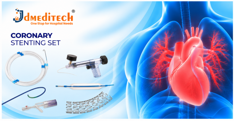
Coronary Stenting Set: Components, Uses, Procedure & Benefits

A Coronary Stenting Set brings together selected medical devices and accessories used during percutaneous coronary intervention (PCI). Interventional cardiologists use compatible coronary stents, balloon catheters, guide wires, and related instruments to treat selected coronary artery narrowings.

Coronary artery disease develops when atherosclerotic plaque builds up inside the arteries supplying blood to the heart muscle. Consequently, blood flow may become restricted, causing symptoms such as chest discomfort or shortness of breath.

In suitable patients, coronary angioplasty and stenting may improve blood flow through a narrowed artery. However, not every patient with coronary artery disease requires stent implantation.

JDMeditech offers selected interventional cardiology devices and catheter-based accessories for hospitals, cardiac centers, and international medical distributors. This guide explains coronary stenting equipment, common components, applications, benefits, and safety considerations.

What Is a Coronary Stenting Set?

A Coronary Stenting Set refers to a collection of specialized devices used to support coronary angioplasty and stent implantation.

During PCI, an interventional cardiologist introduces compatible catheters into the coronary circulation. Subsequently, a suitable balloon-expandable coronary stent may be positioned at a selected narrowing.

The stent acts as a small expandable scaffold that helps keep the treated artery open. As a result, blood flow through the affected segment may improve.

However, coronary stenting is an invasive treatment. Therefore, specialists must evaluate the patient’s condition, coronary anatomy, and available treatment alternatives.

Understanding Coronary Artery Disease

Coronary artery disease occurs when atherosclerotic plaque accumulates inside the coronary arteries.

Over time, plaque may narrow the arterial passage and reduce blood supply to the heart muscle.

For example, some patients develop angina during physical activity. In addition, sudden plaque disruption and clot formation may cause an acute coronary syndrome, including a heart attack.

Nevertheless, treatment depends on the severity of disease, symptoms, clinical presentation, and individual risk factors.

Why Is Coronary Stenting Performed?

Coronary stenting may be performed to treat selected significant coronary artery narrowings.

In particular, PCI can play an important role during certain acute coronary syndromes and in selected patients with persistent symptoms despite appropriate medical therapy.

Moreover, coronary stenting may be considered when coronary anatomy and clinical circumstances favor catheter-based treatment.

However, some patients benefit more from medication or coronary artery bypass grafting. Consequently, clinicians must choose treatment according to evidence-based guidelines and individual needs.

Common Components of a Coronary Stenting Set

Coronary intervention requires specialized devices that must work together safely. However, the exact components of a commercial set vary by manufacturer and configuration.

1. Coronary Stent Delivery System

A coronary stent delivery system carries a compatible stent to the intended treatment site.

Most contemporary coronary stents are balloon-expandable devices. Therefore, the stent is expanded using the delivery balloon during implantation.

Moreover, the stent diameter, length, and approved indications must match the patient’s clinical requirements.

2. Coronary Balloon Catheter

A coronary balloon catheter supports selected angioplasty procedures.

For example, specialists may use an appropriate balloon to prepare a narrowed lesion before stent placement.

In addition, selected noncompliant balloons may support post-dilation when clinically necessary.

3. PTCA Guide Wire

A percutaneous transluminal coronary angioplasty (PTCA) guide wire provides a pathway for compatible coronary intervention devices.

Furthermore, different guide wire designs support different anatomical and procedural requirements.

However, clinicians must select a guide wire according to its approved specifications.

4. Guiding Catheter

A guiding catheter provides access and support for selected coronary intervention devices.

Moreover, its configuration depends on the coronary anatomy and planned procedure.

5. Inflation Device

A compatible inflation device supports controlled inflation and deflation of selected angioplasty balloons.

In addition, many inflation devices include a pressure gauge.

Therefore, the clinical team must confirm compatibility with the selected balloon catheter and follow the approved pressure limits.

6. Hemostatic Y-Connector

A compatible Y-connector helps manage the catheter access system during selected interventions.

Furthermore, its valve design supports device passage while helping limit blood loss.

7. Introducer Sheath

A suitable introducer sheath provides vascular access for compatible catheter-based equipment.

However, the sheath dimensions and access configuration depend on the selected procedure.

8. Torque Device

A compatible torque device may support controlled handling of selected guide wires.

Consequently, clinicians should verify its compatibility with the intended guide wire.

9. Contrast Injection Accessories

Selected procedures use suitable syringes, manifolds, and connection tubing for contrast administration.

Moreover, contrast administration requires appropriate clinical assessment and monitoring.

10. Intravascular Imaging Equipment

Intravascular ultrasound (IVUS) or optical coherence tomography (OCT) may support selected coronary interventions.

For example, intravascular imaging can help assess vessel dimensions and stent expansion.

Importantly, imaging systems are generally supplied separately from disposable coronary stenting kits.

Coronary Stenting Set Components and Functions

The following table summarizes commonly associated PCI devices and their primary functions.

Component Primary Function
Coronary Stent Delivery System Delivers and expands a coronary stent
Coronary Balloon Catheter Supports lesion preparation or selected post-dilation
PTCA Guide Wire Provides a pathway for compatible intervention devices
Guiding Catheter Provides coronary access and device support
Inflation Device Supports controlled balloon inflation
Hemostatic Y-Connector Supports catheter access and helps manage blood loss
Introducer Sheath Provides vascular access
Torque Device Supports selected guide wire handling
Contrast Accessories Support contrast administration
Intravascular Imaging Supports selected vessel and stent assessments

Importantly, this table is educational and does not represent a verified JDMeditech packing list. Customers should confirm which components are supplied together and which require separate purchase.

Types of Coronary Stents

Different coronary stent technologies are available for selected clinical applications.

1. Drug-Eluting Stents

Drug-eluting stents release medication intended to reduce excessive tissue growth within the treated segment.

Consequently, contemporary drug-eluting stents are widely used during suitable PCI procedures.

However, device selection must follow the approved indications and relevant clinical guidance.

2. Bare-Metal Stents

Bare-metal stents provide mechanical support without a drug-eluting coating.

Nevertheless, they are used much less frequently in contemporary coronary intervention because modern drug-eluting stents generally offer better protection against restenosis.

3. Specialized Coronary Stent Designs

Different stent platforms offer various structural designs, materials, and delivery characteristics.

Therefore, specialists select a suitable device according to vessel anatomy, lesion characteristics, and approved product specifications.

Key Features of Coronary Stenting Equipment
1. Balloon-Expandable Stent Delivery

Suitable delivery systems support the placement and expansion of coronary stents.

Moreover, balloon and stent dimensions are selected according to the intended treatment site.

2. Compatibility With Guide Wires

Coronary intervention devices are designed for use with specified guide wire configurations.

Therefore, clinicians must confirm the relevant compatibility requirements.

3. Catheter-Based Treatment

PCI uses vascular access rather than conventional open-chest surgery.

As a result, suitable patients may undergo coronary revascularization through a minimally invasive approach.

4. Different Stent Sizes

Coronary stents are available in different diameters and lengths.

However, exact available dimensions depend on the manufacturer and product model.

5. Compatibility With Imaging Technologies

Selected procedures use angiography, IVUS, or OCT to support device selection and treatment assessment.

Furthermore, imaging may be particularly valuable during complex coronary interventions.

Clinical Applications of Coronary Stenting

Coronary stenting supports selected treatments for coronary artery disease.

1. Acute Coronary Syndromes

PCI is an important treatment option for selected patients experiencing acute coronary syndromes.

For example, emergency PCI may restore blood flow in an occluded coronary artery during certain heart attacks.

2. Symptomatic Coronary Artery Narrowing

Some patients experience angina because of significant coronary artery narrowing.

When appropriate medical therapy does not adequately control symptoms, PCI may provide additional relief in selected cases.

3. Selected Complex Coronary Lesions

Specialists may use PCI for selected complex coronary artery lesions.

However, the choice between PCI and coronary bypass surgery depends on anatomy, clinical risk, and expected outcomes.

4. Selected Restenotic Lesions

Restenosis occurs when a previously treated arterial segment becomes narrowed again.

Consequently, clinicians may consider further intervention according to the cause and severity of the narrowing.

5. Selected Multivessel Coronary Disease

Some patients with disease affecting multiple coronary arteries may undergo PCI.

Nevertheless, coronary bypass surgery may provide better outcomes for certain patients, particularly when coronary anatomy is complex.

How Is Coronary Stenting Performed?

A qualified interventional cardiologist performs coronary stenting in an appropriately equipped cardiac catheterization laboratory.

First, the clinical team reviews the patient’s symptoms, diagnostic findings, and overall medical condition.

Next, specialists select suitable vascular access equipment and coronary intervention devices.

During the procedure, coronary angiography helps identify the relevant arterial narrowing.

Afterward, the operator may use compatible guide wires and balloon catheters to prepare the selected treatment area.

When clinically appropriate, a suitable coronary stent is deployed within the narrowed segment.

Subsequently, the specialist assesses the result using angiography and, in selected cases, intravascular imaging.

Finally, the healthcare team monitors the patient and establishes an appropriate post-procedure treatment plan.

However, the exact technique depends on coronary anatomy, lesion characteristics, and clinical circumstances.

Potential Benefits of Coronary Stenting
1. Improved Coronary Blood Flow

Successful stent implantation can widen a significantly narrowed coronary artery.

As a result, blood flow through the treated segment may improve.

2. Relief of Angina Symptoms

PCI may reduce angina in selected patients with significant coronary artery disease.

Consequently, some patients experience improved exercise tolerance and quality of life.

3. Important Role During Heart Attack Treatment

Emergency PCI can be lifesaving in appropriate acute coronary syndromes.

Moreover, timely restoration of coronary blood flow may limit heart muscle injury in suitable patients.

4. Minimally Invasive Revascularization

Coronary stenting uses catheter-based vascular access.

Therefore, suitable patients may avoid open-chest surgery for selected coronary lesions.

5. Different Device Options

Modern PCI equipment includes various stent platforms, balloon catheters, and guide wires.

However, device selection must reflect clinical evidence and approved specifications.

Drug-Eluting Stent vs Bare-Metal Stent

Both stent types provide mechanical support to the treated coronary artery. However, their characteristics differ.

Feature Drug-Eluting Stent Bare-Metal Stent
Medication Coating Contains a drug-eluting system No drug-eluting coating
Primary Function Supports the artery and reduces tissue overgrowth Provides mechanical support
Restenosis Generally lower with contemporary devices Generally higher
Current Clinical Use Widely preferred for suitable PCI Used much less frequently
Selection Based on clinical and device requirements Based on selected circumstances

Importantly, the patient’s treatment plan must account for bleeding risk, antiplatelet medication requirements, and clinical circumstances.

Coronary Angioplasty vs Coronary Stenting

Balloon angioplasty and coronary stenting are related procedures, but they are not identical.

Balloon angioplasty uses a compatible balloon catheter to expand a narrowed coronary segment.

In contrast, coronary stenting also places a scaffold within the treated artery.

Moreover, balloon angioplasty may be performed before stent implantation or during selected procedures without an additional stent.

Therefore, clinicians must select the appropriate treatment strategy according to the patient’s coronary disease.

Risks and Safety Considerations

Coronary stenting is an invasive procedure that carries important risks.

Potential complications include bleeding, vascular injury, coronary artery dissection, perforation, and contrast-related adverse effects.

In addition, serious complications may include heart attack, stroke, or death.

Furthermore, a stent may become blocked by a blood clot, known as stent thrombosis.

Restenosis can also develop when the treated artery becomes narrowed again.

Consequently, clinicians must carefully assess the patient and select appropriate equipment, medications, and monitoring.

Why Is Antiplatelet Therapy Important After Coronary Stenting?

Antiplatelet medication helps reduce the risk of clot formation after coronary stent implantation.

For many patients, clinicians prescribe a combination of antiplatelet medicines for a defined period following PCI.

However, the medication choice and duration depend on the clinical presentation, bleeding risk, and other medical conditions.

Therefore, patients should never stop prescribed antiplatelet treatment without advice from their treating healthcare professional.

Recovery After Coronary Stenting

Recovery depends on the patient’s clinical condition and whether PCI was performed electively or during an emergency.

After the procedure, the healthcare team monitors the vascular access site, cardiac status, and potential complications.

Moreover, patients receive individualized instructions about medications, activity, wound care, and follow-up appointments.

Long-term management also includes attention to cholesterol, blood pressure, smoking, diabetes, and other cardiovascular risk factors.

However, new or severe chest pain, breathlessness, fainting, or other concerning symptoms require urgent medical evaluation.

How to Choose a Coronary Stenting Set

Hospitals and medical distributors should review the intended coronary intervention before purchasing equipment.

First, confirm whether the requirement involves a coronary stent delivery system, a PTCA accessory kit, or a broader PCI equipment configuration.

Next, verify the approved specifications and compatibility of all components.

Important Equipment Selection Factors
  • Approved intended use
  • Stent type and delivery system
  • Stent diameter and length
  • Balloon catheter dimensions
  • Balloon pressure ratings
  • Guide wire compatibility
  • Guiding catheter compatibility
  • Introducer sheath requirements
  • Inflation device specifications
  • Y-connector compatibility
  • Radiopaque marker configuration
  • Sterile packaging and labeling
  • Single-use device classification
  • Product shelf life
  • Manufacturer instructions for use
  • Applicable regulatory documentation

Furthermore, purchasing teams should confirm the exact packing list, available sizes, and product model before ordering.

Coronary Stenting Equipment and Cardiology Devices by JDMeditech

JDMeditech offers selected interventional cardiology devices and accessories for hospitals, cardiac catheterization laboratories, and international medical distributors.

Our cardiology product range includes selected PTCA guide wires, balloon-related accessories, introducer sheaths, connectors, and other catheter-based devices.

Moreover, customers can contact JDMeditech to discuss coronary stenting equipment availability, compatible product configurations, and technical documentation.

For more information, visit www.jdmeditech.com.

Frequently Asked Questions About Coronary Stenting Set
1. What is a Coronary Stenting Set?

A Coronary Stenting Set refers to selected medical devices used to support coronary stent implantation during PCI. However, exact components vary by manufacturer.

2. What is the purpose of a coronary stent?

A coronary stent helps maintain the opening of a treated coronary artery and supports blood flow through the segment.

3. Which instruments are used during coronary stenting?

Common equipment includes a coronary stent delivery system, guide wire, guiding catheter, balloon catheter, and compatible inflation accessories.

4. What is a drug-eluting stent?

A drug-eluting stent releases medication intended to reduce excessive tissue growth within the treated arterial segment.

5. Is coronary stenting the same as angioplasty?

Not exactly. Angioplasty involves balloon dilation, while stenting additionally places a scaffold inside the artery.

6. Does every coronary stenting kit include a stent?

Not necessarily. Product configurations vary. Therefore, customers must verify the actual supplied components.

7. Can a coronary artery become narrowed again after stenting?

Yes. Restenosis may occur, although contemporary drug-eluting stents reduce this risk compared with older bare-metal designs.

8. What is stent thrombosis?

Stent thrombosis is the formation of a blood clot within or near an implanted stent. It is a potentially serious complication.

9. Are antiplatelet medicines required after stenting?

Antiplatelet therapy is generally an important part of care after PCI. However, the exact regimen depends on the patient’s clinical circumstances.

10. Where can hospitals inquire about JDMeditech cardiology devices?

Hospitals and medical distributors can visit JDMeditech to request available product details and quotations.

Conclusion

A Coronary Stenting Set supports selected catheter-based procedures used to treat significant coronary artery narrowings.

Moreover, compatible stent delivery systems, balloon catheters, guide wires, and related accessories are important components of modern PCI.

However, coronary stenting carries significant risks and is not appropriate for every patient with coronary artery disease.

Therefore, healthcare professionals must select suitable devices and treatment approaches according to patient needs, clinical guidelines, and manufacturer specifications.

Contact JDMeditech for interventional cardiology instruments, PCI accessories, and international medical device inquiries.

Medical Disclaimer: This article provides general educational information and is not a procedural training guide. Qualified healthcare professionals must make all treatment and device-selection decisions according to clinical judgment, applicable guidelines, and approved manufacturer instructions.
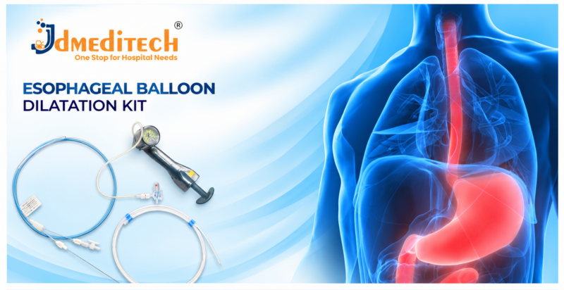
Esophageal Balloon Dilatation Kit: Components, Uses, Features & Benefits

An Esophageal Balloon Dilatation Kit is a collection of medical devices used to support selected esophageal dilation procedures. Gastroenterologists use compatible balloon catheters to widen narrowed areas of the esophagus when clinically indicated.

Esophageal narrowing can make swallowing difficult and may affect a patient’s ability to eat comfortably. Therefore, specialists investigate the underlying cause before recommending treatment.

Moreover, suitable balloon catheters, inflation devices, and accessories support controlled dilation during selected endoscopic procedures. However, device selection depends on the patient’s condition and the manufacturer’s approved specifications.

JDMeditech offers selected gastroenterology instruments and endoscopy accessories for hospitals, clinics, and international medical distributors. This guide explains esophageal balloon dilation, commonly associated kit components, clinical applications, benefits, and safety considerations.

What Is an Esophageal Balloon Dilatation Kit?

An Esophageal Balloon Dilatation Kit refers to a selected collection of devices used during esophageal balloon dilation.

The procedure involves expanding a compatible balloon within a narrowed section of the esophagus. As a result, the treatment may improve the passage of food and liquids in suitable patients.

In addition, specialists may use endoscopic or fluoroscopic guidance according to the type and complexity of the narrowing.

Nevertheless, esophageal dilation is an invasive intervention. Therefore, qualified healthcare professionals must evaluate its benefits, limitations, and potential complications.

Understanding Esophageal Strictures

An esophageal stricture is an abnormal narrowing of the esophagus, the muscular tube connecting the throat to the stomach.

For example, chronic acid reflux may cause inflammation and scarring that narrow the esophageal passage.

Furthermore, other conditions can produce esophageal narrowing, including eosinophilic esophagitis, previous surgery, radiation injury, and corrosive injury.

Consequently, patients may experience difficulty swallowing, food sticking, or discomfort during meals.

However, swallowing difficulties can also result from motility disorders or malignancy. Therefore, clinicians must identify the cause before selecting a treatment.

Why Is Esophageal Balloon Dilatation Performed?

Esophageal balloon dilation is commonly used to manage selected symptomatic esophageal strictures.

During treatment, the specialist expands a suitable balloon within the narrowed region to increase the available passage.

As a result, some patients experience improved swallowing and reduced obstruction-related symptoms.

However, the treatment does not always correct the underlying cause. Consequently, additional medication, repeat dilation, or other management may be necessary.

Common Components of an Esophageal Balloon Dilatation Kit

Different manufacturers offer different balloon dilation configurations. Therefore, the following components represent commonly associated equipment rather than a confirmed JDMeditech packing list.

1. Esophageal Balloon Dilatation Catheter

The balloon dilation catheter is the primary instrument used to expand a selected narrowed section of the esophagus.

Moreover, balloon catheters are available in different diameters, lengths, and designs for specific applications.

However, clinicians must use each catheter only within its approved specifications.

2. Balloon Inflation Device

A compatible inflation device allows the operator to manage balloon inflation during selected dilation procedures.

In addition, some inflation devices incorporate a pressure gauge for monitoring inflation pressure.

Therefore, the device must be compatible with the selected balloon catheter and its rated pressure limits.

3. Pressure Gauge

A pressure gauge displays the inflation pressure when incorporated into a compatible inflation system.

Consequently, the clinical team can monitor pressure according to the manufacturer’s instructions.

However, pressure readings alone do not establish the balloon’s exact expanded diameter unless supported by the device specifications.

4. Connecting Tubing

Compatible tubing may connect the balloon catheter to an inflation device.

Furthermore, secure connections are important for proper system operation.

5. Guide Wire

Selected esophageal balloon catheters are designed for use with compatible guide wires.

Moreover, guide wire selection depends on the catheter design and procedural requirements.

Nevertheless, not every balloon catheter requires a separate guide wire.

6. Endoscopic Visualization Equipment

A suitable endoscope may provide direct visualization during balloon dilation.

In addition, fluoroscopic imaging may be appropriate for selected complex strictures.

Importantly, imaging equipment is generally supplied separately from disposable dilation kits.

7. Connection Accessories

Some configurations include compatible connectors, valves, or other accessories.

However, the actual supplied components depend on the manufacturer’s packing list.

Esophageal Balloon Dilatation Kit Components and Functions

The following table summarizes commonly associated equipment and its primary functions.

Component Primary Function
Balloon Dilatation Catheter Expands a selected narrowed esophageal segment
Inflation Device Supports controlled balloon inflation and deflation
Pressure Gauge Displays inflation pressure
Connecting Tubing Connects compatible inflation components
Guide Wire Supports selected wire-guided dilation systems
Endoscope Provides direct visualization during suitable procedures
Fluoroscopy Equipment Supports imaging guidance when clinically indicated
Connection Accessories Support specified system connections

Importantly, this table is educational. Customers must confirm the exact JDMeditech product contents, available sizes, and compatibility information before ordering.

Key Features of Esophageal Balloon Dilatation Equipment
1. Controlled Balloon Expansion

Compatible dilation balloons expand within the selected esophageal narrowing.

Therefore, specialists can perform dilation according to the approved device characteristics and clinical requirements.

2. Different Balloon Configurations

Esophageal dilation balloons are available in different designs and dimensions.

Moreover, certain products provide multiple specified dilation diameters.

However, available sizes and pressure ratings vary by product.

3. Compatibility With Endoscopic Procedures

Selected balloon catheters are designed for use with compatible endoscopes.

Consequently, hospitals must confirm working channel requirements and catheter dimensions.

4. Pressure Monitoring

Suitable inflation systems may include pressure-monitoring capabilities.

Furthermore, clinicians must follow the approved pressure limits for the specific balloon catheter.

5. Different Clinical Applications

Esophageal balloon dilation supports selected treatment approaches for esophageal narrowing.

Nevertheless, the correct device depends on the underlying condition and procedural plan.

Clinical Applications of Esophageal Balloon Dilatation

Specialists may use esophageal balloon dilation in selected conditions involving narrowing of the esophagus.

1. Benign Esophageal Strictures

Benign strictures can develop because of inflammation, scarring, or previous treatment.

For suitable patients, balloon dilation may help widen the narrowed region.

2. Peptic Esophageal Strictures

Long-standing gastroesophageal reflux can contribute to peptic stricture formation.

Therefore, treatment may combine dilation with appropriate acid-suppressing medication.

3. Schatzki Ring

A Schatzki ring is a narrowing near the lower esophagus that can cause intermittent difficulty swallowing solid foods.

In selected symptomatic patients, dilation may help improve swallowing.

4. Eosinophilic Esophagitis-Related Narrowing

Eosinophilic esophagitis can produce inflammation and fibrotic narrowing.

Consequently, some patients require dilation in addition to treatment of the underlying inflammatory condition.

5. Postoperative Esophageal Strictures

Esophageal narrowing may develop after selected surgical procedures.

However, dilation requires careful evaluation of the patient’s surgical history and anatomy.

6. Radiation-Related or Caustic Strictures

Radiation injury and corrosive exposure can cause complex esophageal strictures.

Nevertheless, these cases may carry higher procedural risks and require specialized planning.

7. Selected Malignant Esophageal Narrowing

Some patients with malignant narrowing may undergo dilation as part of carefully planned management.

However, these cases require particular caution because perforation risk may be higher.

How Is Esophageal Balloon Dilatation Performed?

A qualified gastroenterologist performs esophageal balloon dilation in an appropriately equipped clinical setting.

First, the healthcare team evaluates the patient’s swallowing symptoms, medical history, and diagnostic findings.

Next, the specialist selects an appropriate dilation approach and compatible equipment.

During the procedure, the clinician identifies the narrowed esophageal region using suitable imaging or endoscopic visualization.

Afterward, a compatible balloon catheter is positioned for the planned treatment.

The specialist then performs dilation according to the patient’s condition and the approved device instructions.

Finally, the clinical team assesses the treatment area and monitors the patient during recovery.

However, the exact procedure depends on the cause, severity, and complexity of the narrowing.

Potential Benefits of Esophageal Balloon Dilatation
1. Improved Swallowing in Suitable Patients

Balloon dilation may increase the opening of a narrowed esophageal segment.

As a result, some patients experience improved swallowing and reduced food obstruction symptoms.

2. Endoscopic Treatment Option

Selected strictures can be treated using an endoscopic approach.

Therefore, suitable patients may avoid more invasive interventions for certain types of narrowing.

3. Compatibility With Different Balloon Sizes

Clinicians can select a balloon configuration according to the stricture and approved product dimensions.

Moreover, certain systems offer specified multiple-diameter options.

4. Support for Different Stricture Types

Balloon dilation may be used in several types of benign esophageal narrowing.

However, the treatment plan must account for the underlying cause and associated risks.

5. Repeat Treatment When Necessary

Some strictures recur or require more than one dilation session.

Consequently, clinicians may recommend further treatment according to symptoms and clinical findings.

Balloon Dilatation vs Bougie Dilatation

Balloon and bougie dilators are established options for selected esophageal strictures. However, their designs and mechanisms differ.

Feature Balloon Dilatation Bougie Dilatation
Device Type Expandable balloon catheter Solid or semi-rigid dilator
Primary Mechanism Radial expansion Mechanical passage through the narrowing
Common Guidance Endoscopic or selected fluoroscopic guidance Often guide-wire assisted
Available Designs Different balloon diameters and lengths Different dilator diameters and configurations
Selection Based on stricture and device requirements Based on stricture and device requirements

Importantly, clinical evidence supports both balloon and wire-guided bougie dilation for suitable benign strictures. Therefore, the choice depends on the patient’s condition and the specialist’s assessment.

Esophageal Balloon Dilatation vs Achalasia Pneumatic Dilatation

Although both treatments use balloons, conventional esophageal stricture dilation and pneumatic dilation for achalasia are not identical.

Esophageal stricture dilation aims to widen a narrowed segment of the esophagus.

In contrast, achalasia pneumatic dilation uses specialized equipment to disrupt the lower esophageal sphincter’s resistance to opening.

Moreover, achalasia treatment often requires different balloon dimensions and clinical protocols.

Therefore, an ordinary esophageal balloon dilation catheter must not be assumed suitable for achalasia pneumatic dilation.

Risks and Safety Considerations

Esophageal balloon dilation is an invasive procedure with potentially serious complications.

For example, adverse events may include esophageal perforation, bleeding, aspiration, and pain.

Furthermore, complex strictures and certain underlying diseases can increase procedural risk.

Consequently, specialists must evaluate the patient’s anatomy, clinical condition, and available treatment alternatives.

Active or incompletely healed esophageal perforation is a contraindication to dilation.

In addition, patients with recent esophageal surgery, previous perforation, or significant bleeding disorders require careful assessment.

After treatment, severe or persistent chest pain, fever, breathlessness, or rapid deterioration may indicate a complication and require urgent medical evaluation.

Recovery After Esophageal Balloon Dilatation

Recovery depends on the patient’s condition, sedation, and procedure complexity.

After dilation, the healthcare team monitors the patient and provides instructions about food, fluids, medications, and follow-up.

Some patients may experience temporary throat or chest discomfort.

However, persistent or worsening symptoms require prompt medical assessment.

Furthermore, repeat dilation or additional treatment may be necessary if narrowing returns.

How to Choose an Esophageal Balloon Dilatation Kit

Hospitals and medical distributors should confirm the intended clinical application before purchasing balloon dilation equipment.

First, identify whether the requirement involves a conventional esophageal stricture balloon or a specialized pneumatic dilation system.

Next, review the approved device dimensions and compatible inflation equipment.

Important Equipment Selection Factors
  • Approved intended use
  • Balloon diameter options
  • Balloon working length
  • Catheter working length
  • Catheter outer diameter
  • Endoscope working channel compatibility
  • Guide wire compatibility
  • Rated balloon pressure limits
  • Inflation device compatibility
  • Pressure gauge configuration
  • Connection tubing specifications
  • Radiopaque marker availability
  • Sterile packaging requirements
  • Single-use or reusable classification
  • Manufacturer instructions for use
  • Available technical documentation

Moreover, purchasing teams should verify the exact kit contents and available sizes before placing an order.

Esophageal Balloon Dilatation Instruments by JDMeditech

JDMeditech offers selected gastroenterology instruments and endoscopy accessories for hospitals, endoscopy centers, and international medical distributors.

Our gastroenterology product range includes selected balloon dilation devices, endoscopic catheters, guide wires, and related medical accessories.

Furthermore, customers can contact JDMeditech to discuss available product configurations, compatibility requirements, and technical specifications.

For additional information, visit www.jdmeditech.com.

Frequently Asked Questions About Esophageal Balloon Dilatation Kit
1. What is an Esophageal Balloon Dilatation Kit?

An Esophageal Balloon Dilatation Kit contains selected devices used to support esophageal balloon dilation. However, exact components vary by product configuration.

2. What is the purpose of esophageal balloon dilation?

The procedure aims to widen selected narrowed areas of the esophagus and improve swallowing in suitable patients.

3. Which instruments are commonly used?

Common equipment includes a balloon dilation catheter, compatible inflation device, and selected accessories.

4. What is an esophageal stricture?

An esophageal stricture is an abnormal narrowing that may cause difficulty swallowing.

5. Does every kit include an inflation device?

No. Inflation devices may be supplied separately. Therefore, customers should verify the packing list.

6. Is balloon dilation performed under endoscopic guidance?

Many procedures use endoscopic guidance. In addition, fluoroscopy may be appropriate for selected complex strictures.

7. Can esophageal strictures return after dilation?

Yes. Some strictures recur and require further treatment or repeat dilation.

8. Is esophageal balloon dilation the same as achalasia pneumatic dilation?

No. Achalasia pneumatic dilation may require specialized balloons and different clinical protocols.

9. What are the main risks of esophageal dilation?

Potential complications include esophageal perforation, bleeding, aspiration, and pain.

10. Where can hospitals inquire about JDMeditech gastroenterology devices?

Hospitals and medical distributors can visit JDMeditech to request information about available products and quotations.

Conclusion

An Esophageal Balloon Dilatation Kit supports selected procedures intended to widen narrowed sections of the esophagus.

Moreover, compatible balloon catheters and inflation devices help specialists perform dilation according to clinical requirements.

However, esophageal dilation carries important risks and does not always eliminate the underlying cause of narrowing.

Therefore, healthcare professionals must select suitable equipment and treatment approaches according to patient needs, clinical guidelines, and manufacturer specifications.

Contact JDMeditech for gastroenterology instruments, endoscopic balloon devices, and international medical device inquiries.

Medical Disclaimer: This article provides general educational information and is not a procedural training guide. Qualified healthcare professionals must make all treatment and device-selection decisions according to clinical judgment, applicable guidelines, and approved manufacturer instructions.
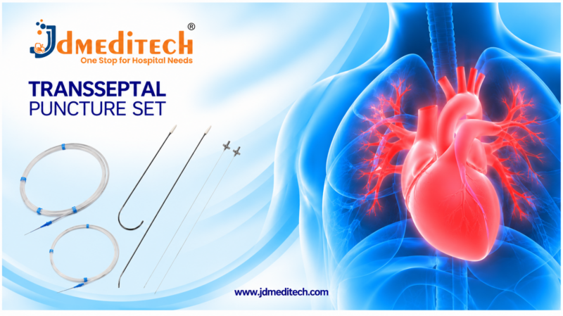
Transseptal Puncture Set: Components, Uses, Features & Benefits

A Transseptal Puncture Set is a specialized collection of medical instruments used to support access to the left atrium during selected catheter-based cardiac procedures. Interventional cardiologists and electrophysiologists use compatible devices to cross the interatrial septum when clinically indicated.

Transseptal access plays an important role in modern cardiac electrophysiology and structural heart interventions. For example, specialists may require left atrial access during atrial fibrillation ablation and selected mitral valve procedures.

Moreover, suitable transseptal needles, sheaths, dilators, and related accessories support the procedure. However, component selection depends on patient anatomy, procedural requirements, and approved device compatibility.

JDMeditech offers selected cardiology devices and catheter-based accessories for hospitals, cardiac centers, and international medical distributors. This guide explains the components, applications, features, benefits, and safety considerations associated with transseptal puncture equipment.

What Is a Transseptal Puncture Set?

A Transseptal Puncture Set refers to a collection of specialized devices used during transseptal catheterization.

The procedure involves creating a controlled passage through the interatrial septum, which separates the right and left atria of the heart.

Consequently, specialists can introduce compatible catheters into the left atrium for selected diagnostic or interventional procedures.

However, transseptal puncture is an invasive cardiac procedure. Therefore, it requires appropriate clinical expertise, imaging guidance, and safety monitoring.

Understanding Transseptal Catheterization

Transseptal catheterization provides a route to the left atrium through the venous circulation and interatrial septum.

In particular, the technique is important when clinicians need left atrial access for electrophysiological mapping, ablation, or selected structural heart interventions.

Furthermore, imaging technologies such as fluoroscopy, transesophageal echocardiography, and intracardiac echocardiography may support procedural planning and guidance.

Nevertheless, the selected imaging approach depends on the clinical procedure, patient anatomy, and available equipment.

Why Is Transseptal Puncture Performed?

Transseptal puncture allows specialists to access the left atrium during procedures that require catheter-based intervention.

For example, the technique may support selected atrial fibrillation ablation procedures.

In addition, clinicians may use transseptal access during left atrial appendage closure and certain percutaneous mitral valve interventions.

However, transseptal puncture is not required for every cardiac catheterization procedure. Consequently, specialists must evaluate the indication and potential risks before proceeding.

Common Components of a Transseptal Puncture Set

Different manufacturers offer different transseptal device configurations. Therefore, the following components represent commonly associated equipment rather than a confirmed packing list.

1. Transseptal Puncture Needle

A transseptal puncture needle is designed for use during controlled access across the interatrial septum.

For example, selected procedures use a conventional Brockenbrough-type needle with a compatible sheath and dilator.

However, the needle design, dimensions, and curvature must match the intended system and approved application.

2. Transseptal Sheath

A transseptal sheath provides a compatible access pathway for selected catheters and interventional devices.

Moreover, different sheath designs may offer fixed or steerable configurations depending on the intended procedure.

3. Transseptal Dilator

A compatible dilator works with the selected sheath during transseptal access.

Therefore, hospitals must verify that the dilator and sheath are approved for use together.

4. Guide Wire

A suitable guide wire may support selected vascular access and catheter exchange tasks.

In addition, clinicians must confirm its diameter, length, and compatibility with the intended devices.

5. Hemostatic Valve

A compatible hemostatic valve helps manage blood loss and air entry during selected catheter-based procedures.

However, its presence and configuration depend on the specific sheath system.

6. Sidearm and Connection Tubing

Selected transseptal sheaths include a sidearm or compatible connection tubing.

Furthermore, these components may support specified flushing or pressure-monitoring arrangements.

7. Vascular Access Accessories

Some procedural configurations require separate access needles, introducers, or other vascular access devices.

Nevertheless, these accessories may not be included in a transseptal puncture set.

8. Imaging and Monitoring Equipment

Fluoroscopy and echocardiographic imaging may support safe transseptal access.

In addition, appropriate hemodynamic and cardiac monitoring equipment is essential during the procedure.

Importantly, imaging systems and monitors are generally separate from disposable transseptal puncture sets.

Transseptal Puncture Set Components and Functions

The following table summarizes commonly associated components and their functions.

Component Primary Function
Transseptal Needle Supports controlled puncture of the interatrial septum
Transseptal Sheath Provides a compatible catheter access pathway
Transseptal Dilator Works with the sheath during transseptal access
Guide Wire Supports selected access and exchange tasks
Hemostatic Valve Helps manage blood loss and air entry
Connection Tubing Supports specified fluid or monitoring connections
Vascular Access Accessories Support selected vascular access requirements
Imaging Equipment Supports visualization and procedural guidance

Importantly, this table is educational. Customers must verify the exact JDMeditech set contents, dimensions, and device specifications before ordering.

Key Features of Transseptal Puncture Equipment
1. Specialized Left Atrial Access

Transseptal instruments support selected procedures requiring access to the left atrium.

Consequently, compatible systems play an important role in cardiac electrophysiology and structural heart interventions.

2. Compatible Needle and Sheath Configurations

Transseptal needles and sheaths are available in different designs.

Therefore, clinicians must select a compatible combination according to the intended procedure.

3. Catheter-Based Access

Suitable sheaths provide an access route for selected cardiac catheters.

Moreover, catheter compatibility depends on sheath dimensions and approved device specifications.

4. Imaging-Guided Procedures

Modern transseptal procedures may use fluoroscopy or echocardiographic guidance.

As a result, specialists can assess anatomy and confirm important procedural findings.

5. Different Procedural Configurations

Hospitals may require different transseptal systems for electrophysiology and structural heart procedures.

However, no single configuration is appropriate for every application.

Clinical Applications of Transseptal Puncture

Transseptal puncture supports selected procedures involving the left atrium.

1. Atrial Fibrillation Ablation

Catheter ablation for atrial fibrillation often requires access to the left atrium.

Therefore, transseptal access is an important part of many pulmonary vein isolation procedures.

2. Left Atrial Electrophysiology Procedures

Selected electrophysiology procedures require left atrial mapping or catheter intervention.

In addition, transseptal access may provide a suitable route for specific left-sided arrhythmia treatments.

3. Left Atrial Appendage Closure

Selected left atrial appendage closure procedures use transseptal access.

Consequently, clinicians can introduce compatible closure equipment into the left atrium.

4. Percutaneous Mitral Valve Interventions

Certain catheter-based mitral valve procedures require transseptal access.

Moreover, the required puncture location and equipment configuration depend on the planned intervention.

5. Selected Structural Heart Procedures

Other structural heart interventions may also require left atrial access.

However, clinicians must confirm the specific procedural indication and equipment requirements.

How Does Transseptal Puncture Work?

A qualified interventional cardiologist or electrophysiologist performs transseptal puncture in an appropriately equipped clinical setting.

First, the clinical team evaluates the patient’s anatomy, procedure requirements, and potential risks.

Next, specialists select suitable vascular access equipment and compatible transseptal devices.

During the procedure, the team uses appropriate imaging and monitoring to guide the transseptal system toward the interatrial septum.

Subsequently, the operator establishes and confirms the intended left atrial access.

Afterward, compatible interventional catheters may be introduced according to the planned procedure.

Finally, the healthcare team monitors the patient and evaluates for potential complications.

However, the exact technique depends on anatomy, equipment, and clinical protocols. This overview is not intended as procedural training.

Benefits of Transseptal Access Systems
1. Access to the Left Atrium

Transseptal systems provide an established route to the left atrium for selected procedures.

As a result, specialists can perform interventions that require access to left atrial structures.

2. Support for Catheter Ablation

Compatible transseptal devices support many left atrial electrophysiology procedures.

Furthermore, they form part of the equipment used during selected atrial fibrillation ablation procedures.

3. Support for Structural Heart Interventions

Selected transseptal systems support catheter-based structural heart procedures.

For example, certain mitral valve and left atrial appendage interventions require this access route.

4. Compatibility With Specialized Catheters

Suitable transseptal sheaths provide a pathway for approved catheter systems.

However, the operator must verify the compatibility of each component.

5. Different Device Options

Conventional and alternative transseptal access technologies are available for selected clinical situations.

Therefore, specialists can evaluate device options according to procedural requirements and patient anatomy.

Conventional vs Radiofrequency Transseptal Puncture

Different technologies can support transseptal access. However, their mechanisms and equipment requirements vary.

Feature Conventional Needle RF Transseptal System
Access Mechanism Mechanical needle puncture Radiofrequency-assisted puncture
Equipment Compatible needle, sheath, and dilator Compatible RF device and associated equipment
Clinical Use Selected transseptal procedures Selected transseptal procedures
Energy Source No RF energy required for puncture Requires compatible RF equipment
Selection Based on anatomy and clinical requirements Based on anatomy and clinical requirements

Importantly, RF devices are not interchangeable with conventional needles. Therefore, hospitals must verify approved compatibility and system requirements.

Risks and Safety Considerations

Transseptal puncture is an invasive procedure that can cause serious complications.

Potential risks include cardiac perforation, pericardial bleeding, cardiac tamponade, vascular injury, and thromboembolic events.

In addition, inadvertent puncture of an unintended cardiac or vascular structure may require emergency treatment.

Furthermore, air embolism, device-related complications, and access-site bleeding are important safety concerns.

Consequently, trained specialists must use appropriate imaging, anticoagulation management, and clinical monitoring according to established protocols.

Moreover, healthcare facilities should have suitable emergency equipment and complication-management arrangements available.

How to Choose a Transseptal Puncture Set

Hospitals and medical distributors should evaluate the intended cardiac procedure before selecting a transseptal system.

First, confirm whether the requirement involves a conventional transseptal needle, a complete sheath system, or an alternative access technology.

Next, review the approved compatibility of all selected components.

Important Equipment Selection Factors
  • Approved intended use
  • Transseptal needle design
  • Needle curvature and usable length
  • Transseptal sheath diameter and length
  • Fixed or steerable sheath configuration
  • Dilator compatibility
  • Guide wire compatibility
  • Hemostatic valve configuration
  • Sidearm and connector design
  • Radiopacity and imaging compatibility
  • Sterile packaging requirements
  • Single-use or reusable classification
  • Manufacturer instructions for use
  • Relevant technical documentation

Furthermore, purchasing teams should confirm the actual set contents and available sizes before placing an order.

Transseptal Puncture Instruments by JDMeditech

JDMeditech offers selected cardiology devices and catheter-based accessories for hospitals, cardiac care centers, and international medical distributors.

Our cardiology product range includes selected introducer sheaths, guide wires, and related interventional accessories.

Moreover, customers can contact JDMeditech to discuss transseptal puncture equipment availability, compatible configurations, and product specifications.

For more information, visit www.jdmeditech.com.

Frequently Asked Questions About Transseptal Puncture Set
1. What is a Transseptal Puncture Set?

A Transseptal Puncture Set is a collection of specialized devices used to support access across the interatrial septum during selected cardiac procedures.

2. What is the purpose of transseptal puncture?

Transseptal puncture provides access to the left atrium for selected catheter-based diagnostic and interventional procedures.

3. Which instruments are commonly used?

Common components include a compatible transseptal needle, sheath, dilator, and selected accessories.

4. What is a Brockenbrough needle?

A Brockenbrough needle is a conventional transseptal needle design used with compatible equipment during selected transseptal procedures.

5. Is transseptal puncture used during atrial fibrillation ablation?

Yes. Transseptal access is commonly used during left atrial catheter ablation procedures, including many atrial fibrillation treatments.

6. What is the function of a transseptal sheath?

A transseptal sheath provides a compatible pathway for selected catheters and interventional devices.

7. Are transseptal needles and sheaths interchangeable?

No. Clinicians must confirm that the selected components are approved and compatible with one another.

8. What imaging is used during transseptal puncture?

Depending on the procedure, clinicians may use fluoroscopy, transesophageal echocardiography, or intracardiac echocardiography.

9. What are the major risks of transseptal puncture?

Potential complications include cardiac perforation, tamponade, vascular injury, and thromboembolic events.

10. Where can hospitals inquire about JDMeditech transseptal equipment?

Hospitals and medical distributors can visit JDMeditech to request information about available cardiology devices and quotations.

Conclusion

A Transseptal Puncture Set supports selected cardiac procedures that require access to the left atrium.

Moreover, compatible needles, sheaths, dilators, and related accessories are important components of conventional transseptal systems.

However, transseptal puncture carries significant procedural risks and requires specialized training, imaging guidance, and monitoring.

Therefore, hospitals should select appropriate equipment according to clinical requirements, manufacturer specifications, and professional guidelines.

Contact JDMeditech for cardiology instruments, catheter-based accessories, and international medical device inquiries.

Medical Disclaimer: This article is intended for general educational purposes and does not provide procedural instructions. Qualified healthcare professionals must make treatment and device-selection decisions according to clinical judgment, applicable guidelines, and approved manufacturer instructions.
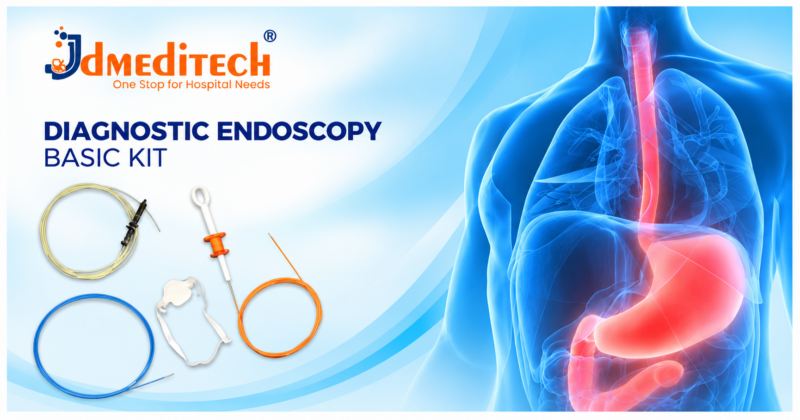
Diagnostic Endoscopy Basic Kit: Components, Uses, Features & Benefits

A Diagnostic Endoscopy Basic Kit brings together selected medical accessories that support gastrointestinal endoscopy procedures. Depending on the configuration, these accessories may help clinicians perform tissue sampling, fluid management, and other diagnostic tasks.

Gastrointestinal endoscopy allows specialists to examine the digestive tract using a suitable endoscope. Moreover, compatible accessories extend the capabilities of the examination when additional diagnostic procedures are necessary.

However, not every diagnostic endoscopy kit contains the same instruments. Therefore, hospitals and medical distributors should verify the intended use, dimensions, and exact supplied components before purchasing.

JDMeditech offers selected gastroenterology instruments and endoscopy accessories for hospitals, endoscopy centers, and international medical distributors. This guide explains common kit components, clinical applications, features, benefits, and safety considerations.

What Is a Diagnostic Endoscopy Basic Kit?

A Diagnostic Endoscopy Basic Kit is a collection of selected accessories intended to support compatible diagnostic endoscopy procedures.

For example, a gastrointestinal endoscopy accessory configuration may include devices for tissue sampling, specimen collection, or selected catheter-based tasks.

In addition, healthcare professionals may use these accessories with suitable gastroscopes, colonoscopes, or other endoscopic equipment, depending on their approved applications.

Nevertheless, the exact kit configuration varies. Consequently, purchasing teams must confirm the manufacturer’s packing list and compatibility information.

Understanding Diagnostic Gastrointestinal Endoscopy

Diagnostic gastrointestinal endoscopy allows clinicians to examine selected parts of the digestive tract.

During upper gastrointestinal endoscopy, a specialist may inspect the esophagus, stomach, and duodenum. In contrast, colonoscopy allows examination of the colon and, when appropriate, the terminal ileum.

Furthermore, suitable accessories may support biopsy collection or other investigations when clinically indicated.

However, accessory selection depends on the procedure and the endoscope’s working channel dimensions.

Common Accessories Associated With Diagnostic Endoscopy

Different endoscopy procedures require different accessories. Therefore, the following instruments represent examples rather than a confirmed list of components in every kit.

1. Endoscopic Biopsy Forceps

Endoscopic biopsy forceps support tissue sampling from selected areas of the gastrointestinal tract.

During a suitable procedure, the clinician introduces compatible forceps through the endoscope’s working channel.

Afterward, the healthcare team handles the tissue specimen according to the relevant laboratory protocol.

2. Endoscopic Cytology Brush

A cytology brush supports collection of cellular samples from selected gastrointestinal or ductal sites when indicated.

Moreover, the clinician selects the brush according to the intended procedure and device compatibility.

3. Diagnostic Endoscopy Catheter

Selected endoscopic catheters support fluid delivery, aspiration, or other approved diagnostic applications.

However, the catheter’s function depends on its specific design and approved intended use.

4. Endoscopic Guide Wire

A compatible guide wire may support selected catheter-based endoscopic procedures.

In addition, guide wire diameter, length, and tip configuration must match the intended accessories.

5. Aspiration Accessories

Suitable aspiration accessories may support the collection or removal of fluids during selected procedures.

Consequently, clinicians must verify connections and intended applications before use.

6. Endoscopic Injection Needle

An endoscopic injection needle supports selected injection procedures when clinically indicated.

However, it is not necessarily part of a basic diagnostic kit and may be used for therapeutic rather than purely diagnostic purposes.

7. Endoscope Cleaning Brush

A compatible cleaning brush supports manual cleaning of designated endoscope channels during reprocessing.

Importantly, cleaning brushes are reprocessing accessories and should not be confused with cytology brushes used for specimen collection.

8. Collection and Connection Accessories

Selected kits may include suitable connectors or collection accessories.

Furthermore, hospitals should verify whether these items are supplied separately or included in the package.

Diagnostic Endoscopy Instruments and Their Functions

The following table summarizes common endoscopy accessories and their potential applications.

Accessory Primary Function
Biopsy Forceps Collect tissue samples during compatible endoscopic procedures
Cytology Brush Collect cellular specimens from selected sites
Endoscopy Catheter Support specified fluid or catheter-based applications
Guide Wire Support compatible catheter positioning
Aspiration Accessory Support selected fluid collection tasks
Injection Needle Support selected endoscopic injection procedures
Cleaning Brush Support endoscope channel cleaning during reprocessing
Collection Accessories Support suitable specimen or fluid handling

Importantly, this table is educational. It does not establish the actual contents of a JDMeditech Diagnostic Endoscopy Basic Kit.

Key Features of Diagnostic Endoscopy Accessories
1. Compatibility With Endoscopic Equipment

Suitable accessories work with specified endoscope models and channel dimensions.

Therefore, clinicians and purchasing teams must confirm compatibility before use.

2. Support for Diagnostic Procedures

Selected accessories allow clinicians to collect tissue or cellular specimens.

As a result, laboratory testing may provide additional diagnostic information.

3. Different Instrument Configurations

Endoscopy accessories are available in different dimensions and designs.

Moreover, device selection depends on the clinical procedure and approved intended use.

4. Catheter-Based Applications

Selected catheter accessories support specific endoscopic tasks.

However, clinicians should not assume that all catheters are interchangeable.

5. Sterile and Reusable Options

Endoscopy accessories may be supplied as single-use or reusable devices.

Consequently, healthcare facilities must follow the manufacturer’s labeling and reprocessing instructions.

Clinical Applications of Diagnostic Endoscopy Accessories

Diagnostic endoscopy accessories support a range of gastrointestinal investigations.

For example, clinicians may use suitable devices during:

  • Upper gastrointestinal endoscopy
  • Colonoscopy
  • Selected endoscopic biopsy procedures
  • Endoscopic cytology sampling
  • Selected fluid aspiration procedures
  • Specified catheter-based diagnostic procedures
  • Other compatible gastrointestinal investigations

However, each accessory has its own approved intended use. Therefore, the clinician must select the appropriate device for the planned examination.

How Are Diagnostic Endoscopy Accessories Used?

A qualified healthcare professional selects diagnostic endoscopy accessories according to the patient’s clinical needs.

First, the endoscopy team reviews the planned examination and relevant patient information.

Next, staff confirm that the selected accessory is compatible with the endoscope and suitable for its intended application.

During the examination, the clinician may introduce an approved accessory through the working channel when indicated.

Afterward, the healthcare team collects and identifies any required specimens.

Finally, staff dispose of single-use accessories or reprocess reusable instruments according to validated instructions.

Nevertheless, actual clinical techniques depend on the procedure, device design, and applicable medical protocols.

Benefits of a Diagnostic Endoscopy Basic Kit
1. Convenient Accessory Organization

A suitably configured kit can bring selected instruments together for a planned procedure.

As a result, purchasing and preparation teams may find it easier to organize compatible accessories.

2. Support for Tissue Sampling

Suitable biopsy instruments help clinicians collect specimens for laboratory examination.

Furthermore, specimen analysis may contribute to the diagnostic process.

3. Support for Different Investigations

Different accessories serve different diagnostic purposes.

Therefore, a carefully selected configuration may support the needs of a particular endoscopy service.

4. Equipment Compatibility Planning

Reviewing accessories as a set can help hospitals assess compatibility with existing endoscopes.

However, every component still requires individual verification.

5. Procurement Convenience

Hospitals may prefer clearly documented accessory configurations when requesting quotations.

Moreover, an accurate packing list helps customers compare available products.

Diagnostic Endoscopy Kit vs Therapeutic Endoscopy Kit

Diagnostic and therapeutic endoscopy accessories have different primary purposes.

Feature Diagnostic Accessories Therapeutic Accessories
Primary Purpose Support examination and diagnostic sampling Support selected endoscopic treatments
Examples Biopsy forceps and cytology brushes Selected snares, clips, and treatment catheters
Clinical Application Diagnostic investigations Endoscopic intervention
Equipment Selection Depends on examination requirements Depends on planned treatment

However, some accessories can serve different roles depending on their approved intended use. Consequently, clinicians should follow the device documentation rather than relying on kit names alone.

Safety and Infection Prevention

Diagnostic endoscopy requires suitable equipment, trained personnel, and appropriate infection prevention practices.

Before use, staff should inspect device packaging, labeling, and compatibility information.

Furthermore, healthcare facilities must follow validated reprocessing instructions for reusable endoscopes and accessories.

Reusable accessories that penetrate mucosal tissue require appropriate sterilization between patients. In contrast, single-use devices must not be reused unless expressly permitted by applicable regulations and validated processes.

Moreover, damaged or incompatible instruments may increase procedural risks.

Therefore, healthcare professionals must follow the approved instructions for use and applicable infection control guidelines.

How to Choose a Diagnostic Endoscopy Basic Kit

Hospitals and medical distributors should review the intended clinical application before purchasing endoscopy accessories.

First, confirm whether the kit is intended for upper gastrointestinal endoscopy, colonoscopy, or another procedure.

Next, compare the available instrument configurations with the hospital’s equipment.

Important Equipment Selection Factors
  • Approved intended use
  • Exact kit components
  • Endoscope channel compatibility
  • Instrument working length
  • Catheter or accessory diameter
  • Biopsy forceps configuration
  • Cytology brush dimensions
  • Guide wire compatibility
  • Connection requirements
  • Sterile packaging and labeling
  • Single-use or reusable classification
  • Reprocessing requirements
  • Manufacturer technical documentation

In addition, purchasing teams should confirm the actual quantity of each supplied component.

Diagnostic Endoscopy Accessories by JDMeditech

JDMeditech offers selected gastroenterology instruments and endoscopy accessories for hospitals, clinics, endoscopy centers, and international medical distributors.

Our gastroenterology product range includes selected biopsy forceps, cytology brushes, endoscopic catheters, guide wires, and related devices.

Furthermore, customers can contact JDMeditech to discuss available product configurations, specifications, and quotations.

For additional product information, visit www.jdmeditech.com.

Frequently Asked Questions About Diagnostic Endoscopy Basic Kit
1. What is a Diagnostic Endoscopy Basic Kit?

A Diagnostic Endoscopy Basic Kit is a selected collection of accessories intended to support compatible endoscopic procedures. However, the contents vary by configuration.

2. What is the purpose of diagnostic endoscopy?

Diagnostic endoscopy allows clinicians to examine selected areas of the digestive tract and investigate suspected abnormalities.

3. Which instruments are used during diagnostic endoscopy?

Depending on the procedure, clinicians may use compatible biopsy forceps, cytology brushes, catheters, and other accessories.

4. Does every diagnostic endoscopy kit contain biopsy forceps?

No. Customers should verify the actual packing list before ordering.

5. What is an endoscopic cytology brush?

A cytology brush is an accessory used to collect cellular specimens from selected anatomical sites.

6. Can the same kit be used for gastroscopy and colonoscopy?

Only when the supplied devices are approved and compatible with the intended endoscopes and procedures.

7. Are diagnostic endoscopy accessories reusable?

Some accessories are reusable, while others are single-use. Therefore, hospitals must follow the manufacturer’s labeling and instructions.

8. Why is working channel compatibility important?

Compatible dimensions help ensure that an accessory can be used with the selected endoscope according to its approved specifications.

9. Does a basic kit include the endoscope itself?

Not necessarily. The endoscope and other equipment may be supplied separately.

10. Where can hospitals inquire about JDMeditech endoscopy accessories?

Hospitals and medical distributors can visit JDMeditech to request product information and quotations.

Conclusion

A Diagnostic Endoscopy Basic Kit can support selected gastrointestinal investigations through compatible diagnostic accessories.

Moreover, suitable biopsy instruments, cytology brushes, and other devices help clinicians perform specified diagnostic tasks.

However, the exact kit contents, device compatibility, and approved applications must be verified before clinical use.

Therefore, hospitals should select endoscopy accessories according to their procedural requirements and manufacturer documentation.

Contact JDMeditech for gastroenterology instruments, endoscopy accessories, and international medical device inquiries.

Medical Disclaimer: This article provides general educational information and does not replace professional medical advice or device instructions. Qualified healthcare professionals must select medical procedures and instruments according to clinical judgment, approved manufacturer instructions, and applicable guidelines.Diagnostic Endoscopy Basic Kit

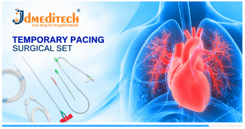
Temporary Pacing Surgical Set: Components, Uses, Features & Benefits

A Temporary Pacing Surgical Set brings together selected medical devices and accessories used during temporary cardiac pacing procedures. These devices support healthcare professionals when short-term electrical stimulation of the heart is clinically necessary.

Temporary cardiac pacing may help manage certain serious heart rhythm disturbances. In particular, specialists may use it when severe bradycardia causes hemodynamic instability or when an immediate pacing requirement is expected.

Moreover, compatible pacing leads, vascular access instruments, and external pacing equipment support selected procedures. Therefore, hospitals must choose suitable components according to clinical requirements and device compatibility.

JDMeditech offers selected cardiology instruments and accessories for hospitals, cardiac care centers, and international medical distributors. This guide explains temporary pacing equipment, applications, benefits, and important safety considerations.

What Is a Temporary Pacing Surgical Set?

A Temporary Pacing Surgical Set refers to a selected collection of instruments and accessories used to establish temporary cardiac pacing.

During temporary transvenous pacing, a qualified specialist positions a compatible pacing lead inside the heart through an appropriate venous access route. Subsequently, the lead connects to a suitable external pulse generator.

The external pacemaker delivers electrical impulses when pacing is clinically indicated. As a result, the system may help maintain an appropriate heart rate in selected patients.

However, temporary pacing is not necessary for every slow heart rhythm. Instead, clinicians consider symptoms, hemodynamic status, underlying causes, and alternative treatments.

Understanding Temporary Cardiac Pacing

Temporary cardiac pacing provides short-term electrical stimulation to support the heartbeat.

For example, clinicians may consider temporary pacing for selected patients with severe symptomatic bradycardia or significant atrioventricular conduction disturbances.

In addition, temporary pacing may serve as a bridge while specialists treat a reversible condition or arrange permanent pacemaker implantation.

Nevertheless, transvenous temporary pacing carries important procedural risks. Therefore, clinical guidelines recommend limiting its use to appropriate indications and maintaining close monitoring.

When Is Temporary Cardiac Pacing Used?

Healthcare professionals may consider temporary cardiac pacing in selected emergency or perioperative situations.

Common clinical circumstances include:

  • Severe bradycardia with hemodynamic compromise
  • Selected high-grade atrioventricular blocks
  • Selected reversible conduction disturbances
  • Bradyarrhythmias associated with certain medication effects
  • Selected rhythm disturbances following cardiac procedures
  • Temporary support while arranging permanent pacing
  • Anticipated pacing requirements during selected procedures

However, these conditions do not automatically require a temporary pacemaker. Consequently, clinicians must assess the patient and follow applicable treatment guidelines.

Common Components of a Temporary Pacing Surgical Set

Different temporary pacing systems require different components. Furthermore, the contents of a commercial surgical set vary by manufacturer and configuration.

1. Temporary Pacing Lead

A temporary pacing lead provides an electrical connection between the external pacing system and the selected cardiac pacing site.

In addition, the lead must be compatible with the intended pulse generator and procedural technique.

2. Balloon-Tipped Temporary Pacing Catheter

Selected transvenous pacing procedures use a balloon-tipped pacing catheter to assist positioning.

Moreover, the catheter configuration depends on the approved intended use and clinical requirements.

3. Vascular Introducer Sheath

A compatible introducer sheath provides a route for selected pacing catheters or leads during transvenous procedures.

Therefore, clinicians must confirm the sheath dimensions and compatibility before use.

4. Guide Wire

A suitable guide wire may support vascular access during selected procedures.

However, guide wire selection must follow the intended application and manufacturer instructions.

5. Dilator

A compatible dilator may support preparation of the vascular access route.

Furthermore, its dimensions must match the selected introducer system.

6. Access Needle

A suitable access needle supports venous entry during selected transvenous procedures.

Nevertheless, the exact needle configuration depends on the access technique and approved device requirements.

7. Connecting Cable

A compatible connecting cable establishes the electrical connection between the pacing lead and external equipment.

Consequently, purchasing teams should verify connector compatibility before selecting accessories.

8. External Temporary Pacemaker

An external temporary pacemaker generates electrical pacing impulses for a compatible temporary pacing system.

In addition, clinicians monitor device function and adjust settings according to the patient’s clinical needs.

Importantly, the external pacemaker may be supplied separately rather than as part of the surgical set.

9. Fixation and Dressing Accessories

Suitable fixation and dressing accessories help secure the access site and associated equipment.

Moreover, appropriate site care supports infection prevention and device stability.

Temporary Pacing Surgical Set Components and Functions

The following table summarizes commonly associated equipment. However, it does not represent a verified JDMeditech packing list.

Component Primary Function
Temporary Pacing Lead Conducts pacing impulses to the selected cardiac site
Balloon-Tipped Pacing Catheter Supports selected transvenous pacing procedures
Introducer Sheath Provides compatible vascular access
Guide Wire Supports selected vascular access techniques
Dilator Supports preparation of the access route
Access Needle Supports initial venous access
Connecting Cable Connects compatible pacing components
External Pacemaker Generates temporary pacing impulses
Fixation Accessories Help secure selected components

Before ordering, customers should confirm which components are included and which items require separate purchase.

Key Features of Temporary Pacing Equipment
1. Short-Term Cardiac Support

Temporary pacing systems provide electrical stimulation when short-term cardiac pacing is clinically necessary.

As a result, suitable systems can support selected patients while clinicians manage the underlying condition.

2. Compatibility With External Pacemakers

Temporary pacing leads connect to suitable external pacing equipment.

Therefore, hospitals must verify electrical and connector compatibility.

3. Different Pacing Lead Configurations

Temporary pacing leads are available in different designs and dimensions.

Moreover, clinicians select a suitable configuration according to the intended procedure.

4. Compatibility With Vascular Access Equipment

Selected transvenous pacing systems work with compatible introducer sheaths and related accessories.

However, sheath dimensions and catheter compatibility must follow the manufacturer’s specifications.

5. Support for Emergency Cardiac Care

Temporary pacing may provide important short-term support during selected cardiac emergencies.

Nevertheless, trained professionals must supervise the system and continuously assess its performance.

Types of Temporary Cardiac Pacing

Healthcare professionals use different temporary pacing approaches according to the patient’s clinical condition.

1. Temporary Transvenous Pacing

Transvenous pacing uses a compatible lead positioned through a venous access route.

Subsequently, an external pulse generator provides electrical stimulation when required.

2. Transcutaneous Pacing

Transcutaneous pacing delivers electrical impulses through external electrode pads.

For example, clinicians may use it as an emergency measure when immediate pacing support is required.

However, discomfort and other limitations may affect prolonged use.

3. Temporary Epicardial Pacing

Epicardial pacing uses electrodes attached to the heart’s outer surface.

In particular, cardiac surgical teams may use temporary epicardial pacing wires following selected heart operations.

Temporary Transvenous vs Transcutaneous Pacing

Both techniques provide temporary cardiac pacing. However, their equipment and clinical applications differ.

Feature Transvenous Pacing Transcutaneous Pacing
Access Method Intravascular pacing lead External electrode pads
Invasive Procedure Yes No vascular insertion required
Main Equipment Pacing lead and external generator Compatible external pacing device and pads
Typical Role Selected temporary cardiac support Rapid emergency pacing support
Monitoring Continuous clinical monitoring Continuous clinical monitoring

Therefore, clinicians must select the approach according to urgency, patient stability, available equipment, and established guidelines.

How Does Temporary Transvenous Pacing Work?

Temporary transvenous pacing uses an electrode lead connected to an external pacing device.

First, the clinical team assesses the patient’s cardiac rhythm and need for temporary support.

Next, a qualified specialist selects appropriate equipment and an access approach.

During the procedure, the clinician positions the pacing lead using suitable clinical guidance.

Afterward, the lead connects to a compatible external pacemaker.

The healthcare team then assesses electrical capture, cardiac response, and overall patient stability.

Finally, clinicians continue appropriate monitoring and reassess whether temporary pacing remains necessary.

However, the exact procedure varies according to the patient’s condition and institutional protocols.

Benefits of Temporary Pacing Systems
1. Immediate Pacing Support When Indicated

Temporary pacing can provide short-term electrical stimulation for selected serious bradyarrhythmias.

Consequently, it may help support cardiac function while clinicians address the underlying problem.

2. Support During Reversible Conditions

Some conduction disturbances improve after treatment of their underlying causes.

Therefore, temporary pacing may provide support during the recovery period when clinically indicated.

3. Bridge to Permanent Pacemaker Implantation

Selected patients require pacing support before permanent pacemaker implantation becomes possible.

In these circumstances, an appropriate temporary system may provide interim support.

4. Different Clinical Approaches

Temporary pacing includes transvenous, transcutaneous, and epicardial techniques.

As a result, specialists can select the most suitable approach for a particular clinical situation.

5. External Pacing Control

Compatible external pacemakers allow trained professionals to manage pacing parameters.

However, continuous monitoring and appropriate device checks remain essential.

Temporary Pacemaker vs Permanent Pacemaker

Temporary and permanent pacemakers have different intended uses.

Feature Temporary Pacemaker Permanent Pacemaker
Primary Purpose Short-term pacing support Long-term pacing therapy
Pulse Generator Usually external Usually implanted
Duration Limited to clinical need Long-term use
Common Application Selected urgent or reversible conditions Persistent pacing indications
Clinical Monitoring Close monitoring required Regular device follow-up

Consequently, clinicians should choose the pacing strategy according to the diagnosis and expected duration of treatment.

Risks and Safety Considerations

Temporary transvenous pacing carries important risks. Therefore, only appropriately trained healthcare professionals should perform and supervise the procedure.

Potential complications include bleeding, infection, vascular injury, cardiac perforation, and arrhythmias.

In addition, lead displacement or electrical malfunction may interrupt effective pacing.

Furthermore, prolonged temporary lead use can increase certain complications.

As a result, clinicians should regularly reassess pacing requirements and remove temporary equipment when it is no longer needed.

How to Choose a Temporary Pacing Surgical Set

Hospitals and medical distributors should review the intended procedure before purchasing temporary pacing equipment.

First, confirm whether the requirement involves a temporary transvenous pacing lead, a complete access kit, or a separate external pacemaker.

Next, review the relevant dimensions, connectors, and device compatibility.

Important Equipment Selection Factors
  • Approved intended use
  • Temporary pacing lead type
  • Lead diameter and usable length
  • Balloon-tipped or non-balloon configuration
  • Introducer sheath compatibility
  • Guide wire and dilator specifications
  • External pacemaker compatibility
  • Electrical connector configuration
  • Radiopacity and imaging requirements
  • Sterile packaging and labeling
  • Single-use or reusable classification
  • Instructions for use
  • Manufacturer technical documentation

Moreover, purchasing teams should confirm the exact product contents and available specifications before placing an order.

Temporary Pacing Equipment and Cardiology Devices by JDMeditech

JDMeditech offers selected cardiology devices and vascular access accessories for hospitals, cardiac care centers, and international medical distributors.

Our cardiology product range includes selected temporary pacing leads, introducer sheaths, guide wires, and related medical devices.

Furthermore, customers can contact our team to discuss available configurations, compatibility requirements, and product specifications.

For additional information, visit www.jdmeditech.com.

Frequently Asked Questions About Temporary Pacing Surgical Set
1. What is a Temporary Pacing Surgical Set?

A Temporary Pacing Surgical Set includes selected instruments and accessories used during temporary cardiac pacing procedures. However, exact kit contents vary.

2. What is the purpose of temporary cardiac pacing?

Temporary pacing provides short-term electrical stimulation when clinically necessary, particularly in selected serious bradyarrhythmias.

3. Which components are commonly used?

Common equipment includes a temporary pacing lead, compatible vascular access accessories, connecting cables, and an external pacemaker.

4. What is a temporary pacing lead?

A temporary pacing lead conducts electrical impulses between the external pacing system and the selected cardiac pacing site.

5. What is a balloon-tipped temporary pacing catheter?

A balloon-tipped pacing catheter is a specialized device used during selected temporary transvenous pacing procedures.

6. Is temporary pacing the same as permanent pacing?

No. Temporary pacing provides short-term support, while permanent pacemakers are intended for long-term treatment of suitable conditions.

7. Does every temporary pacing kit include an external pacemaker?

No. External pacemakers may be supplied separately. Therefore, customers should confirm the exact packing list.

8. What are the risks of temporary transvenous pacing?

Potential risks include bleeding, infection, arrhythmias, cardiac perforation, lead displacement, and pacing malfunction.

9. Who performs temporary transvenous pacing?

Qualified healthcare professionals with appropriate training perform the procedure in suitable clinical settings.

10. Where can hospitals inquire about JDMeditech cardiology devices?

Hospitals and distributors can visit JDMeditech to request product details and quotations.

Conclusion

A Temporary Pacing Surgical Set supports selected procedures that provide short-term electrical cardiac stimulation.

Moreover, compatible pacing leads, vascular access devices, and external pacemakers help healthcare professionals establish appropriate temporary pacing systems.

However, temporary transvenous pacing carries important risks and requires careful patient selection and monitoring.

Therefore, hospitals should select suitable equipment according to clinical requirements, approved device specifications, and professional guidelines.

Contact JDMeditech for temporary pacing leads, cardiology instruments, and international medical device inquiries.

Medical Disclaimer: This article provides general educational information and is not a procedural training guide. Qualified healthcare professionals must make all treatment and device-selection decisions according to clinical judgment, applicable guidelines, and approved manufacturer instructions.
Ovarian Drilling Surgery: Procedure, Instruments, Benefits & Risks

Ovarian Drilling Surgery is a laparoscopic procedure that may help restore ovulation in selected patients with polycystic ovary syndrome (PCOS). Specialists usually consider this treatment when appropriate fertility medications have not produced the desired response.

During the procedure, a gynecological surgeon uses laparoscopic instruments and a controlled energy device to treat selected areas of ovarian tissue. Consequently, the treatment may improve ovulatory function in suitable patients.

However, ovarian drilling is not the first treatment for most patients with PCOS-related infertility. Therefore, clinicians carefully assess the patient’s fertility history, previous treatments, and potential surgical risks.

JDMeditech offers selected gynecology and laparoscopic surgical instruments for hospitals, fertility centers, and medical distributors. Moreover, this guide explains ovarian drilling, common instruments, clinical applications, benefits, and safety considerations.

What Is Ovarian Drilling Surgery?

Ovarian Drilling Surgery, also called laparoscopic ovarian drilling (LOD), is a surgical treatment for selected cases of anovulatory infertility associated with PCOS.

During laparoscopy, a surgeon makes small abdominal incisions to access and visualize the pelvic organs. Subsequently, a suitable energy instrument creates controlled treatment points on the ovary.

The procedure aims to improve ovulation in patients who meet appropriate clinical criteria. However, treatment outcomes vary, and pregnancy is not guaranteed.

Understanding PCOS and Anovulatory Infertility

Polycystic ovary syndrome is a hormonal condition that can affect ovulation, menstrual cycles, and reproductive health.

For example, some patients with PCOS experience irregular or absent ovulation. Consequently, natural conception may become more difficult.

In addition, PCOS can involve metabolic and hormonal changes that require appropriate medical assessment.

Nevertheless, not every patient with PCOS experiences infertility. Therefore, clinicians must evaluate individual symptoms and reproductive goals before recommending treatment.

When Is Ovarian Drilling Considered?

Specialists may consider laparoscopic ovarian drilling for selected patients with PCOS-related anovulatory infertility.

In particular, the procedure may be an option when suitable ovulation-induction medications have not worked and other significant infertility factors are absent.

However, international guidelines generally recommend pharmacological ovulation induction before considering surgery.

Therefore, fertility specialists should discuss medical treatment, injectable gonadotrophins, surgery, and assisted reproductive options according to individual circumstances.

Common Instruments Used in Ovarian Drilling Surgery

Ovarian drilling requires suitable laparoscopic instruments and appropriate energy equipment. Moreover, the exact instrument configuration depends on the surgeon’s technique.

1. Laparoscope

A laparoscope provides visualization of the abdominal and pelvic organs during minimally invasive surgery.

In addition, the laparoscope connects to compatible imaging and illumination equipment.

2. Laparoscopic Trocar and Cannula

Compatible trocars and cannulas provide access for selected laparoscopic instruments.

Therefore, surgeons choose suitable access devices according to the planned procedure.

3. Laparoscopic Grasping Forceps

Grasping forceps support controlled handling of selected tissues during laparoscopy.

Furthermore, atraumatic designs may help reduce unnecessary tissue compression when used appropriately.

4. Laparoscopic Scissors

Laparoscopic scissors support selected tissue dissection and preparation.

However, surgeons must choose instruments that match the intended procedure and access system.

5. Monopolar Electrosurgical Instrument

Selected ovarian drilling techniques use a compatible monopolar electrosurgical instrument to deliver controlled energy to ovarian tissue.

Nevertheless, excessive energy application can damage ovarian tissue. Therefore, only appropriately trained surgeons should perform the procedure.

6. Electrosurgical Generator

An electrosurgical generator supplies energy to compatible instruments during selected laparoscopic procedures.

Moreover, clinicians must follow approved device instructions and established surgical safety protocols.

7. Camera System and Monitor

A suitable camera system displays laparoscopic images on a monitor.

Consequently, the surgical team can observe the operative field and guide compatible instruments.

8. Light Source and Light Cable

A compatible light source provides illumination during laparoscopic procedures.

In addition, the selected light cable must match the laparoscope and illumination equipment.

9. Insufflation System

An insufflator provides controlled gas delivery to create a working space inside the abdomen.

Therefore, the surgical team must monitor the system according to appropriate clinical protocols.

10. Suction and Irrigation Instruments

Selected laparoscopic procedures use suction and irrigation instruments to manage fluids and maintain visualization.

However, not every ovarian drilling configuration includes the same accessories.

Ovarian Drilling Surgery Instruments and Functions

The following table describes instruments commonly associated with laparoscopic ovarian drilling. However, the actual supplied components depend on the selected configuration.

Instrument Primary Function
Laparoscope Provides visualization of pelvic anatomy
Trocar and Cannula Provide laparoscopic instrument access
Grasping Forceps Support selected tissue handling
Laparoscopic Scissors Support tissue dissection
Electrosurgical Instrument Delivers controlled energy during selected techniques
Electrosurgical Generator Supplies compatible surgical energy
Camera and Monitor Display the operative field
Light Source Provides laparoscopic illumination
Insufflation System Supports abdominal working space
Suction and Irrigation System Supports fluid management and visualization

Importantly, these are examples of clinical equipment rather than a confirmed JDMeditech kit. Customers should verify the actual packing list before ordering.

Key Features of Laparoscopic Ovarian Drilling Instruments
1. Minimally Invasive Access

Laparoscopic instruments support selected procedures through small abdominal incisions.

As a result, surgeons can visualize and access the ovaries without a conventional large abdominal incision.

2. Compatible Energy Delivery

Selected instruments allow controlled energy delivery during ovarian drilling.

However, safe use requires appropriate training and adherence to manufacturer instructions.

3. Direct Surgical Visualization

Laparoscopic imaging systems provide visualization of the pelvic organs.

Moreover, compatible illumination equipment supports the surgical team’s view.

4. Different Instrument Configurations

Hospitals can select instruments according to the surgeon’s planned technique.

Therefore, purchasing teams should review instrument compatibility before ordering.

How Is Ovarian Drilling Surgery Performed?

A qualified gynecological surgeon performs ovarian drilling in an appropriately equipped operating room.

First, the healthcare team reviews the patient’s fertility history, previous treatment, and relevant medical conditions.

Next, the patient receives appropriate anesthesia and surgical preparation.

During laparoscopy, the surgeon introduces a camera and compatible instruments through small abdominal incisions.

Afterward, the surgeon identifies the ovaries and assesses the pelvic anatomy.

When clinically appropriate, a compatible energy instrument treats selected areas of ovarian tissue.

Finally, the surgical team completes the procedure and arranges postoperative monitoring.

However, the exact technique and recovery plan depend on the patient’s condition and surgical findings.

Potential Benefits of Ovarian Drilling Surgery
1. Possible Improvement in Ovulation

Ovarian drilling may help restore ovulation in selected patients with PCOS-related anovulatory infertility.

Consequently, some patients may experience more regular ovulatory cycles after treatment.

2. Alternative to Further Ovulation-Induction Treatment

For suitable patients, ovarian drilling provides an alternative to selected medication-based treatment approaches.

However, clinicians must compare the potential benefits with surgical risks and available nonsurgical options.

3. Potential Fertility Benefits

Restored ovulation may support the possibility of pregnancy in selected patients.

Nevertheless, successful ovulation does not guarantee conception or live birth.

4. Laparoscopic Surgical Approach

The procedure uses minimally invasive abdominal access.

Therefore, suitable patients may experience a different recovery profile compared with open abdominal surgery.

5. Individualized Treatment Planning

Ovarian drilling may form part of a broader fertility management plan.

Moreover, specialists can consider additional reproductive treatments when necessary.

Ovarian Drilling vs Ovulation-Induction Medication

Ovarian drilling and medication-based ovulation induction aim to improve ovulation. However, their treatment methods differ.

Feature Ovarian Drilling Ovulation-Induction Medication
Treatment Type Laparoscopic surgery Pharmacological treatment
Common Role Selected second-line option Usually first-line approach
General Anesthesia Usually required Not routinely required
Potential Risks Surgical complications and ovarian tissue damage Medication-specific adverse effects
Clinical Selection Depends on prior treatment and patient factors Depends on diagnosis and treatment response

Importantly, international PCOS guidelines recommend letrozole as a first-line pharmacological treatment for suitable patients with anovulatory infertility and no other infertility factors.

In contrast, laparoscopic ovarian surgery may be considered as a second-line option in selected cases.

Is Ovarian Drilling Suitable for Every Patient With PCOS?

No. Most patients with PCOS do not require ovarian drilling.

Instead, clinicians usually begin with individualized medical and fertility management.

Furthermore, ovarian drilling may be unsuitable when other significant infertility factors are present or surgical risks outweigh potential benefits.

Therefore, fertility specialists should assess each patient’s reproductive goals and previous treatment response.

Risks and Safety Considerations

Ovarian drilling involves potential surgical and reproductive risks.

For example, complications may include bleeding, infection, injury to nearby structures, and anesthesia-related problems.

In addition, postoperative adhesions may develop after pelvic surgery.

Excessive ovarian tissue damage may also affect ovarian reserve. Consequently, careful patient selection and appropriate surgical technique remain essential.

Moreover, patients should understand that ovulation and pregnancy outcomes vary after treatment.

Recovery After Ovarian Drilling Surgery

Recovery depends on the patient’s health, surgical findings, and clinical circumstances.

After laparoscopy, patients may experience temporary abdominal discomfort, shoulder-tip pain, or bloating.

Furthermore, the healthcare team provides individualized instructions about wound care, activities, and follow-up appointments.

However, severe pain, fever, heavy bleeding, or other concerning symptoms require prompt medical assessment.

How to Choose Instruments for Ovarian Drilling Surgery

Hospitals and medical distributors should evaluate the intended laparoscopic procedure before purchasing equipment.

First, confirm the surgeon’s preferred surgical approach and approved instrument requirements.

Next, review the dimensions, energy compatibility, and available accessories.

Important Equipment Selection Factors
  • Approved intended use
  • Laparoscope diameter and viewing angle
  • Trocar and cannula compatibility
  • Grasping instrument design
  • Laparoscopic scissors configuration
  • Electrosurgical instrument compatibility
  • Electrosurgical generator requirements
  • Camera and monitor compatibility
  • Light source and cable connections
  • Insufflation equipment requirements
  • Instrument material and construction
  • Reusable or single-use classification
  • Cleaning and sterilization instructions
  • Manufacturer technical documentation

Furthermore, purchasing teams should verify the actual supplied components and product specifications.

Gynecology and Laparoscopic Instruments by JDMeditech

JDMeditech offers selected gynecology and laparoscopic surgical instruments for hospitals, surgical centers, fertility clinics, and international medical distributors.

Our product range includes selected laparoscopic instruments, hysteroscopy accessories, and related surgical equipment.

Moreover, customers can contact our team to discuss available instrument configurations and technical specifications.

For more information, visit www.jdmeditech.com.

Frequently Asked Questions About Ovarian Drilling Surgery
1. What is ovarian drilling surgery?

Ovarian drilling is a laparoscopic procedure that may help restore ovulation in selected patients with PCOS-related infertility.

2. Why is ovarian drilling performed?

Specialists may consider the procedure when suitable ovulation-induction treatments have not worked and the patient meets appropriate clinical criteria.

3. Is ovarian drilling a first-line treatment for PCOS?

No. Medical treatment is generally preferred before considering laparoscopic ovarian drilling.

4. Which instruments are used for ovarian drilling?

Common equipment includes a laparoscope, trocars, compatible grasping instruments, an electrosurgical device, and a suitable imaging system.

5. Can ovarian drilling improve ovulation?

Yes. Some patients experience improved ovulation after treatment. However, individual outcomes vary.

6. Does ovarian drilling guarantee pregnancy?

No. Pregnancy depends on several reproductive factors, and ovarian drilling does not guarantee conception.

7. Is ovarian drilling performed laparoscopically?

Yes. The procedure commonly uses laparoscopic access through small abdominal incisions.

8. What are the possible complications?

Potential complications include bleeding, infection, adhesions, injury to nearby structures, and ovarian tissue damage.

9. Can ovarian drilling affect ovarian reserve?

Excessive ovarian tissue damage may reduce ovarian reserve. Therefore, careful technique and appropriate patient selection are important.

10. Where can hospitals inquire about JDMeditech laparoscopic instruments?

Hospitals and medical distributors can visit JDMeditech for product information and quotations.

Conclusion

Ovarian Drilling Surgery may provide a treatment option for selected patients with PCOS-related anovulatory infertility.

Moreover, compatible laparoscopic instruments and energy systems support the procedure when performed by trained surgeons.

However, ovarian drilling is not suitable for every patient. Therefore, clinicians must evaluate previous treatments, potential benefits, and surgical risks.

Contact JDMeditech for gynecology instruments, laparoscopic surgical accessories, and international medical device inquiries.

Medical Disclaimer: This article provides general educational information. Qualified healthcare professionals must select treatment methods and medical devices according to clinical judgment, applicable guidelines, and approved manufacturer instructions.
Egg Retrieval in IVF Surgery: Procedure, Instruments, Benefits & Risks

Egg Retrieval in IVF Surgery is an important step in assisted reproductive treatment. During this procedure, a fertility specialist collects eggs from ovarian follicles for laboratory fertilization.

Egg retrieval is also called oocyte retrieval or ovum pick-up (OPU). In most IVF cycles, specialists perform the procedure using transvaginal ultrasound guidance and a compatible aspiration needle.

Moreover, suitable medical instruments support follicular aspiration and specimen collection. This guide explains the procedure, commonly used equipment, benefits, risks, and important considerations for healthcare professionals.

What Is Egg Retrieval in IVF Surgery?

Egg Retrieval in IVF Surgery is a medical procedure that collects eggs from the ovaries during an IVF treatment cycle.

Before retrieval, patients usually receive fertility medications to stimulate the development of multiple ovarian follicles. Subsequently, the fertility team monitors follicular growth using ultrasound and selected blood tests.

When the follicles reach an appropriate stage, the clinical team schedules egg retrieval after administering a final maturation medication.

During retrieval, the specialist uses ultrasound guidance to position a suitable aspiration needle and collect follicular fluid. An embryology laboratory then examines the collected fluid to identify eggs.

Why Is Egg Retrieval Important in IVF?

Egg retrieval allows fertility specialists to collect oocytes for assisted reproductive procedures.

After collection, embryologists assess the retrieved eggs and prepare them for conventional IVF or intracytoplasmic sperm injection (ICSI), as appropriate.

However, not every follicle contains a retrievable egg. In addition, not every collected egg is mature or suitable for fertilization.

Therefore, treatment outcomes depend on egg quality, sperm factors, patient characteristics, and laboratory conditions.

Common Instruments Used During IVF Egg Retrieval

Egg retrieval requires specialized equipment and trained clinical personnel. Furthermore, equipment configurations may differ between fertility centers.

1. IVF Aspiration Needle

An IVF aspiration needle supports the collection of follicular fluid during ultrasound-guided egg retrieval.

Specialists select a compatible needle according to its approved intended use, dimensions, and procedural requirements.

2. Transvaginal Ultrasound Probe

A transvaginal ultrasound probe provides real-time imaging of the ovaries and surrounding anatomy.

Consequently, the specialist can visualize ovarian follicles during the procedure.

3. Needle Guide

A compatible needle guide helps align the aspiration needle with the ultrasound probe.

However, the guide must match the specific probe and approved device configuration.

4. Follicular Aspiration Tubing

Suitable aspiration tubing connects compatible collection equipment to the aspiration system.

Moreover, the tubing must meet the applicable requirements for handling follicular fluid.

5. Aspiration Pump

An aspiration pump provides controlled suction during selected egg retrieval procedures.

Therefore, clinicians must follow the device manufacturer’s instructions and established clinical protocols.

6. Collection Tubes

Suitable collection tubes receive aspirated follicular fluid for laboratory examination.

In addition, the embryology team follows validated handling and identification procedures.

7. Ultrasound Imaging System

An ultrasound system displays images that guide the retrieval procedure.

Furthermore, the clinical team uses the images to assess follicle location and surrounding structures.

8. Laboratory Equipment

Embryology laboratories use specialized equipment to identify and handle retrieved oocytes.

For example, appropriate microscopes and controlled laboratory systems support egg assessment and subsequent treatment.

IVF Egg Retrieval Instruments and Their Functions

The following table describes commonly used equipment. However, the exact supplied instruments depend on the clinical configuration.

Instrument Primary Function
IVF Aspiration Needle Supports follicular fluid collection
Transvaginal Ultrasound Probe Provides real-time ovarian imaging
Needle Guide Supports compatible needle positioning
Aspiration Tubing Connects compatible aspiration equipment
Aspiration Pump Provides controlled suction
Collection Tubes Receive follicular fluid
Ultrasound System Displays the procedure images
Embryology Laboratory Equipment Supports egg identification and handling

Importantly, this table describes general clinical equipment rather than a confirmed JDMeditech kit. Customers should verify product availability and specifications before purchasing.

How Is Egg Retrieval in IVF Surgery Performed?

A qualified fertility specialist performs egg retrieval in an appropriately equipped medical setting.

First, the fertility team monitors ovarian follicle development during the stimulation phase.

Next, clinicians schedule retrieval after the final maturation medication. The timing follows the patient’s treatment protocol.

Before the procedure, the healthcare team reviews consent, anesthesia or sedation requirements, and relevant medical conditions.

During retrieval, the specialist uses transvaginal ultrasound guidance to identify suitable ovarian follicles.

Then, a compatible aspiration needle collects follicular fluid from the selected follicles.

Meanwhile, the embryology team receives the collected samples and examines them for oocytes.

Finally, the clinical team monitors the patient during recovery and provides postoperative instructions.

However, the exact technique, duration, and recovery plan vary according to individual circumstances.

What Happens After Egg Retrieval?

After retrieval, embryologists examine the follicular fluid to identify the collected eggs.

Subsequently, the laboratory evaluates the oocytes and prepares them for the planned fertilization method.

Depending on the treatment plan, the laboratory may perform conventional IVF or ICSI.

Furthermore, embryologists monitor fertilization and embryo development under appropriate laboratory conditions.

However, some retrieved eggs may be immature or unsuitable for fertilization. Therefore, the number of retrieved eggs does not equal the number of embryos produced.

Benefits of Egg Retrieval in IVF Treatment
1. Collection of Oocytes

Egg retrieval allows specialists to collect oocytes for assisted reproductive treatment.

As a result, the laboratory can assess the eggs and proceed with suitable fertilization techniques.

2. Ultrasound-Guided Procedure

Transvaginal ultrasound provides real-time visualization during most egg retrieval procedures.

Consequently, specialists can identify ovarian follicles and guide compatible equipment.

3. Support for Different Fertility Treatments

Retrieved eggs may be used for conventional IVF, ICSI, or selected fertility preservation procedures.

However, the appropriate treatment depends on clinical indications and patient preferences.

4. Possibility of Egg Freezing

Selected patients may choose to freeze suitable retrieved eggs for future reproductive use.

Nevertheless, egg freezing does not guarantee a future pregnancy or live birth.

5. Individualized Treatment Planning

Fertility specialists can tailor stimulation and retrieval planning to the patient’s circumstances.

Moreover, clinical monitoring helps the team assess treatment response and safety.

Egg Retrieval vs Embryo Transfer

Egg retrieval and embryo transfer are different stages of assisted reproductive treatment.

Feature Egg Retrieval Embryo Transfer
Purpose Collect eggs from ovarian follicles Place an embryo into the uterus
Typical Timing After ovarian stimulation After embryo preparation
Common Guidance Transvaginal ultrasound Often abdominal ultrasound
Main Instrument Follicular aspiration needle Embryo transfer catheter
Clinical Goal Obtain oocytes Transfer a selected embryo

Therefore, fertility centers require different instruments and clinical arrangements for these procedures.

Is Egg Retrieval Painful?

Patients may experience discomfort during or after egg retrieval.

However, fertility centers commonly provide sedation, anesthesia, or other appropriate pain management.

After the procedure, mild pelvic discomfort, bloating, or light spotting may occur.

Nevertheless, severe or worsening pain requires prompt medical assessment.

How Long Does Egg Retrieval Take?

The egg retrieval procedure often takes approximately 15 to 30 minutes. However, the actual duration varies according to the number and accessibility of follicles.

In addition, patients usually require preparation and recovery time at the fertility center.

Therefore, the healthcare team should provide individualized instructions regarding discharge and activities after treatment.

Risks and Safety Considerations

Egg retrieval is an established fertility procedure. Nevertheless, it involves potential complications.

Possible risks include bleeding, infection, injury to surrounding structures, and anesthesia-related complications.

In addition, ovarian stimulation may cause ovarian hyperstimulation syndrome (OHSS) in susceptible patients.

Consequently, clinicians must monitor treatment response and follow appropriate safety protocols.

Patients should seek urgent medical advice if they experience severe abdominal pain, heavy bleeding, fever, breathing difficulties, or rapidly worsening abdominal swelling.

How to Choose Instruments for IVF Egg Retrieval

Hospitals and fertility centers should review their clinical requirements before purchasing egg retrieval equipment.

First, confirm the intended procedure and the approved use of each device.

Next, review equipment dimensions, compatibility, and handling requirements.

Important Equipment Selection Factors
  • Approved intended use for oocyte retrieval
  • Aspiration needle gauge and usable length
  • Needle design and construction
  • Ultrasound probe compatibility
  • Needle guide compatibility
  • Aspiration tubing dimensions
  • Aspiration pump compatibility
  • Suitable collection tubes
  • Sterile packaging requirements
  • Single-use or reusable classification
  • Instructions for use
  • Relevant manufacturer documentation

Furthermore, purchasing teams should confirm the exact specifications and contents of the selected products.

Gynecology and IVF-Related Instruments by JDMeditech

JDMeditech offers selected gynecology and hysteroscopy instruments for hospitals, surgical centers, and international medical distributors.

Our gynecology product range includes selected diagnostic instruments, hysteroscopy accessories, and related medical devices.

Customers seeking equipment for IVF-related procedures can contact our team to discuss product availability, specifications, and compatibility requirements.

For more information, visit www.jdmeditech.com.

Frequently Asked Questions About Egg Retrieval in IVF Surgery
1. What is egg retrieval in IVF surgery?

Egg retrieval is a procedure that collects eggs from ovarian follicles for assisted reproductive treatment.

2. What is another name for egg retrieval?

Egg retrieval is also called oocyte retrieval or ovum pick-up (OPU).

3. Which instrument is used for IVF egg retrieval?

Specialists commonly use a compatible follicular aspiration needle under transvaginal ultrasound guidance.

4. Is ultrasound used during egg retrieval?

Yes. Most egg retrieval procedures use transvaginal ultrasound to guide the aspiration needle.

5. How long does egg retrieval take?

The retrieval itself often takes approximately 15 to 30 minutes. However, procedure duration and recovery time vary.

6. Does egg retrieval require anesthesia?

Many fertility centers provide sedation or anesthesia. Nevertheless, the approach depends on the patient and clinical protocol.

7. Are all retrieved eggs mature?

No. Some retrieved eggs may be immature or unsuitable for fertilization.

8. Can eggs be frozen after retrieval?

Yes. Suitable eggs may undergo cryopreservation according to the patient’s fertility treatment plan.

9. What are the possible complications of egg retrieval?

Potential complications include bleeding, infection, injury to surrounding structures, and anesthesia-related risks. Ovarian stimulation also carries a risk of OHSS.

10. Where can hospitals inquire about JDMeditech gynecology instruments?

Hospitals and distributors can visit JDMeditech to request product information and quotations.

Conclusion

Egg Retrieval in IVF Surgery is an essential step in many assisted reproductive treatment cycles.

Moreover, ultrasound guidance and compatible aspiration equipment support the collection of oocytes for laboratory fertilization or fertility preservation.

However, successful egg retrieval does not guarantee fertilization, embryo development, pregnancy, or live birth.

Therefore, fertility specialists must select suitable treatment methods and medical devices according to individual patient needs.

Contact JDMeditech for gynecology instruments, hysteroscopy accessories, and international medical device inquiries.

Medical Disclaimer: This article provides general educational information and does not replace professional medical advice. Qualified healthcare professionals must select procedures and medical devices according to clinical judgment, approved manufacturer instructions, and relevant guidelines.

Tubal Recanalization Surgery: Procedure, Instruments, Uses & Benefits

What Is Tubal Recanalization Surgery?

Tubal Recanalization Surgery refers to procedures intended to reopen selected blocked fallopian tubes.

In particular, selective tubal cannulation may help treat certain proximal obstructions. Furthermore, specialists may perform the procedure under fluoroscopic or hysteroscopic guidance.

During the procedure, a specialist uses a compatible catheter system to access the opening of the fallopian tube. When appropriate, a small guide wire and catheter may help traverse a proximal obstruction.

However, tubal recanalization is not suitable for every type of blockage. Therefore, clinicians must evaluate the patient’s tubal anatomy and other fertility factors before recommending treatment.

Understanding Fallopian Tube Blockage

The fallopian tubes help transport the egg and provide the usual location where fertilization occurs.

However, obstruction may interfere with the movement of reproductive cells through the tube.

In general, tubal blockage may involve the proximal section near the uterus or the distal section closer to the ovary.

For example, proximal obstruction can result from temporary tubal spasm, mucus, debris, or structural disease.

By comparison, distal tubal obstruction may involve scarring, adhesions, or hydrosalpinx.

Consequently, different causes of blockage require different treatment approaches.

Benefits of Tubal Recanalization Surgery

For suitable patients, tubal recanalization may provide an additional option for managing proximal fallopian tube obstruction.

Furthermore, selective tubal cannulation can help confirm certain suspected blockages. In some cases, the procedure may also restore passage through the affected tube.

As a result, successful recanalization may support further fertility management. Nevertheless, restored tubal patency does not guarantee pregnancy.

Hysteroscopy for Infertility Evaluation: Procedure, Instruments & Benefits

Hysteroscopy for Infertility Evaluation is a gynecological procedure that helps healthcare professionals examine the uterine cavity. During the examination, a clinician uses a hysteroscope to inspect the inside of the uterus and identify selected abnormalities that may affect fertility.

Moreover, hysteroscopy provides direct visualization of the uterine cavity. Therefore, specialists may recommend it when medical history, symptoms, or earlier investigations suggest an intrauterine problem.

JDMeditech offers hysteroscopy instruments and accessories for hospitals, gynecology clinics, and medical distributors. In addition, this guide explains the procedure, common equipment, applications, benefits, and important selection factors.

What Is Hysteroscopy for Infertility Evaluation?

Hysteroscopy is a procedure that allows a gynecologist to examine the inside of the uterus using a thin instrument called a hysteroscope.

The hysteroscope passes through the vagina and cervix into the uterine cavity. Consequently, the clinician can inspect the uterine lining and identify selected structural abnormalities.

For example, hysteroscopy may help identify endometrial polyps, submucosal fibroids, intrauterine adhesions, and certain uterine cavity abnormalities.

However, hysteroscopy is not necessary for every infertility evaluation. Instead, clinicians select the procedure according to the patient’s medical history, symptoms, and earlier test results.

Why Is Hysteroscopy Used During Infertility Evaluation?

The uterine cavity plays an important role in reproductive health. Therefore, abnormalities affecting its structure may contribute to difficulty conceiving or recurrent pregnancy loss.

During selected fertility investigations, hysteroscopy helps specialists examine the uterine cavity directly.

Furthermore, clinicians may use hysteroscopy to investigate abnormalities identified during ultrasound or other diagnostic tests.

Nevertheless, infertility can involve many factors, including ovulation, tubal function, sperm factors, and reproductive health conditions. Consequently, hysteroscopy represents only one part of a comprehensive fertility evaluation.

Common Conditions Evaluated Through Hysteroscopy
1. Endometrial Polyps

Endometrial polyps are growths that develop from the uterine lining.

Hysteroscopy allows clinicians to visualize these growths. In addition, suitable operative instruments may support polyp removal when treatment is indicated.

2. Submucosal Fibroids

Submucosal fibroids are uterine muscle growths that extend into or distort the uterine cavity.

Consequently, selected fibroids may affect reproductive outcomes. Hysteroscopy helps clinicians assess their location and suitability for treatment.

3. Intrauterine Adhesions

Intrauterine adhesions are bands of scar tissue within the uterine cavity.

These adhesions may develop after certain uterine procedures or infections. Therefore, hysteroscopy can help identify and assess them.

4. Uterine Septum

A uterine septum is a congenital structural difference involving tissue that divides part or all of the uterine cavity.

Hysteroscopy may help evaluate the cavity. However, additional imaging is often necessary to distinguish a septate uterus from other uterine anomalies.

5. Other Uterine Cavity Abnormalities

Hysteroscopy can also reveal selected irregularities of the endometrium or uterine cavity.

Moreover, the clinician may recommend a biopsy or further investigation when appropriate.

Common Instruments Used in Hysteroscopy for Infertility Evaluation

Different hysteroscopy procedures require different instrument configurations. Therefore, hospitals should confirm the intended diagnostic or operative application before selecting equipment.

1. Diagnostic Hysteroscope

A diagnostic hysteroscope provides direct visualization of the uterine cavity.

In addition, its optical system allows the clinician to examine the uterine lining and identify selected abnormalities.

2. Hysteroscopy Telescope

A hysteroscopy telescope supports visualization during compatible procedures.

Moreover, telescope diameter and viewing angle depend on the selected equipment configuration.

3. Diagnostic Hysteroscopy Sheath

A compatible sheath supports the hysteroscope and may provide channels for uterine distension fluid.

Therefore, clinicians must select a sheath that matches the telescope and intended procedure.

4. Hysteroscopy Grasping Forceps

Selected hysteroscopy grasping forceps support tissue manipulation and retrieval during compatible operative procedures.

However, the instrument must match the working channel and the approved intended use.

5. Hysteroscopy Scissors

Hysteroscopy scissors support selected operative procedures involving delicate tissue division.

For example, clinicians may use suitable scissors during treatment of selected intrauterine adhesions.

6. Biopsy Forceps

Compatible biopsy forceps allow clinicians to obtain selected tissue samples from the uterine cavity.

Afterward, the healthcare team may send the sample for laboratory examination.

7. Camera System and Monitor

A compatible camera system displays hysteroscopic images on a monitor.

Consequently, the clinical team can observe the uterine cavity during examination and treatment.

8. Light Source and Light Cable

A suitable light source provides illumination for compatible hysteroscopic visualization systems.

Furthermore, the selected light cable must match the telescope and equipment connections.

9. Hysteroscopy Irrigation Pump

Selected hysteroscopy procedures use an irrigation or fluid management system to support uterine cavity distension.

However, fluid pressure, inflow, outflow, and fluid deficit require appropriate clinical monitoring.

10. Operative Hysteroscopy Accessories

Operative hysteroscopy may require additional instruments for selected intrauterine treatments.

Nevertheless, the complete configuration depends on the intended procedure and equipment compatibility.

Hysteroscopy Instruments and Their Functions

The following table explains commonly associated hysteroscopy instruments. However, not every component is included in every equipment package.

Instrument Primary Function
Diagnostic Hysteroscope Provides direct visualization of the uterine cavity
Hysteroscopy Telescope Supports optical examination
Diagnostic Sheath Supports compatible hysteroscopic access and fluid channels
Grasping Forceps Support selected tissue handling procedures
Hysteroscopy Scissors Support selected tissue division procedures
Biopsy Forceps Support tissue sampling when indicated
Camera and Monitor Display the operative field
Light Source Provides illumination
Irrigation System Supports controlled uterine cavity distension
Operative Accessories Support selected hysteroscopic treatments

Importantly, customers should verify the actual JDMeditech product specifications and packing list before ordering.

Key Features of Hysteroscopy Instruments
1. Direct Uterine Visualization

Hysteroscopic instruments allow clinicians to examine the uterine cavity directly.

As a result, the procedure can provide information about selected structural abnormalities.

2. Transcervical Access

Hysteroscopy generally accesses the uterine cavity through the vagina and cervix.

Therefore, routine diagnostic hysteroscopy does not require an abdominal incision.

3. Diagnostic and Operative Options

Clinicians may use hysteroscopy for examination or selected intrauterine treatments.

However, diagnostic and operative procedures require different instruments and clinical preparation.

4. Compatible Imaging Systems

Hysteroscopes can connect to suitable imaging and illumination equipment.

Moreover, a monitor helps the clinical team view the examination.

5. Different Instrument Configurations

Hysteroscopy instruments are available in different configurations for selected applications.

Consequently, purchasing teams should confirm compatibility with existing equipment.

How Is Hysteroscopy for Infertility Evaluation Performed?

A qualified gynecologist performs hysteroscopy in an appropriate clinical setting.

First, the healthcare professional reviews the patient’s history, symptoms, previous investigations, and suitability for the procedure.

Next, the clinical team discusses the examination and available pain management options.

During the procedure, the clinician introduces the hysteroscope through the cervix into the uterine cavity.

Then, a suitable distension medium helps create space for visualization.

Afterward, the clinician examines the uterine lining and identifies any visible abnormalities.

When indicated, a compatible instrument may support biopsy or selected treatment.

Finally, the healthcare team provides recovery guidance and explains any further investigations or follow-up requirements.

However, the procedure varies according to patient needs, clinical findings, and the selected hysteroscopy technique.

Benefits of Hysteroscopy for Infertility Evaluation
1. Direct Examination of the Uterine Cavity

Hysteroscopy allows clinicians to observe the uterine cavity directly.

Consequently, the examination can help identify selected abnormalities that other investigations suggest.

2. Evaluation of Structural Abnormalities

The procedure helps clinicians assess polyps, submucosal fibroids, adhesions, and certain cavity abnormalities.

Moreover, direct visualization can help guide appropriate further management.

3. Possibility of Targeted Biopsy

Selected hysteroscopy procedures allow clinicians to obtain tissue samples.

Therefore, the healthcare team may request laboratory evaluation when necessary.

4. Selected Treatment Options

Operative hysteroscopy allows treatment of certain intrauterine abnormalities.

However, treatment decisions depend on the diagnosis, symptoms, reproductive goals, and clinical evidence.

5. No Routine Abdominal Incision

Standard hysteroscopy uses transcervical access.

As a result, diagnostic hysteroscopy generally does not require an abdominal incision.

Diagnostic Hysteroscopy vs Operative Hysteroscopy

Diagnostic and operative hysteroscopy have different primary purposes.

Feature Diagnostic Hysteroscopy Operative Hysteroscopy
Primary Purpose Examination of the uterine cavity Treatment of selected intrauterine conditions
Common Equipment Hysteroscope and imaging accessories Hysteroscope with compatible operative instruments
Tissue Sampling May be performed when indicated May be performed as part of treatment
Examples Evaluation of suspected uterine abnormalities Selected polyp, fibroid, or adhesion treatment
Procedure Planning Depends on diagnostic needs Depends on the planned intervention

Therefore, clinicians should choose the appropriate equipment according to the intended diagnostic or therapeutic procedure.

Hysteroscopy vs Hysterosalpingography for Infertility Evaluation

Hysteroscopy and hysterosalpingography, also called HSG, provide different information during fertility investigations.

Hysteroscopy allows direct examination of the uterine cavity. In contrast, HSG uses contrast imaging to assess the uterine cavity and fallopian tube patency.

Moreover, hysteroscopy can support selected tissue sampling or intrauterine treatment.

However, routine hysteroscopy does not establish whether the fallopian tubes are fully patent.

Consequently, clinicians may use different investigations according to the suspected cause of infertility.

Is Hysteroscopy Necessary for Every Infertility Patient?

No. Hysteroscopy is not a routine requirement for every person undergoing infertility evaluation.

Instead, clinicians consider medical history, ultrasound findings, symptoms, previous treatment, and other reproductive investigations.

For example, suspected intrauterine pathology may justify hysteroscopic examination.

However, a normal hysteroscopy does not exclude every possible cause of infertility.

Safety Considerations During Hysteroscopy

Hysteroscopy requires suitable patient selection, appropriate equipment, and trained clinical personnel.

Before the procedure, clinicians should exclude pregnancy and assess relevant medical conditions.

Moreover, the clinical team should discuss pain relief, possible discomfort, and procedure-related risks.

Potential complications include infection, bleeding, cervical injury, and uterine perforation.

In addition, fluid-related complications may occur during selected procedures that use uterine distension media.

Therefore, appropriate monitoring and adherence to established clinical guidelines remain essential.

How to Choose Hysteroscopy Instruments for Infertility Evaluation

Hospitals and medical distributors should evaluate the intended procedure before purchasing hysteroscopy equipment.

First, confirm whether the instruments are required for diagnostic or operative hysteroscopy.

Next, review the available equipment and compatibility requirements.

Important Equipment Selection Factors
  • Diagnostic or operative intended use
  • Hysteroscope diameter and configuration
  • Telescope viewing angle
  • Compatible diagnostic or operative sheath
  • Working channel dimensions
  • Grasping forceps compatibility
  • Scissors and biopsy instrument compatibility
  • Camera and monitor connections
  • Light source and light cable compatibility
  • Distension fluid management requirements
  • Reusable or single-use classification
  • Cleaning and sterilization instructions
  • Manufacturer documentation and approved intended use

Furthermore, purchasing teams should verify the actual supplied components and technical specifications.

As a result, hospitals can select instruments appropriate for their clinical requirements.

Hysteroscopy Instruments by JDMeditech

JDMeditech offers hysteroscopy surgical instruments and accessories for hospitals, gynecology clinics, surgical centers, and international medical distributors.

Our hysteroscopy product range includes selected diagnostic sheaths, telescopes, grasping forceps, scissors, and related equipment.

Moreover, customers can contact our team to request available product configurations, technical specifications, and quotations.

For more information about hysteroscopy instruments for infertility evaluation, visit www.jdmeditech.com.

Frequently Asked Questions About Hysteroscopy for Infertility Evaluation
1. What is hysteroscopy for infertility evaluation?

Hysteroscopy is a procedure that allows clinicians to examine the uterine cavity directly. It may help identify selected structural abnormalities during infertility investigations.

2. Why is hysteroscopy recommended during fertility investigations?

Clinicians may recommend hysteroscopy when symptoms or earlier tests suggest an intrauterine abnormality.

3. Can hysteroscopy identify uterine polyps?

Yes. Hysteroscopy allows direct visualization of endometrial polyps and other selected cavity abnormalities.

4. Can hysteroscopy detect intrauterine adhesions?

Yes. Clinicians may use hysteroscopy to identify and assess adhesions within the uterine cavity.

5. Is hysteroscopy painful?

Some patients experience significant discomfort or pain. Therefore, clinicians should discuss appropriate pain relief options before the procedure.

6. Does hysteroscopy require an abdominal incision?

No. Standard hysteroscopy uses access through the vagina and cervix rather than an abdominal incision.

7. Can hysteroscopy improve fertility?

Hysteroscopy itself is primarily an examination. However, treatment of selected intrauterine abnormalities may benefit certain patients. Fertility outcomes depend on the diagnosis and other reproductive factors.

8. Can hysteroscopy check whether fallopian tubes are blocked?

Hysteroscopy can show the tubal openings inside the uterus. However, it does not routinely establish full fallopian tube patency.

9. What instruments are commonly used during hysteroscopy?

Common equipment includes a hysteroscope, compatible sheath, imaging system, light source, and selected operative accessories.

10. Where can hospitals inquire about JDMeditech hysteroscopy instruments?

Hospitals and distributors can visit JDMeditech to request available hysteroscopy product details and quotations.

Conclusion

Hysteroscopy for Infertility Evaluation provides direct visualization of the uterine cavity and helps clinicians investigate selected structural abnormalities.

Moreover, compatible hysteroscopy instruments support examination, targeted biopsy, and selected intrauterine procedures.

However, hysteroscopy represents only one part of a complete infertility evaluation. Therefore, clinicians must select investigations according to individual patient needs.

Contact JDMeditech for hysteroscopy instruments, gynecology surgical accessories, and international medical device inquiries.

Medical Disclaimer: This article provides general educational information. Qualified healthcare professionals must select diagnostic procedures, treatment options, and medical devices according to clinical judgment and approved manufacturer instructions.

Femoral Introducer Sheath vs Transradial Sheath: Key Differences

Femoral Introducer Sheath vs Transradial Sheath is an important comparison in interventional cardiology. Both devices support vascular access during selected catheter-based procedures. However, they differ in access location, vessel anatomy, and clinical applications.A femoral introducer sheath provides access through a suitable femoral vessel in the groin. In contrast, a transradial sheath supports access through the radial artery near the wrist.

JDMeditech offers cardiology instruments and vascular access accessories for hospitals, catheterization laboratories, and medical distributors. Moreover, this guide explains the differences, features, benefits, and selection factors associated with these devices.

What Is a Femoral Introducer Sheath?

A Femoral Introducer Sheath is a vascular access device that provides a pathway for compatible catheters, guide wires, and other instruments during selected procedures.

Clinicians commonly use femoral arterial access during cardiovascular interventions. In addition, appropriate femoral venous sheaths support selected venous procedures.

The device typically includes a sheath tube, proximal hub, and compatible dilator. Depending on the model, it may also include a hemostatic valve and sidearm.

However, the actual components and dimensions vary between manufacturers and product configurations.

What Is a Transradial Sheath?

A Transradial Sheath, also called a radial introducer sheath, supports vascular access through the radial artery near the wrist.

Cardiologists commonly use radial access during coronary angiography and selected percutaneous coronary interventions (PCI).

Moreover, compatible transradial systems allow clinicians to introduce suitable diagnostic and interventional catheters.

However, the smaller radial artery requires careful assessment of vessel size, sheath dimensions, and device compatibility.

Femoral Introducer Sheath vs Transradial Sheath: Main Differences

Both devices provide vascular access. Nevertheless, their anatomical locations and procedural requirements differ.

Feature Femoral Introducer Sheath Transradial Sheath
Access Location Groin Wrist
Common Arterial Access Femoral artery Radial artery
Common Applications Selected coronary, peripheral, and structural interventions Coronary angiography and selected PCI procedures
Vessel Characteristics Generally larger access vessel Generally smaller access vessel
Sheath Selection Depends on vessel size and procedural requirements Depends on radial artery size and catheter compatibility
Bleeding Considerations Greater access-site bleeding risk in many coronary intervention comparisons Lower access-site bleeding risk in many coronary intervention comparisons
Patient Mobility May require longer postprocedure mobility restrictions Often allows earlier ambulation
Large-Bore Access Suitable for selected large-bore procedures More limited by radial artery dimensions

Importantly, these differences describe general clinical practice. Therefore, the treating clinician must select the appropriate approach for each patient.

Common Components of Vascular Introducer Sheaths

Femoral and transradial introducer systems may share several design elements. However, their dimensions and configurations differ.

1. Introducer Sheath Tube

The sheath tube provides a temporary access channel into the selected blood vessel.

Moreover, it allows compatible instruments to pass through the vascular access site.

2. Dilator

A compatible dilator supports introduction of the sheath into the selected vessel.

Therefore, the dilator and sheath must match the manufacturer’s specifications.

3. Hemostatic Valve

Many introducer systems include a hemostatic valve at the proximal hub.

This component helps limit blood leakage during compatible catheter and guide wire exchanges.

4. Sidearm and Stopcock

Selected introducer sheaths include a sidearm with a stopcock.

In addition, the sidearm may support flushing or other approved procedural functions.

5. Guide Wire

A compatible guide wire supports selected vascular access techniques.

However, guide wire diameter, length, and intended use must match the selected introducer system.

6. Access Needle

Some introducer kits include an access needle.

Nevertheless, hospitals should verify whether the selected package includes this component.

Key Features of a Femoral Introducer Sheath
1. Femoral Vascular Access

A femoral introducer sheath provides access through a suitable femoral artery or vein, depending on the device’s intended use.

Consequently, clinicians can perform selected catheter-based procedures through the groin.

2. Compatibility With Different Catheters

Femoral access can accommodate a range of compatible diagnostic and interventional devices.

Moreover, selected procedures may require larger sheath configurations.

3. Support for Complex Interventions

Femoral arterial access remains important for certain complex cardiovascular procedures.

For example, some large-bore interventions require access through a suitable femoral artery.

4. Different Configuration Options

Hospitals can select sheath dimensions and accessories according to procedural needs.

However, clinicians must confirm vessel suitability and equipment compatibility.

Key Features of a Transradial Sheath
1. Wrist-Based Arterial Access

A transradial sheath supports access through the radial artery.

Therefore, clinicians can perform selected coronary procedures without using the femoral artery.

2. Lower Access-Site Bleeding Risk

Clinical studies associate radial access with fewer access-site bleeding complications during many coronary interventions.

Moreover, major cardiology guidelines prefer radial access for suitable patients undergoing PCI.

3. Earlier Patient Ambulation

Radial access often allows patients to walk sooner after the procedure.

As a result, suitable patients may experience fewer mobility restrictions.

4. Dedicated Radial Equipment

Transradial procedures use compatible sheaths, guide wires, and catheters.

Furthermore, clinicians must consider radial artery diameter and the risk of arterial spasm.

Applications of Femoral Introducer Sheaths

Femoral introducer sheaths support various catheter-based procedures when clinicians select an appropriate device.

  • Selected coronary angiography procedures
  • Selected percutaneous coronary interventions
  • Peripheral vascular interventions
  • Selected structural heart interventions
  • Procedures requiring suitable large-bore arterial access
  • Selected venous catheter-based procedures using compatible femoral venous sheaths

However, arterial and venous introducer devices are not automatically interchangeable. Therefore, the medical team must verify the approved intended use.

Applications of Transradial Sheaths

Transradial sheaths primarily support selected arterial catheterization procedures through the wrist.

  • Coronary angiography
  • Selected percutaneous coronary interventions
  • Diagnostic cardiac catheterization
  • Selected coronary stent procedures
  • Other compatible arterial interventions when clinically appropriate

However, radial access may not suit every patient. Consequently, the operator must evaluate vascular anatomy and procedural requirements.

Benefits of Femoral Introducer Sheaths
1. Access to Larger Vessels

The femoral artery generally offers a larger access vessel than the radial artery.

Therefore, suitable femoral access may accommodate larger compatible devices.

2. Broad Procedural Applications

Femoral introducer systems support selected coronary, peripheral, and structural interventions.

Moreover, appropriate femoral venous access remains important for many catheter-based procedures.

3. Alternative Access Route

Clinicians may choose femoral access when radial access is unsuitable.

For example, certain anatomical or technical conditions can limit the radial approach.

Benefits of Transradial Sheaths
1. Reduced Bleeding Complications

Radial access reduces access-site bleeding and vascular complications in many PCI patient populations.

Therefore, current clinical guidelines recommend radial access for suitable coronary intervention patients.

2. Improved Patient Mobility

Patients can often walk earlier after radial catheterization.

In addition, the wrist access site may require fewer postprocedure movement restrictions.

3. Convenient Access-Site Monitoring

The wrist access site allows clinicians to observe and manage local bleeding with suitable compression techniques.

However, the team must also assess hand circulation and radial artery patency.

Radial vs Femoral Access: Which Is Better for Coronary Angiography?

Radial access is generally preferred for suitable patients undergoing coronary angiography and PCI, particularly in acute coronary syndrome care.

For example, the 2025 ACC/AHA guideline recommends radial access over femoral access for PCI in patients with acute coronary syndromes.

Nevertheless, femoral access remains important when radial access is unsuitable or the procedure requires different equipment.

Therefore, the best access site depends on the patient’s anatomy, clinical condition, and procedural requirements.

Potential Complications of Femoral Access

Femoral arterial access requires careful technique and appropriate monitoring.

Potential complications include bleeding, hematoma, pseudoaneurysm, arterial injury, and, rarely, retroperitoneal bleeding.

Moreover, clinicians should assess access-site suitability and follow established procedural safety practices.

Potential Complications of Radial Access

Radial arterial access also carries potential risks.

For example, complications may include radial artery spasm, radial artery occlusion, hematoma, or vascular injury.

Therefore, clinicians should use appropriate equipment and follow suitable postprocedure monitoring protocols.

How to Choose the Right Introducer Sheath

Hospitals and medical distributors should evaluate the complete equipment configuration before purchasing vascular introducer sheaths.

First, confirm the intended access site and vessel type. Next, review sheath dimensions and catheter compatibility.

Important Selection Factors
  • Femoral or radial access requirements
  • Arterial or venous intended use
  • Sheath French size
  • Usable sheath length
  • Inner and outer diameter specifications
  • Compatible guide wire diameter
  • Dilator compatibility
  • Hemostatic valve configuration
  • Sidearm and stopcock requirements
  • Catheter and device compatibility
  • Material and coating specifications
  • Sterility and single-use classification
  • Approved manufacturer instructions

Furthermore, purchasing teams should confirm the actual supplied components and technical specifications.

As a result, hospitals can select appropriate vascular access equipment for their intended procedures.

Femoral Introducer Sheath and Transradial Sheath by JDMeditech

JDMeditech offers cardiology and vascular access products for hospitals, catheterization laboratories, and international medical distributors.

Our cardiology product range includes selected introducer sheaths, guide wires, and related catheterization accessories.

Moreover, customers can contact our team to request available sizes, technical specifications, and quotations.

For more information about Femoral Introducer Sheath vs Transradial Sheath, visit www.jdmeditech.com.

Frequently Asked Questions About Femoral and Transradial Sheaths
1. What is the difference between a femoral introducer sheath and a transradial sheath?

A femoral introducer sheath provides access through a suitable femoral vessel in the groin. In contrast, a transradial sheath provides access through the radial artery near the wrist.

2. What is a femoral introducer sheath used for?

Clinicians use suitable femoral introducer sheaths during selected coronary, peripheral, structural, and venous catheter-based procedures.

3. What is a transradial sheath used for?

A transradial sheath supports radial artery access during coronary angiography and selected interventional procedures.

4. Which access route has a lower bleeding risk?

Radial access generally has a lower access-site bleeding risk than femoral access during many coronary interventions.

5. Why do some procedures require femoral access?

Some interventions require larger devices or different vascular access arrangements. In addition, radial access may be unsuitable for certain patients.

6. Can both sheath types use the same guide wire?

Not necessarily. Guide wire compatibility depends on the sheath dimensions, selected access technique, and manufacturer specifications.

7. What does French size mean in introducer sheaths?

French size is a catheter sizing convention. However, introducer sheath labeling may describe compatible catheter capacity rather than the actual outer diameter.

8. Is radial access always better than femoral access?

No. Although radial access is preferred for many coronary interventions, femoral access remains necessary or advantageous in selected clinical situations.

9. What should hospitals check before purchasing an introducer sheath?

Hospitals should confirm the intended access site, dimensions, guide wire compatibility, sterile packaging, and approved manufacturer instructions.
10. Where can I inquire about JDMeditech vascular introducer sheaths?

Hospitals and distributors can visit JDMeditech for product information, available configurations, and quotations.

Conclusion

Femoral Introducer Sheath vs Transradial Sheath highlights two important vascular access approaches in modern interventional cardiology.

Femoral introducer sheaths support selected procedures through the groin. Meanwhile, transradial sheaths provide wrist-based arterial access for suitable coronary procedures.

Therefore, clinicians and purchasing teams should consider anatomy, procedural requirements, device compatibility, and patient safety before selecting an introducer sheath.

Contact JDMeditech for femoral introducer sheaths, transradial sheaths, cardiology accessories, and international medical device inquiries.

Medical Disclaimer: This article provides general educational information. Qualified healthcare professionals must select vascular access methods and medical devices according to clinical judgment, current guidelines, and approved manufacturer instructions.
Pyeloplasty Surgery Set: Instruments, Uses & Benefits

A Pyeloplasty Surgery Set includes specialized instruments that support reconstructive urology procedures. Surgeons use pyeloplasty to treat selected cases of ureteropelvic junction (UPJ) obstruction, which affects urine flow from the kidney into the ureter.During surgery, the surgeon reconstructs the narrowed connection between the renal pelvis and ureter. In addition, compatible instruments support tissue handling, dissection, suturing, and urinary drainage.

JDMeditech offers urology surgical instruments and accessories for hospitals, surgical centers, and international medical distributors. Moreover, this guide explains common pyeloplasty instruments, clinical applications, benefits, and important equipment selection factors.

What Is a Pyeloplasty Surgery Set?

A Pyeloplasty Surgery Set contains compatible surgical instruments and accessories for selected ureteropelvic junction reconstruction procedures.

The renal pelvis collects urine from the kidney before it enters the ureter. However, narrowing at the ureteropelvic junction can interfere with normal urinary drainage.

As a result, urine may accumulate in the kidney and cause hydronephrosis. Some patients experience pain, recurrent infections, or reduced kidney function.

Therefore, urologists evaluate the underlying condition before recommending surgical reconstruction.

Understanding Pyeloplasty and UPJ Obstruction

Pyeloplasty reconstructs the connection between the renal pelvis and ureter. Its primary goal is to improve urinary drainage across the affected junction.

UPJ obstruction, also called PUJ obstruction, may result from congenital narrowing, abnormal tissue, scarring, or external compression.

For example, an abnormal crossing blood vessel can contribute to obstruction in selected patients.

Depending on the clinical findings, surgeons may recommend open, laparoscopic, or robotic-assisted pyeloplasty.

However, not every patient with hydronephrosis requires surgery. Consequently, specialists assess symptoms, drainage, kidney function, and imaging results before selecting treatment.

Common Instruments Used in a Pyeloplasty Surgery Set

Different surgical approaches require different instruments. Therefore, hospitals should confirm the complete equipment configuration before purchasing.

1. Atraumatic Grasping Forceps

Atraumatic grasping forceps help surgeons handle delicate tissue during reconstructive procedures.

In addition, suitable instrument designs support controlled tissue manipulation during dissection and suturing.

2. Surgical Scissors

Compatible surgical scissors support tissue dissection and preparation during pyeloplasty.

Moreover, surgeons select the scissors according to the operative approach and tissue requirements.

3. Needle Holder

A needle holder supports controlled handling of surgical needles during reconstruction.

As a result, the surgeon can place suitable sutures while creating the connection between the renal pelvis and ureter.

4. Dissecting Forceps

Dissecting forceps support tissue separation and handling within the operative field.

Furthermore, the instrument configuration depends on whether the surgeon uses an open or minimally invasive approach.

5. Double J Ureteral Stent

A Double J ureteral stent may support internal urine drainage across the reconstructed ureteropelvic junction.

However, stenting practices vary. Therefore, the surgeon determines whether a stent is necessary and selects its dimensions and duration.

6. Ureteric Catheter

A ureteric catheter may support selected ureteral access or drainage techniques.

In addition, catheter selection depends on the patient’s anatomy and the planned procedure.

7. Urology Guide Wire

A compatible guide wire may support selected catheter or ureteral stent placement techniques.

Nevertheless, the surgeon determines its use according to the clinical situation and equipment requirements.

8. Laparoscopic Instruments

Laparoscopic pyeloplasty requires compatible minimally invasive instruments.

For example, surgeons may use laparoscopic graspers, scissors, needle holders, and dissection instruments.

However, not every pyeloplasty set includes a complete laparoscopic instrument system.

9. Endoscopic Camera and Monitor

During laparoscopic or robotic-assisted procedures, an appropriate imaging system provides visualization of the operative field.

Furthermore, the surgical team uses the displayed images to guide compatible instruments.

10. Urinary Drainage Accessories

Selected pyeloplasty procedures require urinary drainage devices during postoperative recovery.

However, the treating surgeon determines the appropriate drainage arrangement according to the reconstruction and clinical findings.

Pyeloplasty Surgery Set Components and Functions

The following table explains common pyeloplasty instruments and their functions. However, the actual supplied components depend on the selected surgical configuration.

Instrument Primary Function
Atraumatic Grasping Forceps Support delicate tissue handling
Surgical Scissors Support tissue dissection and preparation
Needle Holder Supports surgical suturing
Dissecting Forceps Support tissue manipulation and dissection
Double J Ureteral Stent Supports internal urinary drainage when indicated
Ureteric Catheter Supports selected ureteral access techniques
Guide Wire Supports selected catheter and stent placement
Laparoscopic Instruments Support selected minimally invasive procedures
Camera and Monitor Provide visualization during compatible procedures
Urinary Drainage Accessories Support postoperative drainage when required

Importantly, these are examples of commonly associated reconstructive urology instruments. Customers should confirm the actual JDMeditech packing list before ordering.

Key Features of a Pyeloplasty Surgery Set
1. Reconstructive Urology Support

The instrument set supports selected procedures that reconstruct the ureteropelvic junction.

Moreover, compatible surgical instruments assist surgeons with dissection, tissue handling, and suturing.

2. Controlled Tissue Handling

Suitable forceps and dissecting instruments support controlled handling of delicate urinary tract tissue.

As a result, surgeons can perform the required reconstruction using instruments appropriate for the selected approach.

3. Suturing Capability

Needle holders and compatible surgical instruments support suturing during reconstruction.

Furthermore, the surgeon selects suitable sutures according to the operative requirements.

4. Urinary Drainage Options

Selected procedures use ureteral stents or other drainage accessories during postoperative healing.

Consequently, the surgical team can select an appropriate drainage arrangement.

5. Different Surgical Approaches

Surgeons may perform pyeloplasty through open, laparoscopic, or robotic-assisted techniques.

However, each approach requires compatible instruments and appropriate surgical expertise.

Applications of a Pyeloplasty Surgery Set

A Pyeloplasty Surgery Set supports selected reconstructive procedures involving the renal pelvis and ureteropelvic junction.

For example, a urologist may recommend pyeloplasty when UPJ obstruction causes clinically significant symptoms or impaired urinary drainage.

  • Selected ureteropelvic junction obstruction procedures
  • Reconstruction of the renal pelvis and proximal ureter connection
  • Management of suitable congenital UPJ abnormalities
  • Selected symptomatic hydronephrosis associated with UPJ obstruction
  • Selected cases of obstruction affecting kidney drainage
  • Open, laparoscopic, or robotic-assisted pyeloplasty procedures

However, pyeloplasty does not suit every patient with hydronephrosis. Therefore, qualified urologists must evaluate the underlying condition before recommending surgery.

How Is Pyeloplasty Surgery Performed?

Trained urologists perform pyeloplasty in an appropriately equipped surgical setting.

First, the healthcare team evaluates the patient’s symptoms, medical history, kidney function, and relevant imaging findings.

Next, the surgeon selects the appropriate reconstruction technique and compatible surgical instruments.

During surgery, the surgeon identifies the affected ureteropelvic junction and prepares the tissue for reconstruction.

Afterward, the surgeon creates a suitable connection between the renal pelvis and ureter.

When indicated, a compatible ureteral stent supports internal urinary drainage during healing.

Finally, the clinical team evaluates the reconstruction and arranges appropriate postoperative monitoring.

However, the exact technique and recovery plan depend on the patient’s anatomy and surgical findings.

Benefits of Pyeloplasty Surgery
1. Improved Urinary Drainage

Pyeloplasty aims to restore appropriate urine flow from the kidney into the ureter.

Consequently, successful reconstruction can improve drainage across the affected junction.

2. Treatment of Selected UPJ Obstruction

Surgeons use pyeloplasty to treat suitable ureteropelvic junction obstruction.

Moreover, the procedure can address certain anatomical causes of urinary drainage problems.

3. Different Surgical Options

Open, laparoscopic, and robotic-assisted techniques provide different surgical approaches.

Therefore, the treating team can evaluate the most suitable method for each patient.

4. Kidney Function Preservation

Appropriate treatment of significant obstruction may help protect kidney function.

However, the outcome depends on existing kidney function, obstruction severity, and other clinical factors.

5. Reconstructive Treatment Approach

Pyeloplasty addresses the affected ureteropelvic junction through surgical reconstruction.

Nevertheless, postoperative follow-up remains important because persistent or recurrent obstruction can occur.

Open vs Laparoscopic Pyeloplasty

Open and laparoscopic pyeloplasty both aim to reconstruct the ureteropelvic junction.

However, they use different surgical access methods.

Feature Open Pyeloplasty Laparoscopic Pyeloplasty
Surgical Access Open incision Small access incisions
Visualization Direct operative visualization Camera-assisted visualization
Common Instruments Open reconstructive instruments Compatible laparoscopic instruments
Suturing Open surgical suturing Laparoscopic suturing
Clinical Selection Depends on patient and surgeon factors Depends on patient and surgeon factors

Both approaches have established clinical applications. Therefore, the surgeon should select the appropriate method according to patient needs and available expertise.

Pyeloplasty vs Ureteral Stenting

Pyeloplasty and ureteral stenting serve different purposes in urinary tract management.

Pyeloplasty reconstructs the narrowed ureteropelvic junction. In contrast, ureteral stenting provides an internal pathway for urine drainage.

Moreover, surgeons may use a Double J stent as part of selected pyeloplasty procedures.

However, stenting alone does not necessarily correct the underlying anatomical obstruction. Consequently, the treating urologist must determine whether reconstruction is necessary.

How to Choose the Right Pyeloplasty Surgery Set

Hospitals and medical distributors should evaluate the complete equipment configuration before purchasing a Pyeloplasty Surgery Set.

First, confirm the intended surgical approach. Next, review the required reconstructive instruments and compatible urinary drainage accessories.

Important Selection Factors
  • Open, laparoscopic, or robotic-assisted procedure requirements
  • Grasping forceps design and dimensions
  • Dissecting instrument compatibility
  • Surgical scissors configuration
  • Needle holder design
  • Ureteric catheter size and type
  • Double J stent dimensions when required
  • Guide wire compatibility
  • Endoscopic imaging requirements
  • Instrument material and construction
  • Reusable or single-use classification
  • Cleaning and sterilization instructions
  • Approved intended use and manufacturer documentation

Furthermore, purchasing teams should confirm the actual supplied instruments and technical specifications.

As a result, hospitals can select equipment that matches their procedural requirements.

Safety and Maintenance Considerations

Pyeloplasty requires trained surgical personnel, suitable equipment, and appropriate patient selection.

Before surgery, the healthcare team should review infection risk, urinary anatomy, and relevant medical conditions.

Moreover, clinicians must inspect reusable instruments and follow validated reprocessing instructions.

Potential complications include bleeding, infection, urinary leakage, persistent obstruction, and recurrent narrowing.

In addition, selected patients may require imaging or kidney function assessments after reconstruction.

Therefore, appropriate postoperative monitoring remains important.

Pyeloplasty Surgery Set by JDMeditech

JDMeditech offers urology surgical instruments and accessories for hospitals, surgical centers, and international medical distributors.

Our urology product range includes selected reconstructive instruments, ureteral catheters, Double J stents, and related accessories.

Moreover, customers can contact our team to request available configurations, technical specifications, and quotations.

For more information about the Pyeloplasty Surgery Set, visit www.jdmeditech.com.

Frequently Asked Questions About Pyeloplasty Surgery Sets
1. What is a Pyeloplasty Surgery Set?

A Pyeloplasty Surgery Set contains compatible reconstructive instruments and accessories. Together, these instruments support selected ureteropelvic junction reconstruction procedures.

2. What is pyeloplasty surgery?

Pyeloplasty reconstructs the connection between the renal pelvis and ureter. As a result, the procedure aims to improve urinary drainage across an obstructed junction.

3. What is UPJ obstruction?

UPJ obstruction refers to narrowing or blockage at the connection between the renal pelvis and ureter.

4. Which instruments are commonly used during pyeloplasty?

Common instruments include suitable grasping forceps, dissecting instruments, scissors, needle holders, and selected urinary drainage accessories.

5. Does every pyeloplasty procedure require a Double J stent?

No. The surgeon determines whether a ureteral stent is necessary according to the reconstruction technique and clinical circumstances.

6. Can surgeons perform pyeloplasty laparoscopically?

Yes. Surgeons may use laparoscopic techniques for suitable patients. However, the procedure requires appropriate instruments and expertise.

7. What is the difference between pyeloplasty and ureteral stenting?

Pyeloplasty reconstructs the narrowed ureteropelvic junction. In contrast, ureteral stenting provides internal urinary drainage.

8. Is pyeloplasty suitable for every patient with hydronephrosis?

No. The appropriate treatment depends on the underlying cause, symptoms, kidney function, and other clinical factors.

9. What complications can occur after pyeloplasty?

Possible complications include infection, bleeding, urinary leakage, and recurrent obstruction. Therefore, follow-up care remains important.

10. Where can I inquire about JDMeditech pyeloplasty instruments?

Hospitals and distributors can visit JDMeditech for product details, available configurations, and quotations.

Conclusion

A Pyeloplasty Surgery Set supports selected reconstructive procedures involving the renal pelvis and ureteropelvic junction.

Moreover, compatible surgical instruments help surgeons perform tissue handling, dissection, suturing, and selected urinary drainage procedures.

Therefore, hospitals should carefully review instrument compatibility, technical specifications, and clinical requirements before selecting equipment.

Contact JDMeditech for pyeloplasty instruments, reconstructive urology accessories, and international medical device inquiries.

Medical Disclaimer: This article provides general educational information. Qualified healthcare professionals must select suitable procedures and medical devices according to clinical judgment and approved manufacturer instructions.
Ureteric Reimplantation Surgery Set: Instruments, Uses & Benefits

A Ureteric Reimplantation Surgery Set includes specialized instruments that support reconstructive urology procedures. Surgeons use ureteric reimplantation to create a new connection between the ureter and urinary bladder when a patient’s condition requires surgical reconstruction.During the procedure, the surgeon prepares the affected ureter and establishes an appropriate connection with the bladder. In addition, compatible instruments help support tissue handling, suturing, and urinary drainage.

Moreover, JDMeditech offers urology surgical instruments and accessories for hospitals, surgical centers, and medical distributors. This guide explains common ureteric reimplantation instruments, clinical applications, benefits, and important equipment selection factors.

What Is a Ureteric Reimplantation Surgery Set?

A Ureteric Reimplantation Surgery Set contains compatible surgical instruments and accessories for selected ureteral reconstruction procedures.

The ureters carry urine from the kidneys to the urinary bladder. However, certain conditions can affect normal urine flow or the connection between a ureter and the bladder.

For example, a patient may require ureteric reimplantation because of a distal ureteric stricture, ureteral injury, or selected cases of vesicoureteral reflux.

Therefore, surgeons choose an appropriate reconstruction technique according to the patient’s anatomy and medical condition.

Understanding Ureteric Reimplantation Surgery

Ureteric reimplantation, also called ureteroneocystostomy, creates a new connection between the ureter and the urinary bladder.

Depending on the clinical situation, surgeons may perform the procedure through an open, laparoscopic, or robotic-assisted approach.

In addition, selected reconstructive procedures may require bladder mobilization or other surgical techniques to achieve a suitable connection.

However, the surgical approach and instrument requirements vary. Consequently, hospitals should select equipment according to the planned procedure.

Common Instruments Used in a Ureteric Reimplantation Surgery Set

Different surgical approaches require different instrument configurations. Therefore, hospitals should confirm the complete equipment list before purchasing.

1. Ureteric Catheter

A ureteric catheter may help surgeons identify or access the ureter during selected reconstructive procedures.

Moreover, catheter selection depends on the surgical approach and the patient’s anatomy.

2. Double J Ureteral Stent

A Double J ureteral stent may support urine drainage across a reconstructed ureterovesical connection.

In addition, the treating surgeon determines whether a stent is necessary and selects its size and duration.

3. Ureteric Guide Wire

A compatible guide wire may support selected ureteral access and stent placement techniques.

However, guide wire use depends on the procedure and the surgeon’s clinical judgment.

4. Atraumatic Grasping Forceps

Atraumatic grasping forceps support delicate tissue handling during reconstructive surgery.

Furthermore, suitable instrument tips help surgeons manipulate tissue according to the operative requirements.

5. Surgical Scissors

Compatible surgical scissors support tissue dissection and preparation during reconstruction.

Moreover, surgeons select the instrument design according to the surgical approach and tissue requirements.

6. Needle Holder

A needle holder supports controlled handling of surgical needles during suturing.

As a result, surgeons can place suitable sutures while constructing the ureterovesical connection.

7. Dissecting Forceps

Dissecting forceps support tissue handling and preparation of the operative field.

In addition, surgeons may use different forceps designs according to the selected reconstruction technique.

8. Laparoscopic Instruments

Selected laparoscopic ureteric reimplantation procedures require compatible minimally invasive instruments.

For example, the surgical team may use laparoscopic graspers, scissors, needle holders, and dissection instruments.

However, these components are not necessarily included in every ureteric reimplantation set.

9. Endoscopic Camera and Monitor

Minimally invasive procedures require a suitable imaging system for visualization.

Furthermore, the camera and monitor help the surgical team observe the operative field.

10. Urinary Drainage Accessories

Selected procedures may require bladder drainage or other postoperative urinary drainage arrangements.

However, the surgeon determines the appropriate drainage method according to the reconstruction and clinical findings.

Ureteric Reimplantation Surgery Set Components and Functions

The following table explains common instruments and their functions. Nevertheless, the actual supplied equipment depends on the selected surgical configuration.

Instrument Primary Function
Ureteric Catheter Supports selected ureteral access and identification
Double J Ureteral Stent Supports internal urinary drainage when indicated
Ureteric Guide Wire Supports selected catheter and stent placement techniques
Atraumatic Grasping Forceps Support delicate tissue handling
Surgical Scissors Support tissue dissection and preparation
Needle Holder Supports suturing during reconstruction
Dissecting Forceps Support tissue manipulation and dissection
Laparoscopic Instruments Support selected minimally invasive procedures
Camera and Monitor Provide visualization for compatible procedures
Urinary Drainage Accessories Support postoperative drainage when required

Importantly, these instruments represent commonly associated reconstructive urology equipment. Therefore, customers should verify the actual JDMeditech packing list before ordering.

Key Features of a Ureteric Reimplantation Surgery Set
1. Reconstructive Urology Support

The instrument set supports selected surgical procedures that reconnect the ureter to the urinary bladder.

Moreover, compatible instruments assist surgeons with tissue handling and reconstruction.

2. Precise Tissue Handling

Suitable forceps and surgical instruments support controlled handling of delicate urinary tract tissue.

As a result, surgeons can perform the required reconstruction using instruments appropriate for the selected approach.

3. Suturing Capability

Needle holders and compatible instruments support suturing during ureteric reconstruction.

Furthermore, surgeons select suitable sutures according to the operative requirements.

4. Urinary Drainage Options

Selected ureteric reconstruction procedures use ureteral stents or other drainage accessories.

Consequently, the surgeon can select an appropriate postoperative drainage arrangement.

5. Different Surgical Approaches

Surgeons may perform ureteric reimplantation through open, laparoscopic, or robotic-assisted techniques.

However, each approach requires a compatible instrument configuration and appropriate surgical expertise.

Applications of a Ureteric Reimplantation Surgery Set

A Ureteric Reimplantation Surgery Set supports selected reconstructive procedures involving the ureter and urinary bladder.

For example, surgeons may recommend reconstruction when a distal ureteric problem requires a new connection to the bladder.

  • Selected distal ureteric stricture reconstruction
  • Management of suitable distal ureteral injuries
  • Selected vesicoureteral reflux procedures
  • Reconstruction after selected ureteral surgical complications
  • Selected congenital ureterovesical abnormalities
  • Ureteroneocystostomy procedures

However, not every ureteral condition requires reimplantation. Therefore, a qualified urologist must evaluate the patient’s anatomy and treatment options.

How Is Ureteric Reimplantation Surgery Performed?

Qualified urologists perform ureteric reimplantation in an appropriate surgical setting.

First, the healthcare team evaluates the patient’s urinary condition, medical history, and relevant imaging findings.

Next, the surgeon selects the appropriate reconstruction technique and compatible instruments.

During surgery, the surgeon prepares the affected ureter and bladder according to the planned approach.

Afterward, the surgeon creates a suitable connection between the ureter and bladder.

When indicated, a compatible ureteral stent may support internal urinary drainage.

Finally, the clinical team evaluates the reconstruction and arranges postoperative monitoring.

However, the exact technique and recovery plan depend on the patient’s condition and surgical findings.

Benefits of Ureteric Reimplantation Surgery
1. Restoration of Urinary Drainage

Ureteric reimplantation aims to restore an appropriate urine drainage pathway from the ureter into the bladder.

Consequently, suitable patients may experience improvement in urinary drainage after successful reconstruction.

2. Treatment of Selected Ureteral Problems

Surgeons may use ureteric reimplantation to address selected distal ureteral injuries or strictures.

Moreover, the procedure can form part of reconstructive treatment for certain urinary tract abnormalities.

3. Different Surgical Options

Open, laparoscopic, and robotic-assisted approaches offer different reconstruction options.

Therefore, the treating team can evaluate the most suitable method for each patient.

4. Internal Drainage Support

Selected procedures use ureteral stents to support drainage during postoperative healing.

However, the need for a stent depends on the surgeon’s assessment.

5. Reconstructive Treatment Approach

Ureteric reimplantation provides a reconstructive option for suitable ureterovesical conditions.

Nevertheless, outcomes depend on the underlying condition, surgical technique, and patient factors.

Open vs Laparoscopic Ureteric Reimplantation

Open and laparoscopic ureteric reimplantation both aim to reconstruct the ureterovesical connection.

However, the procedures use different surgical access methods.

Feature Open Reimplantation Laparoscopic Reimplantation
Surgical Access Open incision Small access incisions
Visualization Direct operative visualization Camera-assisted visualization
Common Instruments Open reconstructive surgical instruments Compatible laparoscopic instruments
Suturing Open surgical suturing Laparoscopic suturing
Clinical Selection Depends on patient and surgeon factors Depends on patient and surgeon factors

Both approaches have clinical applications. Therefore, the surgeon should select the appropriate method according to patient needs and available expertise.

Ureteric Reimplantation vs Ureteral Stenting

Ureteric reimplantation and ureteral stenting serve different purposes in urinary tract management.

Ureteric reimplantation reconstructs the connection between the ureter and bladder. In contrast, ureteral stenting provides an internal drainage pathway.

Moreover, surgeons may use a ureteral stent as part of selected reimplantation procedures.

However, stenting alone does not replace reconstructive surgery when the patient’s condition requires surgical repair.

How to Choose the Right Ureteric Reimplantation Surgery Set

Hospitals and medical distributors should evaluate the complete equipment configuration before purchasing a Ureteric Reimplantation Surgery Set.

First, confirm the intended surgical approach. Next, review the required reconstructive instruments and compatible urinary drainage accessories.

Important Selection Factors
  • Open, laparoscopic, or robotic-assisted procedure requirements
  • Grasping forceps design and dimensions
  • Dissecting instrument compatibility
  • Surgical scissors configuration
  • Needle holder design
  • Ureteric catheter size and type
  • Double J stent dimensions when required
  • Guide wire compatibility
  • Endoscopic imaging requirements
  • Instrument material and construction
  • Reusable or single-use classification
  • Cleaning and sterilization instructions
  • Approved intended use and manufacturer documentation

Furthermore, purchasing teams should confirm the actual supplied instruments and product specifications.

As a result, hospitals can select equipment that matches their procedural requirements.

Safety and Maintenance Considerations

Ureteric reimplantation requires trained surgical personnel, suitable equipment, and appropriate patient selection.

Before surgery, the healthcare team should review infection risk, urinary anatomy, and relevant medical conditions.

Moreover, clinicians must inspect reusable instruments and follow validated reprocessing instructions.

Potential complications include bleeding, infection, urinary leakage, ureteral narrowing, and persistent or recurrent urinary obstruction.

In addition, selected patients may require imaging or other follow-up assessments after reconstruction.

Therefore, appropriate postoperative monitoring remains important.

Ureteric Reimplantation Surgery Set by JDMeditech

JDMeditech offers urology surgical instruments and accessories for hospitals, surgical centers, and international medical distributors.

Our urology product range includes selected reconstructive instruments, ureteral catheters, Double J stents, and related accessories.

Moreover, customers can contact our team to request available configurations, technical specifications, and quotations.

For more information about the Ureteric Reimplantation Surgery Set, visit www.jdmeditech.com.

Frequently Asked Questions About Ureteric Reimplantation Surgery Sets
1. What is a Ureteric Reimplantation Surgery Set?

A Ureteric Reimplantation Surgery Set contains compatible reconstructive instruments and accessories. Together, these instruments support selected ureterovesical reconstruction procedures.

2. What is ureteric reimplantation surgery?

Ureteric reimplantation creates a new connection between the ureter and urinary bladder. As a result, the procedure aims to restore appropriate urinary drainage or address selected reflux conditions.

3. What is ureteroneocystostomy?

Ureteroneocystostomy is the surgical creation of a new connection between the ureter and bladder.

4. Which instruments are commonly used during ureteric reimplantation?

Common instruments include suitable grasping forceps, dissecting instruments, scissors, needle holders, and selected urinary drainage accessories.

5. Does every ureteric reimplantation procedure require a Double J stent?

No. The surgeon determines whether a ureteral stent is necessary according to the reconstruction technique and clinical circumstances.

6. Can surgeons perform ureteric reimplantation laparoscopically?

Yes. Surgeons may use laparoscopic techniques for suitable patients. However, the procedure requires appropriate instruments and expertise.

7. What is the difference between ureteric reimplantation and ureteral stenting?

Reimplantation reconstructs the ureterovesical connection. In contrast, ureteral stenting provides internal urinary drainage.

8. Is ureteric reimplantation suitable for every ureteral stricture?

No. The appropriate treatment depends on stricture location, length, anatomy, and other clinical factors.

9. What complications can occur after ureteric reimplantation?

Possible complications include infection, bleeding, urinary leakage, recurrent narrowing, and urinary obstruction. Therefore, follow-up care remains important.

10. Where can I inquire about JDMeditech ureteric reimplantation instruments?

Hospitals and distributors can visit JDMeditech for product details, available configurations, and quotations.

Conclusion

A Ureteric Reimplantation Surgery Set supports selected reconstructive procedures involving the ureter and urinary bladder.

Moreover, compatible surgical instruments help surgeons perform tissue handling, suturing, and selected urinary drainage procedures.

Therefore, hospitals should carefully review instrument compatibility, technical specifications, and clinical requirements before selecting equipment.

Contact JDMeditech for ureteric reimplantation instruments, reconstructive urology accessories, and international medical device inquiries.

Medical Disclaimer: This article provides general educational information. Qualified healthcare professionals must select suitable procedures and medical devices according to clinical judgment and approved manufacturer instructions.
Cystolithotripsy Surgery Set: Instruments, Uses & Benefits

Cystolithotripsy Surgery Set: Instruments, Uses & Benefits

A Cystolithotripsy Surgery Set includes specialized urology instruments that support endoscopic bladder stone treatment. Urologists use cystolithotripsy to break bladder stones into smaller fragments and remove them through a suitable surgical approach.

During the procedure, the surgeon examines the bladder using a compatible endoscopic system. Afterward, appropriate lithotripsy equipment helps fragment the stones for removal.

Moreover, JDMeditech offers urology surgical instruments and accessories for hospitals, surgical centers, and international medical distributors. This guide explains common cystolithotripsy instruments, applications, benefits, and important equipment selection factors.

What Is a Cystolithotripsy Surgery Set?

A Cystolithotripsy Surgery Set combines compatible endoscopic instruments and stone fragmentation equipment for selected bladder stone procedures.

Bladder stones develop when minerals crystallize and form solid deposits inside the urinary bladder. In addition, incomplete bladder emptying can contribute to stone formation.

During cystolithotripsy, the surgeon uses suitable instruments to locate and fragment bladder stones. Subsequently, the clinical team removes the fragments using compatible equipment.

However, the surgical approach depends on stone size, bladder anatomy, patient condition, and available technology.

Understanding Cystolithotripsy and Cystolitholapaxy

Cystolithotripsy refers to the fragmentation of bladder stones. Meanwhile, cystolitholapaxy commonly describes an endoscopic procedure that breaks and removes bladder stones.

Both terms appear in discussions of minimally invasive bladder stone treatment. However, the exact technique may differ between hospitals and surgical systems.

For example, urologists may use laser, pneumatic, ultrasonic, or mechanical stone fragmentation methods.

Therefore, the surgeon selects the appropriate technology according to clinical requirements and equipment compatibility.

Common Instruments Used in a Cystolithotripsy Surgery Set

Different cystolithotripsy systems require different instruments. Consequently, hospitals should confirm the complete equipment configuration before purchasing.

1. Cystoscope

A cystoscope allows the surgeon to examine the urinary bladder and identify bladder stones.

Moreover, compatible cystoscopes provide access for selected surgical instruments. Therefore, the endoscopic system plays an important role in bladder stone procedures.

2. Cystoscope Sheath

A compatible cystoscope sheath supports endoscopic access and instrument passage.

In addition, selected sheath configurations accommodate irrigation and working instruments. However, the sheath dimensions must match the chosen system.

3. Telescope

A suitable telescope provides direct visualization of the bladder during endoscopic procedures.

As a result, surgeons can identify stones and guide compatible lithotripsy instruments.

4. Lithotripsy Generator

A lithotripsy generator provides energy for compatible stone fragmentation equipment.

For example, different systems may use laser, pneumatic, or ultrasonic technology. Therefore, hospitals should verify the generator type and approved application.

5. Lithotripsy Probe or Laser Fiber

A compatible probe or laser fiber delivers the selected fragmentation energy to the bladder stone.

Moreover, its dimensions and operating limits must match the generator and endoscopic working channel.

6. Stone Retrieval Forceps

Stone retrieval forceps help surgeons remove suitable bladder stone fragments.

In addition, the selected forceps must match the endoscope and procedural requirements.

7. Irrigation Accessories

Irrigation accessories help maintain visibility during endoscopic bladder procedures.

Furthermore, appropriate fluid management supports surgical visualization and fragment evacuation.

8. Evacuation Accessories

Compatible evacuation instruments help remove selected stone fragments from the bladder.

However, the required equipment depends on the stone fragmentation method and surgical approach.

9. Camera and Light Source

A compatible camera displays endoscopic images on a monitor. Meanwhile, the light source provides illumination for the operative field.

Together, these systems support direct visualization during bladder stone treatment.

10. Urinary Catheter

Healthcare professionals may use a urinary catheter after selected cystolithotripsy procedures.

However, the treating clinician determines whether the patient needs catheterization and how long it should continue.

Cystolithotripsy Surgery Set Components and Functions

The following table explains common instruments and their primary functions. Nevertheless, the actual supplied components depend on the selected equipment configuration.

Instrument Primary Function
Cystoscope Supports bladder examination and endoscopic access
Cystoscope Sheath Provides access for compatible instruments
Telescope Supports visualization of the bladder
Lithotripsy Generator Provides energy for compatible stone fragmentation
Lithotripsy Probe or Laser Fiber Delivers fragmentation energy to the stone
Stone Retrieval Forceps Support removal of suitable stone fragments
Irrigation Accessories Support visibility and fluid management
Evacuation Accessories Help remove selected stone fragments
Camera and Light Source Provide imaging and illumination
Urinary Catheter Supports postoperative drainage when indicated

Importantly, these instruments represent commonly associated cystolithotripsy equipment. Therefore, customers should confirm the actual JDMeditech packing list before ordering.

Key Features of a Cystolithotripsy Surgery Set
1. Endoscopic Bladder Access

Compatible endoscopic instruments allow surgeons to examine the bladder and locate stones.

Moreover, selected systems support treatment through the natural urinary passage.

2. Multiple Stone Fragmentation Technologies

Cystolithotripsy may use laser, pneumatic, ultrasonic, or mechanical fragmentation equipment.

However, each technology requires compatible instruments and appropriate clinical expertise.

3. Direct Visualization

A suitable optical system provides a view of the bladder during treatment.

As a result, surgeons can guide compatible instruments and monitor stone fragmentation.

4. Stone Fragment Removal

Selected retrieval and evacuation instruments help remove stone fragments.

Furthermore, the surgeon may assess the bladder for remaining fragments after treatment.

5. Instrument Configuration Options

Hospitals can review different cystoscopes, lithotripsy systems, and accessories.

Therefore, purchasing teams should evaluate the complete system rather than individual components alone.

Applications of a Cystolithotripsy Surgery Set

A Cystolithotripsy Surgery Set primarily supports selected bladder stone treatment procedures.

For example, urologists may recommend endoscopic treatment for symptomatic bladder stones when the approach suits the patient’s condition.

  • Endoscopic treatment of selected bladder stones
  • Fragmentation of suitable urinary bladder calculi
  • Removal of selected bladder stone fragments
  • Transurethral cystolithotripsy procedures
  • Selected percutaneous bladder stone procedures using compatible equipment
  • Management of bladder stones when clinical assessment supports intervention

However, cystolithotripsy does not suit every patient or stone configuration. Therefore, a qualified urologist must evaluate the appropriate treatment approach.

How Is Cystolithotripsy Surgery Performed?

Trained urologists perform cystolithotripsy in an appropriately equipped clinical setting.

First, the healthcare team evaluates the patient’s symptoms, medical history, and diagnostic findings.

Next, the surgeon selects an appropriate access method and compatible endoscopic system.

After establishing access, the surgeon identifies the bladder stones under direct visualization.

Then, suitable lithotripsy equipment fragments the stones into smaller pieces.

Afterward, compatible instruments help remove the fragments from the bladder.

Finally, the clinical team evaluates the treatment result and arranges appropriate postoperative care.

However, the exact technique depends on the stone characteristics, patient anatomy, and selected equipment.

Benefits of Endoscopic Cystolithotripsy
1. Minimally Invasive Treatment Options

Endoscopic cystolithotripsy provides a less invasive alternative to open bladder stone surgery for many suitable patients.

As a result, clinicians can often treat bladder stones without a large surgical incision.

2. Direct Stone Visualization

A compatible cystoscope allows surgeons to view bladder stones during treatment.

Therefore, the surgeon can guide the fragmentation instruments under direct visualization.

3. Different Lithotripsy Methods

Several fragmentation technologies support endoscopic bladder stone treatment.

Moreover, equipment selection allows clinicians to match the method to the clinical situation.

4. Stone Fragment Retrieval

Compatible retrieval and evacuation accessories support removal of suitable fragments.

Consequently, the surgeon can assess the bladder after stone fragmentation.

5. Treatment of Suitable Symptomatic Stones

Endoscopic cystolithotripsy offers an established treatment option for selected symptomatic bladder stones.

Nevertheless, individual outcomes depend on stone burden, patient health, and procedural factors.

Laser vs Pneumatic Cystolithotripsy

Laser and pneumatic lithotripsy use different energy sources to fragment bladder stones.

However, both methods require suitable instruments and trained operators.

Feature Laser Lithotripsy Pneumatic Lithotripsy
Energy Type Laser energy Mechanical impact energy
Energy Delivery Compatible laser fiber Compatible lithotripsy probe
Stone Fragmentation Laser-based fragmentation Mechanical fragmentation
Equipment Laser generator and accessories Pneumatic generator and accessories
Selection Depends on clinical and system requirements Depends on clinical and system requirements

Both technologies have applications in urinary stone treatment. Therefore, hospitals should consider clinical requirements and equipment compatibility.

Cystolithotripsy vs Open Bladder Stone Surgery

Cystolithotripsy and open cystolithotomy provide different methods of treating bladder stones.

Endoscopic cystolithotripsy uses compatible instruments to fragment and remove stones. In contrast, open cystolithotomy involves a surgical incision to access the bladder.

Moreover, endoscopic treatment may offer shorter recovery in suitable cases. However, large or complex stones and certain anatomical conditions may require alternative approaches.

Therefore, the treating urologist should select the most appropriate procedure.

How to Choose the Right Cystolithotripsy Surgery Set

Hospitals and medical distributors should review the complete equipment configuration before purchasing a Cystolithotripsy Surgery Set.

First, identify the required lithotripsy technology. Next, confirm compatibility between the generator, endoscope, and working instruments.

Important Selection Factors
  • Lithotripsy generator technology
  • Generator operating specifications
  • Cystoscope dimensions and configuration
  • Sheath diameter and working channel
  • Telescope compatibility
  • Laser fiber or lithotripsy probe dimensions
  • Stone retrieval instrument compatibility
  • Fragment evacuation accessories
  • Camera and light source connections
  • Irrigation system requirements
  • Reusable or single-use classification
  • Cleaning and sterilization instructions
  • Approved intended use and manufacturer documentation

Furthermore, purchasing teams should confirm the actual supplied accessories and technical specifications.

As a result, hospitals can select equipment that matches their procedural requirements.

Safety and Maintenance Considerations

Cystolithotripsy requires trained surgical personnel, suitable equipment, and appropriate patient selection.

Before treatment, clinicians should evaluate infection risk, stone characteristics, and relevant urinary tract conditions.

Moreover, healthcare professionals must inspect reusable instruments and follow validated reprocessing instructions.

Potential complications include bleeding, urinary tract infection, bladder injury, urethral injury, and residual stone fragments.

In addition, bladder stones may recur if underlying causes remain untreated.

Therefore, the clinical team should consider follow-up care and management of contributing conditions.

Cystolithotripsy Surgery Set by JDMeditech

JDMeditech offers urology surgical instruments and accessories for hospitals, surgical centers, and international medical distributors.

Our urology product range includes selected endoscopic instruments, lithotripsy equipment, and related accessories.

Moreover, customers can contact our team to request available configurations, technical specifications, and quotations.

For more information about the Cystolithotripsy Surgery Set, visit www.jdmeditech.com.

Frequently Asked Questions About Cystolithotripsy Surgery Sets
1. What is a Cystolithotripsy Surgery Set?

A Cystolithotripsy Surgery Set contains compatible endoscopic instruments and stone fragmentation equipment. Together, these devices support selected bladder stone procedures.

2. What is cystolithotripsy used for?

Urologists use cystolithotripsy to fragment bladder stones. Afterward, suitable instruments help remove the fragments.

3. What is the difference between cystolithotripsy and cystolitholapaxy?

Cystolithotripsy refers to stone fragmentation. Meanwhile, cystolitholapaxy commonly describes endoscopic fragmentation and removal of bladder stones.

4. Which instruments are commonly used during cystolithotripsy?

Common equipment includes a cystoscope, suitable lithotripsy generator, compatible probe or laser fiber, and stone retrieval accessories.

5. Can surgeons use laser technology for bladder stones?

Yes. Compatible laser systems support selected endoscopic bladder stone procedures. However, the equipment must match the approved application.

6. What is the difference between laser and pneumatic lithotripsy?

Laser lithotripsy uses laser energy to fragment stones. In contrast, pneumatic lithotripsy uses mechanical impact energy.

7. Does cystolithotripsy always require an external incision?

No. Transurethral cystolithotripsy uses the natural urinary passage. However, selected percutaneous procedures require a different access route.

8. Can bladder stones return after treatment?

Yes. Bladder stones may recur, particularly when underlying causes persist. Therefore, follow-up assessment remains important.

9. Does every cystolithotripsy set include a laser generator?

No. Different equipment configurations may use laser, pneumatic, ultrasonic, or other compatible fragmentation technologies.

10. Where can I inquire about JDMeditech cystolithotripsy equipment?

Hospitals and distributors can visit JDMeditech for product details, available configurations, and quotations.

Conclusion

A Cystolithotripsy Surgery Set supports selected endoscopic procedures for urinary bladder stone treatment.

Moreover, compatible endoscopic instruments and lithotripsy equipment help surgeons visualize, fragment, and remove suitable bladder stones.

Therefore, hospitals should review instrument compatibility, technical specifications, and clinical requirements before selecting equipment.

Contact JDMeditech for cystolithotripsy instruments, urology accessories, and international medical device inquiries.

Medical Disclaimer: This article provides general educational information. Qualified healthcare professionals must select suitable procedures and medical devices according to clinical judgment and approved manufacturer instructions.
Thulium Laser Enucleation of Prostate Surgery Set: Complete Guide

Thulium Laser Enucleation of Prostate Surgery Set: Instruments, Uses & Benefits

A Thulium Laser Enucleation of Prostate Surgery Set includes specialized urology instruments that support endoscopic prostate surgery. Urologists use thulium laser enucleation to treat selected patients with urinary obstruction caused by benign prostatic hyperplasia (BPH).

During the procedure, a surgeon uses compatible thulium laser equipment to separate obstructing prostate tissue. Afterward, suitable instruments help remove the tissue from the bladder.

Moreover, JDMeditech offers urology instruments and accessories for hospitals, surgical centers, and international medical distributors. This guide explains common thulium laser instruments, applications, benefits, and important equipment selection factors.

What Is a Thulium Laser Enucleation of Prostate Surgery Set?

A Thulium Laser Enucleation of Prostate Surgery Set combines compatible endoscopic instruments and laser equipment for selected prostate enucleation procedures.

Thulium laser enucleation provides a surgical treatment option for urinary obstruction caused by benign prostate enlargement. In addition, surgeons access the prostate through the natural urinary passage.

Therefore, standard thulium laser enucleation does not require an external incision. However, the procedure requires trained personnel, appropriate equipment, and careful patient selection.

Understanding Thulium Laser Technology

Thulium laser technology supports selected endoscopic soft-tissue procedures. Depending on the laser platform, systems may use thulium:YAG or thulium fiber laser technology.

Moreover, these systems deliver laser energy through compatible optical fibers. The specific wavelength and operating characteristics depend on the laser type and equipment model.

During prostate enucleation, the surgeon uses laser energy to separate obstructing tissue. Furthermore, the laser supports coagulation during suitable procedures.

Consequently, healthcare facilities should verify the laser generator, fiber, and endoscope specifications before use.

Common Instruments Used in a Thulium Laser Prostate Surgery Set

Different thulium laser systems require different instrument configurations. Therefore, hospitals should confirm the complete equipment list before purchasing.

1. Thulium Laser Generator

The thulium laser generator provides laser energy for compatible prostate surgery procedures.

Moreover, the available power, operating modes, and technical features depend on the selected model. Therefore, hospitals should review the manufacturer’s specifications.

2. Laser Resectoscope

A compatible laser resectoscope provides transurethral access and supports visualization during prostate enucleation.

In addition, the instrument arrangement accommodates compatible laser accessories.

3. Thulium Laser Fiber

A suitable laser fiber transfers energy from the generator to the operative area.

Furthermore, surgeons use the fiber during selected tissue separation techniques. However, the fiber must match the laser system and approved application.

4. Resectoscope Sheath

A compatible resectoscope sheath provides an access channel for the endoscopic instruments.

Moreover, selected sheath designs support irrigation. Therefore, hospitals should verify the sheath dimensions and compatibility.

5. Telescope

A suitable telescope provides direct visualization of the urethra, prostate, and operative field.

As a result, the surgeon can guide the laser instrument during tissue separation.

6. Working Element

A compatible working element supports the selected endoscopic instrument configuration.

In addition, its design must match the telescope, sheath, and laser fiber arrangement.

7. Tissue Morcellator

A tissue morcellator helps surgeons fragment and remove enucleated prostate tissue from the bladder.

Moreover, suitable morcellation equipment supports tissue removal during many prostate enucleation procedures.

However, a morcellator may belong to a separate equipment package.

8. Irrigation Accessories

Irrigation accessories help maintain visibility and support fluid management during endoscopic surgery.

Furthermore, the clinical team selects the irrigation arrangement according to the equipment and procedural requirements.

9. Camera and Light Source

A compatible camera system displays endoscopic images on a monitor. Meanwhile, a suitable light source provides illumination.

Together, these systems support visualization throughout the procedure.

10. Urinary Catheter

Healthcare professionals commonly use a urinary catheter after prostate enucleation to support bladder drainage.

However, the treating clinician determines the catheter type and duration according to the patient’s condition.

Thulium Laser Enucleation Surgery Set Components and Functions

The following table explains common equipment and its functions. However, the actual supplied components depend on the selected configuration.

Instrument Primary Function
Thulium Laser Generator Provides laser energy for compatible prostate procedures
Laser Resectoscope Supports transurethral access and visualization
Laser Fiber Delivers laser energy to the operative area
Resectoscope Sheath Provides access for compatible instruments
Telescope Supports direct endoscopic visualization
Working Element Supports compatible instrument operation
Tissue Morcellator Supports removal of enucleated tissue
Irrigation Accessories Support visibility and fluid management
Camera and Light Source Provide imaging and illumination
Urinary Catheter Supports postoperative bladder drainage

Importantly, these components represent common thulium laser prostate surgery equipment. Therefore, customers should confirm the actual JDMeditech packing list before ordering.

Key Features of a Thulium Laser Enucleation of Prostate Surgery Set
1. Endoscopic Surgical Access

Thulium laser prostate enucleation uses the natural urinary passage to access the prostate.

Therefore, surgeons can treat selected benign prostate enlargement without making an external incision.

2. Thulium Laser Technology

Compatible thulium laser systems support tissue separation and coagulation during suitable procedures.

Moreover, surgeons select operating parameters according to the manufacturer’s instructions and clinical requirements.

3. Direct Visualization

A compatible endoscopic system allows surgeons to examine the operative field.

As a result, they can guide the laser fiber during tissue separation.

4. Tissue Removal Capability

Thulium laser enucleation commonly combines laser tissue separation with suitable tissue removal techniques.

After enucleation, surgeons use compatible equipment to remove the separated tissue.

5. Instrument Configuration Options

Hospitals can select equipment according to their clinical requirements.

However, every component must match the approved intended use and technical specifications.

Applications of a Thulium Laser Enucleation of Prostate Surgery Set

A Thulium Laser Enucleation of Prostate Surgery Set primarily supports selected endoscopic procedures for benign prostatic obstruction.

For example, urologists may consider this approach for suitable patients with bothersome urinary symptoms caused by BPH.

  • Treatment of selected benign prostatic obstruction
  • Management of suitable enlarged prostate conditions
  • Endoscopic removal of obstructing prostate tissue
  • Selected patients with urinary retention caused by BPH
  • Selected cases requiring surgical relief of bladder outlet obstruction

However, thulium laser enucleation does not treat every prostate condition. In particular, it does not replace treatment for prostate cancer.

Therefore, a qualified urologist should evaluate each patient before recommending surgery.

How Is Thulium Laser Enucleation of the Prostate Performed?

Trained urologists perform thulium laser prostate enucleation in an appropriately equipped surgical setting.

First, the healthcare team reviews the patient’s symptoms, medical history, and relevant investigations.

Next, the surgeon introduces a compatible endoscopic instrument through the urethra.

Afterward, the surgeon uses thulium laser energy to separate obstructing prostate tissue.

Then, suitable equipment helps remove the enucleated tissue from the bladder.

Following tissue removal, the surgeon evaluates the operative field and checks bleeding control.

Finally, the clinical team arranges suitable bladder drainage and postoperative monitoring.

However, the exact technique and recovery plan depend on the patient’s condition and surgical findings.

Benefits of Thulium Laser Enucleation of the Prostate
1. Minimally Invasive Approach

Thulium laser prostate enucleation uses transurethral access rather than an external incision.

As a result, it provides an endoscopic surgical option for suitable patients.

2. Relief of Urinary Obstruction

The procedure removes selected obstructing prostate tissue.

Consequently, suitable patients may experience improved urinary flow and reduced urinary symptoms after recovery.

3. Treatment Across Different Prostate Sizes

Urologists may consider laser enucleation for a range of prostate sizes.

However, patient suitability depends on anatomy, symptoms, medical history, and surgeon expertise.

4. Tissue Coagulation

Thulium laser energy supports tissue coagulation during suitable procedures.

Moreover, appropriate surgical technique and equipment settings help surgeons manage bleeding.

5. Endoscopic Tissue Removal

Compatible endoscopic instruments allow surgeons to separate and remove obstructing prostate tissue.

Furthermore, tissue removal equipment supports the completion of selected enucleation procedures.

Thulium Laser Enucleation vs HoLEP

Thulium laser enucleation and Holmium Laser Enucleation of the Prostate (HoLEP) both provide endoscopic treatment options for benign prostatic obstruction.

However, the two procedures use different laser technologies.

Feature Thulium Laser Enucleation HoLEP
Laser Technology Thulium-based laser platform Holmium:YAG laser
Energy Delivery Compatible optical fiber Compatible optical fiber
Access Through the urethra Through the urethra
Primary Application Selected benign prostatic obstruction Selected benign prostatic obstruction
Tissue Removal Enucleation followed by compatible removal technique Enucleation followed by compatible removal technique
Equipment Selection Depends on laser platform and compatibility Depends on laser platform and compatibility

Both technologies have clinical applications in prostate surgery. Therefore, surgeons should select the appropriate approach according to patient needs and available expertise.

Thulium Laser Enucleation vs TURP

Thulium laser enucleation and transurethral resection of the prostate (TURP) both treat selected urinary obstruction caused by BPH.

However, they use different methods to remove obstructing prostate tissue.

Feature Thulium Laser Enucleation TURP
Energy Source Thulium laser Electrosurgical energy
Technique Laser enucleation Endoscopic tissue resection
Access Through the urethra Through the urethra
Common Equipment Laser endoscope, fiber, tissue removal equipment Resectoscope and compatible electrodes
Clinical Selection Depends on patient and surgeon factors Depends on patient and surgeon factors

Both procedures have established roles in BPH management. Consequently, the urologist should evaluate the most suitable treatment for each patient.

How to Choose the Right Thulium Laser Prostate Surgery Set

Hospitals and medical distributors should evaluate the complete equipment configuration before purchasing a Thulium Laser Enucleation of Prostate Surgery Set.

First, confirm the laser technology and generator model. Next, review the compatibility of the laser fiber, endoscope, and other instruments.

Important Selection Factors
  • Thulium laser generator specifications
  • Laser technology and operating modes
  • Compatible laser fiber dimensions
  • Laser resectoscope configuration
  • Resectoscope sheath diameter
  • Telescope diameter and viewing angle
  • Working element compatibility
  • Tissue morcellator availability and compatibility
  • Camera and light source connections
  • Irrigation system requirements
  • Laser safety requirements
  • Cleaning and sterilization instructions
  • Manufacturer documentation

Furthermore, purchasing teams should confirm the actual supplied accessories and approved intended uses.

As a result, hospitals can select equipment that matches their procedural requirements.

Safety and Maintenance Considerations

Thulium laser prostate enucleation requires trained surgeons, appropriate patient selection, and suitable laser safety procedures.

Before surgery, the clinical team should review infection risk, bleeding risk, and relevant medical conditions.

Moreover, healthcare professionals must inspect the instruments and follow validated reprocessing instructions.

Potential complications include bleeding, urinary tract infection, temporary urinary leakage, urethral stricture, and bladder neck narrowing.

In addition, patients may experience changes in ejaculation after prostate surgery.

Therefore, healthcare professionals should explain potential risks and arrange appropriate postoperative follow-up.

Thulium Laser Enucleation of Prostate Surgery Set by JDMeditech

JDMeditech offers urology surgical instruments and accessories for hospitals, surgical centers, and international medical distributors.

Our urology product range includes selected laser systems, endoscopic instruments, and related urology accessories.

Moreover, customers can contact our team to request available configurations, technical information, and quotations.

For more information about the Thulium Laser Enucleation of Prostate Surgery Set, visit www.jdmeditech.com.

Frequently Asked Questions About Thulium Laser Prostate Surgery
1. What is a Thulium Laser Enucleation of Prostate Surgery Set?

A thulium laser prostate surgery set includes compatible laser equipment and endoscopic instruments. Together, these devices support selected prostate enucleation procedures.

2. What is thulium laser enucleation used for?

Urologists use thulium laser enucleation to treat selected patients with urinary obstruction caused by benign prostate enlargement.

3. What is the difference between thulium laser enucleation and HoLEP?

Thulium laser enucleation uses a thulium-based laser platform. In contrast, HoLEP uses holmium:YAG laser technology.

4. Which instruments are commonly used during thulium laser prostate surgery?

Common equipment includes a laser generator, compatible fiber, endoscopic instruments, irrigation accessories, and tissue removal equipment.

5. Does thulium laser enucleation require an external incision?

No. Standard thulium laser enucleation uses transurethral access. Therefore, surgeons do not need an external incision.

6. Is thulium laser enucleation suitable for every patient with BPH?

No. A urologist must assess the patient’s symptoms, prostate condition, medical history, and treatment options before recommending surgery.

7. Can urinary symptoms return after treatment?

Yes. Although laser enucleation can provide lasting improvement for many patients, some individuals may experience persistent or recurrent urinary symptoms.

8. Does every thulium laser surgery set include a tissue morcellator?

No. Equipment configurations vary. Therefore, customers should confirm whether the selected package includes compatible tissue removal equipment.

9. What safety precautions are important during thulium laser surgery?

Healthcare teams must follow approved laser safety procedures, equipment instructions, and appropriate clinical protocols.

10. Where can I inquire about JDMeditech thulium laser surgery instruments?

Hospitals and distributors can visit JDMeditech for product information, available configurations, and quotations.

Conclusion

A Thulium Laser Enucleation of Prostate Surgery Set supports selected endoscopic procedures for benign prostatic obstruction.

Moreover, compatible thulium laser equipment and surgical instruments help urologists separate and remove obstructing prostate tissue.

Therefore, hospitals should carefully review instrument compatibility, technical specifications, and clinical requirements before selecting equipment.

Contact JDMeditech for thulium laser prostate surgery instruments, urology accessories, and international medical device inquiries.

Medical Disclaimer: This article provides general educational information. Qualified healthcare professionals must select suitable procedures and medical devices according to clinical judgment and approved manufacturer instructions.
Suprapubic Cystostomy Surgery Set: Instruments, Uses & Benefits

Suprapubic Cystostomy Surgery Set: Instruments, Uses & Benefits

A Suprapubic Cystostomy Surgery Set includes specialized urology instruments and accessories that support suprapubic bladder drainage procedures. Healthcare professionals use suprapubic catheterization when selected patients need urinary drainage through the lower abdominal wall.

During the procedure, a qualified clinician establishes a suitable access route into the bladder. The clinician then places a compatible urinary catheter to support continuous or temporary urine drainage.

Moreover, JDMeditech offers urology instruments and accessories for hospitals, surgical centers, and international medical distributors. This guide explains common suprapubic cystostomy instruments, applications, benefits, and important equipment selection factors.

What Is a Suprapubic Cystostomy Surgery Set?

A Suprapubic Cystostomy Surgery Set contains compatible medical instruments that support selected suprapubic catheter placement procedures.

Suprapubic cystostomy creates a drainage route between the urinary bladder and the lower abdominal wall. As a result, urine can drain through a catheter without passing through the urethra.

In addition, clinicians may consider suprapubic drainage when urethral catheterization is unsuitable, unsuccessful, or undesirable for a particular patient.

However, the exact procedure and equipment configuration depend on the patient’s condition and clinical requirements.

Understanding Suprapubic Catheterization

Suprapubic catheterization provides an alternative route for bladder drainage. Unlike a urethral catheter, a suprapubic catheter enters the bladder through the lower abdominal wall.

For example, clinicians may consider this approach for selected patients with urinary retention, urethral injury concerns, or long-term catheterization requirements.

Furthermore, suprapubic drainage may help avoid repeated urethral instrumentation in suitable patients.

Nevertheless, clinicians must assess bladder anatomy, infection risk, bleeding risk, and other factors before selecting the procedure.

Common Instruments Used in a Suprapubic Cystostomy Surgery Set

Different suprapubic catheter systems require different components. Therefore, hospitals should confirm the complete instrument configuration before purchasing.

1. Suprapubic Catheter

A suprapubic catheter provides a drainage channel from the bladder to an external collection system.

Moreover, clinicians select the catheter size and design according to the patient’s requirements and the approved device instructions.

2. Cystostomy Trocar

A compatible cystostomy trocar may support bladder access during selected suprapubic catheter placement techniques.

However, trocar-based access requires careful patient selection and appropriate clinical expertise.

3. Puncture Needle

Selected suprapubic catheter kits include a puncture needle for establishing initial access.

In addition, some catheter placement techniques use a guide wire after the clinician confirms appropriate access.

4. Guide Wire

A compatible guide wire may support selected wire-guided suprapubic catheter placement techniques.

However, not every suprapubic catheter system requires a guide wire. Therefore, clinicians should follow the instructions for the selected device.

5. Dilator

Some catheter placement systems include a compatible dilator to prepare the access tract.

Furthermore, the required dimensions depend on the selected catheter and insertion system.

6. Introducer Sheath

A compatible introducer sheath may support catheter placement in selected suprapubic access systems.

Consequently, healthcare facilities should confirm sheath compatibility before using the equipment.

7. Retention Balloon

Some suprapubic catheters include a retention balloon that helps maintain catheter position within the bladder.

However, retention mechanisms differ between catheter designs. Therefore, clinicians must follow the manufacturer’s instructions.

8. Drainage Tubing

Compatible drainage tubing connects the catheter to a suitable urine collection system.

Moreover, secure connections help maintain a functional drainage pathway.

9. Urine Collection Bag

A urine collection bag collects urine from the drainage catheter.

In addition, appropriate positioning and handling help maintain effective drainage.

10. Catheter Accessories

Selected kits may include connectors, clamps, or other accessories for catheter management.

However, the actual accessories depend on the product configuration.

Suprapubic Cystostomy Surgery Set Components and Functions

The following table explains common components and their functions. Nevertheless, the actual supplied instruments depend on the selected kit.

Instrument Primary Function
Suprapubic Catheter Provides bladder drainage through the abdominal wall
Cystostomy Trocar Supports selected bladder access techniques
Puncture Needle Supports initial access in suitable systems
Guide Wire Supports selected wire-guided catheter placement
Dilator Helps prepare the access tract when required
Introducer Sheath Supports compatible catheter insertion techniques
Retention Balloon Helps retain compatible catheters in position
Drainage Tubing Connects the catheter to the collection system
Urine Collection Bag Collects urine from the drainage catheter
Catheter Accessories Support selected catheter management functions

Importantly, these components represent common suprapubic drainage equipment. Customers should confirm the actual JDMeditech packing list before ordering.

Key Features of a Suprapubic Cystostomy Surgery Set
1. Alternative Bladder Drainage Route

Suprapubic catheterization provides a urinary drainage route through the lower abdominal wall.

As a result, clinicians can avoid urethral catheter placement when the suprapubic approach is appropriate.

2. Compatible Catheter Options

Different suprapubic drainage systems offer various catheter configurations.

Therefore, hospitals can select suitable devices according to clinical requirements and manufacturer specifications.

3. Selected Access Techniques

Healthcare professionals may use different suprapubic access techniques depending on the patient’s condition.

Moreover, some systems support trocar-based access, while others use wire-guided techniques.

4. Urinary Drainage Support

A compatible catheter and collection system provide a pathway for urine drainage.

Furthermore, appropriate catheter management helps support ongoing bladder drainage.

5. Equipment Configuration Options

Hospitals can review different catheter sizes, access devices, and drainage accessories.

However, every component must match the approved intended use and selected clinical technique.

Applications of a Suprapubic Cystostomy Surgery Set

A Suprapubic Cystostomy Surgery Set supports selected urinary drainage procedures.

For example, clinicians may consider suprapubic drainage when urethral catheterization presents difficulties or specific concerns.

  • Selected cases of urinary retention
  • Patients who cannot undergo suitable urethral catheterization
  • Selected urethral injury management situations
  • Long-term bladder drainage when clinically appropriate
  • Selected postoperative urinary drainage requirements
  • Alternative catheterization in suitable clinical circumstances

However, suprapubic catheterization does not suit every patient. Therefore, clinicians must evaluate relevant contraindications and procedural risks.

How Is Suprapubic Cystostomy Performed?

Qualified healthcare professionals perform suprapubic catheter placement in an appropriate clinical setting.

First, the clinical team evaluates the patient’s urinary condition, bladder anatomy, and relevant medical history.

Next, the clinician selects an appropriate access technique and compatible catheter system.

During placement, the healthcare team uses suitable clinical guidance and established safety procedures.

Afterward, the clinician confirms appropriate catheter positioning and urine drainage.

Finally, the healthcare team provides catheter care instructions and arranges follow-up when necessary.

However, the exact technique depends on the patient’s condition and the selected medical device.

Benefits of Suprapubic Catheterization
1. Avoids the Urethral Drainage Route

A suprapubic catheter drains urine through the lower abdominal wall.

Therefore, it offers an alternative when urethral catheterization is unsuitable.

2. Supports Selected Long-Term Drainage Needs

Some patients require urinary drainage for extended periods.

In these situations, clinicians may consider a suprapubic catheter according to the patient’s needs and treatment plan.

3. Alternative for Selected Urethral Conditions

Suprapubic catheterization may provide a suitable option for selected patients with urethral problems.

However, the treating clinician must evaluate the condition before recommending the procedure.

4. Catheter Management Options

Compatible suprapubic catheter systems offer different retention and drainage configurations.

Consequently, healthcare teams can select appropriate equipment according to clinical requirements.

Suprapubic Catheter vs Urethral Catheter

Suprapubic and urethral catheters both provide bladder drainage. However, they use different access routes.

Feature Suprapubic Catheter Urethral Catheter
Access Route Through the lower abdominal wall Through the urethra
Primary Function Bladder drainage Bladder drainage
Placement Requires creation of a suprapubic tract Uses the natural urinary passage
Typical Selection Selected alternative or long-term drainage situations Common short-term or long-term drainage situations
Potential Complications Infection, bleeding, bowel injury, blockage Infection, urethral injury, blockage

Both catheter types have important clinical applications. Therefore, healthcare professionals should select the most appropriate option for each patient.

Suprapubic Cystostomy vs Urethral Catheterization

Suprapubic cystostomy creates a catheter drainage route through the lower abdomen. In contrast, urethral catheterization introduces a catheter through the urethra.

Moreover, suprapubic access may reduce certain urethral complications in selected long-term catheter users.

However, suprapubic placement carries its own procedural risks. Consequently, clinical assessment remains essential.

How to Choose the Right Suprapubic Cystostomy Surgery Set

Hospitals and medical distributors should review the complete equipment configuration before purchasing a Suprapubic Cystostomy Surgery Set.

First, confirm the required catheter type and size. Next, review the compatible access instruments and drainage accessories.

Important Selection Factors
  • Suprapubic catheter size and material
  • Catheter length and design
  • Retention mechanism
  • Cystostomy trocar configuration
  • Puncture needle compatibility
  • Guide wire dimensions when applicable
  • Dilator and introducer sheath compatibility
  • Drainage tubing and connector requirements
  • Urine collection system compatibility
  • Sterility and single-use classification
  • Approved intended use
  • Manufacturer documentation

Furthermore, purchasing teams should verify the actual supplied accessories and product specifications.

As a result, hospitals can select equipment that matches their clinical and operational requirements.

Safety and Catheter Care Considerations

Suprapubic catheterization requires appropriate patient selection and trained clinical personnel.

Before placement, clinicians should assess relevant risks, including bleeding, infection, and potential injury to nearby structures.

Moreover, healthcare professionals must follow appropriate aseptic techniques and manufacturer instructions.
Potential complications include catheter blockage, urinary tract infection, bleeding, leakage, and injury to surrounding organs.

In addition, patients and caregivers should receive suitable catheter care instructions.

Therefore, appropriate monitoring and clinical follow-up remain important.

Suprapubic Cystostomy Surgery Set by JDMeditech

JDMeditech offers urology instruments and accessories for hospitals, surgical centers, and international medical distributors.

Our urology product range includes selected suprapubic catheters, cystostomy instruments, and related urinary drainage accessories.

Moreover, customers can contact our team to request available configurations, technical specifications, and quotations.

For more information about the Suprapubic Cystostomy Surgery Set, visit www.jdmeditech.com.

Frequently Asked Questions About Suprapubic Cystostomy Sets
1. What is a Suprapubic Cystostomy Surgery Set?

A Suprapubic Cystostomy Surgery Set includes compatible instruments and accessories that support selected suprapubic catheter placement procedures.

2. What is suprapubic catheterization?

Suprapubic catheterization provides bladder drainage through a catheter that enters the bladder via the lower abdominal wall.

3. What is a suprapubic catheter used for?

Clinicians use suprapubic catheters for selected urinary drainage requirements. For example, they may consider this approach when urethral catheterization is unsuitable.

4. Which instruments are commonly included in a suprapubic catheter kit?

Depending on the system, common components may include a catheter, trocar or puncture needle, guide wire, dilator, introducer, and drainage accessories.

5. Is suprapubic catheterization suitable for every patient?

No. Clinicians must assess the patient’s anatomy, medical condition, and relevant procedural risks before selecting this approach.

6. What is the difference between a suprapubic and urethral catheter?

A suprapubic catheter enters the bladder through the lower abdomen. In contrast, a urethral catheter passes through the urethra.

7. Can a suprapubic catheter support long-term drainage?

Yes. Clinicians may recommend suprapubic catheterization for selected patients who require long-term urinary drainage.

8. What complications can occur with suprapubic catheterization?

Possible complications include infection, bleeding, catheter blockage, leakage, and injury to nearby structures. Therefore, appropriate clinical care remains essential.

9. Does every suprapubic catheter kit include a trocar?

No. Different systems use different access techniques and components. Therefore, customers should verify the supplied kit configuration.

10. Where can I inquire about JDMeditech suprapubic cystostomy equipment?

Hospitals and distributors can visit JDMeditech for product details, available configurations, and quotations.

Conclusion

A Suprapubic Cystostomy Surgery Set supports selected urinary drainage procedures through the lower abdominal wall.

Moreover, compatible catheters and access instruments help healthcare professionals establish an alternative drainage route when clinically appropriate.

Therefore, hospitals should carefully review catheter compatibility, technical specifications, and clinical requirements before selecting equipment.

Contact JDMeditech for suprapubic cystostomy instruments, urinary drainage accessories, and international medical device inquiries.

Medical Disclaimer: This article provides general educational information. Qualified healthcare professionals must select suitable procedures and medical devices according to clinical judgment and approved manufacturer instructions.
Holmium Laser Enucleation of Prostate Surgery Set: Complete Guide

Holmium Laser Enucleation of Prostate Surgery Set: Instruments, Uses & Benefits

A Holmium Laser Enucleation of Prostate Surgery Set includes specialized urology instruments for endoscopic prostate procedures. Urologists use Holmium Laser Enucleation of the Prostate (HoLEP) to treat selected patients with urinary obstruction caused by benign prostatic hyperplasia (BPH).

During HoLEP, a surgeon uses holmium laser energy to separate enlarged prostate tissue from the surgical capsule. Afterward, compatible equipment helps remove the tissue from the bladder.

In addition, JDMeditech offers urology instruments and accessories for hospitals, surgical centers, and medical distributors. This guide explains the common HoLEP instruments, their applications, and important equipment selection factors.

What Is a Holmium Laser Enucleation of Prostate Surgery Set?

A Holmium Laser Enucleation of Prostate Surgery Set combines compatible endoscopic instruments and laser equipment for selected prostate enucleation procedures.

HoLEP provides a surgical treatment option for urinary obstruction caused by benign prostate enlargement. Moreover, surgeons access the prostate through the natural urinary passage.

Therefore, standard HoLEP does not require an external incision. However, the procedure requires trained personnel, suitable equipment, and appropriate patient selection.

Understanding Holmium Laser Technology

A holmium laser system uses holmium:yttrium-aluminium-garnet (Ho:YAG) laser technology. In addition, the generator delivers laser energy through a compatible optical fiber.

During HoLEP, surgeons use this energy to separate obstructing prostate tissue. Furthermore, the laser supports bleeding control during suitable procedures.

The Ho:YAG laser typically operates at a wavelength of approximately 2,140 nanometers. Consequently, its interaction with water-containing tissue supports endoscopic cutting and coagulation.

Before use, healthcare facilities must confirm laser generator, fiber, and endoscope compatibility.

Common Instruments Used in a HoLEP Surgery Set

Different HoLEP systems require different equipment configurations. Therefore, hospitals should review the complete instrument list before purchasing.

1. Holmium Laser Generator

The holmium laser generator provides laser energy for compatible prostate enucleation procedures.

Moreover, its available power, pulse settings, and operating features depend on the selected model. Hospitals should therefore review the manufacturer’s technical specifications.

2. Laser Resectoscope

A compatible laser resectoscope provides transurethral access and supports visualization during HoLEP.

In addition, its working channel accommodates suitable laser instruments and accessories.

3. Laser Fiber

A compatible laser fiber transfers energy from the generator to the surgical area.

Furthermore, surgeons use the fiber to separate selected prostate tissue under endoscopic visualization. However, fiber dimensions and operating limits must match the laser system.

4. Resectoscope Sheath

A suitable resectoscope sheath provides an access channel for compatible instruments.

Moreover, selected sheath designs support irrigation during the procedure. Therefore, surgeons must use the correct sheath configuration.

5. Telescope

A compatible telescope provides visualization of the urethra, prostate, and operative field.

As a result, surgeons can guide the laser fiber and monitor tissue separation.

6. Working Element

A compatible working element supports the selected endoscopic instrument arrangement.

In addition, its design must match the sheath, telescope, and laser fiber system.

7. Tissue Morcellator

A tissue morcellator helps surgeons fragment and remove enucleated prostate tissue from the bladder.

Moreover, suitable morcellation equipment plays an important role in many HoLEP procedures. However, the morcellator may form part of a separate equipment configuration.

8. Irrigation Accessories

Irrigation accessories support endoscopic visibility and fluid management.

Furthermore, the surgical team monitors irrigation according to the selected system and clinical requirements.

9. Camera and Light Source

A compatible camera system displays endoscopic images on a monitor. Meanwhile, the light source provides illumination for the operative field.

Together, these systems support visualization during endoscopic surgery.

10. Urinary Catheter

Healthcare professionals commonly use a urinary catheter after HoLEP to support postoperative bladder drainage.

However, the treating clinician determines the catheter type and duration according to the patient’s condition.

HoLEP Surgery Set Components and Functions

The following table explains common HoLEP equipment and its functions. However, the actual supplied instruments depend on the selected configuration.

Instrument Primary Function
Holmium Laser Generator Provides laser energy for prostate enucleation
Laser Resectoscope Supports transurethral access and visualization
Laser Fiber Delivers laser energy to prostate tissue
Resectoscope Sheath Provides access for compatible instruments
Telescope Supports direct endoscopic visualization
Working Element Supports compatible instrument operation
Tissue Morcellator Fragments and removes enucleated tissue
Irrigation Accessories Support visibility and fluid management
Camera and Light Source Provide imaging and illumination
Urinary Catheter Supports postoperative drainage

Importantly, these instruments represent common HoLEP equipment. Therefore, customers should confirm the actual JDMeditech packing list before ordering.

Key Features of a Holmium Laser Enucleation of Prostate Surgery Set
1. Endoscopic Surgical Access

HoLEP uses the natural urinary passage to access the prostate.

Therefore, surgeons can treat selected prostate enlargement without making an external incision.

2. Holmium Laser Technology

The Ho:YAG laser supports tissue separation and coagulation during compatible procedures.

Moreover, surgeons can select suitable operating settings according to device instructions and clinical requirements.

3. Direct Visualization

A compatible endoscopic system allows surgeons to examine the operative field.

As a result, they can guide the laser fiber while separating obstructing tissue.

4. Tissue Removal Capability

HoLEP commonly combines laser enucleation with tissue morcellation.

After enucleation, surgeons use suitable equipment to remove tissue from the bladder.

5. Compatible Instrument Configurations

Hospitals can select equipment according to their clinical requirements.

However, every component must match its approved intended use and technical specifications.

Applications of a Holmium Laser Enucleation of Prostate Surgery Set

A Holmium Laser Enucleation of Prostate Surgery Set primarily supports endoscopic treatment of benign prostatic obstruction.

For example, urologists may consider HoLEP for suitable patients with moderate to severe urinary symptoms caused by BPH.

  • Treatment of selected benign prostatic obstruction
  • Management of suitable enlarged prostate conditions
  • Endoscopic removal of obstructing prostate tissue
  • Treatment of selected patients with urinary retention caused by BPH
  • Selected cases requiring surgical relief of bladder outlet obstruction

However, HoLEP does not treat every prostate condition. In particular, it is not a substitute for prostate cancer treatment.

Therefore, a qualified urologist should evaluate each patient before recommending surgery.

How Is HoLEP Surgery Performed?

Trained urologists perform HoLEP in an appropriately equipped surgical setting.

First, the healthcare team reviews the patient’s symptoms, prostate condition, medical history, and relevant investigations.

Next, the surgeon introduces a compatible endoscopic instrument through the urethra.

Afterward, the surgeon uses holmium laser energy to separate obstructing prostate tissue.

Then, suitable morcellation equipment helps remove the enucleated tissue from the bladder.

Following tissue removal, the surgeon evaluates the operative field and checks bleeding control.

Finally, the clinical team arranges appropriate bladder drainage and postoperative monitoring.

However, the exact technique and recovery plan depend on the patient’s condition and surgical findings.

Benefits of Holmium Laser Enucleation of the Prostate
1. Minimally Invasive Approach

HoLEP uses transurethral access rather than an external incision.

As a result, it offers an established endoscopic surgical option for suitable patients.

2. Effective Relief of Obstruction

HoLEP removes selected obstructing prostate tissue.

Consequently, suitable patients may experience improved urinary flow and reduced urinary symptoms after recovery.

3. Treatment Across Different Prostate Sizes

Urologists can consider HoLEP for a range of prostate sizes.

However, patient suitability also depends on anatomy, symptoms, medical history, and surgeon expertise.

4. Bleeding Control

Holmium laser energy supports tissue coagulation during enucleation.

Moreover, clinical studies have reported lower blood loss and fewer transfusions in some comparisons with traditional TURP.

5. Established Long-Term Outcomes

Clinical evidence supports HoLEP as an effective treatment for benign prostatic obstruction.

Furthermore, studies have demonstrated durable improvements in urinary symptoms and flow rates.

Nevertheless, individual outcomes vary, and complications or repeat procedures remain possible.

HoLEP vs TURP: What Is the Difference?

HoLEP and transurethral resection of the prostate (TURP) both treat selected urinary obstruction caused by BPH.

However, they use different methods to remove obstructing tissue.

Feature HoLEP TURP
Full Form Holmium Laser Enucleation of the Prostate Transurethral Resection of the Prostate
Energy Source Holmium laser Electrosurgical energy
Tissue Removal Laser enucleation followed by tissue removal Endoscopic tissue resection
Access Through the urethra Through the urethra
Common Instruments Laser endoscope, fiber, and morcellation equipment Resectoscope and compatible electrodes
Clinical Selection Depends on patient and surgeon factors Depends on patient and surgeon factors

Both procedures have established roles in BPH treatment. Therefore, the urologist should select the most suitable option for each patient.

HoLEP vs Open Simple Prostatectomy

HoLEP and open simple prostatectomy provide different approaches to treating selected large benign prostate enlargement.

HoLEP uses endoscopic access through the urethra. In contrast, open simple prostatectomy requires a surgical incision to remove obstructing prostate tissue.

Moreover, HoLEP often offers shorter hospitalization and catheterization periods in comparative studies.

However, clinical outcomes depend on patient characteristics, surgical expertise, and the treatment setting.

How to Choose the Right HoLEP Surgery Set

Hospitals and medical distributors should evaluate the complete equipment configuration before purchasing a Holmium Laser Enucleation of Prostate Surgery Set.

First, confirm the required laser generator and endoscopic system. Next, review the compatibility of the laser fiber, sheath, and other instruments.

Important Selection Factors
  • Holmium laser generator specifications
  • Laser power and operating capabilities
  • Compatible laser fiber dimensions
  • Laser resectoscope configuration
  • Resectoscope sheath diameter
  • Telescope diameter and viewing angle
  • Working element compatibility
  • Tissue morcellator availability and compatibility
  • Camera and light source connections
  • Irrigation system requirements
  • Laser safety requirements
  • Cleaning and sterilization instructions
  • Manufacturer documentation

Furthermore, purchasing teams should confirm the actual supplied accessories and approved intended uses.

As a result, hospitals can select equipment that matches their procedural requirements.

Safety and Maintenance Considerations

HoLEP requires trained surgeons, appropriate patient selection, and suitable laser safety procedures.

Before surgery, the clinical team should review infection risk, bleeding risk, and relevant medical conditions.

Moreover, healthcare professionals must inspect the instruments and follow validated reprocessing instructions.

Potential complications include bleeding, urinary tract infection, temporary urinary leakage, urethral stricture, and bladder neck narrowing.

In addition, patients may experience changes in ejaculation after prostate surgery.

Therefore, healthcare professionals should explain potential risks and arrange appropriate postoperative follow-up.

Holmium Laser Enucleation of Prostate Surgery Set by JDMeditech

JDMeditech offers urology surgical instruments and accessories for hospitals, surgical centers, and international medical distributors.

Our urology product range includes selected holmium laser systems, endoscopic instruments, and related urology accessories.

Moreover, customers can contact our team to request available configurations, technical information, and quotations.

For more information about the Holmium Laser Enucleation of Prostate Surgery Set, visit www.jdmeditech.com.

Frequently Asked Questions About HoLEP Surgery Sets
1. What is a Holmium Laser Enucleation of Prostate Surgery Set?

For HoLEP procedures, surgeons use compatible endoscopic instruments and holmium laser equipment. Together, these devices support selected prostate enucleation procedures.

2. What does HoLEP stand for?

HoLEP stands for Holmium Laser Enucleation of the Prostate. This endoscopic procedure helps treat selected patients with benign prostatic obstruction.

3. What is HoLEP surgery used for?

Urologists perform HoLEP to relieve urinary obstruction caused by benign prostate enlargement. As a result, suitable patients may experience improved urinary flow.

4. Which instruments are commonly used during HoLEP?

Common equipment includes a holmium laser generator, laser fiber, compatible endoscope, irrigation accessories, and tissue morcellation equipment. However, the exact configuration varies.

5. Does HoLEP require an external incision?

No. Standard HoLEP uses transurethral access. Therefore, the surgeon does not need an external incision.

6. What is the difference between HoLEP and TURP?

HoLEP uses laser energy to enucleate obstructing prostate tissue. In contrast, TURP uses electrosurgical instruments to resect tissue.

7. Is HoLEP suitable for every patient with BPH?

No. A urologist must assess the patient’s symptoms, prostate condition, medical history, and treatment options before recommending surgery.

8. Is tissue morcellation necessary during HoLEP?

Standard HoLEP commonly uses tissue morcellation to remove enucleated prostate tissue. However, the exact tissue removal method depends on the surgical technique and equipment.

9. Can urinary symptoms return after HoLEP?

Yes. Although HoLEP offers durable outcomes for many patients, some individuals may experience persistent or recurrent symptoms.

10. Where can I inquire about JDMeditech HoLEP instruments?

Hospitals and distributors can visit JDMeditech for product information, available configurations, and quotations.

Conclusion

A Holmium Laser Enucleation of Prostate Surgery Set supports selected endoscopic procedures for benign prostatic obstruction. In addition, the equipment provides compatible instruments for prostate enucleation.

Moreover, holmium laser technology allows surgeons to separate obstructing prostate tissue. Afterward, suitable equipment helps remove the enucleated tissue.

Therefore, hospitals should carefully review instrument compatibility, technical specifications, and clinical requirements before selecting equipment. Furthermore, they should confirm the complete equipment configuration with the supplier.

Contact JDMeditech for HoLEP surgical instruments, holmium laser systems, and international medical device inquiries.

Medical Disclaimer: This article provides general educational information. Qualified healthcare professionals must select suitable procedures and medical devices according to clinical judgment and approved manufacturer instructions.
Optical Internal Urethrotomy Surgery Set: Instruments, Uses & Benefits

Optical Internal Urethrotomy Surgery Set: Instruments, Uses & Benefits

An Optical Internal Urethrotomy Surgery Set includes specialized urology instruments that support selected endoscopic procedures for urethral stricture treatment. Medical professionals also call this procedure direct vision internal urethrotomy, or DVIU.

During the procedure, a urologist uses an optical urethrotome to examine a narrowed section of the urethra. The surgeon then uses a compatible cutting instrument to incise the selected stricture under direct vision.

Moreover, JDMeditech offers urology instruments and accessories for hospitals, surgical centers, and medical distributors. This guide explains the common instruments, features, applications, and important selection factors associated with optical internal urethrotomy equipment.

What Is an Optical Internal Urethrotomy Surgery Set?

An Optical Internal Urethrotomy Surgery Set contains compatible endoscopic instruments that help surgeons perform selected internal urethrotomy procedures.

A urethral stricture develops when scar tissue narrows the urethra. Consequently, this narrowing may interfere with normal urine flow and cause urinary symptoms.

Optical internal urethrotomy uses an endoscopic approach to make an incision in selected strictures. However, this treatment does not suit every type of urethral narrowing.

For example, clinical guidelines support considering DVIU for selected primary, single, short, non-obliterative bulbar urethral strictures. Therefore, careful patient selection remains important.

Common Instruments Used in an Optical Internal Urethrotomy Set

Different urethrotomy systems require different instruments. Therefore, hospitals should review the complete equipment configuration before selecting a set.

1. Optical Urethrotome

An optical urethrotome helps surgeons visualize the urethra during internal urethrotomy. Moreover, its optical system supports examination of the narrowed urethral segment.

During treatment, the surgeon uses a compatible cutting mechanism under direct vision. As a result, the instrument supports endoscopic management of selected strictures.

2. Urethrotomy Sheath

A compatible urethrotomy sheath provides an access channel for the optical instrument and selected accessories.

In addition, its diameter and design must match the selected urethrotome system. Therefore, hospitals should verify compatibility before purchasing.

3. Telescope

A compatible rigid telescope allows the surgeon to examine the urethral passage during the procedure.

Furthermore, different optical systems may require specific telescope dimensions and viewing angles. Consequently, the purchasing team should confirm these details.

4. Urethrotomy Knife

Surgeons use a compatible cold-knife urethrotomy instrument to incise selected urethral strictures under direct visualization.

However, different urethrotome systems may require different knife designs. Therefore, users should verify the knife configuration before use.

5. Working Element

A compatible working element supports controlled movement of the cutting instrument within the urethrotomy system.

Moreover, the working element must match the selected optical equipment. Therefore, hospitals should confirm mechanical compatibility before ordering replacement instruments.

6. Urology Guide Wire

A suitable guide wire may help surgeons maintain access during selected urethral procedures.

However, the clinical situation and surgical technique determine whether the surgeon needs a guide wire. In addition, the selected wire must match the intended application.

7. Light Source and Light Cable

A compatible light source and light cable provide illumination for the optical endoscopic system.

As a result, the surgeon can examine the urethral passage under suitable lighting conditions. Furthermore, proper connections support equipment compatibility.

8. Irrigation Accessories

Irrigation accessories may help maintain visibility during optical urethrotomy.

Moreover, the surgical team should select an appropriate irrigation arrangement for the endoscopic system. Therefore, equipment compatibility remains essential.

9. Camera System

A compatible camera system displays endoscopic images on a monitor.

In addition, selected systems may support image recording and procedural documentation. Consequently, healthcare facilities should review their visualization requirements.

10. Urinary Catheter

Healthcare professionals may use a urinary catheter after selected internal urethrotomy procedures.

However, the patient’s condition and surgical findings determine the need for catheterization. Furthermore, the treating clinician decides the appropriate duration.

Optical Internal Urethrotomy Set Components and Functions

The following table explains the common instruments and their functions. However, the actual components depend on the selected equipment configuration.

Instrument Primary Function
Optical Urethrotome Supports direct-vision internal urethrotomy
Urethrotomy Sheath Provides access for compatible instruments
Telescope Supports visualization of the urethra
Urethrotomy Knife Incises selected urethral strictures
Working Element Supports controlled cutting-instrument movement
Guide Wire Supports access when clinically indicated
Light Source and Cable Provide endoscopic illumination
Irrigation Accessories Support visibility during examination
Camera System Displays compatible endoscopic images
Urinary Catheter Supports postoperative drainage when indicated

These instruments commonly support optical urethrotomy procedures. Nevertheless, customers should confirm the actual JDMeditech set contents against the product catalogue.

Key Features of an Optical Internal Urethrotomy Surgery Set

An Optical Internal Urethrotomy Surgery Set supports selected endoscopic procedures through compatible instruments. In addition, its optical components help surgeons examine the urethral passage.

1. Direct Endoscopic Visualization

An optical urethrotome allows the surgeon to view the urethral narrowing during the procedure.

Moreover, the optical system supports direct observation of the treatment area. As a result, the surgeon can guide the compatible cutting instrument.

2. Transurethral Access

Surgeons perform optical internal urethrotomy through the natural urinary passage.

Therefore, the standard procedure does not require an external surgical incision. However, the procedure still requires appropriate clinical preparation.

3. Compatible Cutting Instruments

Cold-knife urethrotomy uses a compatible mechanical cutting instrument.

In addition, other endoscopic incision technologies may offer different treatment options. However, their equipment requirements and clinical applications may differ.

4. Optical System Compatibility

The telescope, sheath, working element, and cutting instrument must work together as a compatible system.

Therefore, hospitals should evaluate the complete equipment configuration. Moreover, compatibility checks help identify the correct replacement components.

5. Configurable Instrument Selection

Hospitals may select instruments according to their clinical requirements and existing endoscopy equipment.

Furthermore, purchasing teams should review the manufacturer’s documentation. As a result, they can confirm whether the selected components match the intended application.

Applications of an Optical Internal Urethrotomy Surgery Set

An Optical Internal Urethrotomy Surgery Set primarily supports endoscopic management of selected urethral strictures.

For example, surgeons may consider the procedure for suitable short bulbar strictures. However, patient selection depends on clinical assessment.

  • Selected short bulbar urethral strictures
  • Selected primary non-obliterative urethral strictures
  • Direct endoscopic visualization of urethral narrowing
  • Cold-knife internal urethrotomy using compatible instruments
  • Selected recurrent strictures when clinically appropriate

Importantly, DVIU does not provide a suitable treatment for every urethral stricture. For example, long, complex, penile, or repeatedly recurrent strictures may require other approaches.

Therefore, a qualified urologist should evaluate the stricture before recommending treatment.

How Is Optical Internal Urethrotomy Performed?

Trained urologists perform optical internal urethrotomy in an appropriate clinical setting.

First, the healthcare team reviews the patient’s symptoms, medical history, and urethral anatomy.

Next, the surgeon introduces a compatible optical urethrotome through the urethra.

Afterward, the surgeon identifies the narrowed section using direct endoscopic visualization.

Then, the surgeon uses an appropriate cutting instrument to incise the selected stricture.

Following the procedure, the clinical team evaluates the result and decides whether the patient needs temporary urinary catheterization.

Finally, follow-up assessments help clinicians monitor urinary symptoms and possible stricture recurrence.

However, the exact technique and postoperative management depend on the patient’s condition.

Benefits of Optical Internal Urethrotomy

Optical internal urethrotomy offers an endoscopic treatment option for selected urethral strictures. Moreover, the procedure combines direct visualization with compatible cutting instruments.

1. Endoscopic Treatment Approach

Surgeons perform DVIU through the natural urinary passage.

As a result, standard internal urethrotomy avoids an external incision. However, patients still require appropriate clinical evaluation.

2. Direct Visualization

The optical system allows the surgeon to view the narrowed urethral segment.

Therefore, the surgeon can guide the cutting instrument under direct endoscopic visualization.

3. Treatment Option for Selected Short Strictures

Urologists may consider DVIU for suitable short bulbar urethral strictures.

However, treatment outcomes depend on stricture length, location, previous procedures, and other clinical factors.

4. Compatible Instrument Options

Different urethrotome systems offer various instrument configurations.

Consequently, hospitals can compare equipment according to their clinical requirements. In addition, they should confirm the compatibility of every selected component.

Optical Internal Urethrotomy vs Urethral Dilation

Optical internal urethrotomy and urethral dilation provide different approaches to selected urethral strictures.

While internal urethrotomy uses an endoscopic incision, dilation gradually expands the narrowed segment.

Feature Optical Internal Urethrotomy Urethral Dilation
Approach Endoscopic incision Gradual expansion of narrowing
Common Equipment Optical urethrotome and compatible knife Suitable dilators or dilation devices
Visualization Direct endoscopic visualization Depends on dilation technique
Clinical Selection Selected suitable strictures Selected suitable strictures
Recurrence Possible Possible

Both treatments may suit selected patients. Nevertheless, neither approach guarantees permanent freedom from recurrence.

Therefore, the treating urologist should consider the patient’s stricture characteristics and treatment history.

Optical Internal Urethrotomy vs Urethroplasty

Optical internal urethrotomy and urethroplasty offer different treatments for urethral stricture disease.

DVIU uses an endoscopic incision to treat selected narrowing. In contrast, urethroplasty uses reconstructive surgery to repair the affected urethral segment.

Although DVIU may suit selected short strictures, urologists often prefer urethroplasty for longer or recurrent strictures when appropriate.

Therefore, specialists should evaluate the stricture and review current clinical guidelines before selecting treatment.

How to Choose the Right Optical Internal Urethrotomy Set

Hospitals and medical distributors should review the complete equipment configuration before purchasing an Optical Internal Urethrotomy Surgery Set.

First, identify the required optical urethrotome system. Next, confirm the dimensions and compatibility of the selected instruments.

Important Selection Factors
  • Optical urethrotome model
  • Urethrotomy sheath diameter
  • Telescope diameter and viewing angle
  • Working element compatibility
  • Urethrotomy knife design
  • Guide wire compatibility
  • Light source and cable connections
  • Camera system compatibility
  • Irrigation accessory requirements
  • Reusable or single-use classification
  • Approved cleaning and sterilization instructions
  • Manufacturer documentation

Furthermore, purchasing teams should verify the actual supplied components and their approved intended uses.

As a result, hospitals can select equipment that matches their clinical and operational requirements.

Safety and Maintenance Considerations

Optical internal urethrotomy requires appropriate patient selection, suitable equipment, and trained surgical personnel.

Before the procedure, the clinical team should assess infection risk and review relevant diagnostic findings.

Moreover, healthcare professionals must clean and sterilize reusable instruments according to validated manufacturer instructions.
Potential complications include bleeding, urinary tract infection, urethral injury, and stricture recurrence.

Furthermore, repeated endoscopic treatment may not suit some patients with recurrent or complex strictures.

Therefore, the healthcare team should arrange appropriate clinical follow-up after treatment.

Optical Internal Urethrotomy Surgery Set by JDMeditech

JDMeditech offers urology surgical instruments and accessories for hospitals, surgical centers, and international medical distributors.

Our urology product range includes selected optical urethrotomy instruments, endoscopic accessories, and related medical equipment.

Moreover, customers can contact our team to request available configurations, technical specifications, and quotations.

For more information about the Optical Internal Urethrotomy Surgery Set, visit www.jdmeditech.com.

Frequently Asked Questions About Optical Internal Urethrotomy Sets
1. What is an Optical Internal Urethrotomy Surgery Set?

An Optical Internal Urethrotomy Surgery Set contains compatible endoscopic instruments that support selected direct vision internal urethrotomy procedures.

2. What does OIU stand for?

OIU stands for Optical Internal Urethrotomy. In addition, medical professionals commonly use the abbreviation DVIU for direct vision internal urethrotomy.

3. What does DVIU stand for?

DVIU stands for Direct Vision Internal Urethrotomy. During this procedure, a urologist uses an optical instrument to examine and treat selected urethral strictures.

4. What is an optical urethrotome used for?

Urologists use an optical urethrotome to visualize the urethra and perform selected internal urethrotomy procedures.

5. Which instruments are commonly used during OIU?

Common instruments include an optical urethrotome, compatible sheath, telescope, working element, cutting knife, and illumination accessories.

6. Is optical internal urethrotomy suitable for every urethral stricture?

No. Urologists generally consider DVIU for selected short, non-obliterative strictures, particularly in the bulbar urethra. However, other strictures may require different treatment approaches.

7. Can a urethral stricture return after OIU?

Yes. A urethral stricture can recur after treatment. Moreover, the risk depends on stricture characteristics, previous procedures, and other clinical factors.

8. What is the difference between OIU and urethroplasty?

OIU uses an endoscopic incision to treat selected strictures. In contrast, urethroplasty uses reconstructive surgery to repair the narrowed urethra.

9. Where can I inquire about JDMeditech OIU instruments?

Hospitals and distributors can visit JDMeditech for product details, available configurations, and quotations.

Conclusion

An Optical Internal Urethrotomy Surgery Set supports selected endoscopic procedures for urethral stricture management.

Moreover, compatible optical instruments allow surgeons to visualize and incise suitable urethral narrowing.

Therefore, selecting the correct equipment configuration and appropriate clinical indications remains important.

Contact JDMeditech for optical urethrotomy instruments, urology accessories, and international medical device inquiries.

Medical Disclaimer: This article provides general educational information. Qualified healthcare professionals must select suitable medical procedures and devices according to clinical judgment and approved manufacturer instructions.
PCNL Surgery Set with Holmium Laser System: Complete Guide

PCNL Surgery Set with Holmium Laser System: Instruments, Uses and Benefits

A PCNL Surgery Set with Holmium Laser System combines specialized urology instruments with compatible laser lithotripsy equipment for kidney stone treatment. Percutaneous nephrolithotomy (PCNL) is a minimally invasive surgical procedure used to remove selected large or complex kidney stones.

During PCNL, a urologist accesses the kidney through a small incision in the back. A nephroscope provides direct visualization, while compatible lithotripsy equipment helps fragment stones for removal.

JDMeditech offers urology instruments and accessories for hospitals, surgical centers, and medical distributors. This guide explains the common components, applications, features, and selection considerations for PCNL equipment with a holmium laser system.

What Is a PCNL Surgery Set with Holmium Laser System?

A PCNL Surgery Set includes compatible surgical instruments used during percutaneous nephrolithotomy procedures.

When combined with a suitable holmium laser system, the equipment may support laser-based stone fragmentation. The surgeon can then remove selected fragments using compatible instruments.

However, the exact equipment configuration depends on the procedure, access tract, endoscope, and clinical requirements.

What Is a Holmium Laser System?

A holmium laser system, commonly known as a Ho:YAG laser, uses laser energy delivered through a compatible optical fiber.

In urology, holmium laser lithotripsy is used to fragment urinary stones during suitable endoscopic procedures.

Unlike pneumatic lithotripsy, which uses mechanical impact, holmium laser lithotripsy delivers laser energy to the target stone.

Therefore, the laser generator, fiber, endoscope, and operating parameters must be compatible with the intended application.

Common Instruments Used in a PCNL Surgery Set

Different PCNL procedures require different instruments. The following components are commonly associated with PCNL and laser lithotripsy.

1. Holmium Laser Generator

The holmium laser generator provides laser energy for compatible lithotripsy applications.

Its available settings and operating capabilities depend on the specific equipment model.

2. Nephroscope

A nephroscope allows the surgeon to visualize the kidney’s collecting system during PCNL.

Moreover, compatible nephroscopes provide working channels for selected surgical instruments.

3. Laser Fiber

A compatible laser fiber transfers laser energy from the generator to the target stone.

Therefore, fiber diameter and operating limits must match the laser system and endoscope.

4. PCN Puncture Needle

A percutaneous puncture needle is used to establish initial access to the kidney’s collecting system.

Access is planned and performed by trained professionals using appropriate imaging guidance.

5. Urology Guide Wire

A guide wire helps establish and maintain the selected access pathway.

In addition, compatible guide wires support the introduction of certain access instruments.

6. Renal Dilator Set

Renal dilators help prepare the percutaneous access tract for the selected endoscopic system.

Different PCNL approaches may require different dilation techniques and instrument sizes.

7. Amplatz Sheath

An Amplatz sheath provides a working access channel during compatible PCNL procedures.

Moreover, it can support instrument passage and fluid outflow.

8. Stone Retrieval Forceps

Compatible stone retrieval forceps help remove selected stone fragments.

However, the instrument dimensions must match the selected nephroscope and access system.

9. Irrigation Accessories

Irrigation equipment helps maintain visibility during endoscopic surgery.

Furthermore, appropriate fluid management is important for procedural safety.

10. Drainage Accessories

A nephrostomy tube or ureteral stent may be used after selected PCNL procedures.

However, drainage requirements depend on the surgical findings and clinical judgment.

PCNL Surgery Set Components and Functions
Instrument Primary Function
Holmium Laser Generator Provides laser energy for compatible stone fragmentation
Nephroscope Supports visualization of the kidney collecting system
Laser Fiber Transfers laser energy to the target stone
PCN Puncture Needle Supports initial percutaneous access
Guide Wire Helps maintain the access pathway
Renal Dilator Set Supports preparation of the access tract
Amplatz Sheath Provides an access channel for compatible instruments
Stone Retrieval Forceps Support removal of suitable stone fragments
Irrigation Accessories Support visibility and fluid management
Drainage Accessories Support postoperative drainage when indicated

These are examples of commonly used instruments, not a confirmed packing list for every PCNL system.

Key Features of a PCNL Surgery Set with Holmium Laser
1. Percutaneous Kidney Access

PCNL provides access to the kidney through a small incision rather than conventional open surgery.

As a result, it is an established minimally invasive treatment option for selected kidney stones.

2. Laser Stone Fragmentation

Compatible holmium laser equipment supports fragmentation of suitable urinary stones.

Moreover, the surgeon can select appropriate laser settings according to the equipment instructions and clinical requirements.

3. Direct Endoscopic Visualization

A suitable nephroscope allows the surgeon to examine the kidney’s collecting system.

Therefore, stone fragmentation and retrieval can be performed under endoscopic visualization.

4. Compatible Stone Retrieval

Selected retrieval instruments support the removal of suitable fragments.

However, the method depends on the stone characteristics and selected surgical system.

5. Multiple Instrument Configurations

PCNL equipment may be configured for standard or miniaturized percutaneous procedures.

Consequently, hospitals should verify the instrument dimensions and system compatibility before purchasing.

Applications of a PCNL Surgery Set

A PCNL Surgery Set with compatible holmium laser equipment may be used during selected kidney stone procedures.

  • Treatment of selected large kidney stones
  • Management of complex renal stone burdens
  • Selected staghorn kidney stone procedures
  • Laser fragmentation of suitable renal stones
  • Endoscopic retrieval of selected stone fragments
  • Selected procedures requiring percutaneous kidney access

PCNL is commonly recommended as a first-line treatment for renal stones larger than 2 cm.

Nevertheless, the final treatment decision depends on stone size, location, anatomy, infection status, and other clinical factors.

How Is PCNL Surgery with Holmium Laser Performed?

PCNL is performed by trained urologists in an appropriately equipped surgical setting.

First, the clinical team evaluates the patient using relevant imaging, laboratory investigations, and infection screening.

Next, the surgeon establishes percutaneous access to the kidney’s collecting system using appropriate guidance.

After preparing the access tract, a compatible nephroscope is introduced.

During the procedure, the surgeon locates the stone under endoscopic visualization.

If appropriate, a holmium laser and compatible fiber are used to fragment the stone.

Afterward, suitable instruments may remove selected fragments.

Finally, the surgeon evaluates the result and determines whether postoperative drainage is necessary.

The exact technique varies according to the patient’s condition and selected procedure.

Benefits of PCNL with Holmium Laser
1. Treatment of Large Kidney Stones

PCNL is an established treatment for selected large or complex renal stones.

In addition, it may be preferred when other treatment approaches are less suitable.

2. Minimally Invasive Surgical Access

PCNL uses a small percutaneous access tract.

However, it remains an invasive surgical procedure requiring anesthesia and appropriate clinical monitoring.

3. Laser Lithotripsy Option

Holmium laser lithotripsy offers an established method of fragmenting suitable urinary stones.

Moreover, laser fibers can be used through compatible endoscopic working channels.

4. Direct Visualization

A nephroscope provides visualization of the operative field.

Therefore, the surgeon can guide compatible instruments during treatment.

5. Instrument Configuration Options

Hospitals can select compatible equipment according to the planned PCNL approach.

Consequently, appropriate equipment selection supports procedural preparation.

Holmium Laser vs Pneumatic Lithotripter
Feature Holmium Laser Pneumatic Lithotripter
Energy Type Laser energy Mechanical impact energy
Energy Delivery Compatible optical fiber Compatible rigid probe
Stone Treatment Laser-based fragmentation Mechanical fragmentation
Endoscopic Compatibility Selected rigid and flexible systems Primarily selected rigid systems
Equipment Selection Based on laser and fiber compatibility Based on generator and probe compatibility

Both technologies have established roles in endoscopic stone surgery.

However, the most appropriate system depends on the procedure, instruments, and surgeon’s clinical judgment.

PCNL vs RIRS: What Is the Difference?
Feature PCNL RIRS
Full Form Percutaneous Nephrolithotomy Retrograde Intrarenal Surgery
Access Method Small incision through the back Natural urinary tract
Common Endoscope Nephroscope Flexible ureteroscope
Stone Treatment Laser, ultrasonic, pneumatic, or combined lithotripsy Commonly laser lithotripsy
Typical Application Selected large or complex kidney stones Selected kidney and upper urinary tract stones

Both procedures are used in modern kidney stone management.

Therefore, treatment selection depends on stone characteristics, patient anatomy, and clinical considerations.

How to Choose the Right PCNL Surgery Set

Hospitals and distributors should review their clinical requirements before purchasing a PCNL system.

First, identify the planned PCNL approach and required nephroscope configuration.

Next, confirm the compatibility of the holmium laser, laser fiber, and endoscopic instruments.

Important Selection Factors
  • Holmium laser generator specifications
  • Compatible laser fiber diameters
  • Nephroscope dimensions and working channel
  • PCN puncture needle configuration
  • Guide wire diameter and length
  • Renal dilator set dimensions
  • Amplatz sheath sizes
  • Stone retrieval instrument compatibility
  • Irrigation and fluid management requirements
  • Laser safety requirements
  • Cleaning and sterilization instructions
  • Manufacturer documentation and approved intended use

Furthermore, hospitals should confirm the exact supplied accessories and available equipment configurations.

Safety and Maintenance Considerations

PCNL with laser lithotripsy requires trained surgical personnel and appropriate laser safety procedures.

Before surgery, the clinical team should assess infection risk, bleeding risk, and relevant kidney anatomy.

Moreover, untreated urinary tract infections require appropriate management before planned stone removal.

Reusable instruments must be reprocessed according to validated manufacturer instructions.
Potential PCNL complications include bleeding, infection, injury to surrounding structures, and residual stones.

Therefore, postoperative monitoring and appropriate follow-up are important.

PCNL Surgery Set with Holmium Laser System by JDMeditech

JDMeditech offers urology surgical instruments and accessories for hospitals, healthcare institutions, and international medical distributors.

Our urology product range includes selected nephroscopes, laser lithotripsy equipment, guide wires, renal dilators, Amplatz sheaths, and related accessories.

In addition, our team can assist with product inquiries, available configurations, and technical specification requests.

For information about the PCNL Surgery Set with Holmium Laser System, contact JDMeditech.

Website: www.jdmeditech.com

Frequently Asked Questions About PCNL Surgery Sets
1. What is a PCNL Surgery Set with Holmium Laser?

It is a configuration of compatible surgical instruments and laser equipment used during selected percutaneous kidney stone procedures.

2. What does PCNL stand for?

PCNL stands for Percutaneous Nephrolithotomy.

3. What is a holmium laser used for in PCNL?

A compatible holmium laser can fragment urinary stones during selected PCNL procedures.

4. Which instruments are commonly used during PCNL?

Common instruments include a nephroscope, puncture needle, guide wire, renal dilators, access sheath, lithotripsy equipment, and stone retrieval accessories.

5. Is PCNL suitable for large kidney stones?

Yes. PCNL is commonly recommended as first-line treatment for many kidney stones larger than 2 cm.

6. What is the difference between holmium laser and pneumatic lithotripsy?

Holmium laser lithotripsy uses laser energy, while pneumatic lithotripsy uses mechanical impact energy to fragment suitable stones.

7. Does PCNL always require a nephrostomy tube?

No. Selected uncomplicated PCNL procedures may be performed without a nephrostomy tube, depending on clinical judgment.

8. What is the difference between PCNL and RIRS?

PCNL accesses the kidney through a small incision in the back, while RIRS reaches the kidney through the natural urinary tract.

9. Where can I inquire about JDMeditech PCNL equipment?

Visit JDMeditech for product information, available configurations, and quotations.

Conclusion

A PCNL Surgery Set with Holmium Laser System supports selected minimally invasive kidney stone procedures.

Moreover, compatible nephroscopes, access instruments, laser equipment, and retrieval accessories support stone visualization, fragmentation, and removal.

Therefore, selecting the correct equipment configuration is important for appropriate clinical use.

Contact JDMeditech for PCNL instruments, holmium laser systems, and international urology equipment inquiries.

Medical Disclaimer: This article is intended for general educational purposes. Medical procedures and device selection must follow qualified clinical judgment and approved manufacturer instructions.
PCNL Surgery Set with Pneumatic Lithotripter: Complete Guide

PCNL Surgery Set with Pneumatic Lithotripter: Instruments, Uses and Benefits

A PCNL Surgery Set with Pneumatic Lithotripter includes specialized urology instruments and equipment used during percutaneous nephrolithotomy. This minimally invasive procedure helps urologists remove selected large or complex kidney stones through a small incision in the back.

Moreover, a pneumatic lithotripter provides mechanical energy to fragment suitable urinary stones. Surgeons can then remove the fragments using compatible instruments.

JDMeditech offers urology surgical instruments and accessories for hospitals, surgical centers, and international medical distributors. This guide explains the common components, features, applications, and selection considerations for PCNL equipment.

What Is a PCNL Surgery Set?

A PCNL Surgery Set is a collection of compatible instruments used during percutaneous nephrolithotomy procedures.

PCNL allows the surgeon to access the kidney’s collecting system through a small percutaneous tract. A nephroscope provides visualization, while suitable instruments support stone fragmentation and removal.

As a result, PCNL is an important treatment option for selected large kidney stones and complex stone burdens.

However, the equipment configuration depends on the clinical procedure, stone characteristics, and surgical system.

What Is a Pneumatic Lithotripter?

A Pneumatic Lithotripter is a medical device that uses mechanical impact energy to break suitable urinary stones into smaller fragments.

During compatible endoscopic procedures, the lithotripter transfers mechanical impulses through a probe to the target stone.

Unlike laser lithotripsy, pneumatic lithotripsy relies on mechanical impact rather than laser energy.

Therefore, the selected probe and operating equipment must be compatible with the intended endoscopic system.

Common Instruments Used in a PCNL Surgery Set

Different PCNL procedures require different instrument configurations. The following components are commonly associated with PCNL surgery.

1. Nephroscope

A nephroscope provides endoscopic visualization inside the kidney’s collecting system.

Moreover, a compatible working channel allows selected instruments to be introduced during the procedure.

2. Pneumatic Lithotripter

The pneumatic lithotripter provides mechanical energy for stone fragmentation.

In addition, suitable probes transfer this energy to the stone under direct endoscopic visualization.

3. Lithotripsy Probe

A compatible lithotripsy probe delivers mechanical impact energy to the target stone.

Therefore, its diameter and operating specifications must match the selected equipment.

4. PCN Puncture Needle

A percutaneous access needle is used to establish initial access to the kidney’s collecting system.

However, access must be planned and performed by qualified healthcare professionals using appropriate imaging guidance.

5. Urology Guide Wire

A guide wire helps establish and maintain the selected access pathway.

Furthermore, its size, length, and stiffness must be suitable for the intended procedure.

6. Renal Dilator Set

Compatible renal dilators help enlarge the percutaneous access tract to accommodate the selected instruments.

For example, different PCNL systems may require different dilation methods and tract sizes.

7. Amplatz Sheath

An Amplatz sheath provides a working access channel for compatible PCNL instruments.

In addition, it may support instrument passage and irrigation outflow during the procedure.

8. Stone Retrieval Forceps

Compatible forceps may help retrieve suitable stone fragments after fragmentation.

Consequently, the selected instrument dimensions must match the nephroscope and access sheath.

9. Irrigation Accessories

Irrigation equipment helps maintain visibility during endoscopic kidney surgery.

Moreover, appropriate irrigation management is important for procedural safety.

10. Drainage Accessories

A nephrostomy tube or ureteral stent may be used after selected PCNL procedures.

However, some uncomplicated procedures may be completed without a nephrostomy tube, depending on clinical judgment.

PCNL Surgery Set Components and Functions
Instrument Primary Function
Nephroscope Provides visualization of the kidney collecting system
Pneumatic Lithotripter Provides mechanical energy for stone fragmentation
Lithotripsy Probe Transfers mechanical impact to the stone
PCN Puncture Needle Supports initial percutaneous access
Guide Wire Helps maintain the access pathway
Renal Dilator Set Supports dilation of the access tract
Amplatz Sheath Provides a working channel for instruments
Stone Retrieval Forceps Supports removal of suitable stone fragments
Irrigation Accessories Support fluid flow and visibility
Drainage Accessories Support postoperative drainage when required

Importantly, these are examples of common PCNL instruments. The exact JDMeditech kit contents must be confirmed from the approved product catalogue.

Key Features of a PCNL Surgery Set with Pneumatic Lithotripter
1. Percutaneous Kidney Access

PCNL provides direct access to the kidney through a small incision.

As a result, surgeons can reach selected stones without conventional open kidney surgery.

2. Mechanical Stone Fragmentation

A pneumatic lithotripter uses mechanical impact energy to fragment suitable stones.

Moreover, compatible probes allow the system to be used during selected endoscopic procedures.

3. Direct Endoscopic Visualization

A suitable nephroscope allows the surgeon to inspect the collecting system and locate stones.

Therefore, stone treatment can be performed under endoscopic visualization.

4. Compatible Stone Retrieval Instruments

Selected forceps and other retrieval instruments help remove stone fragments.

However, the choice of retrieval equipment depends on the surgical system.

5. Multiple Equipment Configurations

Hospitals may require standard, miniaturized, or other selected PCNL instrument configurations.

Consequently, instrument dimensions and compatibility should be verified before purchasing.

Applications of a PCNL Surgery Set

A PCNL Surgery Set supports selected percutaneous kidney stone procedures.

  • Treatment of selected large kidney stones
  • Removal of complex renal stone burdens
  • Management of selected staghorn kidney stones
  • Fragmentation of suitable stones using pneumatic lithotripsy
  • Retrieval of stone fragments during PCNL
  • Selected cases where other stone treatments are unsuitable

In general, PCNL is a first-line treatment for many renal stones larger than 2 cm.

Nevertheless, the final treatment approach depends on stone size, location, anatomy, infection status, and the patient’s overall condition.

How Is PCNL Surgery Performed?

PCNL is performed by trained urologists in an appropriately equipped surgical setting.

First, the clinical team evaluates the patient using suitable imaging and laboratory investigations.

Next, the surgeon establishes percutaneous access to the kidney’s collecting system under appropriate guidance.

After the access tract is prepared, compatible endoscopic instruments are introduced.

During the procedure, the surgeon locates the stones using a nephroscope.

If required, a pneumatic lithotripter fragments the stones into smaller pieces.

Afterward, suitable instruments may be used to remove the fragments.

Finally, the surgical team evaluates the result and determines whether drainage equipment is needed.

The exact technique depends on the patient’s clinical condition and selected surgical approach.

Benefits of PCNL Surgery
1. Treatment of Large Kidney Stones

PCNL is an established treatment option for large or complex kidney stones.

Moreover, it may be preferred when other approaches are less suitable.

2. Minimally Invasive Access

PCNL uses a small percutaneous access tract rather than a conventional open surgical incision.

However, it remains an invasive operation that requires appropriate clinical assessment.

3. Direct Stone Visualization

A nephroscope provides a direct view of the selected kidney structures.

Therefore, the surgeon can identify stones and guide compatible instruments.

4. Stone Fragmentation and Removal

Pneumatic lithotripsy supports mechanical fragmentation of suitable stones.

In addition, retrieval instruments can remove selected fragments during the procedure.

5. Different Instrument Options

PCNL systems may be configured with different access and fragmentation instruments.

Consequently, hospitals can select appropriate equipment based on their clinical requirements.

PCNL vs RIRS: What Is the Difference?
Feature PCNL RIRS
Full Form Percutaneous Nephrolithotomy Retrograde Intrarenal Surgery
Access Through a small incision in the back Through the natural urinary tract
Common Endoscope Nephroscope Flexible Ureteroscope
Stone Fragmentation Pneumatic, ultrasonic, laser or compatible combined systems Commonly laser lithotripsy
Common Application Selected large or complex kidney stones Selected kidney and upper urinary tract stones

Although both procedures treat kidney stones, they use different access methods.

Therefore, the treating urologist selects the most appropriate approach according to the individual case.

Pneumatic Lithotripter vs Laser Lithotripter
Feature Pneumatic Lithotripter Laser Lithotripter
Energy Type Mechanical impact Laser energy
Energy Delivery Compatible rigid probe Compatible laser fiber
Typical Use Selected rigid endoscopic procedures Selected rigid and flexible endoscopic procedures
Fragmentation Mechanical stone fragmentation Laser-based stone fragmentation
Compatibility Requires suitable lithotripter and probe Requires suitable laser system and fiber

Both technologies have established applications in urology. However, the equipment choice depends on the procedure and clinical requirements.

How to Choose the Right PCNL Surgery Set

Hospitals and medical distributors should review the complete equipment configuration before purchasing.

First, confirm the required PCNL procedure type and nephroscope system.

Next, verify the lithotripter specifications and compatible accessories.

Important Selection Factors
  • Nephroscope type and dimensions
  • Pneumatic lithotripter specifications
  • Compatible lithotripsy probe sizes
  • PCN puncture needle configuration
  • Guide wire diameter and length
  • Renal dilator set dimensions
  • Amplatz sheath size
  • Stone retrieval instrument compatibility
  • Irrigation system requirements
  • Reusable or single-use device classification
  • Approved cleaning and sterilization instructions
  • Manufacturer documentation

Furthermore, the purchasing team should confirm the approved intended use of every component.

Safety and Maintenance Considerations

PCNL requires suitable patient assessment, trained surgical personnel, and appropriate equipment.

Before surgery, the clinical team should assess infection risk, bleeding risk, and relevant anatomy.

Moreover, untreated urinary tract infection requires appropriate management before elective stone removal.

Reusable instruments must be cleaned and sterilized according to validated manufacturer instructions.
Potential PCNL complications include bleeding, infection, injury to nearby structures, and residual stones.

Therefore, appropriate monitoring and postoperative care are essential.

PCNL Surgery Set with Pneumatic Lithotripter by JDMeditech

JDMeditech offers urology surgical instruments and accessories for hospitals, surgical centers, and international medical distributors.

Our urology product range includes selected nephroscopes, pneumatic lithotripsy equipment, guide wires, renal dilators, Amplatz sheaths, and related accessories.

In addition, our team can assist with available configurations, product inquiries, and technical specification requests.

For information about the PCNL Surgery Set with Pneumatic Lithotripter, contact JDMeditech.

Website: www.jdmeditech.com

Frequently Asked Questions About PCNL Surgery Sets
1. What is a PCNL Surgery Set?

A PCNL Surgery Set includes selected compatible instruments used during percutaneous nephrolithotomy for kidney stone treatment.

2. What does PCNL stand for?

PCNL stands for Percutaneous Nephrolithotomy.

3. What is a pneumatic lithotripter used for?

A pneumatic lithotripter uses mechanical impact energy to fragment suitable urinary stones during compatible endoscopic procedures.

4. Which instruments are commonly used during PCNL?

Common instruments include a nephroscope, access needle, guide wire, renal dilators, Amplatz sheath, lithotripsy equipment, and stone retrieval instruments.

5. Is PCNL suitable for large kidney stones?

Yes. PCNL is commonly recommended as first-line treatment for many renal stones larger than 2 cm.

6. What is the difference between PCNL and RIRS?

PCNL uses percutaneous kidney access, while RIRS reaches the kidney through the natural urinary tract.

7. Does PCNL always require a nephrostomy tube?

No. Selected uncomplicated PCNL procedures may be completed without a nephrostomy tube, depending on clinical judgment.

8. Can pneumatic lithotripsy be used during PCNL?

Yes. Pneumatic lithotripsy is one of the established fragmentation methods used with compatible PCNL equipment.

9. Where can I inquire about JDMeditech PCNL instruments?

Hospitals and medical distributors can visit JDMeditech for product information, available configurations, and quotations.

Conclusion

A PCNL Surgery Set with Pneumatic Lithotripter supports selected minimally invasive kidney stone procedures.

Moreover, compatible nephroscopes, access instruments, lithotripsy equipment, and retrieval accessories help surgeons perform stone treatment.

Therefore, selecting the correct equipment configuration is important for appropriate clinical use.

Contact JDMeditech for PCNL instruments, pneumatic lithotripters, and international urology equipment inquiries.

Medical Disclaimer: This article provides general educational information. Medical procedures and device selection must follow qualified clinical judgment and approved manufacturer instructions.
Retrograde Intrarenal Surgery Set: Instruments, Uses & Benefits

Retrograde Intrarenal Surgery Set: Instruments, Uses & Benefits

A Retrograde Intrarenal Surgery Set, commonly called an RIRS Set, includes specialized urology instruments used during endoscopic kidney stone treatment. This minimally invasive procedure allows urologists to reach the kidney through the natural urinary tract.

Moreover, RIRS is an established treatment option for selected kidney stones. It commonly involves a flexible ureteroscope and compatible laser lithotripsy equipment to fragment stones inside the kidney.

JDMeditech offers urology instruments and accessories for hospitals, surgical centers, and medical distributors worldwide. This article explains the instruments, features, applications, and important selection factors associated with an RIRS Set.

What Is a Retrograde Intrarenal Surgery Set?

A Retrograde Intrarenal Surgery Set is a collection of compatible endoscopic instruments used during selected procedures involving the kidney’s collecting system.

During RIRS, a urologist introduces a flexible ureteroscope through the urethra and bladder. The instrument then passes through the ureter to reach the kidney.

As a result, the surgeon can visualize selected kidney stones and perform treatment using appropriate instruments.

However, the exact equipment depends on the patient’s anatomy, stone characteristics, and planned procedure.

Common Instruments Used in an RIRS Set

Different RIRS procedures require different instruments. Therefore, hospitals should verify the complete equipment configuration before purchasing.

1. Flexible Ureteroscope

A flexible ureteroscope provides endoscopic access to the upper urinary tract. Its deflectable tip supports navigation within the kidney’s collecting system.

Moreover, a compatible working channel allows the use of selected accessories.

2. Urology Guide Wire

A guide wire helps establish and maintain access during selected endoscopic procedures.

In addition, suitable guide wires may assist with the introduction of compatible instruments. The selected diameter, length, and coating must match the intended use.

3. Ureteral Access Sheath

A ureteral access sheath may facilitate repeated passage of a flexible ureteroscope and compatible accessories.

Furthermore, selected sheath designs support irrigation outflow. However, access sheath placement is not necessary or appropriate in every case.

4. Laser Lithotripsy System

Laser lithotripsy equipment can fragment urinary stones during compatible RIRS procedures.

For example, holmium:YAG and thulium fiber laser systems are used for selected endoscopic stone treatments.

However, laser equipment and fibers must be compatible with the ureteroscope and approved for the intended procedure.

5. Laser Fiber

A compatible laser fiber delivers energy to the target stone during lithotripsy.

Therefore, the fiber diameter and operating specifications must be verified before use.

6. Stone Retrieval Basket

A stone retrieval basket can help capture and remove selected stone fragments.

Moreover, the basket must be compatible with the ureteroscope’s working channel and deflection requirements.

7. Double J Ureteral Stent

A Double J Stent may be placed after selected RIRS procedures to support urine drainage.

Nevertheless, routine stenting is not required after every uncomplicated ureteroscopy procedure.

8. Irrigation Accessories

Irrigation accessories support visibility during endoscopic kidney procedures.

In addition, appropriate fluid management helps the surgical team maintain suitable operating conditions.

9. Camera and Visualization System

Compatible camera equipment displays endoscopic images during surgery.

As a result, the surgeon can visualize the operative field and guide the selected instruments.

10. Additional Endoscopic Accessories

Depending on the procedure, the surgical team may use compatible dilators, connectors, or other accessories.

However, not every RIRS Set includes these components.

RIRS Set Components and Functions
Instrument Primary Function
Flexible Ureteroscope Supports visualization and navigation inside the upper urinary tract
Guide Wire Helps establish and maintain access
Ureteral Access Sheath Facilitates selected instrument passages and irrigation outflow
Laser Lithotripsy System Provides compatible energy for stone fragmentation
Laser Fiber Delivers laser energy to the stone
Stone Retrieval Basket Captures selected stone fragments
Double J Stent Supports drainage when clinically indicated
Irrigation Accessories Support fluid management and visualization
Camera System Displays endoscopic images

Importantly, these are common examples rather than a confirmed packing list for every RIRS Set.

Key Features of a Retrograde Intrarenal Surgery Set
1. Minimally Invasive Access

RIRS provides access to the kidney through the natural urinary tract.

Therefore, standard RIRS does not require an external surgical incision.

2. Flexible Endoscopic Navigation

A flexible ureteroscope supports navigation within selected areas of the kidney’s collecting system.

Moreover, its working channel can accommodate compatible instruments.

3. Laser Stone Fragmentation

Suitable laser equipment can fragment kidney stones during RIRS.

For example, compatible laser fibers deliver energy to the stone under endoscopic visualization.

4. Stone Retrieval Support

Selected retrieval baskets can remove suitable stone fragments.

However, the surgeon determines whether fragment extraction is required.

5. Compatible Instrument Configurations

Hospitals may select different ureteroscope systems and accessories according to their procedural requirements.

Consequently, compatibility verification remains an important purchasing consideration.

Applications of a Retrograde Intrarenal Surgery Set

A Retrograde Intrarenal Surgery Set is primarily associated with selected upper urinary tract stone procedures.

  • Endoscopic treatment of selected kidney stones
  • Flexible ureteroscopic laser lithotripsy
  • Management of selected renal collecting-system stones
  • Retrieval of suitable kidney stone fragments
  • Selected upper ureteral stone procedures
  • Endoscopic examination of the upper urinary tract
  • Selected diagnostic procedures using compatible equipment

However, the choice of treatment depends on stone size, location, anatomy, and the patient’s clinical condition.

How Is Retrograde Intrarenal Surgery Performed?

RIRS is performed by trained urologists using suitable endoscopic equipment.

First, the clinical team evaluates the patient and reviews relevant imaging and laboratory results.

Next, the surgeon introduces appropriate instruments through the urethra and bladder to access the ureter.

During the procedure, a flexible ureteroscope is advanced toward the kidney’s collecting system.

Once the stone is identified, compatible lithotripsy equipment may be used to fragment it.

Afterward, selected fragments may be retrieved using suitable instruments.

Finally, the surgeon determines whether temporary ureteral stenting or additional treatment is necessary.

The exact procedure varies according to the patient’s condition and the selected treatment plan.

Benefits of Retrograde Intrarenal Surgery
1. No External Surgical Incision

RIRS uses natural urinary tract access rather than a conventional external incision.

As a result, it provides a minimally invasive treatment option for suitable patients.

2. Direct Stone Visualization

A flexible ureteroscope allows the surgeon to visualize selected stones inside the kidney.

Moreover, direct visualization supports controlled use of compatible instruments.

3. Laser Lithotripsy Compatibility

RIRS commonly uses suitable laser systems for urinary stone fragmentation.

Therefore, equipment compatibility is essential during procedure planning.

4. Selected Stone Retrieval

Compatible retrieval baskets may help remove selected stone fragments.

However, complete stone clearance cannot be guaranteed for every patient.

5. Alternative Treatment Option

RIRS may be considered alongside other kidney stone treatments.

Nevertheless, treatment suitability depends on clinical factors and professional judgment.

RIRS vs PCNL: What Is the Difference?
Feature RIRS PCNL
Full Form Retrograde Intrarenal Surgery Percutaneous Nephrolithotomy
Access Through the urinary tract Through a small incision in the back
Common Equipment Flexible ureteroscope and compatible accessories Nephroscope and percutaneous access instruments
Stone Treatment Endoscopic fragmentation and selected retrieval Percutaneous fragmentation and removal
Selection Based on stone and patient factors Often preferred for larger or complex renal stone burdens

Although both procedures treat kidney stones, they use different access methods.

Therefore, the treating urologist selects the appropriate approach based on the individual case.

RIRS vs ESWL: Key Differences

Retrograde intrarenal surgery and extracorporeal shock wave lithotripsy are different kidney stone treatment methods.

Feature RIRS ESWL
Technique Endoscopic stone treatment External shock-wave treatment
Access Through the urinary tract No endoscope required
Stone Visualization Endoscopic visualization Imaging-guided targeting
Fragmentation Compatible laser lithotripsy Shock waves
Suitability Depends on clinical factors Depends on clinical factors

Consequently, treatment selection should consider the stone characteristics and patient’s medical condition.

How to Choose the Right RIRS Set

Hospitals and medical distributors should review their procedural requirements before purchasing an RIRS Set.

First, identify the flexible ureteroscope system required for the planned procedures.

Next, confirm the compatibility of the selected instruments and accessories.

Important Selection Factors
  • Flexible ureteroscope diameter and deflection
  • Working-channel specifications
  • Guide wire diameter and length
  • Ureteral access sheath dimensions
  • Laser system and fiber compatibility
  • Stone retrieval basket specifications
  • Irrigation and fluid management requirements
  • Camera and visualization compatibility
  • Reusable or single-use device classification
  • Cleaning and sterilization instructions
  • Manufacturer documentation
  • Available instrument configurations

Furthermore, the purchasing team should verify the approved intended use of each device.

Safety and Maintenance Considerations

RIRS requires trained medical professionals, suitable equipment, and appropriate patient assessment.

Before surgery, the healthcare team should evaluate infection risk and inspect the instruments.

Moreover, reusable equipment requires validated reprocessing according to manufacturer instructions.
Potential complications include urinary tract infection, bleeding, ureteral injury, postoperative discomfort, and, in severe cases, sepsis.

Therefore, appropriate clinical monitoring and follow-up care are essential.

Retrograde Intrarenal Surgery Set by JDMeditech

JDMeditech offers urology surgical instruments and accessories for hospitals, healthcare institutions, and international medical distributors.

Our urology product range includes selected flexible ureteroscopes, guide wires, ureteral access sheaths, stone retrieval accessories, and related equipment.

In addition, our team supports technical inquiries, product configuration requests, and international supply requirements.

For information about available Retrograde Intrarenal Surgery Set configurations, contact JDMeditech.

Website: www.jdmeditech.com

Frequently Asked Questions About RIRS Sets
1. What is a Retrograde Intrarenal Surgery Set?

A Retrograde Intrarenal Surgery Set includes selected compatible instruments used during endoscopic kidney stone procedures.

2. What does RIRS stand for?

RIRS stands for Retrograde Intrarenal Surgery.

3. What is RIRS used for?

RIRS is primarily used to treat selected kidney stones through a minimally invasive endoscopic approach.

4. Which instruments are commonly used during RIRS?

Common instruments include a flexible ureteroscope, guide wire, laser lithotripsy equipment, irrigation accessories, and selected stone retrieval devices.

5. Does RIRS require an external incision?

No. Standard RIRS accesses the kidney through the natural urinary tract.

6. Is a Double J Stent always required after RIRS?

No. Stent placement depends on the clinical circumstances and is not mandatory after every uncomplicated procedure.

7. What is the difference between RIRS and PCNL?

RIRS accesses the kidney through the urinary tract, while PCNL uses a percutaneous approach through a small incision in the back.

8. Can RIRS treat every kidney stone?

No. Treatment suitability depends on stone size, location, anatomy, infection status, and other clinical factors.

9. Where can I inquire about JDMeditech RIRS instruments?

Hospitals and medical distributors can visit JDMeditech for product details, technical specifications, and quotations.

Conclusion

A Retrograde Intrarenal Surgery Set supports selected endoscopic procedures for kidney stone management.

Moreover, compatible instruments allow surgeons to visualize, fragment, and retrieve suitable stones.

Therefore, selecting the correct equipment configuration is important for appropriate clinical use.

Contact JDMeditech for RIRS instruments, flexible ureteroscopy accessories, and international medical device inquiries.

Medical Disclaimer: This article provides general educational information. Device selection and medical procedures must follow qualified clinical judgment and approved manufacturer instructions.
Ureteroscopy Procedure Set: Instruments, Uses & Benefits

Ureteroscopy Procedure Set: Instruments, Uses & Benefits

A Ureteroscopy Procedure Set brings together specialized instruments used during endoscopic examination and treatment of selected conditions affecting the ureter and kidney. Ureteroscopy, also called URS, allows a urologist to examine the urinary tract using a thin endoscope.

In addition, ureteroscopy plays an important role in urinary stone management. Doctors may use suitable instruments to locate stones, break them into smaller pieces, or remove selected fragments.

JDMeditech offers urology instruments and accessories for hospitals, surgical centers, and medical distributors. This article explains the common instruments, features, applications, and selection factors associated with ureteroscopy equipment.

What Is a Ureteroscopy Procedure Set?

A Ureteroscopy Procedure Set is a collection of compatible medical instruments used during ureteroscopic procedures.

During ureteroscopy, the surgeon passes a ureteroscope through the urethra and bladder into the ureter. Depending on the equipment and procedure, the doctor may also examine the kidney’s collecting system.

As a result, the surgeon can identify selected abnormalities and perform appropriate treatment without a conventional external surgical incision.

However, the exact equipment depends on the clinical indication and selected procedure.

Common Instruments Used in a Ureteroscopy Procedure Set

Different procedures require different instruments. Therefore, a hospital should confirm the required configuration before purchasing.

1. Ureteroscope

A ureteroscope is a thin endoscopic instrument that allows the surgeon to examine the ureter. Both rigid or semi-rigid and flexible designs are available.

Moreover, a suitable working channel may allow the use of compatible accessories.

2. Urology Guide Wire

A compatible guide wire helps establish and maintain access during selected urological procedures.

For example, guide wires may support the introduction of other compatible instruments. However, their diameter, length, and coating must match the intended application.

3. Ureteral Access Sheath

A ureteral access sheath may facilitate repeated passage of compatible instruments during selected flexible ureteroscopy procedures.

In addition, an appropriate sheath can support fluid outflow. Nevertheless, its use is not required in every case.

4. Ureteral Dilator

Selected dilators may be used when clinically appropriate to facilitate instrument access.

Therefore, the surgeon must consider the anatomy, planned procedure, and potential risks before using a dilator.

5. Stone Retrieval Basket

A compatible stone retrieval basket helps capture and remove selected urinary stones or fragments.

Moreover, flexible ureteroscopy requires accessories suitable for the instrument’s working channel and deflection requirements.

6. Stone Retrieval Forceps

Selected endoscopic forceps can assist with stone or fragment retrieval when appropriate.

However, the forceps must be compatible with the ureteroscope and clinical application.

7. Lithotripsy Equipment

Lithotripsy equipment is used to fragment urinary stones. Suitable technologies may include laser lithotripsy or other approved systems.

For example, holmium:YAG and thulium fiber lasers are used in compatible ureteroscopic stone procedures.

8. Double J Ureteral Stent

A Double J Stent may be placed after selected ureteroscopic procedures to support urine drainage.

However, a postoperative stent is not necessary after every uncomplicated ureteroscopy.

9. Irrigation Accessories

Irrigation accessories help maintain visibility during endoscopic procedures.

Moreover, the clinical team must manage irrigation and outflow appropriately to reduce procedure-related risks.

10. Camera and Light Source

Compatible visualization equipment supports endoscopic imaging and illumination.

As a result, the surgeon can examine the operative field using the selected ureteroscope system.

Ureteroscopy Procedure Set Components and Functions
Instrument Primary Function
Ureteroscope Visual examination of the ureter and selected upper urinary tract structures
Guide Wire Supports access and instrument placement
Ureteral Access Sheath Facilitates selected repeated instrument passages
Ureteral Dilator Supports selected access requirements
Stone Retrieval Basket Captures selected stones or fragments
Stone Retrieval Forceps Assists with compatible retrieval applications
Lithotripsy Equipment Fragments urinary stones
Double J Stent Supports drainage when clinically indicated
Irrigation Accessories Support visibility and fluid management
Camera and Light Source Provide imaging and illumination

Importantly, these are examples of commonly used instruments. They do not represent a guaranteed packing list for every ureteroscopy set.

Key Features of a Ureteroscopy Procedure Set
1. Endoscopic Access

Ureteroscopy provides access to the ureter through the natural urinary passage.

Therefore, conventional ureteroscopy generally does not require an external incision.

2. Direct Visualization

A compatible ureteroscope allows the surgeon to inspect the ureter and selected areas of the upper urinary tract.

In addition, suitable camera and lighting equipment supports examination of the operative field.

3. Stone Management Options

Selected instruments support urinary stone fragmentation and retrieval.

For example, a suitable lithotripsy system may break a stone into smaller pieces, while a retrieval basket can remove selected fragments.

4. Instrument Compatibility

Different ureteroscopes require different accessories.

Consequently, hospitals should verify working-channel dimensions, guide wire sizes, and connection specifications.

5. Flexible Procedure Configurations

Hospitals may select instruments according to the intended procedure and existing equipment.

However, every selected device must be approved and compatible with the planned application.

Applications of a Ureteroscopy Procedure Set

A Ureteroscopy Procedure Set may support several diagnostic and therapeutic applications.

  • Examination of the ureter
  • Evaluation of selected ureteral obstructions
  • Diagnosis and treatment of selected ureteral stones
  • Endoscopic management of selected kidney stones
  • Retrieval of suitable urinary stone fragments
  • Evaluation of selected upper urinary tract abnormalities
  • Collection of tissue samples when clinically indicated
  • Placement of compatible ureteral stents when required

Nevertheless, the treating urologist determines the appropriate procedure based on the patient’s condition.

How Is a Ureteroscopy Procedure Performed?

Ureteroscopy is performed by trained healthcare professionals using suitable endoscopic equipment.

First, the clinical team evaluates the patient, reviews imaging, and prepares the necessary instruments.

Next, the surgeon introduces a ureteroscope through the urethra and bladder into the ureter.

During the examination, the surgeon identifies the relevant anatomy and any visible abnormality.

If stone treatment is required, compatible instruments may be used to fragment or retrieve the stone.

Afterward, the surgeon determines whether a temporary ureteral stent is appropriate.

Finally, the healthcare team provides postoperative instructions and arranges follow-up care when needed.

The exact technique depends on the clinical indication, anatomy, and selected equipment.

Benefits of Ureteroscopy
1. Minimally Invasive Access

Standard ureteroscopy uses the natural urinary passage to reach the ureter.

As a result, an external surgical incision is generally unnecessary.

2. Direct Examination

Ureteroscopy allows doctors to inspect selected areas of the urinary tract directly.

Moreover, the examination can help identify stones and other visible abnormalities.

3. Stone Treatment

Suitable instruments allow surgeons to treat selected urinary stones.

For example, compatible lithotripsy equipment may fragment stones before removal.

4. Diagnostic and Therapeutic Applications

Ureteroscopy may support both examination and treatment during an appropriate procedure.

However, the available applications depend on the instruments and clinical requirements.

Rigid vs Flexible Ureteroscopy
Feature Rigid or Semi-Rigid URS Flexible URS
Instrument Design Firm endoscope Deflectable endoscope
Common Applications Selected ureteral procedures Selected ureteral and kidney collecting-system procedures
Navigation Relatively straight access Deflectable tip supports navigation
Accessories Compatible rigid URS instruments Compatible flexible URS instruments
Selection Based on anatomy and procedure Based on anatomy and procedure

Both methods have important clinical applications. Therefore, the surgeon selects the approach according to the treatment plan.

Ureteroscopy vs ESWL: What Is the Difference?

Ureteroscopy and extracorporeal shock wave lithotripsy (ESWL) are different approaches to urinary stone treatment.

Feature Ureteroscopy ESWL
Approach Endoscopic access through the urinary tract Shock waves delivered from outside the body
Visualization Direct endoscopic visualization Imaging-guided targeting
Stone Treatment Fragmentation or retrieval Shock-wave fragmentation
Instrument Access Requires an endoscope Does not require a ureteroscope
Suitability Depends on stone and patient factors Depends on stone and patient factors

Importantly, neither approach is appropriate for every patient. Treatment selection depends on stone size, location, composition, and clinical circumstances.

How to Choose the Right Ureteroscopy Procedure Set

Before purchasing, hospitals should identify the instruments required for their planned procedures.

First, confirm whether the intended application requires a rigid, semi-rigid, or flexible ureteroscope.

Next, verify accessory compatibility and technical requirements.

Important Selection Factors
  • Ureteroscope type and dimensions
  • Working-channel diameter
  • Guide wire diameter and length
  • Stone retrieval basket compatibility
  • Ureteral access sheath specifications
  • Lithotripsy system compatibility
  • Irrigation and visualization requirements
  • Reusable or single-use classification
  • Approved cleaning and sterilization instructions
  • Manufacturer documentation
  • Required accessories and packing configuration

Furthermore, healthcare professionals should verify the approved intended use of each component.

Safety and Maintenance Considerations

Ureteroscopy requires appropriate training, clinical assessment, and suitable equipment.

Before the procedure, healthcare professionals should evaluate infection risk and inspect the selected instruments.

Moreover, reusable devices require validated reprocessing according to manufacturer instructions.

Potential complications include urinary tract infection, bleeding, ureteral injury, and postoperative discomfort.

Therefore, appropriate clinical monitoring and follow-up are important.

Ureteroscopy Procedure Set by JDMeditech

JDMeditech supplies urology instruments and accessories for hospitals, surgical centers, and international medical distributors.

Our urology product range includes selected ureteroscopes, guide wires, ureteral access sheaths, stone retrieval accessories, and related instruments.

In addition, our team can assist with product inquiries, available configurations, and technical specification requests.

For information about the Ureteroscopy Procedure Set, visit www.jdmeditech.com.

Frequently Asked Questions About Ureteroscopy Procedure Sets
1. What is a Ureteroscopy Procedure Set?

A Ureteroscopy Procedure Set contains selected compatible instruments used during endoscopic examination and treatment of the ureter and upper urinary tract.

2. What is ureteroscopy used for?

Ureteroscopy is used to investigate selected urinary tract conditions and treat suitable ureteral or kidney stones.

3. Which instruments are commonly used during ureteroscopy?

Common instruments include a ureteroscope, guide wire, irrigation accessories, and selected stone treatment devices.

4. Is ureteroscopy used for kidney stones?

Yes. Suitable flexible ureteroscopy systems can be used to treat selected stones in the kidney’s collecting system.

5. Is a Double J Stent always required after ureteroscopy?

No. A stent may be used when clinically indicated, but routine stenting is not required after every uncomplicated procedure.

6. What is the difference between rigid and flexible ureteroscopy?

Rigid or semi-rigid ureteroscopes have a firm shaft, while flexible ureteroscopes have a deflectable design that supports navigation in selected areas.

7. Can ureteroscopy remove urinary stones?

Yes. Suitable instruments can fragment or retrieve selected stones during ureteroscopy.

8. Where can I inquire about JDMeditech ureteroscopy instruments?

Hospitals and distributors can contact JDMeditech for product information, specifications, and quotations.

Conclusion

A Ureteroscopy Procedure Set supports endoscopic examination and treatment of selected urinary tract conditions.

Moreover, compatible instruments can assist with urinary stone visualization, fragmentation, and retrieval.

Therefore, selecting the right equipment configuration is important for appropriate clinical use.

Contact JDMeditech for ureteroscopy instruments, urology accessories, and international medical device supply inquiries.

Medical Disclaimer: This article provides general educational information and is not a substitute for clinical guidance. Device selection and medical procedures must follow professional judgment and approved manufacturer instructions.

Diagnostic Cystoscopy Set: Uses, Instruments & Benefits

Diagnostic Cystoscopy Set: Uses, Instruments & Benefits

A Diagnostic Cystoscopy Set includes specialized instruments that help urologists examine the urethra and urinary bladder. This examination is called cystoscopy. It allows doctors to inspect the bladder lining and investigate selected urinary symptoms.

Moreover, cystoscopy plays an important role in modern urology. It helps healthcare professionals assess bladder conditions, investigate bleeding, and identify certain abnormalities.

JDMeditech offers urology instruments and accessories for hospitals, medical centers, and international distributors. In this article, we explain the uses, common instruments, features, and benefits of a diagnostic cystoscopy set.

What Is a Diagnostic Cystoscopy Set?

A Diagnostic Cystoscopy Set is a collection of compatible endoscopic instruments used to examine the lower urinary tract.

During cystoscopy, a doctor introduces a cystoscope through the urethra. The instrument provides a view of the urethra and inside the bladder.

As a result, the doctor can examine the bladder lining and investigate visible abnormalities.

Diagnostic cystoscopy may use either rigid or flexible instruments. However, the selected equipment depends on the procedure and clinical requirements.

Common Instruments in a Diagnostic Cystoscopy Set

The components of a cystoscopy set depend on the selected configuration. However, several instruments are commonly used during rigid diagnostic cystoscopy.

1. Cystoscope Sheath

The cystoscope sheath provides an access channel for compatible endoscopic instruments. In addition, suitable sheath designs support fluid flow during the examination.

2. Cystoscope Telescope

A rigid telescope helps the urologist view the urinary tract. Different telescope designs may offer different viewing angles.

Therefore, hospitals should confirm the telescope diameter and viewing angle before purchasing.

3. Obturator

A compatible obturator may support the introduction of a rigid cystoscope sheath. Its design must match the selected sheath system.

4. Irrigation Accessories

Irrigation equipment helps maintain bladder distension and visibility during cystoscopy.

Moreover, the healthcare team selects the appropriate irrigation arrangement according to the procedure and equipment instructions.

5. Light Source and Light Cable

A suitable light source illuminates the examination area. A compatible light cable transfers illumination to the endoscope.

Consequently, proper compatibility is important for effective visualization.

6. Endoscopic Camera System

A compatible camera system can display cystoscopic images on a monitor. In addition, certain systems may support image recording or documentation.

7. Diagnostic Accessories

Selected cystoscopy procedures may require additional accessories, such as compatible biopsy forceps.

However, these accessories are not included in every diagnostic cystoscopy set.

Diagnostic Cystoscopy Set Components and Functions
Component Function
Cystoscope Sheath Provides endoscopic access
Telescope Supports internal visualization
Obturator Supports insertion of a compatible sheath
Irrigation Accessories Support fluid flow and visibility
Light Source Provides illumination
Light Cable Connects compatible illumination equipment
Camera System Displays endoscopic images
Optional Biopsy Forceps Support tissue sampling when indicated

In short, each instrument has a specific role. Therefore, the selected components must be compatible with the complete cystoscopy system.

Key Features of a Diagnostic Cystoscopy Set
1. Endoscopic Visualization

A diagnostic cystoscope allows healthcare professionals to inspect the urethra and bladder directly.

As a result, doctors can identify visible changes that may require further evaluation.

2. Transurethral Access

Cystoscopy is performed through the urethra. Therefore, a standard diagnostic examination does not require an external surgical incision.

3. Compatible Instrument Options

Different cystoscopy systems may offer various sheath sizes, telescope designs, and accessory options.

Consequently, hospitals should select instruments according to their clinical needs.

4. Irrigation Support

Suitable irrigation helps expand the bladder and maintain a clear view.

Moreover, the selected system should follow the manufacturer’s instructions for use.

5. Diagnostic Accessory Compatibility

Some cystoscopy systems support additional compatible accessories.

For example, selected instruments may help collect tissue samples when clinically indicated.

Applications of a Diagnostic Cystoscopy Set

A Diagnostic Cystoscopy Set supports the evaluation of various urinary tract conditions.

  • Investigation of blood in the urine (hematuria)
  • Evaluation of selected recurrent urinary symptoms
  • Examination of suspected bladder abnormalities
  • Assessment of certain urethral conditions
  • Investigation of selected urinary tract obstructions
  • Bladder tumor surveillance when clinically indicated
  • Evaluation of selected bladder stones
  • Further examination after abnormal diagnostic findings

However, cystoscopy is not necessary for every urinary symptom. The treating urologist determines whether the examination is appropriate.

How Is Diagnostic Cystoscopy Performed?

Diagnostic cystoscopy is performed by a trained healthcare professional.

First, the clinical team reviews the patient’s condition and prepares the required instruments.

Next, the healthcare professional introduces a suitable cystoscope through the urethra using the appropriate clinical technique.

During the examination, the doctor inspects the urethra and bladder lining. Irrigation may be used to support visibility.

Afterward, the doctor reviews the findings and recommends further tests or treatment when needed.

The examination method, anesthesia, and recovery requirements depend on the patient’s condition and the type of cystoscopy performed.

Benefits of Diagnostic Cystoscopy
1. Direct Examination

Cystoscopy allows direct visualization of the urethra and bladder lining.

Therefore, it can provide information that complements imaging and laboratory investigations.

2. Supports Clinical Diagnosis

The examination helps doctors investigate selected urinary tract conditions.

In addition, suspicious findings may guide further testing or tissue sampling.

3. No External Incision

Standard diagnostic cystoscopy uses transurethral access.

As a result, an external incision is generally unnecessary.

4. Supports Follow-Up Care

Cystoscopy may also help monitor certain previously diagnosed bladder conditions.

However, follow-up schedules depend on the individual clinical diagnosis.

Rigid vs Flexible Diagnostic Cystoscopy
Feature Rigid Cystoscopy Flexible Cystoscopy
Instrument Design Rigid metal endoscope Flexible endoscope
Access Through the urethra Through the urethra
Visualization Direct endoscopic viewing Flexible endoscopic viewing
Common Setting Clinical or operating-room setting Frequently used in outpatient settings
Accessory Options Depend on the selected system Depend on the selected system

Both methods allow examination of the lower urinary tract. Nevertheless, the choice depends on the patient’s condition, planned procedure, and available equipment.

How to Choose the Right Diagnostic Cystoscopy Set

Hospitals and distributors should review several factors before selecting a cystoscopy system.

Important Selection Factors
  • Rigid or flexible cystoscopy requirements
  • Cystoscope sheath diameter
  • Telescope viewing angle
  • Optical and camera compatibility
  • Light source connection
  • Irrigation system compatibility
  • Required diagnostic accessories
  • Instrument material and construction
  • Cleaning and sterilization instructions
  • Manufacturer documentation
  • Availability of replacement components

Furthermore, the purchasing team should confirm the approved intended use of each instrument.

Therefore, selecting a compatible configuration is essential for appropriate clinical use.

Cleaning, Sterilization and Safety

Reusable cystoscopy instruments require appropriate reprocessing according to validated manufacturer instructions.

Before use, healthcare professionals should inspect instruments for damage and verify their compatibility.

In addition, the clinical team should follow infection prevention procedures and applicable facility protocols.

Potential cystoscopy-related complications include urinary discomfort, bleeding, and urinary tract infection. Serious complications are less common but may occur.

Therefore, patients should receive appropriate clinical advice before and after the procedure.

Diagnostic Cystoscopy Set by JDMeditech

JDMeditech provides urology surgical instruments and accessories for healthcare institutions and international medical distributors.

Our urology product range includes selected cystoscopy instruments, endoscopic accessories, and related medical equipment.

Moreover, customers can contact our team to request available configurations, technical information, and quotations.

For product inquiries, visit www.jdmeditech.com.

Frequently Asked Questions About Diagnostic Cystoscopy Sets
1. What is a Diagnostic Cystoscopy Set?

A Diagnostic Cystoscopy Set contains compatible instruments used to examine the urethra and urinary bladder.

2. What is diagnostic cystoscopy used for?

Doctors use diagnostic cystoscopy to investigate selected urinary symptoms, examine the bladder lining, and assess certain urinary tract abnormalities.

3. Which instruments are used in rigid cystoscopy?

Common instruments include a cystoscope sheath, telescope, obturator, light source, and suitable irrigation accessories.

4. Is cystoscopy a surgical procedure?

Cystoscopy is an endoscopic medical procedure. It may be performed for diagnostic examination or as part of selected therapeutic procedures.

5. Is diagnostic cystoscopy painful?

Patients may experience discomfort. However, the level of discomfort varies, and suitable anesthesia or pain-control methods may be used.

6. What is the difference between rigid and flexible cystoscopy?

Rigid cystoscopy uses a firm endoscope, while flexible cystoscopy uses a bendable instrument. Both provide views of the lower urinary tract.

7. Can cystoscopy detect bladder tumors?

Cystoscopy can reveal suspicious bladder lesions. However, tissue examination may be required to establish a diagnosis.

8. Where can I inquire about a JDMeditech Diagnostic Cystoscopy Set?

Visit JDMeditech to request product details, available configurations, and quotations.

Conclusion

A Diagnostic Cystoscopy Set supports the examination of the urethra and urinary bladder using specialized endoscopic instruments.

Moreover, cystoscopy helps healthcare professionals investigate selected urinary symptoms and identify visible abnormalities.

Choosing compatible instruments is important for safe and appropriate clinical use.

Contact JDMeditech for diagnostic cystoscopy instruments, urology equipment, and international supply inquiries.

Medical Disclaimer: This article is intended for general educational information. Medical procedures and device selection should follow professional clinical judgment and approved manufacturer instructions.

TURBT Set: Instruments, Uses, Features & Benefits

TURBT Set: Instruments, Uses, Features & Benefits
Introduction to the TURBT Set

A TURBT Set includes selected urology instruments used during transurethral resection of bladder tumors.

This procedure helps doctors remove visible bladder tumors and collect tissue for laboratory examination. In addition, the findings help the medical team plan further treatment.

Moreover, TURBT uses specialized endoscopic instruments to reach the bladder through the urethra. Therefore, the procedure does not usually require an external surgical incision.

JDMeditech offers urology instruments and accessories for hospitals, surgical centers, and medical distributors worldwide.

What Is a TURBT Set?

A TURBT Set is a collection of compatible instruments used during transurethral bladder tumor resection.

For example, a typical configuration may include a resectoscope sheath, telescope, working element, resection electrode, and related accessories.

In addition, the surgical team may use an electrosurgical generator and suitable irrigation equipment.

However, the exact contents depend on the selected system and manufacturer specifications.

Understanding Transurethral Resection of Bladder Tumor

Transurethral resection of bladder tumor, or TURBT, is an important procedure for diagnosing and treating many bladder tumors.

During TURBT, a urologist introduces a suitable endoscopic instrument through the urethra to examine the bladder.

Next, the surgeon removes or samples suspicious tissue using an approved technique.

Afterward, a laboratory examines the collected tissue to help determine the tumor type and stage.

Therefore, TURBT provides information that helps guide further bladder cancer care.

Common Instruments Used in a TURBT Set

Different surgical configurations require different instruments. The following are common examples.

1. Resectoscope Sheath

The resectoscope sheath provides access for compatible endoscopic instruments.

Moreover, selected sheath designs support fluid flow during surgery.

2. TURBT Working Element

A working element supports controlled movement of a compatible resection electrode.

In addition, the design must match the selected resectoscope system.

3. Resectoscope Telescope

The telescope allows the surgeon to view the bladder during the procedure.

Therefore, hospitals should confirm the telescope size and compatibility before purchasing.

4. TURBT Loop Electrode

A compatible loop electrode supports the removal of selected bladder tumor tissue.

Meanwhile, other electrode designs may support approved coagulation applications.

However, the electrode type must match the electrosurgical system.

5. Electrosurgical Generator

An electrosurgical generator supplies suitable energy to compatible instruments.

Moreover, different systems support monopolar or bipolar applications.

6. High-Frequency Cable

A high-frequency cable connects compatible surgical instruments to the generator.

Consequently, the connection type and electrical specifications require verification.

7. Irrigation Accessories

Irrigation accessories help maintain fluid flow and visibility during surgery.

For example, the required fluid depends on the approved electrosurgical system.

8. Tissue Collection Accessories

Suitable accessories may help collect resected tissue for laboratory examination.

Therefore, the surgical team should prepare appropriate specimen collection equipment.

TURBT Set Components and Functions
Instrument Function
Resectoscope Sheath Provides endoscopic access
Working Element Supports electrode movement
Telescope Provides surgical visualization
Loop Electrode Supports tissue resection
Electrosurgical Generator Supplies compatible surgical energy
High-Frequency Cable Connects instruments and generator
Irrigation Accessories Support fluid flow and visualization
Specimen Collection Accessories Support tissue collection

In short, each component performs a specific function. Therefore, clinicians should confirm the required configuration before surgery.

Key Features of a TURBT Set
1. Endoscopic Surgical Access

A suitable resectoscope provides access to the bladder through the urethra.

As a result, standard TURBT usually avoids an external incision.

2. Tissue Resection Support

Compatible electrodes help surgeons remove selected bladder tumor tissue.

Moreover, tissue samples can be examined by a pathologist.

3. Surgical Visualization

A compatible telescope supports visualization of the bladder during the procedure.

In addition, suitable irrigation helps maintain a clear operative field.

4. Different Instrument Options

Hospitals may require different sheath sizes, electrodes, and working elements.

Therefore, the selected instruments should match the approved surgical system.

Applications of a TURBT Set

A TURBT Set supports selected diagnostic and treatment procedures involving bladder tumors.

  • Transurethral resection of bladder tumors
  • Removal of selected visible bladder lesions
  • Collection of bladder tumor tissue for pathology
  • Selected repeat bladder tumor resections
  • Other approved endoscopic bladder procedures

However, the final treatment plan depends on the tumor characteristics and patient condition.

How Is a TURBT Procedure Performed?

TURBT is performed by trained urologists using suitable surgical equipment.

First, the clinical team evaluates the patient and prepares the required instruments.

Next, the surgeon examines the bladder using an appropriate endoscopic system.

During the procedure, the surgeon removes or samples suspicious tissue.

Afterward, the tissue is sent for pathology examination.

Finally, the healthcare team reviews the findings and determines whether further treatment or monitoring is required.

However, the exact technique depends on the clinical indication.

Benefits of Using a Suitable TURBT Set
1. Supports Bladder Tumor Removal

Compatible surgical instruments support the removal of selected visible bladder tumors.

2. Supports Pathological Diagnosis

Tissue collected during TURBT can be examined in a laboratory.

As a result, the findings help doctors determine further care.

3. Provides Endoscopic Access

Suitable instruments allow surgeons to access the bladder through the urethra.

Therefore, standard TURBT generally does not require an external incision.

4. Supports Procedure Preparation

A defined instrument configuration helps surgical teams organize the required equipment.

Moreover, hospitals can verify compatibility before ordering.

TURBT vs TURP: What Is the Difference?
Feature TURBT TURP
Full Form Transurethral Resection of Bladder Tumor Transurethral Resection of Prostate
Main Target Bladder tumor tissue Obstructing prostate tissue
Primary Purpose Diagnosis, staging, and treatment of bladder tumors Relief of urinary obstruction from selected prostate conditions
Surgical Access Through the urethra Through the urethra
Instrument Selection Bladder resection configuration Prostate resection configuration

Although both procedures use transurethral instruments, they treat different conditions.

Therefore, hospitals should confirm the intended use and compatibility of each instrument.

How to Choose the Right TURBT Set

Choosing suitable instruments requires a review of the planned surgical procedure.

First, the clinical team identifies the required resectoscope system. Next, it confirms the working element and electrode type.

In addition, purchasing teams should verify compatibility with existing equipment.

Important Selection Factors
  • Resectoscope sheath size
  • Working element configuration
  • Telescope diameter and viewing angle
  • Resection electrode type
  • Monopolar or bipolar compatibility
  • Electrosurgical generator requirements
  • High-frequency cable connection
  • Irrigation system requirements
  • Material and packaging specifications
  • Reusable or single-use labeling
  • Cleaning and sterilization instructions

Furthermore, surgeon preferences may influence the selected configuration.

Therefore, qualified healthcare professionals should approve the final equipment selection.

Safety and Maintenance Considerations

TURBT requires trained healthcare professionals and appropriate surgical equipment.

Before use, the team should inspect the instruments and verify their compatibility.

Moreover, reusable devices require validated cleaning and sterilization according to their instructions.

Possible procedure-related complications include bleeding, infection, and bladder perforation.

Therefore, clinical monitoring and appropriate postoperative care remain essential.

JDMeditech TURBT Set

JDMeditech offers urology surgical instruments and accessories for hospitals, surgical centers, and international medical distributors.

Our urology product range includes selected resectoscope instruments, working elements, electrodes, and related accessories.

In addition, our team supports product inquiries, technical specification requests, and international supply requirements.

For available TURBT Set configurations, contact JDMeditech.

Website: www.jdmeditech.com

Frequently Asked Questions About the TURBT Set
1. What is a TURBT Set?

A TURBT Set includes selected surgical instruments used during transurethral resection of bladder tumors.

2. What is TURBT used for?

TURBT is used to remove or sample bladder tumors and obtain tissue for pathological examination.

3. What instruments are commonly used in TURBT?

Common instruments include a resectoscope sheath, working element, telescope, resection electrode, and compatible accessories.

4. Is TURBT the same as TURP?

No. TURBT targets bladder tumors, while TURP removes selected prostate tissue to relieve urinary obstruction.

5. Can TURBT use monopolar or bipolar instruments?

Yes. Suitable TURBT systems may use monopolar or bipolar electrosurgical technology.

6. Is tissue examination important after TURBT?

Yes. Pathology examination helps determine the tumor characteristics and guide further treatment.

7. Where can I inquire about JDMeditech TURBT instruments?

Hospitals and medical distributors can visit JDMeditech for product information and quotations.

Conclusion

A TURBT Set supports selected bladder tumor procedures using compatible endoscopic instruments.

For example, a configuration may include a resectoscope sheath, working element, telescope, resection electrode, and related accessories.

Moreover, TURBT plays an important role in bladder tumor diagnosis and treatment.

Therefore, hospitals should verify the selected instrument configuration and approved intended use before purchasing.

Contact JDMeditech for TURBT instruments, urology surgical accessories, and international medical device inquiries.

Medical Disclaimer: This article is for general educational purposes. All medical procedures and device selections require qualified clinical judgment and approved manufacturer instructions.

Monopolar TURP Set: Instruments, Uses & Benefits

Monopolar TURP Set: Instruments, Uses & Benefits
Introduction to the Monopolar TURP Set

A Monopolar TURP Set includes surgical instruments used during transurethral resection of the prostate.

This procedure helps remove prostate tissue that blocks normal urine flow. In particular, doctors may recommend TURP for suitable patients with symptoms caused by benign prostatic hyperplasia (BPH).

Moreover, monopolar TURP uses electrical energy and compatible resectoscope instruments to remove selected prostate tissue.

Therefore, choosing suitable surgical equipment is important for safe procedure preparation and instrument compatibility.

What Is a Monopolar TURP Set?

A Monopolar TURP Set is a collection of compatible urology instruments used during monopolar transurethral prostate resection.

For example, a set may include a resectoscope sheath, working element, telescope, monopolar loop electrode, and suitable electrosurgical accessories.

In addition, the procedure requires compatible irrigation and electrical return systems.

However, the exact contents depend on the manufacturer and approved product configuration.

Understanding Transurethral Resection of the Prostate

Transurethral resection of the prostate is an endoscopic procedure used to remove obstructing prostate tissue.

During TURP, a surgeon introduces a resectoscope through the urethra.

Next, the surgeon uses a suitable electrode to remove tissue while viewing the operative area.

As a result, the procedure can help improve urine flow in appropriately selected patients.

However, TURP involves surgical risks. Therefore, treatment decisions require evaluation by a qualified urologist.

Common Components of a Monopolar TURP Set

A monopolar TURP configuration may include several specialized instruments.

The reference image shows a control unit, packaged accessories, and resectoscope-related instruments. However, it does not confirm every component name or technical specification.

Therefore, the following list describes common monopolar TURP components rather than a verified JDMeditech kit inventory.

1. Monopolar Resectoscope Sheath

A resectoscope sheath provides an access pathway for compatible surgical instruments.

Moreover, suitable sheath designs support irrigation during the procedure.

2. Monopolar Working Element

A monopolar working element supports controlled movement of the resection electrode.

In addition, the working element connects with the selected resectoscope system.

3. Resectoscope Telescope

A suitable telescope provides visualization of the operative area.

Therefore, hospitals should confirm compatibility with the selected sheath and working element.

4. Monopolar TURP Loop Electrode

A monopolar loop electrode supports cutting and removal of selected prostate tissue.

Meanwhile, compatible coagulation electrodes may support bleeding control during the procedure.

However, electrode selection depends on the approved surgical system.

5. Monopolar Electrosurgical Unit

A compatible electrosurgical unit supplies electrical energy to the selected monopolar instruments.

Moreover, the generator must support the specific electrode and working element configuration.

6. TURP High-Frequency Cable

A suitable high-frequency cable connects compatible instruments to the electrosurgical generator.

Therefore, the surgical team must verify cable connections and approved electrical ratings.

7. Patient Return Electrode

Monopolar electrosurgery requires a suitable patient return electrode system.

As a result, the electrical circuit can be completed according to the generator’s approved operating requirements.

However, return electrode selection and placement must follow the manufacturer’s instructions.

8. Irrigation Accessories

Irrigation accessories support visualization during suitable TURP procedures.

Traditional monopolar TURP uses compatible non-electrolyte irrigation fluids rather than normal saline.

Therefore, clinicians must follow the approved fluid requirements for the selected electrosurgical system.

Monopolar TURP Set: Instrument Functions
Instrument Main Function
Resectoscope Sheath Provides an access pathway for compatible instruments
Monopolar Working Element Supports movement of the resection electrode
Resectoscope Telescope Provides visualization during surgery
Monopolar Loop Electrode Supports prostate tissue resection
Electrosurgical Generator Supplies compatible monopolar electrical energy
High-Frequency Cable Connects compatible instruments and generator
Patient Return Electrode Completes the monopolar electrical circuit
Irrigation Accessories Support fluid flow and operative visualization

In short, each component performs a different function. Therefore, clinicians should verify the complete set configuration before use.

Key Features of a Monopolar TURP Set
1. Monopolar Electrosurgical Technology

Monopolar TURP uses compatible electrosurgical instruments for prostate tissue resection.

Moreover, the system requires a suitable patient return electrode.

2. Endoscopic Surgical Access

The resectoscope provides access through the urethra.

As a result, the surgeon can perform the procedure without an external abdominal incision.

3. Visualization and Irrigation Support

A compatible telescope and irrigation system support visualization during surgery.

Therefore, hospitals should confirm that the selected components work together.

4. Different Instrument Configurations

Different surgical systems require different sheath sizes, working elements, and electrodes.

Consequently, healthcare professionals should verify product specifications before ordering.

Applications of a Monopolar TURP Set

A Monopolar TURP Set supports selected transurethral prostate procedures.

  • Monopolar transurethral resection of the prostate
  • Selected surgical treatment of benign prostatic hyperplasia
  • Removal of obstructing prostate tissue
  • Compatible monopolar endoscopic prostate procedures
  • Other approved urology applications

However, not every instrument is approved for every urology procedure. Therefore, clinicians must follow the intended use of each device.

How Does Monopolar TURP Work?

Monopolar TURP is performed by trained urologists using suitable endoscopic and electrosurgical equipment.

First, the surgical team prepares the patient and confirms the equipment configuration.

Next, the surgeon introduces a compatible resectoscope through the urethra.

During the procedure, a suitable monopolar electrode removes obstructing prostate tissue.

Meanwhile, compatible irrigation fluid helps maintain visualization.

Finally, the surgical team assesses the operative area and provides appropriate postoperative care.

However, the exact technique depends on the patient’s condition and the selected surgical system.

Benefits of a Monopolar TURP Set
1. Established Surgical Technique

Monopolar TURP is a well-established surgical technique for selected patients with prostate obstruction.

Moreover, it has a long history of clinical use.

2. Endoscopic Tissue Resection

Monopolar TURP allows surgeons to remove selected prostate tissue through the urethra.

Therefore, the standard procedure does not require an external abdominal incision.

3. Electrosurgical Cutting and Coagulation

Compatible monopolar electrodes support tissue cutting and coagulation.

As a result, surgeons can perform selected resection and bleeding control tasks using the approved system.

4. Procedure-Based Instrument Selection

Hospitals can select compatible components based on their surgical requirements.

However, instrument and generator compatibility must be confirmed before use.

Monopolar TURP vs Bipolar TURP
Feature Monopolar TURP Bipolar TURP
Energy system Monopolar electrosurgery Bipolar electrosurgery
Typical irrigation fluid Compatible non-electrolyte solution Normal saline
Patient return electrode Required Generally not required as a separate return pad
Classic dilutional TUR syndrome Recognized risk Lower risk
Procedure access Through the urethra Through the urethra

In summary, both methods can treat suitable patients with prostate obstruction.

However, monopolar TURP uses a different electrical circuit and irrigation system from bipolar TURP.

How to Choose the Right Monopolar TURP Set

Choosing suitable instruments requires careful review of the planned surgical procedure.

First, clinicians identify the required resectoscope system. Next, they review the working element and electrode design.

In addition, hospitals should confirm compatibility with the electrosurgical generator.

Important Selection Factors
  • Resectoscope sheath size
  • Working element type
  • Telescope diameter and viewing angle
  • Monopolar electrode compatibility
  • Electrosurgical generator compatibility
  • High-frequency cable connections
  • Patient return electrode system
  • Approved irrigation fluid requirements
  • Product material specifications
  • Reusable or single-use labeling
  • Cleaning and sterilization instructions
  • Manufacturer’s approved instructions for use

Furthermore, surgeon preferences and patient requirements may affect the final configuration.

Therefore, qualified healthcare professionals should approve the selected equipment.

Safety and Maintenance Considerations

Monopolar TURP requires trained medical professionals and suitable surgical equipment.

Before use, staff should inspect the instruments and confirm compatibility.

Moreover, reusable instruments must undergo validated cleaning and sterilization according to their instructions.

Possible surgical complications include bleeding, infection, urethral stricture, urinary incontinence, and fluid-related complications.

In addition, absorption of non-electrolyte irrigation fluid can cause dilutional TUR syndrome.

Therefore, appropriate patient assessment, fluid management, and postoperative monitoring remain essential.

JDMeditech Monopolar TURP Set

JDMeditech offers urology surgical instruments and accessories for hospitals, surgical centers, and international medical distributors.

Our urology product range includes selected TURP instruments, working elements, resectoscope accessories, and related equipment.

Furthermore, our team supports product inquiries, technical specification requests, and international supply requirements.

For available Monopolar TURP Set configurations, contact JDMeditech.

Website: www.jdmeditech.com

Frequently Asked Questions
1. What is a Monopolar TURP Set?

A Monopolar TURP Set includes selected instruments and accessories used during compatible monopolar transurethral prostate resection procedures.

2. What is monopolar TURP used for?

Monopolar TURP is used to remove obstructing prostate tissue in selected patients, commonly those with symptoms caused by benign prostatic hyperplasia.

3. What instruments are commonly included in a monopolar TURP set?

Common components include a resectoscope sheath, working element, telescope, monopolar electrode, and compatible accessories.

4. What is the difference between monopolar and bipolar TURP?

Monopolar TURP uses a patient return electrode and compatible non-electrolyte irrigation fluid. In contrast, bipolar TURP generally supports normal saline irrigation.

5. Can monopolar TURP use normal saline?

Traditional monopolar TURP generally requires compatible non-electrolyte irrigation fluids. Therefore, clinicians should follow the approved system instructions.

6. Are all monopolar TURP instruments interchangeable?

No. Different systems may use different connections, electrodes, and working elements.

7. Where can I inquire about JDMeditech TURP instruments?

Hospitals and medical distributors can visit JDMeditech for product information and quotations.

Conclusion

A Monopolar TURP Set supports selected prostate resection procedures through compatible endoscopic and electrosurgical instruments.

For example, a configuration may include a resectoscope sheath, working element, telescope, monopolar electrode, and suitable accessories.

Moreover, monopolar TURP remains an established surgical approach for selected patients.

Therefore, hospitals should verify equipment compatibility, product specifications, and approved intended use before purchasing.

Contact JDMeditech for monopolar TURP instruments, urology surgical accessories, and international medical device inquiries.

Medical Disclaimer: This article provides general educational information. Device selection and use must follow qualified clinical judgment and approved manufacturer instructions.

Bipolar TURP Set: Instruments, Uses & Benefits

Introduction

A Bipolar TURP Set includes selected surgical instruments and equipment used during transurethral resection of the prostate.

TURP is a surgical procedure used to remove prostate tissue that blocks urine flow. In many cases, doctors recommend it for suitable patients with symptoms caused by benign prostatic hyperplasia (BPH).

Moreover, bipolar TURP technology allows surgeons to perform tissue resection using compatible bipolar electrosurgical equipment and saline irrigation.

Therefore, selecting suitable instruments is important for surgical preparation and device compatibility.

What Is a Bipolar TURP Set?

A Bipolar TURP Set is a collection of compatible urology instruments used during bipolar transurethral prostate resection.

For example, a configuration may include a resectoscope sheath, working element, telescope, bipolar electrode, and suitable electrosurgical accessories.

In addition, the procedure requires compatible irrigation and electrosurgical systems.

However, the exact set contents depend on the manufacturer and approved device configuration.

Understanding Transurethral Resection of the Prostate

Transurethral resection of the prostate is a procedure used to remove selected prostate tissue through the urethra.

During TURP, the surgeon uses a resectoscope to reach the prostate without making an external abdominal incision.

Next, a suitable electrode removes obstructing tissue while the surgeon views the area through the telescope.

As a result, the procedure can help improve urine flow in appropriately selected patients.

However, TURP involves surgical risks. Therefore, treatment decisions require evaluation by a qualified urologist.

Common Components of a Bipolar TURP Set

A bipolar TURP configuration may include several specialized instruments.

The reference image shows a control unit and selected urology accessories. However, the picture does not confirm every component name, model, or technical specification.

Therefore, the following list describes common bipolar TURP components rather than a verified JDMeditech kit inventory.

1. Bipolar Resectoscope Sheath

The resectoscope sheath provides an access pathway for compatible surgical instruments.

Moreover, suitable sheath designs support irrigation during the procedure.

2. Bipolar Working Element

A bipolar working element supports controlled movement of a compatible resection electrode.

In addition, the working element connects with the selected resectoscope system.

3. Resectoscope Telescope

A suitable telescope provides visualization of the operative area.

Therefore, clinicians must confirm compatibility with the sheath and working element.

4. Bipolar TURP Electrode

A bipolar loop electrode supports the cutting and removal of selected prostate tissue.

Meanwhile, compatible coagulation electrodes may support bleeding control during the procedure.

However, electrode selection depends on the approved surgical system.

5. Bipolar Electrosurgical Unit

A compatible electrosurgical unit supplies electrical energy to the selected bipolar instruments.

Moreover, the generator must support the specific electrode and working element configuration.

6. TURP High-Frequency Cable

A suitable high-frequency cable connects compatible instruments to the electrosurgical unit.

Therefore, the surgical team must verify connector compatibility and approved device ratings.

7. Irrigation Accessories

Irrigation accessories help maintain visualization and fluid flow during suitable TURP procedures.

For bipolar TURP, compatible normal saline irrigation is commonly used.

However, the clinical team must follow the approved instructions for the selected system.

Bipolar TURP Set: Instrument Functions
Instrument Main Function
Resectoscope Sheath Provides an access pathway for compatible instruments
Bipolar Working Element Supports movement of the resection electrode
Resectoscope Telescope Provides visualization of the operative area
Bipolar Loop Electrode Supports prostate tissue resection
Electrosurgical Unit Supplies compatible bipolar electrical energy
High-Frequency Cable Connects compatible instruments and generator
Irrigation Accessories Support irrigation and operative visualization

In short, each component has a different role. Therefore, the surgical team should verify the complete configuration before use.

Key Features of a Bipolar TURP Set
1. Bipolar Electrosurgical Technology

Bipolar TURP uses compatible bipolar electrosurgical instruments for prostate tissue resection.

Moreover, bipolar systems allow the use of normal saline irrigation when approved by the manufacturer.

2. Endoscopic Surgical Access

The resectoscope provides access through the urethra.

As a result, the surgeon can perform the procedure without an external abdominal incision.

3. Visualization and Irrigation Support

A compatible telescope and irrigation system support visualization during surgery.

Therefore, hospitals should confirm that the selected components work together.

4. Different Instrument Configurations

Different surgical systems require different sheath sizes, working elements, and electrodes.

Consequently, healthcare professionals should verify product specifications before ordering.

Applications of a Bipolar TURP Set

A Bipolar TURP Set supports selected transurethral prostate procedures.

  • Bipolar transurethral resection of the prostate
  • Selected surgical treatment of benign prostatic hyperplasia
  • Resection of obstructing prostate tissue
  • Compatible bipolar endoscopic prostate procedures
  • Other approved urology applications

However, not every instrument is approved for every urology procedure. Therefore, clinicians must follow the intended use of each device.

How Does Bipolar TURP Work?

Bipolar TURP is performed by trained urologists using suitable endoscopic and electrosurgical equipment.

First, the surgical team prepares the patient and confirms the selected equipment configuration.

Next, the surgeon introduces a compatible resectoscope through the urethra.

During the procedure, the surgeon uses a suitable bipolar electrode to remove obstructing prostate tissue.

Meanwhile, compatible saline irrigation helps maintain visualization.

Finally, the surgical team assesses the operative area and provides appropriate postoperative care.

However, the exact technique depends on the patient’s condition and the selected surgical system.

Benefits of Bipolar TURP Technology
1. Saline Irrigation Compatibility

Bipolar TURP systems generally support the use of normal saline irrigation.

As a result, they reduce the risk of classic dilutional TUR syndrome associated with non-electrolyte irrigation fluids used in traditional monopolar TURP.

However, fluid absorption and other complications can still occur.

2. Endoscopic Tissue Resection

Bipolar TURP allows surgeons to remove selected prostate tissue through the urethra.

Therefore, no external abdominal incision is required for the standard procedure.

3. Electrosurgical Coagulation

Compatible bipolar instruments can support tissue coagulation during the procedure.

Moreover, the surgeon can select approved electrodes according to the clinical requirements.

4. Procedure-Based Instrument Selection

Hospitals can select compatible components based on the required surgical configuration.

However, compatibility must be confirmed before clinical use.

Bipolar TURP vs Monopolar TURP
Feature Bipolar TURP Monopolar TURP
Energy system Bipolar electrosurgery Monopolar electrosurgery
Typical irrigation fluid Normal saline Compatible non-electrolyte solution
Electrode system Bipolar-compatible electrode Monopolar-compatible electrode
Classic dilutional TUR syndrome Lower risk Recognized risk
Procedure access Through the urethra Through the urethra

In summary, both methods can treat suitable patients with prostate obstruction.

However, bipolar TURP uses a different energy system and typically allows saline irrigation.

How to Choose the Right Bipolar TURP Set

Choosing a suitable set requires careful review of the planned surgical procedure.

First, clinicians identify the required resectoscope system. Next, they review the compatible working element and electrode design.

In addition, hospitals should confirm electrosurgical generator compatibility.

Important Selection Factors
  • Resectoscope sheath size
  • Working element type
  • Telescope diameter and viewing angle
  • Bipolar electrode compatibility
  • Electrosurgical generator compatibility
  • High-frequency cable connections
  • Irrigation system requirements
  • Product material specifications
  • Reusable or single-use labeling
  • Cleaning and sterilization instructions
  • Approved manufacturer’s instructions for use

Furthermore, surgeon preferences and patient requirements may affect the final configuration.

Therefore, qualified healthcare professionals should approve the selected equipment.

Safety and Maintenance Considerations

Bipolar TURP requires trained medical professionals and suitable surgical equipment.

Before use, staff should inspect the instruments and confirm compatibility.

Moreover, reusable instruments must undergo validated cleaning and sterilization according to their instructions.

Possible surgical complications include bleeding, infection, urethral stricture, urinary incontinence, and other procedure-related risks.

Therefore, appropriate patient assessment and postoperative monitoring remain essential.

JDMeditech Bipolar TURP Set

JDMeditech offers urology surgical instruments and accessories for hospitals, surgical centers, and international medical distributors.

Our urology product range includes selected TURP instruments, working elements, resectoscope accessories, and related equipment.

Furthermore, our team supports product inquiries, technical specification requests, and international supply requirements.

For available Bipolar TURP Set configurations, contact JDMeditech.

Website: www.jdmeditech.com

Frequently Asked Questions
1. What is a Bipolar TURP Set?

A Bipolar TURP Set includes selected instruments and accessories used during compatible bipolar transurethral prostate resection procedures.

2. What is bipolar TURP used for?

Bipolar TURP is used to remove obstructing prostate tissue in selected patients, commonly those with symptoms caused by benign prostatic hyperplasia.

3. What instruments are commonly included in a TURP set?

Common components include a resectoscope sheath, working element, telescope, bipolar electrode, and compatible accessories.

4. What is the difference between bipolar and monopolar TURP?

Bipolar TURP uses bipolar electrical energy and typically supports saline irrigation. In contrast, monopolar TURP uses monopolar energy and compatible non-electrolyte irrigation fluid.

5. Can bipolar TURP use normal saline?

Yes. Compatible bipolar TURP systems commonly use normal saline irrigation.

6. Are all bipolar TURP instruments interchangeable?

No. Different systems may use different connections, electrodes, and working elements.

7. Where can I inquire about JDMeditech TURP instruments?

Hospitals and medical distributors can visit JDMeditech for product information and quotations.

Conclusion

A Bipolar TURP Set supports selected prostate resection procedures through compatible endoscopic and electrosurgical instruments.

For example, a configuration may include a resectoscope sheath, working element, telescope, bipolar electrode, and suitable accessories.

Moreover, bipolar TURP technology commonly supports saline irrigation.

Therefore, hospitals should verify equipment compatibility, product specifications, and approved intended use before purchasing.

Contact JDMeditech for bipolar TURP instruments, urology surgical accessories, and international medical device inquiries.

Medical Disclaimer: This article provides general educational information. Device selection and use must follow qualified clinical judgment and approved manufacturer instructions.